Embed Size (px)
Citation preview
Int J Infect. inpress(inpress):e42127.
Published online 2016 November 13.
doi: 10.17795/iji-42127.
Case Report
Invasive Rhinosinusitis due to Alternaria alternata and Rhizopus
arrhizus Mixed Infection: A Case Report and Review
Brijesh Sharma,1,* Ulka Kamble,1 Gopal Ghosh,1 Akhilandeswari Prasad,2 and Anuradha Chowdhary3
1Department of Medicine, Dr. RML Hospital & PGIMER, New Delhi, India2Department of Radiology , Dr. RML Hospital & PGIMER, New Delhi, India3Department of Medical Mycolgy ,Vallabh Bhai Patel Chest Institute , Institute, University Of Delhi, India
*Corresponding author: Brijesh Sharma, Department of Medicine, Dr. RML Hospital & PGIMER, New Delhi, India. Tel: +91-9013262275, E-mail: [email protected]
Received 2016 September 25; Revised 2016 October 14; Accepted 2016 October 14.
Abstract
Introduction: Rhizopuse Sp. are common infectious agents of invasive rhinosinusitis. Though Alternaria is known to cause allergicrhinosinusitis, invasive sinusitis because of Alternaria Sp. is rare. Coinfections with bacteria and fungus causing rhinosinusitis havebeen reported as have the Rhizopus infection with other fungus such as Aspergillus, Candida, and Exserohillum. However Rhizopuscoinfection with Alternaria has not been reported to the best of our knowledge.Case Presentation: A sixty-year- old woman was admitted with periorbital pain, swelling and complete restriction of movementof her right eye. She had diabetes mellitus for fifteen years. Diagnosis of invasive sinusitis was confirmed by hyperintensities inher right maxillary and bilateral ethmoid sinuses on the MRI on her brain and demonstration of hyphal forms from sinus mucosa.Culture confirmed the presence of Alternariaalternata and Rhizopus arrhizus. Patient responded to treatment with Amphotercin Band had complete resolution.Conclusions: Co-infection with bacteria and mixed fungal infection should be looked for as treatment strategies may differ with dif-ferent organisms. Presumptive treatment should include agents, which can cover the broadest possible range of organism knownto cause invasive sinusitis.
Keywords: Sinusitis, Coinfection, Alternariosis, Mucormycosis
1. Introduction
Rhizopuses Sp. are common infectious agents of inva-sive rhinosinusitis (1). Though Alternaria is known to causeallergic rhinosinusitis, invasive sinusitis because of Al-ternaria Sp. is less common (2). Coinfections with bacteriaand fungus causing invasive rhinosinusitis (3) have beenreported as have beenRhizopus infection with other fungussuch as Aspergillus, Candida, and Exserohillum. HoweverRhizopus coinfection withAlternariahas not been reportedto the best of our knowledge. We report a case of Rhizopusand Alternaria coinfection causing invasive rhinosinusitis.Patients consent was taken to report the case.
2. Case Presentation
A 60-year-old female with diabetes mellitus for the last15 years was presented to the medical outpatient depart-ment of a tertiary care teaching hospital in New Delhion August 18, 2013 with complaints of her right perior-bital swelling, pain in the right eye and inability to movethe right eye for last 4 to 5 days. Initially she developed
pain and mild swelling around the right eye with doublevision but over the next 4 - 5 days, she developed com-plete restriction of movement in the right eye, with in-creased periorbital puffiness and pain over the right eye.She had no history of fever, diminution of vision, rednessor watering from her eye. She had no history of recur-rent rhinosinusitis. On her physical examination, she hadswelling around the right eye and movement in all direc-tions were absent. An ophthalmological consultation con-ferred a probable case of orbital cellulitis but the possibil-ity of early cavernous sinus thrombosis could not be ruledout. In view of the probability of cavernous sinus thrombo-sis, intravenous antibiotics, Ceftriaxone 2 gm intravenoustwice a day and Vancomycin 1 gram intravenous twice aday were initiated. On the next day after admission she de-veloped pain around the right half of her face, when ex-animated, she had diminished pain and touch sensationaround the distribution of ophthalmic (V1), maxillary (V2)and mandibular (V3) nerve.
A complete blood count revealed haemoglobin of 13gm% and a total count of 17,000 cells/mm3. The differen-tial count revealed polymorphonuclear leukocytosis (neu-
Copyright © 2016, Infectious Diseases and Tropical Medicine Research Center. This is an open-access article distributed under the terms of the Creative CommonsAttribution-NonCommercial 4.0 International License (http://creativecommons.org/licenses/by-nc/4.0/) which permits copy and redistribute the material just innoncommercial usages, provided the original work is properly cited.
Sharma B et al.
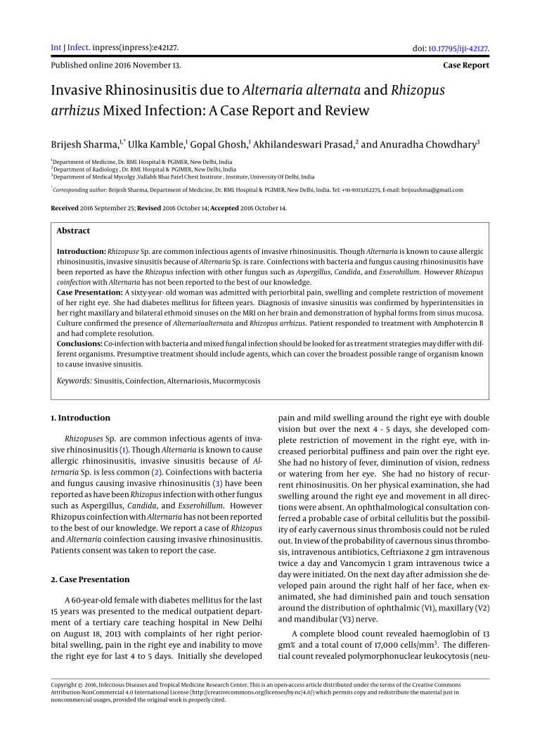
trophils 90%, lymphocytes 8%, monocytes 1%, eosinophil1%) and ESR 25 mm. Total IgE was 25 kU/L. Biochemical in-vestigations revealed fasting blood sugar 134 mg/dL withpost-prandial blood sugar 182 mg/dL. Blood urea nitro-gen levels and serum levels of creatinine, bilirubin, ala-nine aminotransferase aspartate aminotransferase, alka-line phosphate, sodium and potassium were normal. MRIon the brain including an orbit showed hyperintensties inright maxillary and bilateral ethmoid sinuses on T2 axialsequences (Figure 1A and B).
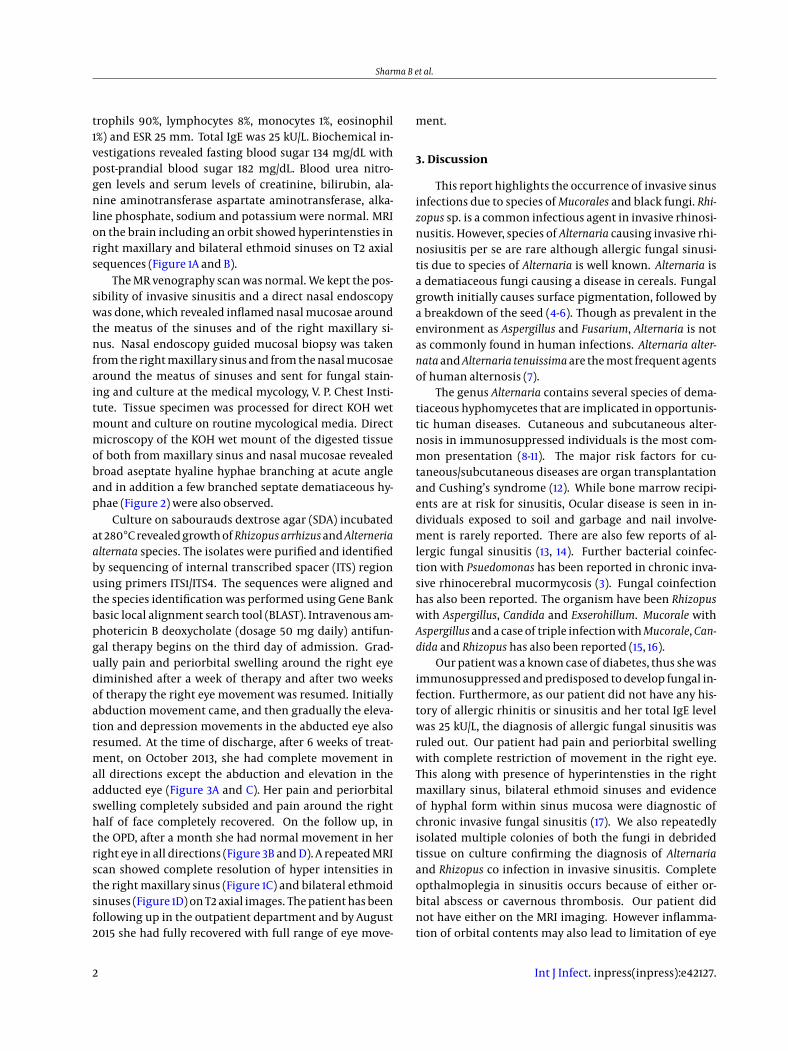
The MR venography scan was normal. We kept the pos-sibility of invasive sinusitis and a direct nasal endoscopywas done, which revealed inflamed nasal mucosae aroundthe meatus of the sinuses and of the right maxillary si-nus. Nasal endoscopy guided mucosal biopsy was takenfrom the right maxillary sinus and from the nasal mucosaearound the meatus of sinuses and sent for fungal stain-ing and culture at the medical mycology, V. P. Chest Insti-tute. Tissue specimen was processed for direct KOH wetmount and culture on routine mycological media. Directmicroscopy of the KOH wet mount of the digested tissueof both from maxillary sinus and nasal mucosae revealedbroad aseptate hyaline hyphae branching at acute angleand in addition a few branched septate dematiaceous hy-phae (Figure 2) were also observed.
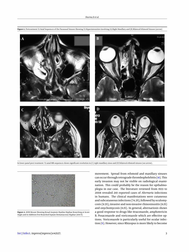
Culture on sabourauds dextrose agar (SDA) incubatedat 280°C revealed growth of RhizopusarrhizusandAlterneriaalternata species. The isolates were purified and identifiedby sequencing of internal transcribed spacer (ITS) regionusing primers ITS1/ITS4. The sequences were aligned andthe species identification was performed using Gene Bankbasic local alignment search tool (BLAST). Intravenous am-photericin B deoxycholate (dosage 50 mg daily) antifun-gal therapy begins on the third day of admission. Grad-ually pain and periorbital swelling around the right eyediminished after a week of therapy and after two weeksof therapy the right eye movement was resumed. Initiallyabduction movement came, and then gradually the eleva-tion and depression movements in the abducted eye alsoresumed. At the time of discharge, after 6 weeks of treat-ment, on October 2013, she had complete movement inall directions except the abduction and elevation in theadducted eye (Figure 3A and C). Her pain and periorbitalswelling completely subsided and pain around the righthalf of face completely recovered. On the follow up, inthe OPD, after a month she had normal movement in herright eye in all directions (Figure 3B and D). A repeated MRIscan showed complete resolution of hyper intensities inthe right maxillary sinus (Figure 1C) and bilateral ethmoidsinuses (Figure 1D) on T2 axial images. The patient has beenfollowing up in the outpatient department and by August2015 she had fully recovered with full range of eye move-
ment.
3. Discussion
This report highlights the occurrence of invasive sinusinfections due to species of Mucorales and black fungi. Rhi-zopus sp. is a common infectious agent in invasive rhinosi-nusitis. However, species of Alternaria causing invasive rhi-nosiusitis per se are rare although allergic fungal sinusi-tis due to species of Alternaria is well known. Alternaria isa dematiaceous fungi causing a disease in cereals. Fungalgrowth initially causes surface pigmentation, followed bya breakdown of the seed (4-6). Though as prevalent in theenvironment as Aspergillus and Fusarium, Alternaria is notas commonly found in human infections. Alternaria alter-nata andAlternaria tenuissima are the most frequent agentsof human alternosis (7).
The genus Alternaria contains several species of dema-tiaceous hyphomycetes that are implicated in opportunis-tic human diseases. Cutaneous and subcutaneous alter-nosis in immunosuppressed individuals is the most com-mon presentation (8-11). The major risk factors for cu-taneous/subcutaneous diseases are organ transplantationand Cushing’s syndrome (12). While bone marrow recipi-ents are at risk for sinusitis, Ocular disease is seen in in-dividuals exposed to soil and garbage and nail involve-ment is rarely reported. There are also few reports of al-lergic fungal sinusitis (13, 14). Further bacterial coinfec-tion with Psuedomonas has been reported in chronic inva-sive rhinocerebral mucormycosis (3). Fungal coinfectionhas also been reported. The organism have been Rhizopuswith Aspergillus, Candida and Exserohillum. Mucorale withAspergillus and a case of triple infection withMucorale,Can-dida and Rhizopus has also been reported (15, 16).
Our patient was a known case of diabetes, thus she wasimmunosuppressed and predisposed to develop fungal in-fection. Furthermore, as our patient did not have any his-tory of allergic rhinitis or sinusitis and her total IgE levelwas 25 kU/L, the diagnosis of allergic fungal sinusitis wasruled out. Our patient had pain and periorbital swellingwith complete restriction of movement in the right eye.This along with presence of hyperintensties in the rightmaxillary sinus, bilateral ethmoid sinuses and evidenceof hyphal form within sinus mucosa were diagnostic ofchronic invasive fungal sinusitis (17). We also repeatedlyisolated multiple colonies of both the fungi in debridedtissue on culture confirming the diagnosis of Alternariaand Rhizopus co infection in invasive sinusitis. Completeopthalmoplegia in sinusitis occurs because of either or-bital abscess or cavernous thrombosis. Our patient didnot have either on the MRI imaging. However inflamma-tion of orbital contents may also lead to limitation of eye
2 Int J Infect. inpress(inpress):e42127.
Sharma B et al.
Figure 1. Pretreatment T2 Axial Sequences of the Paranasal Sinuses Showing T2 Hyperintensities Involving (A) Right Maxillary and (B) Bilateral Ethmoid Sinuses (arrow)
In lower panel post treatment: T2 axial MR sequences shows significant resolution in (C) right maxillary sinus and (D) bilateral ethmoid sinuses (see arrow).
Figure 2. KOH Mount Showing Broad Aseptate Hyaline Hyphae Branching at AcuteAngle and in Addition Few Branched Septate Dematiaceous Hyphae (400 X)
movement. Spread from ethmoid and maxillary sinusescan occur through retrograde thrombophelebitis (18). Thisearly invasion may not be visible on radiological exami-nation. This could probably be the reason for opthalmo-plegia in our case. The literature reviewed from 1933 to2008 revealed 210 reported cases of Alternaria infectionsin humans. The clinical manifestations were cutaneousand subcutaneous infections (74.3%), followed by oculomy-cosis (9.5%), invasive and non-invasive rhinosinusitis (8.1%)and onychomycosis (8.1%). In general, alternariosis showsa good response to drugs like itraconazole, amphotericinB. Posaconazole and voriconazole which are effective op-tions. Voriconazole is particularly useful for ocular infec-tion (6). However, since Rhizopus is more likely to become
Int J Infect. inpress(inpress):e42127. 3
Sharma B et al.
Figure 3. Movement of Eyes Mid Treatment and on Completion of Treatment
Left column, A, and C are mid treatment showing restriction of adduction and elevation of adducted right eye respectively. Right column, B and D show full recovery ofadduction and elevation of adducted eye
disseminated on treatment with voriconazole and our casehad a mixed Rhizopus arrhizus and Alternaria fungal infec-tion, we treated the patient with Amphotericin B deoxy-cholate. The patient recovered completely after treatmentwith Amphotericin B.
3.1. Conclusion
Dematiaceous fungi are increasingly being reportedfor different diseases. The case reported here is rare be-cause Alternaria, a dematiaceous fungus, is not a commonorganism causing invasive rhinosinusitis. Further Mixedinvasive rhinosinusitis with Alternaria has not been re-ported to the best of our knowledge. Signs of invasionin orbit may not be visible on radiological examinationin early invasive sinusitis. When suspected on clinicalgrounds, fungal culture and histopathological examina-tion is a must in invasive sinusitis. Co-infection with bac-teria and mixed fungal infection should be looked for astreatment strategies may differ with different organisms.Presumptive treatment should include agents, which cancover the broadest possible range of organism known tocause invasive sinusitis.
References
1. Michael RC, Michael JS, Ashbee RH, Mathews MS. Mycological pro-file of fungal sinusitis: An audit of specimens over a 7-year periodin a tertiary care hospital in Tamil Nadu. Indian J Pathol Microbiol.2008;51(4):493–6. doi: 10.4103/0377-4929.43738. [PubMed: 19008573].
2. Malani PN, Kauffman CA. Invasive and Allergic Fungal Sinusitis. CurrInfect Dis Rep. 2002;4(3):225–32. [PubMed: 12015915].
3. Dimaka K, Mallis A, Naxakis SS, Marangos M, Papadas TA, StathasT, et al. Chronic rhinocerebral mucormycosis: a rare case reportand review of the literature. Mycoses. 2014;57(11):699–702. doi:10.1111/myc.12219. [PubMed: 25039925].
4. Hasan HA. Phytotoxicity of pathogenic fungi and their mycotoxinsto cereal seedling viability. Mycopathologia. 1999;148(3):149–55. doi:10.1023/A:1007164617175. [PubMed: 11189766].
5. Li F, Yoshizawa T. Alternaria mycotoxins in weathered wheat fromChina. J Agric Food Chem. 2000;48(7):2920–4. doi: 10.1021/jf0000171.[PubMed: 10898645].
6. Morrison VA, McGlave PB. Mucormycosis in the BMT population. BoneMarrow Transplant. 1993;11(5):383–8. [PubMed: 8504272].
7. Brandt ME, Warnock DW. Epidemiology, clinical manifestations, andtherapy of infections caused by dematiaceous fungi. J Chemother.2003;15 Suppl 2:36–47. doi: 10.1179/joc.2003.15.Supplement-2.36.[PubMed: 14708965].
8. Gene J, Azon-Masoliver A, Guarro J, Ballester F, Pujol I, Llovera M, etal. Cutaneous phaeohyphomycosis caused by Alternaria longipes inan immunosuppressed patient. J Clin Microbiol. 1995;33(10):2774–6.[PubMed: 8567925].
9. Pastor FJ, Guarro J. Alternaria infections: laboratory diagnosis andrelevant clinical features. ClinMicrobiol Infect. 2008;14(8):734–46. doi:10.1111/j.1469-0691.2008.02024.x. [PubMed: 18727797].
10. Pereiro M, Pereiro Ferreiros MM, De Hoog GS, Toribio J. Cutaneous in-fection caused by Alternaria in patients receiving tacrolimus.MedMy-col. 2004;42(3):277–82. doi: 10.1080/13693780310001610047. [PubMed:15283243].
11. Wiest PM, Wiese K, Jacobs MR, Morrissey AB, Abelson TI, Witt W,et al. Alternaria infection in a patient with acquired immunodefi-ciency syndrome: case report and review of invasive alternaria in-fections. Rev Infect Dis. 1987;9(4):799–803. doi: 10.1093/clinids/9.4.799.[PubMed: 3326127].
12. Gilaberte M, Bartralot R, Torres JM, Reus FS, Rodriguez V, Alomar A,et al. Cutaneous alternariosis in transplant recipients: clinicopatho-logic review of 9 cases. J Am Acad Dermatol. 2005;52(4):653–9. doi:10.1016/j.jaad.2004.10.875. [PubMed: 15793517].
4 Int J Infect. inpress(inpress):e42127.
Sharma B et al.
13. Bartynski JM, McCaffrey TV, Frigas E. Allergic fungal sinusi-tis secondary to dermatiaceous fungi-Curvularia lunata andAlternaria. Otolaryngol Head Neck Surg. 1990;103:32–9. doi:10.1177/019459989010300105.
14. Chowdhary A, Agarwal K, Randhawa HS, Kathuria S, Gaur SN, Na-jafzadeh MJ, et al. A rare case of allergic bronchopulmonary myco-sis caused by Alternaria alternata. Med Mycol. 2012;50(8):890–6. doi:10.3109/13693786.2012.682320. [PubMed: 22563857].
15. Talmi YP, Goldschmied-Reouven A, Bakon M, Barshack I, Wolf M,Horowitz Z, et al. Rhino-orbital and rhino-orbito-cerebral mu-cormycosis. Otolaryngol Head Neck Surg. 2002;127(1):22–31. doi:10.1067/mhn.2002.126587. [PubMed: 12161726].
16. Monroe MM, McLean M, Sautter N, Wax MK, Andersen PE, SmithTL, et al. Invasive fungal rhinosinusitis: a 15-year experience with29 patients. Laryngoscope. 2013;123(7):1583–7. doi: 10.1002/lary.23978.[PubMed: 23417294].
17. deShazo RD, O’Brien M, Chapin K, Soto-Aguilar M, Gardner L, Swain R.A new classification and diagnostic criteria for invasive fungal sinusi-tis. Arch Otolaryngol Head Neck Surg. 1997;123(11):1181–8. doi: 10.1001/ar-chotol.1997.01900110031005. [PubMed: 9366697].
18. El-Beltagy Y, Hamdy TAH, Hasaballah MS. Orbital complications fol-lowing sinusitis still a problem: Our experience and results. EgyptianJ Ear, Nose, Throat Allied Sci. 2014;15:189–95.
Int J Infect. inpress(inpress):e42127. 5