Embed Size (px)
Citation preview
Introductory Microbiology
Dr. Heather Townsend

Characteristics of Life
• Growth and development
• Reproduction and heredity
• Metabolism
• Movement and/or irritability
• Cell support, protection, and storage mechanisms
• Transport of nutrients and waste
• Living things are made of cells!!

All cells…….
• Have an outer plasma membrane
• Contain DNA– Enclosed within the cell somewhere
• Contain cytoplasm– Everything between the plasma membrane
and the region of DNA– Gives cells their shape– Assist in movement of cell and organelles

Characteristics of Microbes• Prokaryotic cells
– Smaller – Lack special structures such as a nucleus and organelles– All prokaryotic cells are microorganisms
• Some microorganisms are eukaryotic• Viruses?

“Micro”organisms

Characteristics of Cells
Eukaryotic cells– Animals, plants, fungi,
and protists– contain double-
membrane bound nucleus with DNA
– contain membrane-bound organelles
– 10–100 µm in diameter


Characteristics of Cells
Prokaryotic cells– ~1.0 µm in diameter– Bacteria and archaea– no nucleus – no membrane-bound
organelles


Microbiology• The study of of organisms
(microorganisms or microbes) too small to be seen without magnification
• This includes:1. Bacteria
2. Viruses
3. Fungi
4. Protozoa
5. Helminths (worms)
6. Algae

The Microbes
• 1. Bacteria– Single-celled
organisms – Various shapes
• Spherical• Rod• Spiral shapes
– Cellular– Lack membrane-
enclosed cellular structures
– Widely distributed in nature
Klebsiella pneumoniae, bacteria that causes
pneumonia in humans

The Microbes
• 2. Viruses– Acellular– Composed of nucleic
acid and a few proteins
– Replicate themselves to display other properties of living organisms when they invade living cells

The Microbes• 3. Fungi– Yeasts and molds
• Single-celled, microscopic
– Mushrooms• Multicellular,
macroscopic
– Cell nucleus and other cellular structures
– Absorb nutrients from their environment
– Widely distributed in water and soil
– Act as decomposers of dead organisms

The Microbes• 4. Protozoa– Single-celled,
microscopic organisms
– Have at least one nucleus and many cellular structures
– Obtain food by engulfing or ingesting smaller organisms
– Most can move– Found in many
different environments
Amoeba

The Microbes
• 5. Helminths– Large, multicellular– Parasitize host
tissues – Organs for
reproduction, digestion, movement, protection
– Mouthparts– Ingestion of larvae or
eggs in food Tapeworm Head

The Microbes• 6. Algae– Single-celled
microscopic organisms
– Have a nucleus and many membrane-enclosed cellular structures
– Photosynthesize their own food
– Widely distributed in fresh and salt water
– Important source of food for other organisms
Micrasterias, a green algae living in fresh water

General cell characteristics
• Locomotor appendages
• External boundaries
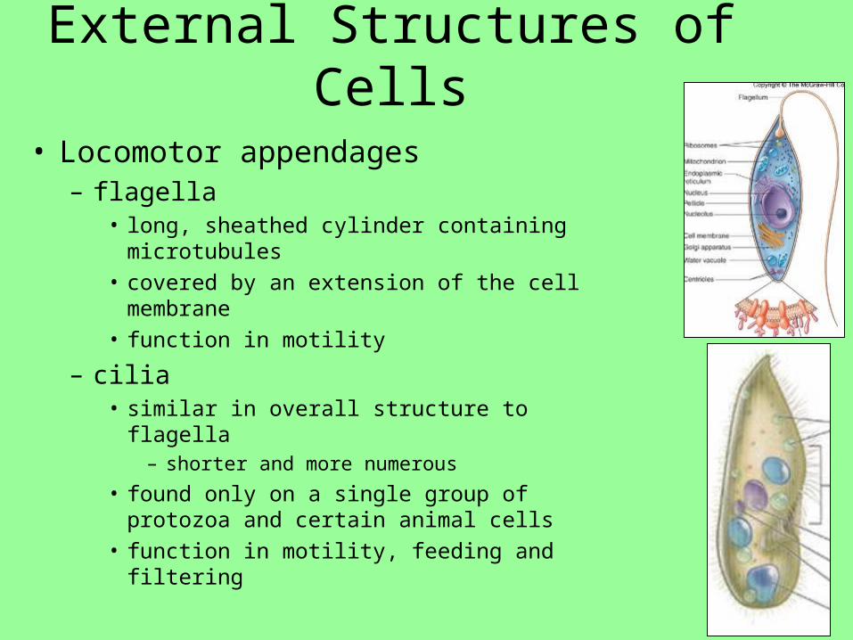
External Structures of Cells
• Locomotor appendages– flagella
• long, sheathed cylinder containing microtubules
• covered by an extension of the cell membrane • function in motility
– cilia • similar in overall structure to flagella
– shorter and more numerous
• found only on a single group of protozoa and certain animal cells
• function in motility, feeding and filtering

External Boundary Structures
• Plasma (cell) membrane– typical bilayer of
phospholipids and proteins
– serves as selectively permeable barrier in transport

External Structures of Cells
• Glycocalyx– an outermost boundary that comes into direct contact
with environment– usually composed of polysaccharides– appears as a network of fibers, a slime layer or a
capsule– functions in adherence, protection, and signal reception– Beneath the glycocalyx:
• Fungi and most algae - cell wall• Protozoa, a few algae, and all animal cells – cell
membrane

External Boundary Structures
• Cell wall– Fungi
• thick inner layer of polysaccharide fibers • composed of chitin or cellulose and a thin layer of mixed
glycans
– Algae • varies in chemical composition• substances include cellulose, pectin, mannans, silicon dioxide,
and calcium carbonate
– Bacteria!!!• Dependent on cell wall composition

Prokaryote Eukaryote

Branches of Study Within Microbiology
• Immunology: – studies immune chemicals and cells that are produced in response
to infection
• Public health microbiology & epidemiology: – aim to monitor and control the spread of diseases (CDC)
• Food, dairy and aquatic microbiology: – examine the ecological and practical roles of microbes in food and
water
• Biotechnology: – ranges from bread making to gene therapy
• Genetic engineering & recombinant DNA technology: – altering the genetic makeup of organisms

Microbes Are Involved In:
• nutrient production & energy flow – i.e., photosynthesis
• decomposition and nutrient recycling
• production of foods, drugs & vaccines
• bioremediation • causing disease

Impact of Pathogens
• Pathogens – Diseases-causing agents
• Nearly 2,000 different microbes cause diseases in the human body
• 10 B infections/year worldwide
• 13 M deaths from infections/year worldwide– killing about 1/3 of the U.S. population each
year

Impact of Pathogens
• Emerging diseases– Becoming more prominent over the years– Zoonosis
• SARS
• Reemerging– Older diseases increasing in occurrence
• TB• Malaria• Hepatitis

Historical Microbiology
• 1546 – physician suggest that invisible organisms may be involved with disease
• Abiogenesis vs biogenesis
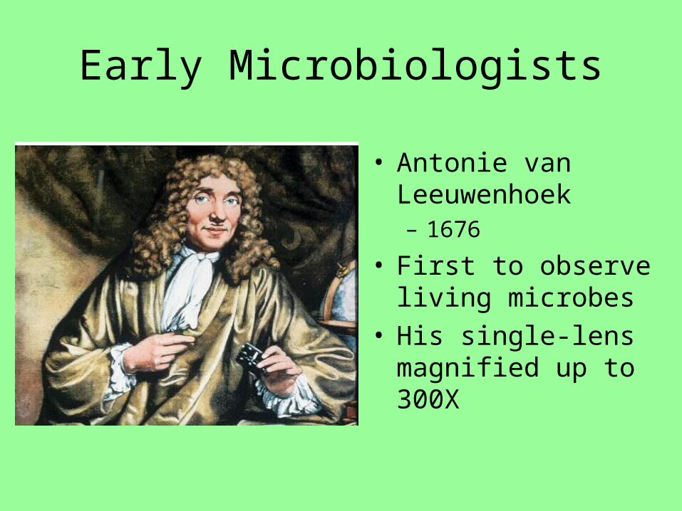
Early Microbiologists
• Antonie van Leeuwenhoek– 1676
• First to observe living microbes
• His single-lens magnified up to 300X

Early Medical Microbiology
• Francesco Redi– 1688– Spontaneous generation
• “Living things arise from nonliving things”
• Belief that some forms of life could arise from vital forces present in nonliving or decomposing matter
• Debate over spontaneous generation led in part to development of scientific method

Science
• Scientific method– 1. Observe some aspect of the natural world and ask
questions about it
– 2. Hypothesis
– 3. Make predictions
– 4. Test the predictions
– 5. Repeat the tests or develop new ones
– 6. Analyze and report the test results and conclusions
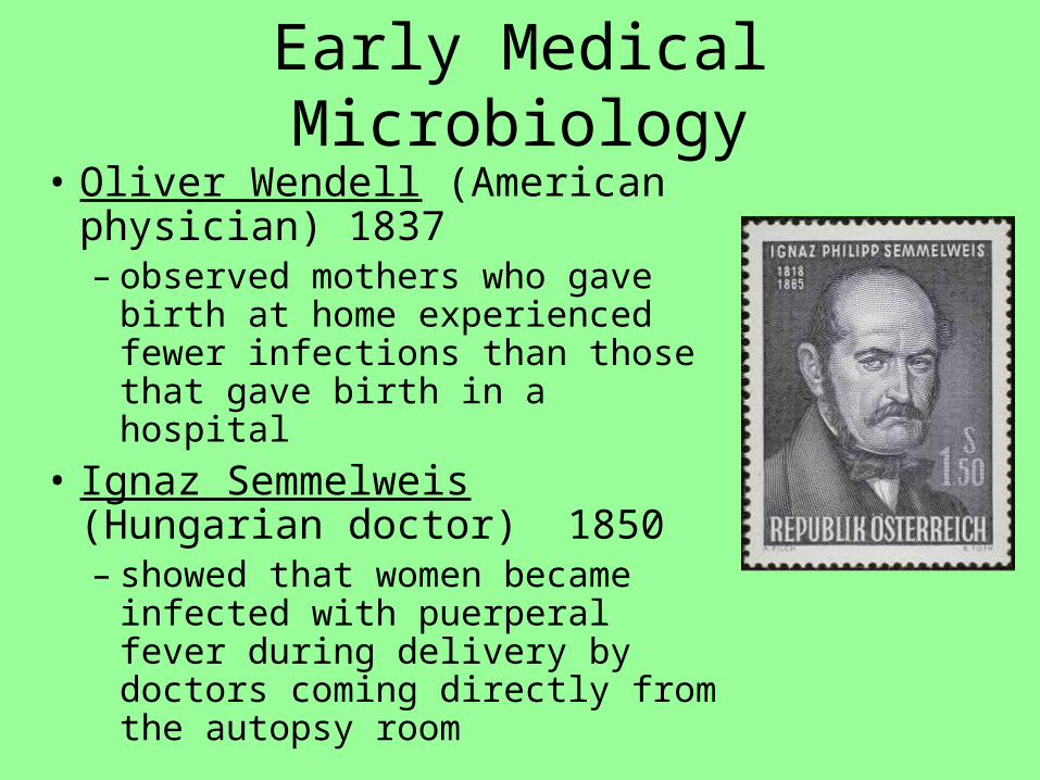
Early Medical Microbiology
• Oliver Wendell (American physician) 1837– observed mothers who gave birth
at home experienced fewer infections than those that gave birth in a hospital
• Ignaz Semmelweis (Hungarian doctor) 1850– showed that women became
infected with puerperal fever during delivery by doctors coming directly from the autopsy room

Early Medical Microbiology• Louis Pasteur - 1861
– Worked in the wine industry • Had knowledge about yeast
producing alcohol
– Swan-neck flasks– Tipping the flask would allow
the microbes to enter the infusion
• Cause them to become cloudy• Main experiment that helped
disprove spontaneous generation
– Developed Pasteurization– Developed a rabies vaccine

Early Medical Microbiology• Joseph Lister (English surgeon) 1865
– Introduced aseptic techniques • Aimed at reducing microbes in a medial setting
and preventing wound infections
– Improved sanitation • Promotes use of carbolic acid on bandages and
medical instruments

Early Medical Microbiology
• Robert Koch (German) 1871– Linked a microscopic
organism with a specific disease (anthrax)
– Developed method to grow bacteria in pure cultures (cultures containing only one kind of organism)
• Used solidified gelatin from potato slices mixed with agar
• Creates a firm surface that microbes could grow on

Koch’s Postulates
1. The specific causative agent must be found in every case of the disease
2. The disease organism must be isolated in pure culture
3. Inoculation of a sample of the culture into a healthy, susceptible animal must produce the same disease
4. The disease must be recovered from the inoculated animal

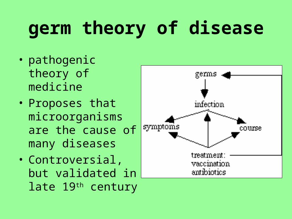
germ theory of disease
• pathogenic theory of medicine
• Proposes that microorganisms are the cause of many diseases
• Controversial, but validated in late 19th century

1900s and beyond…..
• Paul Ehrlich 1900– First to use dyes to ID bacteria– Named concept of chemotherapy– Treatment for syphilis– “magic bullet”
• Compound could be made to selectively target a disease-causing organism

1900’s and beyond…
• Alexander Fleming 1929– observed that a species of Penicillium mold
killed bacterial cells– led to the development of penicillin
• Two types of cells recognized!!!

Microbiology—Now
• Microbiology continues to face many challenges– A pathogen can cause more than one disease– Pathogens are becoming resistant to antimicrobials– Pathogens can be used intentionally to infect large
numbers of people through bioterrorism

Microscopy• Micrometer Size Range
– Most bacterial and archaeal cells are 1-5 micrometers (µm) in length


How to view microbes?
• Light Microscopy – Visible light passes through multiple lenses and through the
specimen– Light microscopes usually have at least 3 lenses
• Scanning (4X)• low-power (10X)• high-power (45X)• oil-immersion (100X)

How to view microbes?
• Staining techniques – simple stain technique – negative stain technique – Special stains
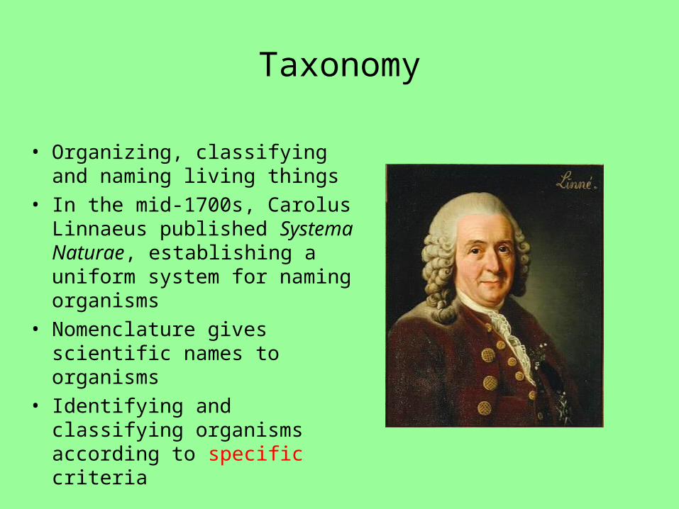
Taxonomy
• Organizing, classifying and naming living things
• In the mid-1700s, Carolus Linnaeus published Systema Naturae, establishing a uniform system for naming organisms
• Nomenclature gives scientific names to organisms
• Identifying and classifying organisms according to specific criteria

Taxonomy
• Domain• Kingdom• Phylum• Class• Order• Family• Genus• species

3 Domains
• Eubacteria – true bacteria– peptidoglycan
• Archaea – odd bacteria that live in extreme environments– high salt, heat, etc. (usually called extremophiles)
• Eukarya– have a nucleus & organelles– Protista
• Algae
– Fungi– Plantae– Animalia

Naming “Most” Micoorganisms
• Binomial (scientific) nomenclature • Gives each microbe 2 names:
– Genus - noun, always capitalized– species - adjective, lowercase
• Both italicized or underlined****– Staphylococcus aureus (S. aureus)– Bacillus subtilis (B. subtilis)– Escherichia coli (E. coli)
Microorganisms in history….
• Edward Jenner– Smallpox vaccine– Tested it on his son and neighborhood children
• Japan– Plague infected fleas covered with cholera
• Tuskegee Syphilis experiment– 1932 - 1972
• Guatemala's National Mental Health Hospital - 1946– US Infected patients with syphilis– Vector - prostitutes

Microorganisms in history…• University of PA – 1950
– Infected 200 female prisoners with hepatitis
• Biological warfare– CIA – whooping cough in FL –
12 killed– US Army – mosquitoes in South
• Plum Island, NY– Lyme Disease??

What to expect……..
• Different microorganisms
• How to detect microorganisms
• Common disease caused by microorganisms
• How to control the spread of microorganisms
• Immune system


![Richard Townsend [315] - UW Madison Astronomy …townsend/tree/scrapbooks/315.pdf* Hungerford connections with Barbara Townsend [210] and Richard Townsend [315]. ** Catherine daughter](https://img.pdfslide.us/doc/110x75/5fe02ca86168ca636365ffc9/richard-townsend-315-uw-madison-astronomy-townsendtreescrapbooks315pdf-.jpg)