Embed Size (px)
Citation preview
Introduction to Physiology
Body Cells
Cell:
Basic living unit of structure & fx of the body. > 100 trillion cells in body. very small (10-5 m in diameter). highly organized. variety of shapes & sizes. each type of cells has a special fx.
Cell (continued)
All Cells share certain characteristics:
general cell structure & components. general mechanisms for changing nutrients to Energy. deliver end products into their surrounding fluid. almost all have the ability to reproduce.
General Cell structure:
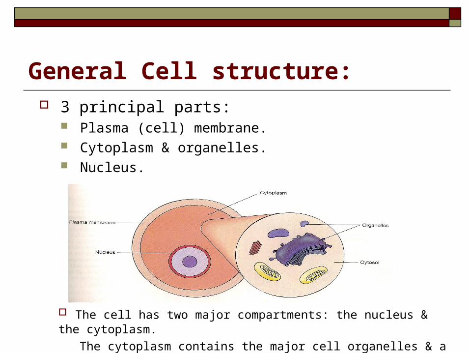
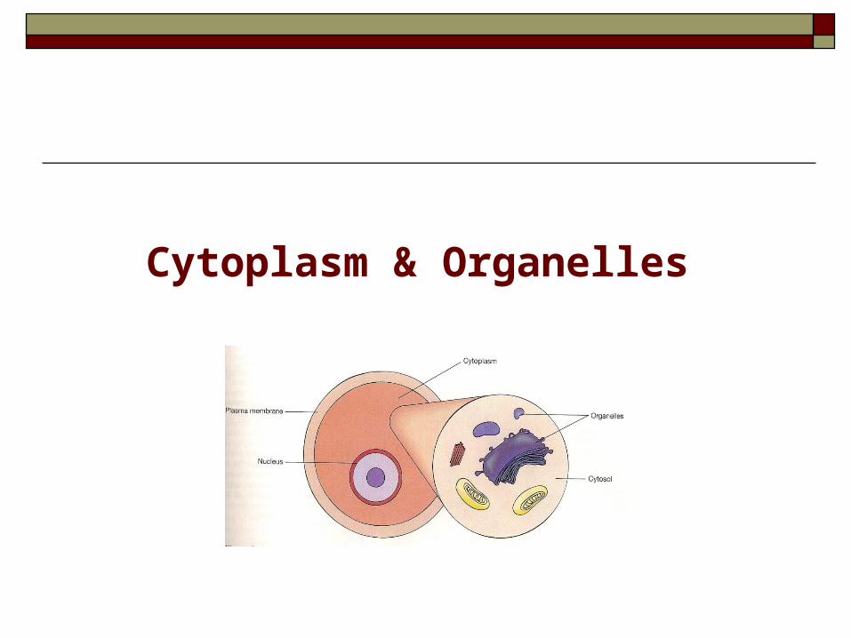
The cell has two major compartments: the nucleus & the cytoplasm.
The cytoplasm contains the major cell organelles & a fluid called cytosol.
3 principal parts: Plasma (cell) membrane. Cytoplasm & organelles. Nucleus.
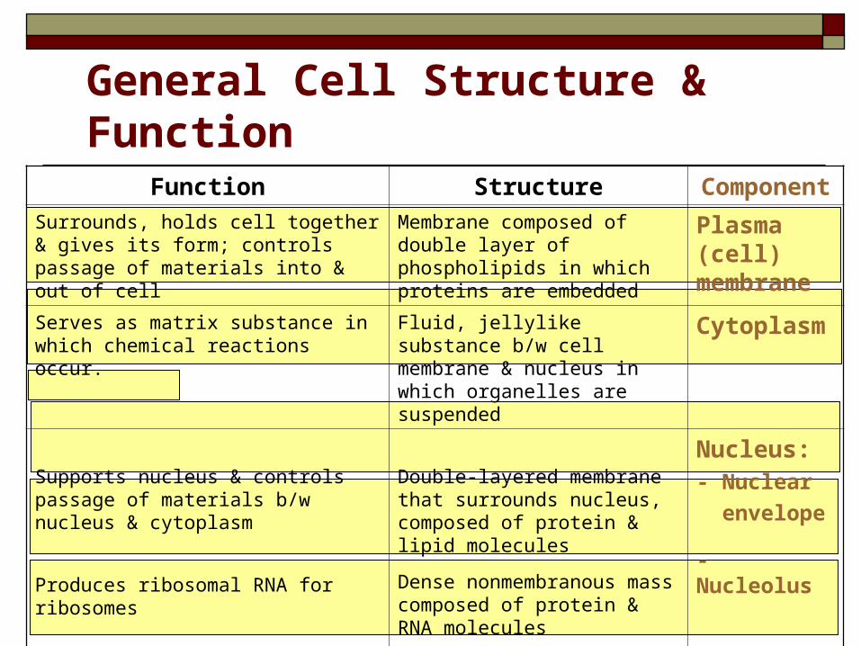
General Cell Structure & FunctionComponentStructureFunction
Plasma (cell) membrane
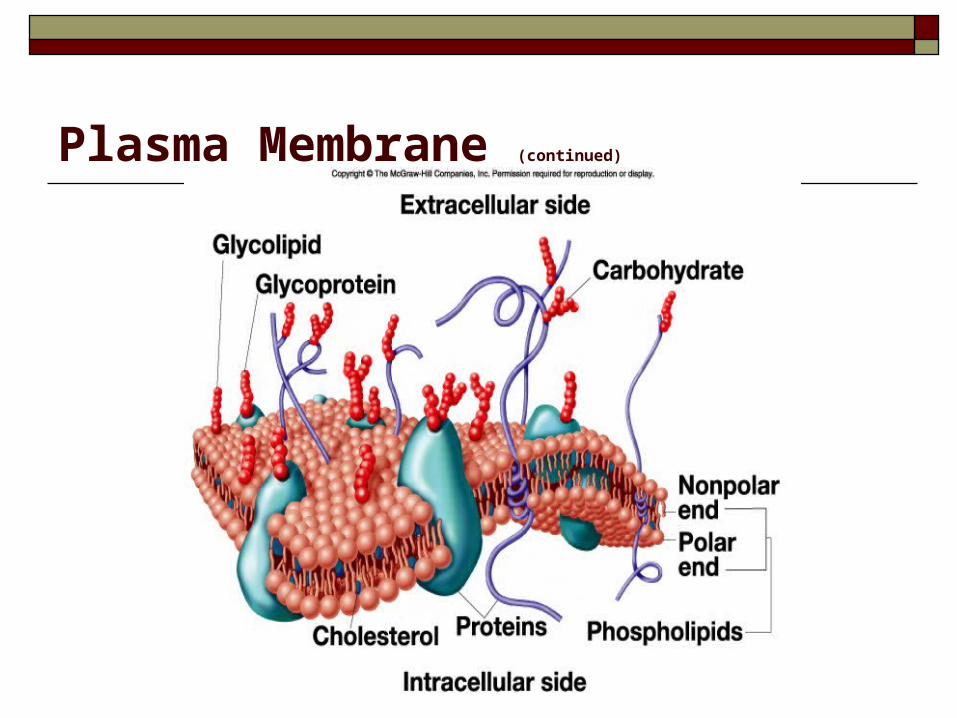
Membrane composed of double layer of phospholipids in which proteins are embedded
Surrounds, holds cell together & gives its form; controls passage of materials into & out of cell
CytoplasmFluid, jellylike substance b/w cell membrane & nucleus in which organelles are suspended
Serves as matrix substance in which chemical reactions occur.
Nucleus:- Nuclear
envelope
- Nucleolus
- Chromatin
Double-layered membrane that surrounds nucleus, composed of protein & lipid molecules
Dense nonmembranous mass composed of protein & RNA molecules
Fibrous strands composed of protein & DNA
Supports nucleus & controls passage of materials b/w nucleus & cytoplasm
Produces ribosomal RNA for ribosomes
Contains genetic code that determines which proteins (including enzymes) will be manufactured by the cell
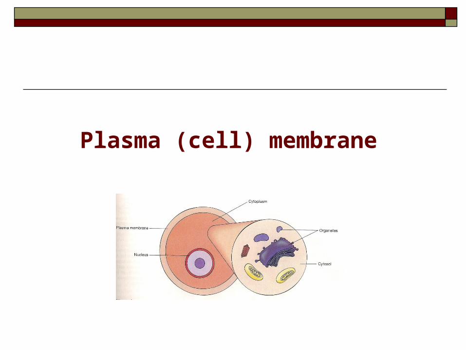
Plasma (cell) membrane
Plasma membrane:
Surrounds, holds cell together & gives its form. 10 nanometer thick. Not solid. Separates cell’s internal structures from extracellular
environment. Is selectively permeable, & controls passage of
materials into & out of cell. Participates in intracellular communication.
Plasma (Cell) Membrane
Composed of:
Double layer of phospholipids (hydrophobic/ hydrophilic parts).
Proteins span, or partially span the membrane.
Negatively charged carbohydrates attach to the outer surface.
Plasma Membrane (continued)
General composition of cell membrane
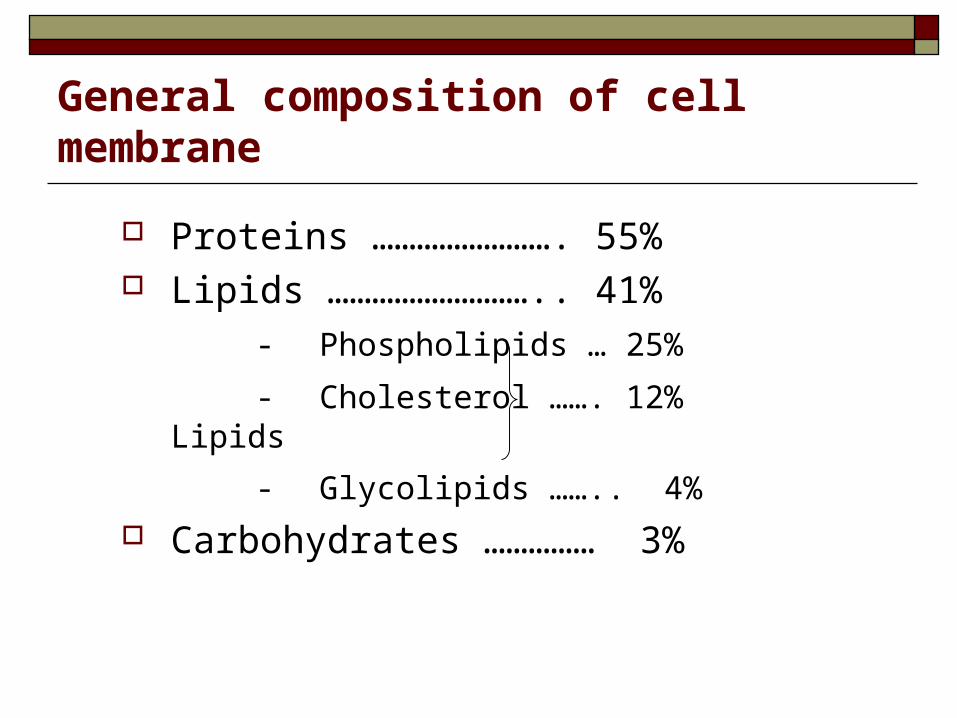
Proteins ……………………. 55% Lipids ……………………….. 41%
- Phospholipids … 25%
- Cholesterol ……. 12% Lipids
- Glycolipids …….. 4%
Carbohydrates …………… 3%
Cell membrane phospholipids
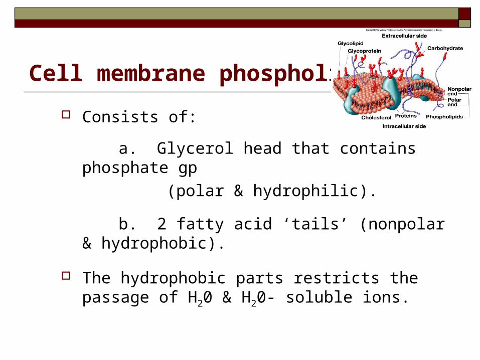
Consists of:
a. Glycerol head that contains phosphate gp
(polar & hydrophilic).
b. 2 fatty acid ‘tails’ (nonpolar & hydrophobic).
The hydrophobic parts restricts the passage of H20 & H20- soluble ions.
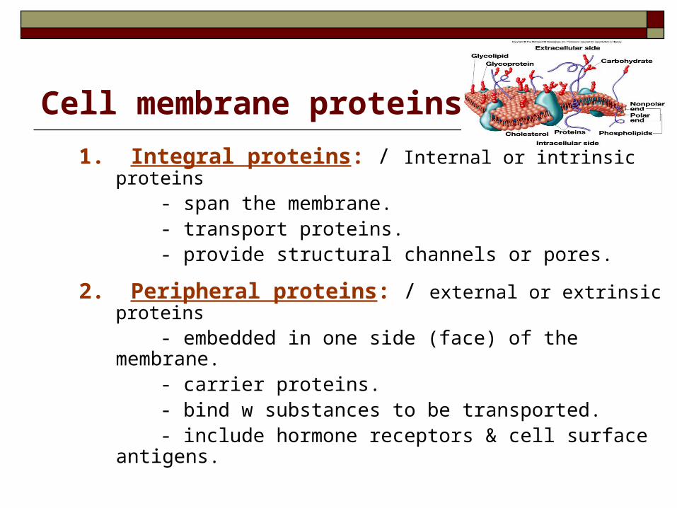
1. Integral proteins: / Internal or intrinsic proteins
- span the membrane. - transport proteins. - provide structural channels or pores.
2. Peripheral proteins: / external or extrinsic proteins
- embedded in one side (face) of the membrane. - carrier proteins. - bind w substances to be transported. - include hormone receptors & cell surface antigens.
Cell membrane proteins
General functions of cell membrane proteins
1. Provide structural support.2. Transport molecules across the membrane.3. Enzymatic control of chemical reactions at cellular surface.4. Some fx as receptors for hormones.5. Some fx as regulatory molecules, that arrive at outer surface of the membrane.6. Some serve as ‘markers’ (antigens), that identify bl & tissue type of an individual.
Cell membrane carbohydrates
Primarily attached to the outer surface of the membrane as:
- Glycoproteins … (most of it).
- Glycolipids …… (1/10).
1. Attach cells to each other.
2. Act as receptor substances.
3. Some enter in immune reactions.
4. Give most of cells overall –ve surface charge, which affects the interaction of regulatory molecules w the membrane.
General functions of cell membrane carbohydrates
Cytoplasm & Organelles
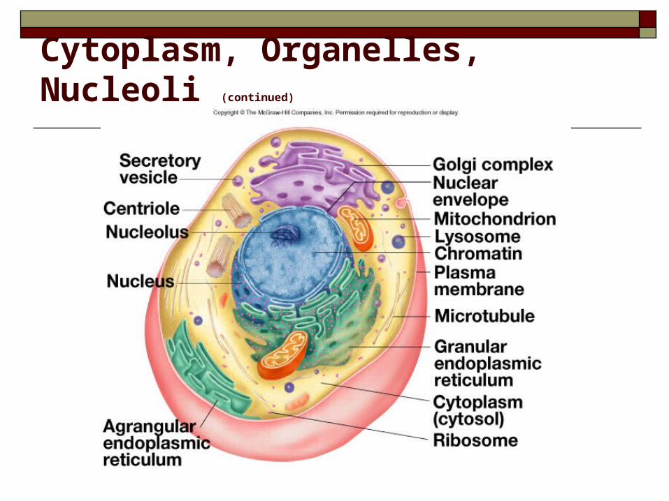
Cytoplasm, Organelles, Nucleoli (continued)

Cytoplasm
The aqueous content of a cell (fluid, jellylike
substance), that lies b/w cell membrane & nucleus
in which organelles are suspended.
Serves as matrix substance in which chemical
reactions occur.
‘cytosol’ is the term used to describe fluid portion of
the cytoplasm.
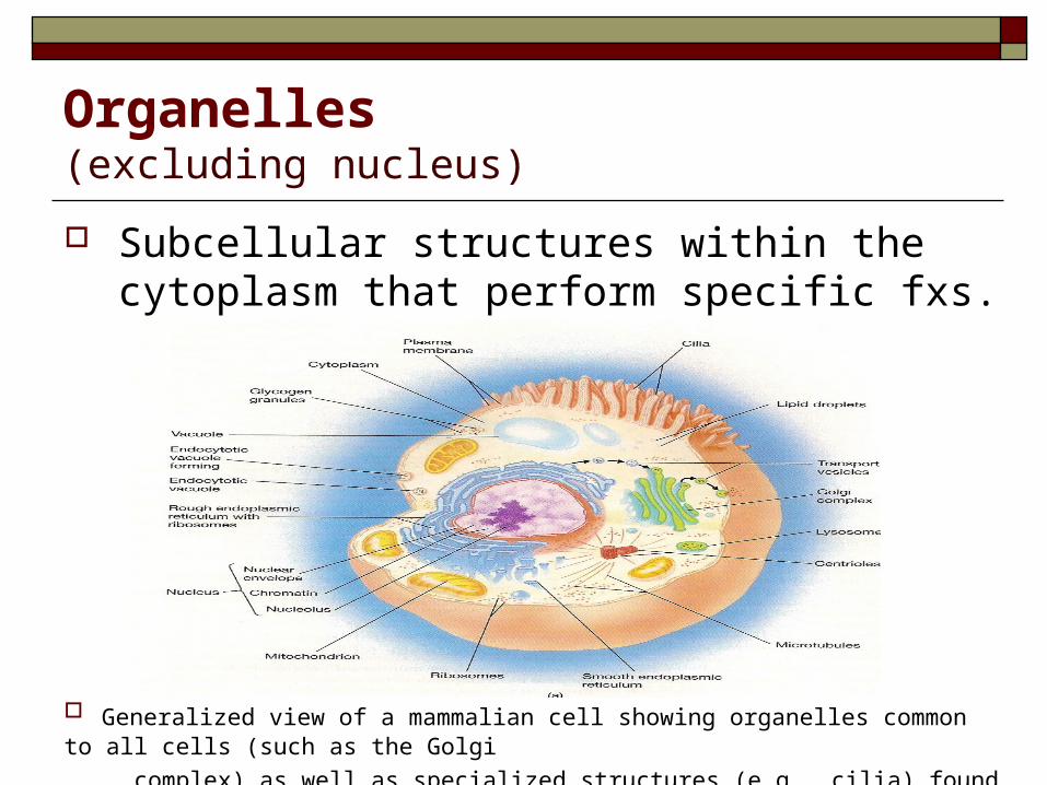
Organelles(excluding nucleus)
Subcellular structures within the cytoplasm that perform specific fxs.
Generalized view of a mammalian cell showing organelles common to all cells (such as the Golgi
complex) as well as specialized structures (e.g., cilia) found only in some cells.
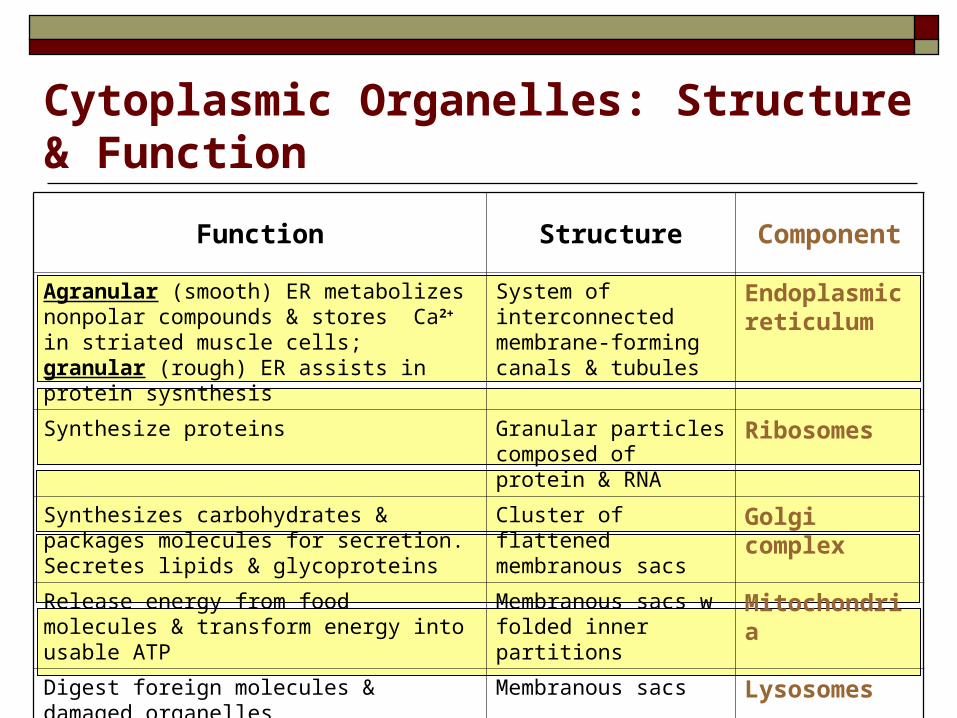
Cytoplasmic Organelles: Structure & Function
ComponentStructureFunction
Endoplasmic reticulum
System of interconnected membrane-forming canals & tubules
Agranular (smooth) ER metabolizes nonpolar compounds & stores Ca2+ in striated muscle cells; granular (rough) ER assists in protein sysnthesis
RibosomesGranular particles composed of protein & RNA
Synthesize proteins
Golgi complexCluster of flattened membranous sacs
Synthesizes carbohydrates & packages molecules for secretion. Secretes lipids & glycoproteins
MitochondriaMembranous sacs w folded inner partitions
Release energy from food molecules & transform energy into usable ATP
LysosomesMembranous sacsDigest foreign molecules & damaged organelles
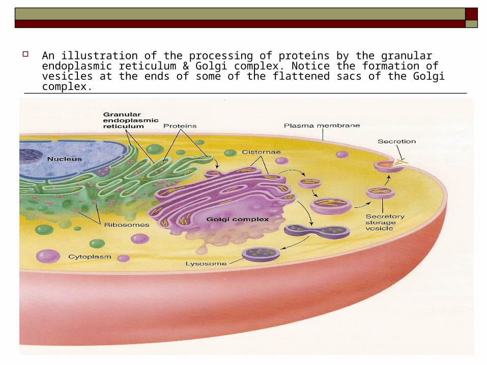
An illustration of the processing of proteins by the granular endoplasmic reticulum & Golgi complex. Notice the formation of vesicles at the ends of some of the flattened sacs of the Golgi complex.
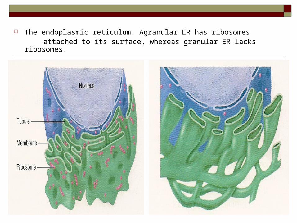
The endoplasmic reticulum. Agranular ER has ribosomes attached to its surface, whereas granular ER lacks ribosomes.
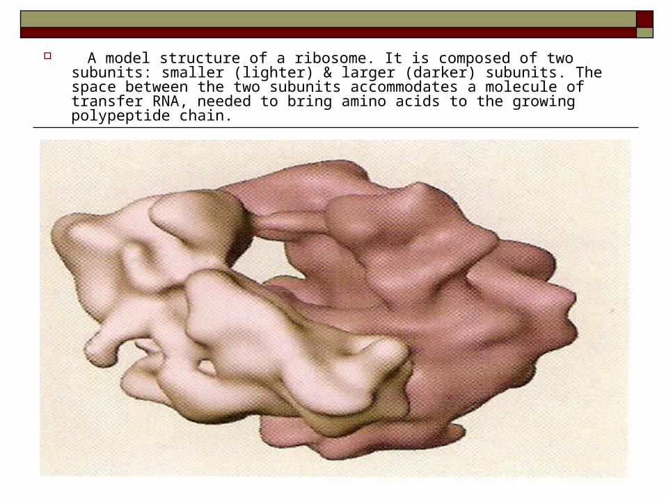
A model structure of a ribosome. It is composed of two subunits: smaller (lighter) & larger (darker) subunits. The space between the two subunits accommodates a molecule of transfer RNA, needed to bring amino acids to the growing polypeptide chain.
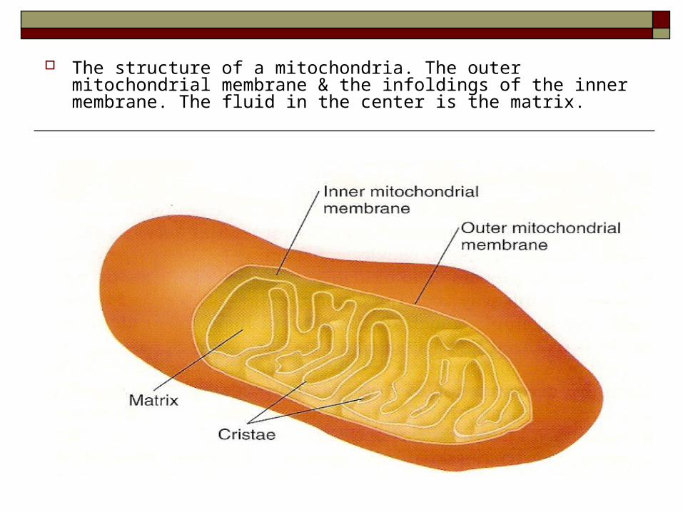
The structure of a mitochondria. The outer mitochondrial membrane & the infoldings of the inner membrane. The fluid in the center is the matrix.
ComponentStructureFunction
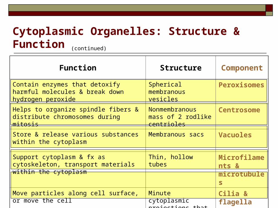
PeroxisomesSpherical membranous vesicles
Contain enzymes that detoxify harmful molecules & break down hydrogen peroxide
CentrosomeNonmembranous mass of 2 rodlike centrioles
Helps to organize spindle fibers & distribute chromosomes during mitosis
VacuolesMembranous sacsStore & release various substances within the cytoplasm
Microfilaments & microtubules
Thin, hollow tubesSupport cytoplasm & fx as cytoskeleton, transport materials within the cytoplasm
Cilia & flagellaMinute cytoplasmic projections that extend from the cell surface
Move particles along cell surface, or move the cell
Cytoplasmic Organelles: Structure & Function (continued)
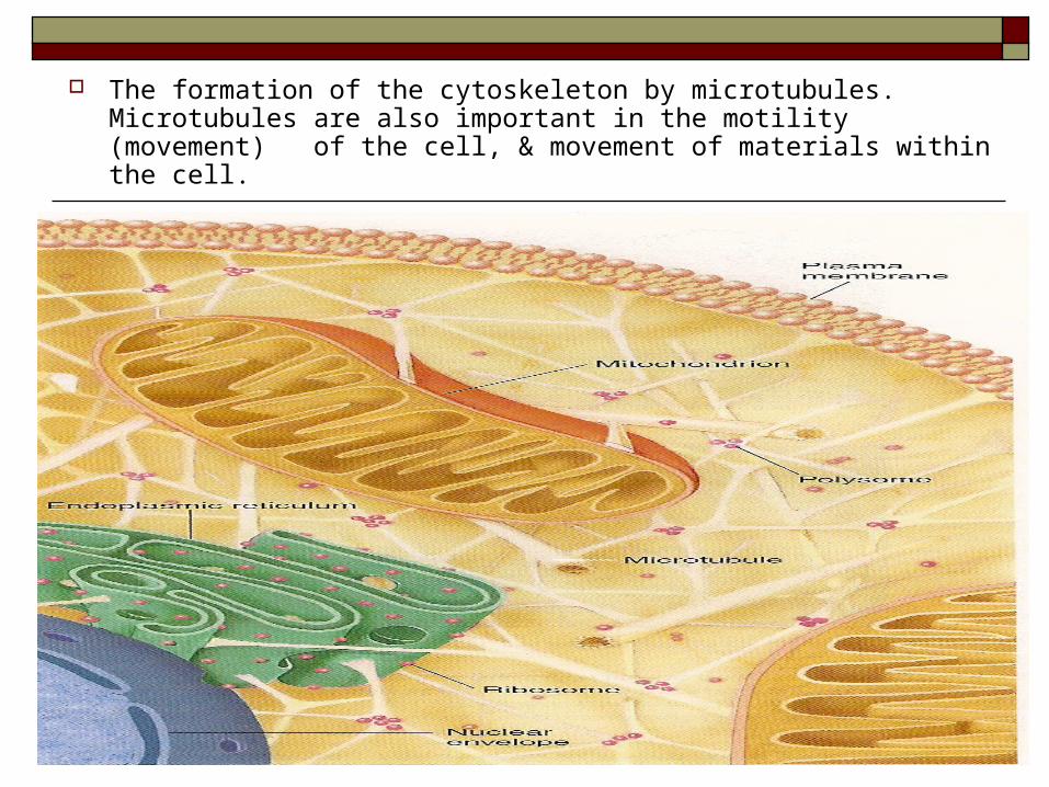
The formation of the cytoskeleton by microtubules. Microtubules are also important in the motility (movement) of the cell, & movement of materials within the cell.
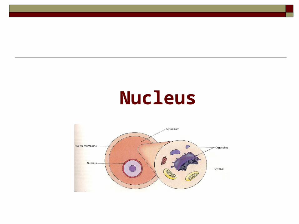
Nucleus
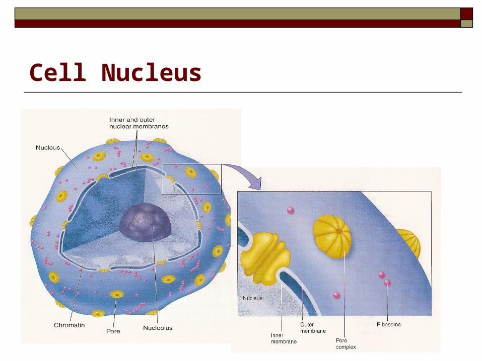
Cell Nucleus Is a large spheroid body. Largest of organelles. Contains the genetic material (DNA). Most cells have a single nucleus. Enclosed by inner & outer membrane (nuclear
envelope). Outer membrane is continuous w ER.
Nuclear pore complexes fuse inner & outer membranes together. Selective active transport of proteins & RNA.
Cell Nucleus
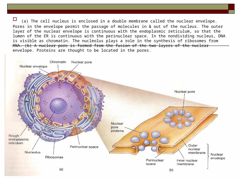
(a) The cell nucleus is enclosed in a double membrane called the nuclear envelope. Pores in the envelope permit the passage of molecules in & out of the nucleus. The outer layer of the nuclear envelope is continuous with the endoplasmic reticulum, so that the lumen of the ER is continuous with the perinuclear space. In the nondividing nucleus, DNA is visible as chromatin. The nucleolus plays a role in the synthesis of ribosomes from RNA. (b) A nuclear pore is formed from the fusion of the two layers of the nuclear envelope. Proteins are thought to be located in the pores.
Cell Nucleus (continued)
Nucleoli: Dark areas within the nucleus, not surrounded by
membrane. Centers for production of ribosomes.
Chromatin: Threadlike material that makes up chromosomes.
Body Fluids
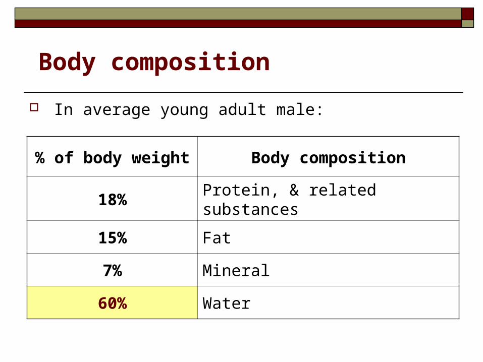
In average young adult male:
Body composition
Body composition% of body weight
Protein, & related substances18%
Fat15%
Mineral7%
Water60%
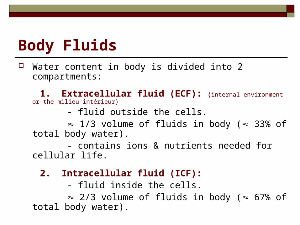
Body Fluids Water content in body is divided into 2 compartments:
1. Extracellular fluid (ECF): (internal environment or the milieu intérieur)
- fluid outside the cells. 1/3 volume of fluids in body ( 33% of total body water). - contains ions & nutrients needed for cellular life.
2. Intracellular fluid (ICF): - fluid inside the cells. 2/3 volume of fluids in body ( 67% of total body water).
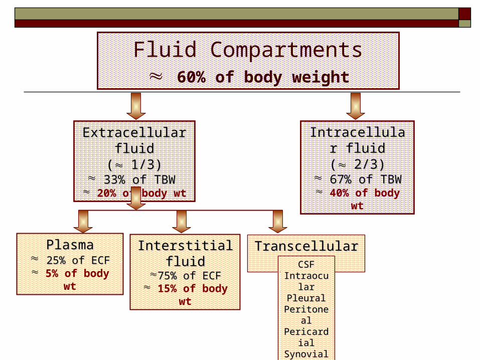
Fluid Compartments 60% of body weight
Extracellular Extracellular fluidfluid
(( 1/3)1/3) 33% of TBW33% of TBW
20% of body wt
Intracellular Intracellular fluidfluid
(( 2/3)2/3) 67% of TBW67% of TBW
40% of body wt
Interstitial Interstitial fluidfluid
75% of ECF75% of ECF 15% of body
wt
PlasmaPlasma 25% of ECF25% of ECF 5% of body
wt
Transcellular Transcellular fluidfluidCSFCSF
IntraoculaIntraocularr
PleuralPleuralPeritoneaPeritonea
llPericardiaPericardia
llSynovialSynovialDigestive Digestive secretionsecretion
ss
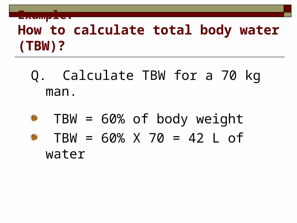
Example: How to calculate total body water (TBW)?
Q. Calculate TBW for a 70 kg man.
TBW = 60% of body weight
TBW = 60% X 70 = 42 L of water
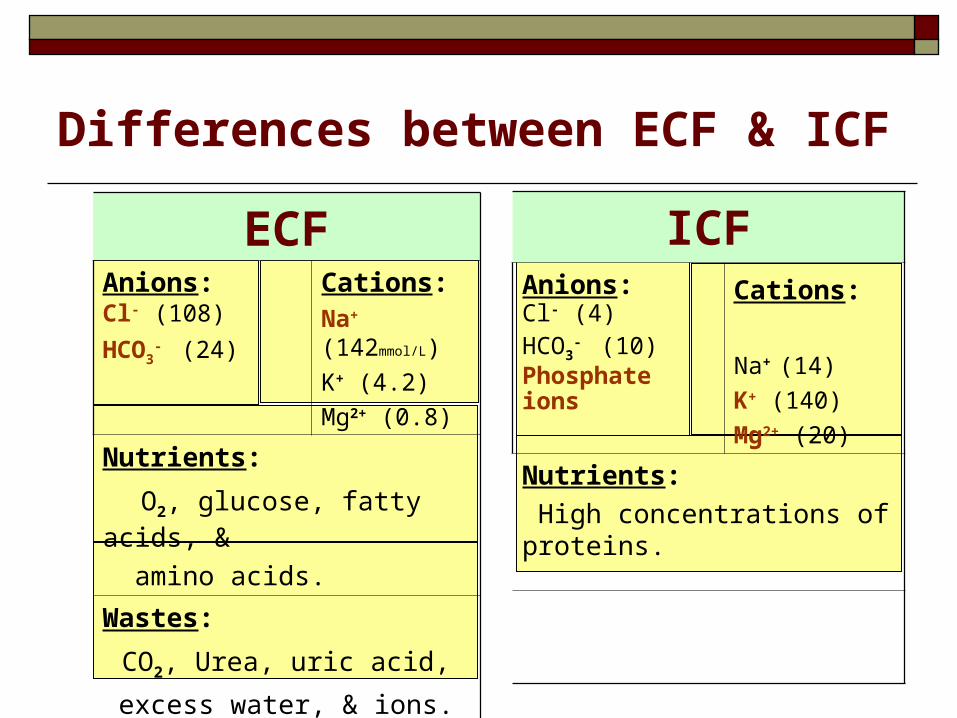
Differences between ECF & ICF
ECFCations:Na+ (142mmol/L)
K+ (4.2)
Mg2+ (0.8)
Anions:Cl- (108)
HCO3- (24)
Nutrients:
O2, glucose, fatty acids, &
amino acids.
Wastes:
CO2, Urea, uric acid,
excess water, & ions.
ICFCations: Na+ (14)
K+ (140)
Mg2+ (20)
Anions:Cl- (4)HCO3
- (10)
Phosphate ions
Nutrients:
High concentrations of proteins.
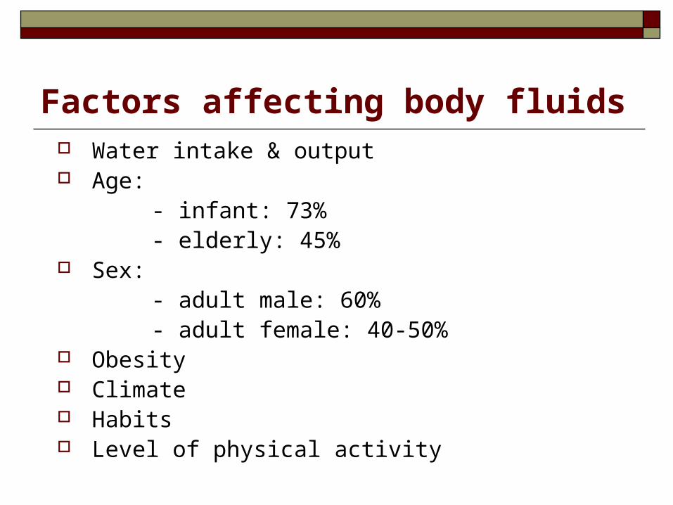
Factors affecting body fluids Water intake & output Age: - infant: 73% - elderly: 45% Sex: - adult male: 60% - adult female: 40-50% Obesity Climate Habits Level of physical activity
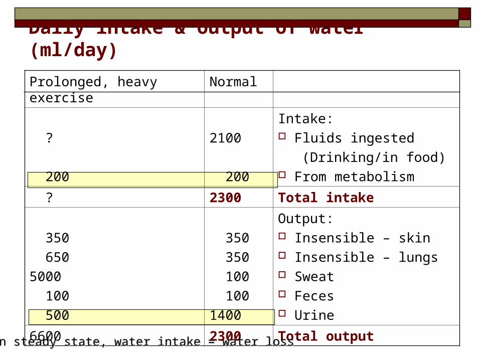
Daily intake & output of water (ml/day)
NormalProlonged, heavy exercise
Intake: Fluids ingested
(Drinking/in food) From metabolism
2100
200
?
200
Total intake2300 ?
Output: Insensible – skin Insensible – lungs Sweat Feces Urine
350
350
100
100
1400
350
650
5000
100
500
Total output23006600
In steady state, water intake = water lossIn steady state, water intake = water loss
Control of body fluids
Thirst Sweating Renal control (aldosterone) Neuronal (osmoreceptors, baroreceptors)
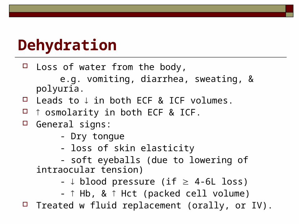
Dehydration Loss of water from the body, e.g. vomiting, diarrhea, sweating, & polyuria. Leads to in both ECF & ICF volumes. osmolarity in both ECF & ICF. General signs: - Dry tongue - loss of skin elasticity - soft eyeballs (due to lowering of intraocular tension) - blood pressure (if 4-6L loss) - Hb, & Hct (packed cell volume) Treated w fluid replacement (orally, or IV).
Transport through the cell membrane
Transport through the cell membrane
Cell membrane is selectively permeable to some molecules & ions. Not permeable to proteins, nucleic acids, & other
molecules.
Lipid or fat-soluble substances, e.g. O2, CO2, OH; enter directly into cell membrane through the lipid bilayer.
Water-soluble substances, e.g. ions, glucose, water; enter through proteins of the cell membrane.
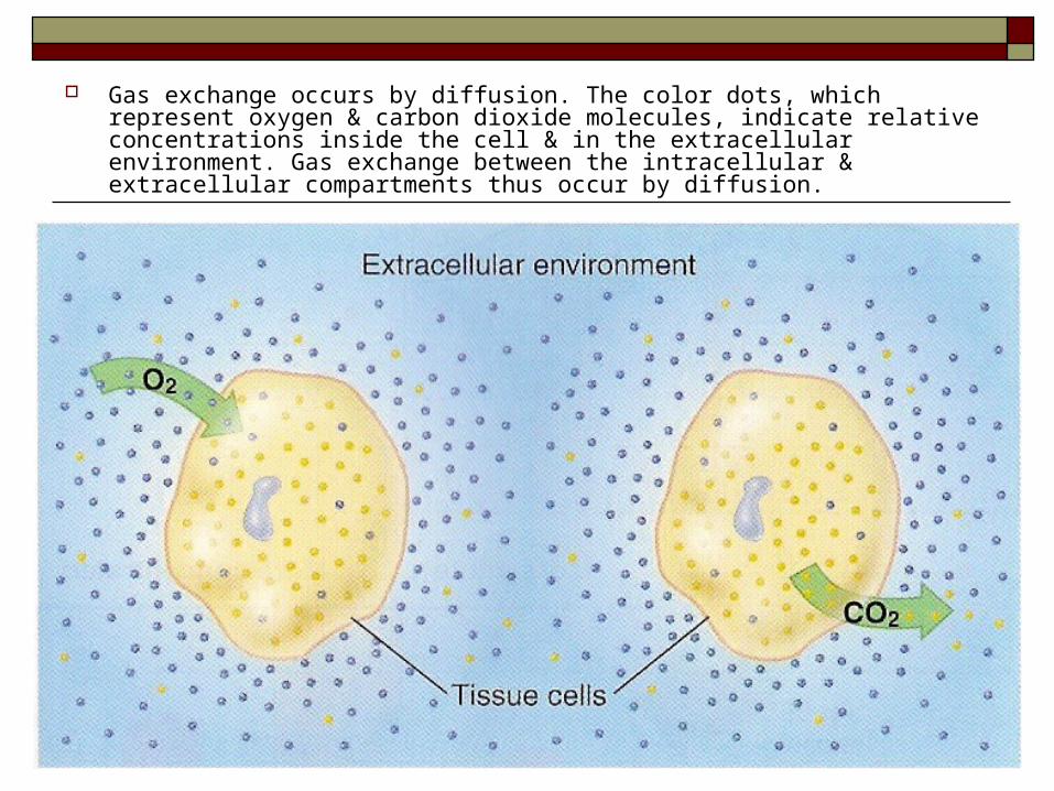
Gas exchange occurs by diffusion. The color dots, which represent oxygen & carbon dioxide molecules, indicate relative concentrations inside the cell & in the extracellular environment. Gas exchange between the intracellular & extracellular compartments thus occur by diffusion.
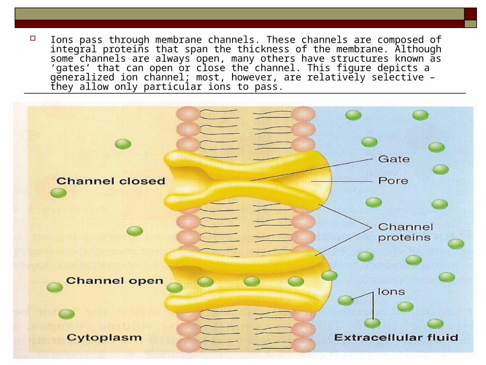
Ions pass through membrane channels. These channels are composed of integral proteins that span the thickness of the membrane. Although some channels are always open, many others have structures known as ‘gates’ that can open or close the channel. This figure depicts a generalized ion channel; most, however, are relatively selective – they allow only particular ions to pass.
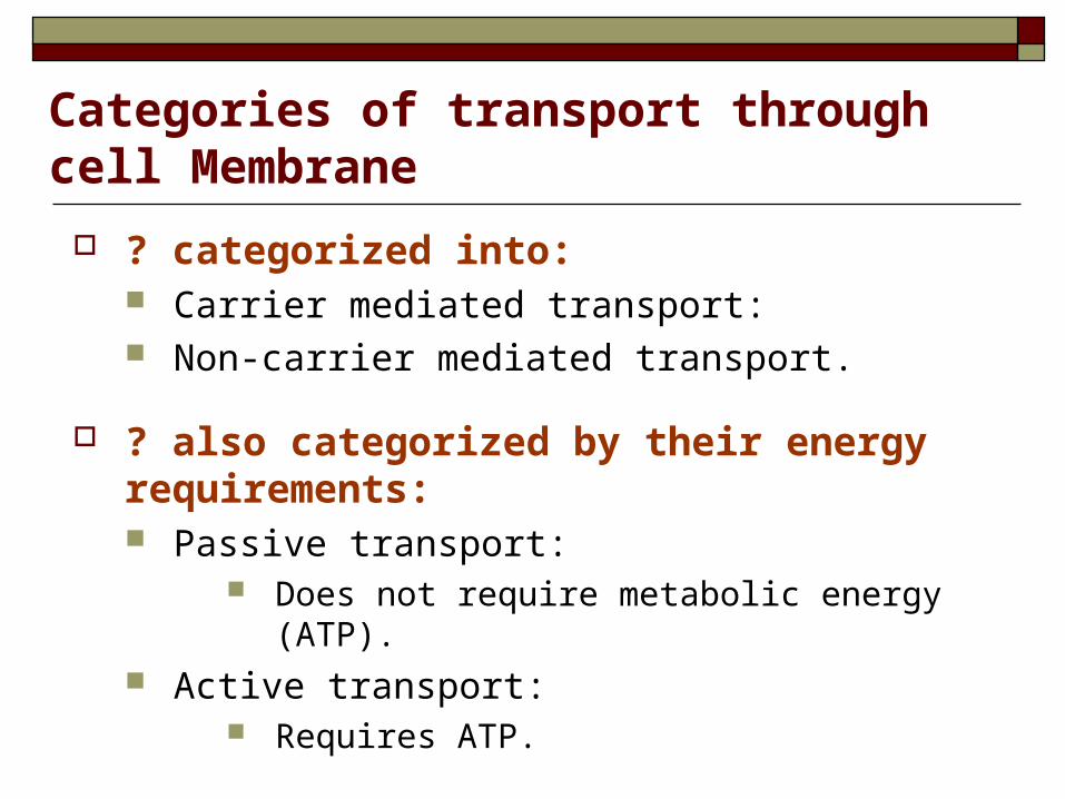
Categories of transport through cell Membrane
? categorized into: Carrier mediated transport: Non-carrier mediated transport.
? also categorized by their energy requirements: Passive transport:
Does not require metabolic energy (ATP). Active transport:
Requires ATP.
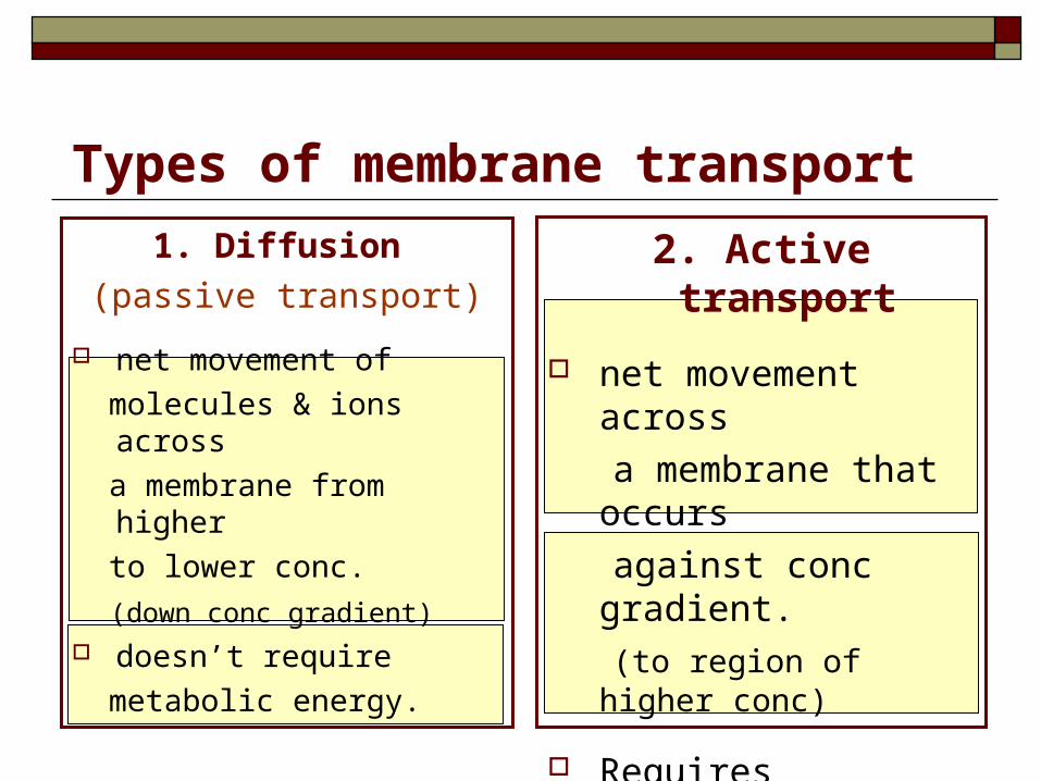
Types of membrane transport
1. Diffusion
(passive transport)
net movement of
molecules & ions across
a membrane from higher
to lower conc.
(down conc gradient)
doesn’t require
metabolic energy.
2. Active transport
net movement across
a membrane that occurs
against conc gradient.
(to region of higher conc)
Requires metabolic energy (ATP), & involves specific carrier proteins.
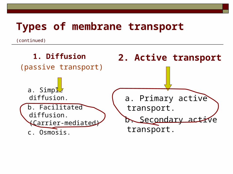
Types of membrane transport (continued)
1. Diffusion
(passive transport)
a. Simple diffusion.
b. Facilitated diffusion. (Carrier-mediated)
c. Osmosis.
2. Active transport
a. Primary active transport.
b. Secondary active transport.
1. Diffusion (passive transport)
1. Diffusion (passive transport) Random movement of substance through the
membrane, either directly or in combination w carrier protein down an electrochemical gradient.
a. simple diffusion
b. facilitated diffusion
c. osmosis
a. Simple diffusion Non-Carrier mediated transport.
Involves net transport down an electrochemical gradient (from higher to lower conc).
Does not need cellular metabolism energy. However, it’s powered by thermal energy of the diffusing molecules.
Net diffusion stops when the conc is equal on both sides of the membrane.
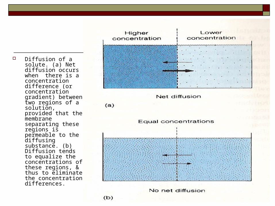
Diffusion of a solute. (a) Net diffusion occurs when there is a concentration difference (or concentration gradient) between two regions of a solution, provided that the membrane separating these regions is permeable to the diffusing substance. (b) Diffusion tends to equalize the concentrations of these regions, & thus to eliminate the concentration differences.
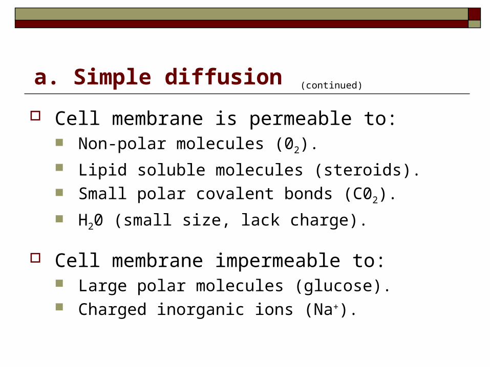
Cell membrane is permeable to: Non-polar molecules (02).
Lipid soluble molecules (steroids). Small polar covalent bonds (C02).
H20 (small size, lack charge).
Cell membrane impermeable to: Large polar molecules (glucose). Charged inorganic ions (Na+).
a. Simple diffusion (continued)
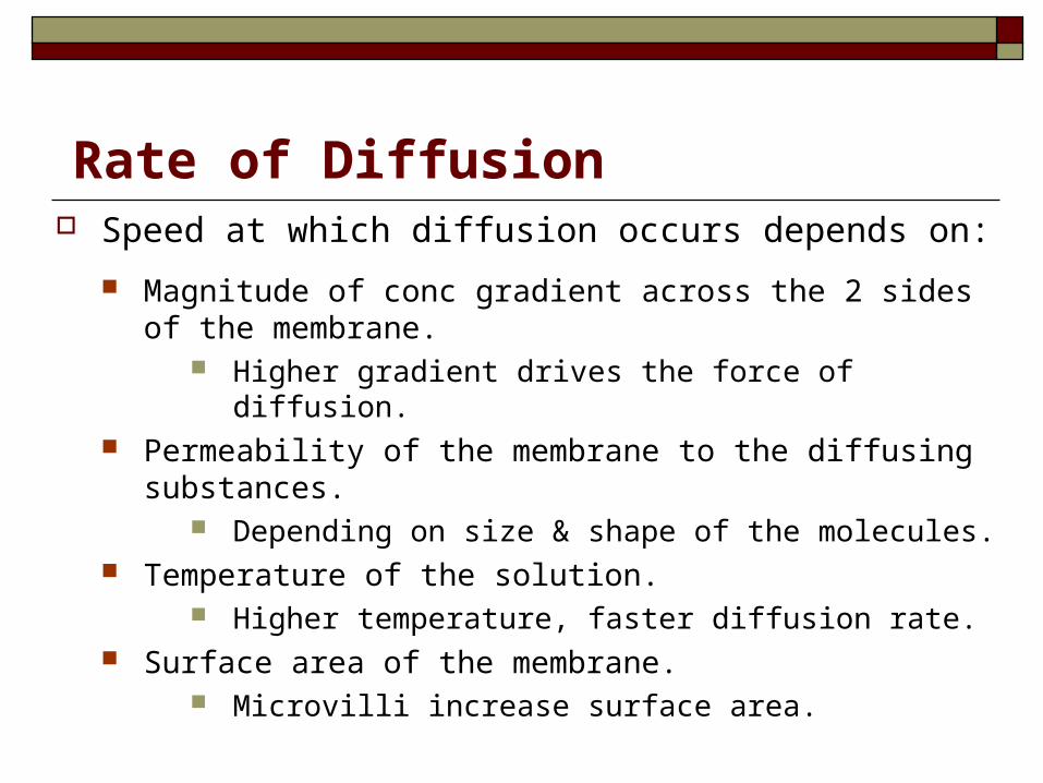
Rate of Diffusion Speed at which diffusion occurs depends on:
Magnitude of conc gradient across the 2 sides of the membrane.
Higher gradient drives the force of diffusion. Permeability of the membrane to the diffusing substances.
Depending on size & shape of the molecules. Temperature of the solution.
Higher temperature, faster diffusion rate. Surface area of the membrane.
Microvilli increase surface area.
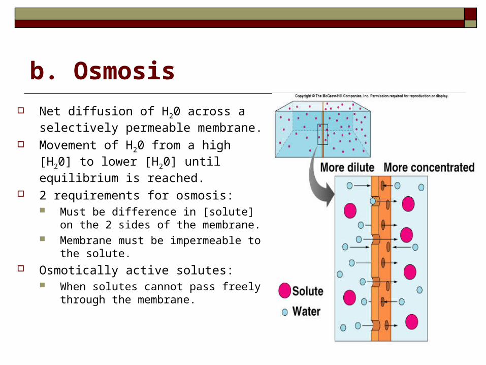
b. Osmosis
Net diffusion of H20 across a selectively permeable membrane.
Movement of H20 from a high [H20] to lower [H20] until equilibrium is reached.
2 requirements for osmosis: Must be difference in [solute] on the 2
sides of the membrane. Membrane must be impermeable to the
solute. Osmotically active solutes:
When solutes cannot pass freely through the membrane.
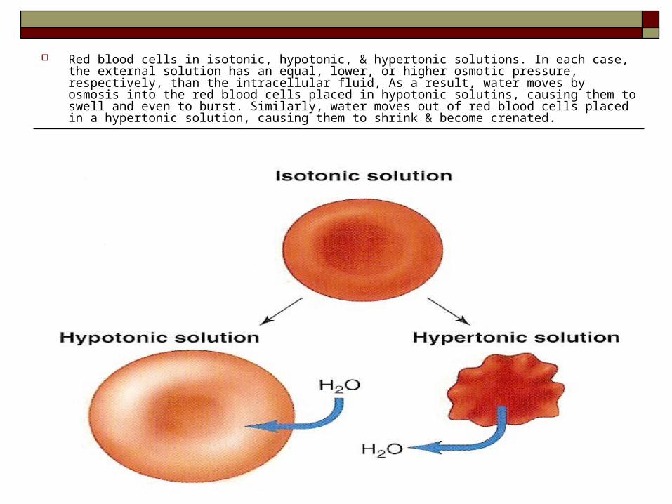
Red blood cells in isotonic, hypotonic, & hypertonic solutions. In each case, the external solution has an equal, lower, or higher osmotic pressure, respectively, than the intracellular fluid, As a result, water moves by osmosis into the red blood cells placed in hypotonic solutins, causing them to swell and even to burst. Similarly, water moves out of red blood cells placed in a hypertonic solution, causing them to shrink & become crenated.
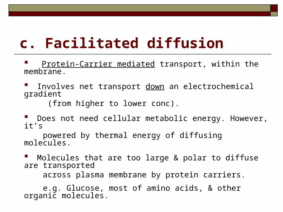
c. Facilitated diffusion Protein-Carrier mediated transport, within the membrane.
Involves net transport down an electrochemical gradient (from higher to lower conc).
Does not need cellular metabolic energy. However, it’s powered by thermal energy of diffusing molecules.
Molecules that are too large & polar to diffuse are transported across plasma membrane by protein carriers.
e.g. Glucose, most of amino acids, & other organic molecules.
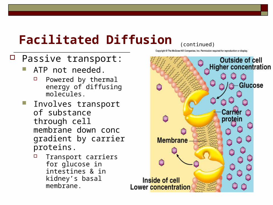
Facilitated Diffusion (continued)
Passive transport: ATP not needed.
Powered by thermal energy of diffusing molecules.
Involves transport of substance through cell membrane down conc gradient by carrier proteins. Transport carriers for
glucose in intestines & in kidney’s basal membrane.
2. Active transport
2. Active transport:
Protein-Carrier mediated transport.
Involves net transport (uphill), i.e. against electrochemical gradient (from lower to higher conc).
Requires metabolic energy (ATP).
Types of active transport
I. Primary active transport
II. Secondary active transport
I. Primary Active Transport
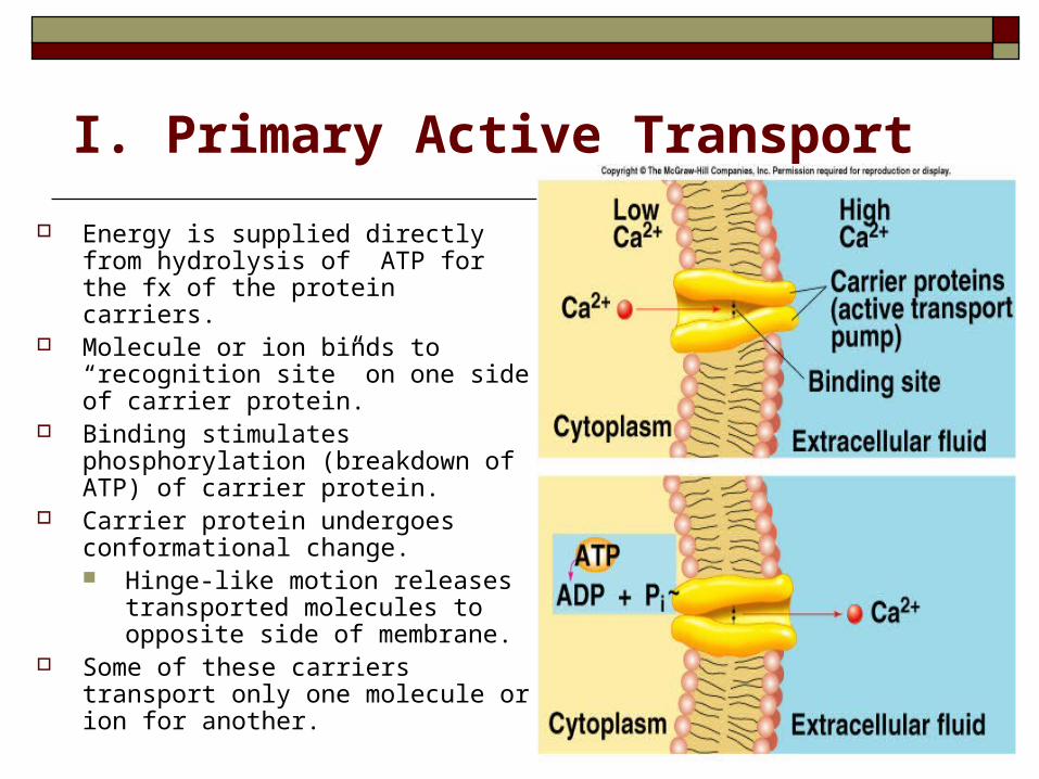
Energy is supplied directly from hydrolysis of ATP for the fx of the protein carriers.
Molecule or ion binds to “recognition site” on one side of carrier protein.
Binding stimulates phosphorylation (breakdown of ATP) of carrier protein.
Carrier protein undergoes conformational change. Hinge-like motion releases
transported molecules to opposite side of membrane.
Some of these carriers transport only one molecule or ion for another.
Primary active transport (continued)
Examples:
a. Sodium-Potassium pump (Na+/K+ pump).
b. Primary active transport of calcium (Ca2+ ATPase).
c. Primary active transport of hydrogen ions
(H+/K+ ATPase)
Sodium-Potassium pump (Na+/K+ pump): Present in most cell membranes.
e.g. in basolateral membrane of the kidneys, & in
intestines.
Energy dependent transport, because both ions are moved against their conc gradient.
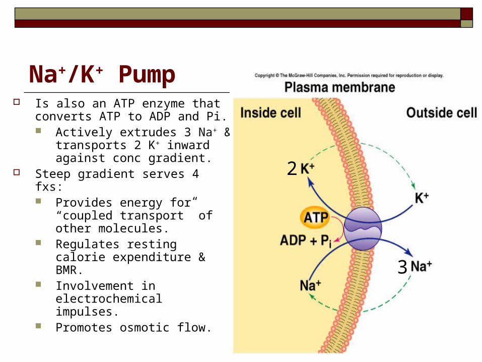
Na+/K+ Pump Is also an ATP enzyme that
converts ATP to ADP and Pi. Actively extrudes 3 Na+ &
transports 2 K+ inward against conc gradient.
Steep gradient serves 4 fxs: Provides energy for “coupled
transport” of other molecules. Regulates resting calorie
expenditure & BMR. Involvement in
electrochemical impulses. Promotes osmotic flow.
3
2
II. Secondary active transport: (Coupled Transport)
Transport of one or more solutes against an electrochemical gradient, coupled to the transport of another solute down an electrochemical gradient.
Energy needed for “uphill” movement obtained from “downhill” transport of Na+.
Hydrolysis of ATP by Na+/K+ pump required indirectly to maintain [Na+] gradient.
If the other molecule or ion is moved in the same direction as Na+ (into the cell), the coupled transport is called either: ‘cotransport’ or ‘symport’.
If the other molecule or ion is moved in the opposite direction as Na+ (out of the cell), the process is called either: ‘countertransport’ or ‘antiport’.
Secondary Active Transport (continued)
a. Co-transport (Symport) All solutes move in the same direction “to the
inside of the cell”
e.g.
- Na+– glucose Co transport
- Na+– amino acid Co transport In the intestinal tract, & kidney’s brush borders.
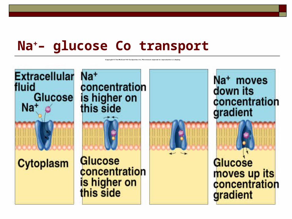
Na+– glucose Co transport
b. Counter transport (Antiport)
Na+ is moving to the interior causing other substance to move out.
e.g.
- Ca2+– Na+ exchange
… (present in many cell membranes)
- Na+– H+ exchange in the kidney
- Cl-– HCO3- exchange across RBCs.