Embed Size (px)
Citation preview
Introduction to PET/CT in Oncology:Practical Aspects
Jeffrey T. Yap, PhD
Department of ImagingDana-Farber Cancer Institute
Outline
• Image registration and fusion• Visualization of PET and CT images• Acquisition considerations• Potential artifacts• Considerations for Quantitative Imaging• Standardized Uptake Value (SUV)

Whole-body PET/CT protocol
Clinical Review
CT TopogramSpiral CT WB PET Fused PET/CT
Patient prep18FDG injectionUptake period
ROI Definitio
n
SUV Analysis
CT PET
Definitions
• Image registration: Process of matching the spatial coordinates between two or more images
• Image fusion: Process of combining multiple images of a scene to obtain a single composite image
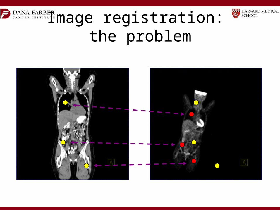
Image registration: the problem

Image registration: zoom

Image registration: rotate
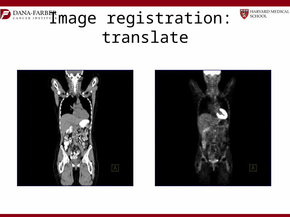
Image registration: translate

Image registration: complete

2D display methods
• 2 dimensional reconstructed planes: transaxial, sagittal, coronal, oblique
• Gray scale versus color tables• Windowing (contrast enhancement)– PET: linear scale specified by min & max– CT: linear scale specified by center and width
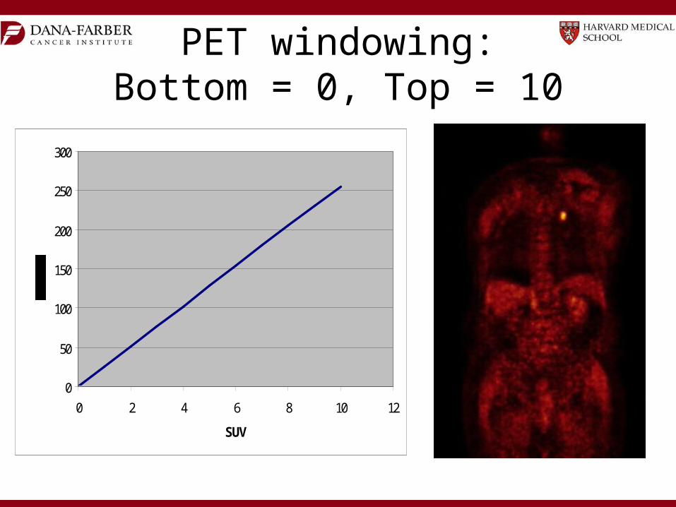
PET windowing:Bottom = 0, Top = 10
0
50
100
150
200
250
300
0 2 4 6 8 10 12
SUV

PET windowing:Bottom = 2, Top = 10
0
50
100
150
200
250
300
0 2 4 6 8 10 12
SUV

PET windowing:Bottom = 2, Top = 8
0
50
100
150
200
250
300
0 2 4 6 8 10 12
SUV

CT windowing notation:Center = 5, Width = 10
0
50
100
150
200
250
300
0 2 4 6 8 10 12
SUV
C=5
W=10

CT Windowing
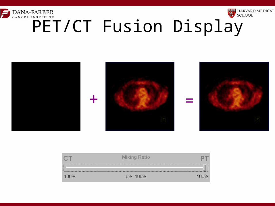
=
PET/CT Fusion Display
+

3D display methods• Maximum intensity projection: Displays the
maximum intensity along each projection• Shaded surface: Displays lighted surface of
segmented voxels • Volume rendering: Displays multiple rendered
objects with various transparencies and color tables

Maximum Intensity Projection (MIP)
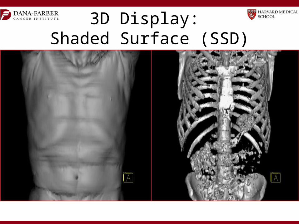
3D Display: Shaded Surface (SSD)
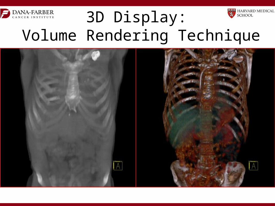
3D Display: Volume Rendering Technique

Acquisition Considerations

Options with CT Acquisition
• Breathing protocols• Arms up for thorax/abdomen• Arms down for head/neck• Dose options: attenuation correction only,
anatomical localization, full diagnostic• I.V. and/or oral contrast• Gated (cardiac or respiratory)• Dynamic (e.g. perfusion imaging)

Options with PET acquisition• Static• Whole body (multiple bed positions)– Step-and-shoot– Continuous bed movement
• List-mode• Dynamic• Gated (cardiac and/or respiratory)

Potential Artifacts

CT-Based attenuation artifacts: respiratory motion

Arms downArms down Arms upArms upArms downArms down Arms upArms up
PET image qualityPET image quality

Intravenous contrast mediaIntravenous contrast media
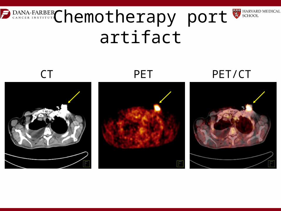
Chemotherapy port artifact
CT PET PET/CT
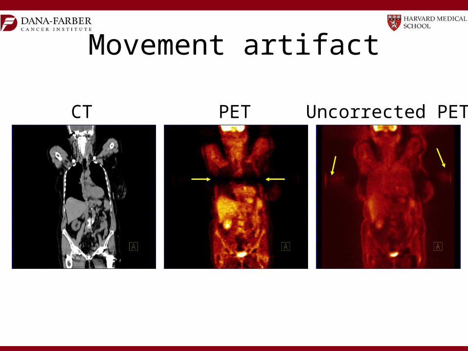
Movement artifact
CT PET Uncorrected PET

Considerations for Quantitative Imaging
• Calibration of all instrumentation is required at commissioning and regular intervals (PET/CT scanners, dose calibrators, scales, clocks)
• Consistent patient preparation is critical (e.g. fasting)
• Technical acquisition should be standardized and critical parameters should be recorded

Hardware/Software Requirements for Accurate SUV Quantification
• Dose calibrator accuracy – traceable standard• Scanner normalization (detector efficiency)• Scanner calibration• PET corrections: attenuation, scatter, randoms,
decay (images and doses)• Partial volume correction for small objects• Appropriate reconstruction algorithm• Daily/weekly/monthly scanner QC

Requirements for Reproducible SUV Quantification
• PET technique: 18FDG dose, 18FDG uptake period, emission scan length, scanning range, scanning direction (e.g. head to toe)
• Patient preparation: fasting, resting, medication• Reconstruction parameters: slice thickness,
filters• Region-of-interest definition methods (mostly
manual or semi-automated)• Consistency is the most important factor!

Mandatory measurements• Acquisition parameters• Patient height, weight• Injected activity, residual, and time• Circulating glucose• Infiltrated doses• Patient compliance (e.g. fasting state, movement)• Protocol deviations– Injection time/scan delays – Injected activity

Standardized Uptake Value
• Under certain circumstances, 18FDG SUV correlates with metabolic rate of glucose and/or the number of viable tumor cells
• Simplified semi-quantitative measure that can be routinely performed in clinical PET studies
• Adjusts for differences in patient size and injected activity
SUV (time) = Radioactive Concentration x Weight Injected Activity
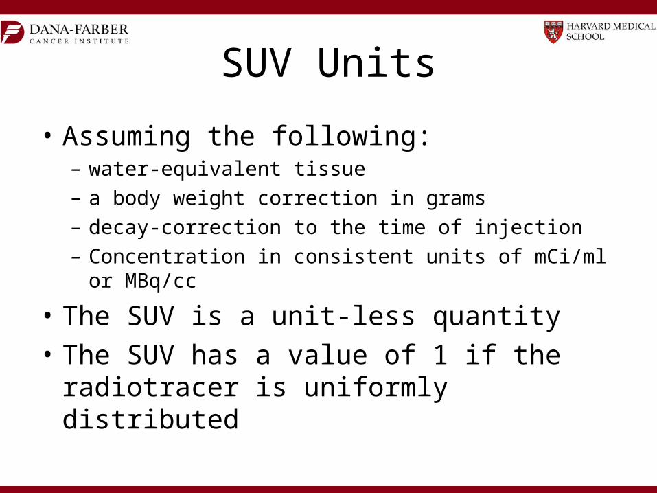
SUV Units
• Assuming the following:– water-equivalent tissue– a body weight correction in grams– decay-correction to the time of injection– Concentration in consistent units of mCi/ml or MBq/cc
• The SUV is a unit-less quantity• The SUV has a value of 1 if the radiotracer is
uniformly distributed

SUV Example
• Consider 0.8 ml volume containing 12 mCi of 18FDG is “injected” into a 1.5 liter volume of water
• SUV = Radioactive Conc. x Weight Injected Activity
• SUV = (12 mCi / 1500 ml) * 1500 g (1 ml/g) 12 mCi
SUV = 1
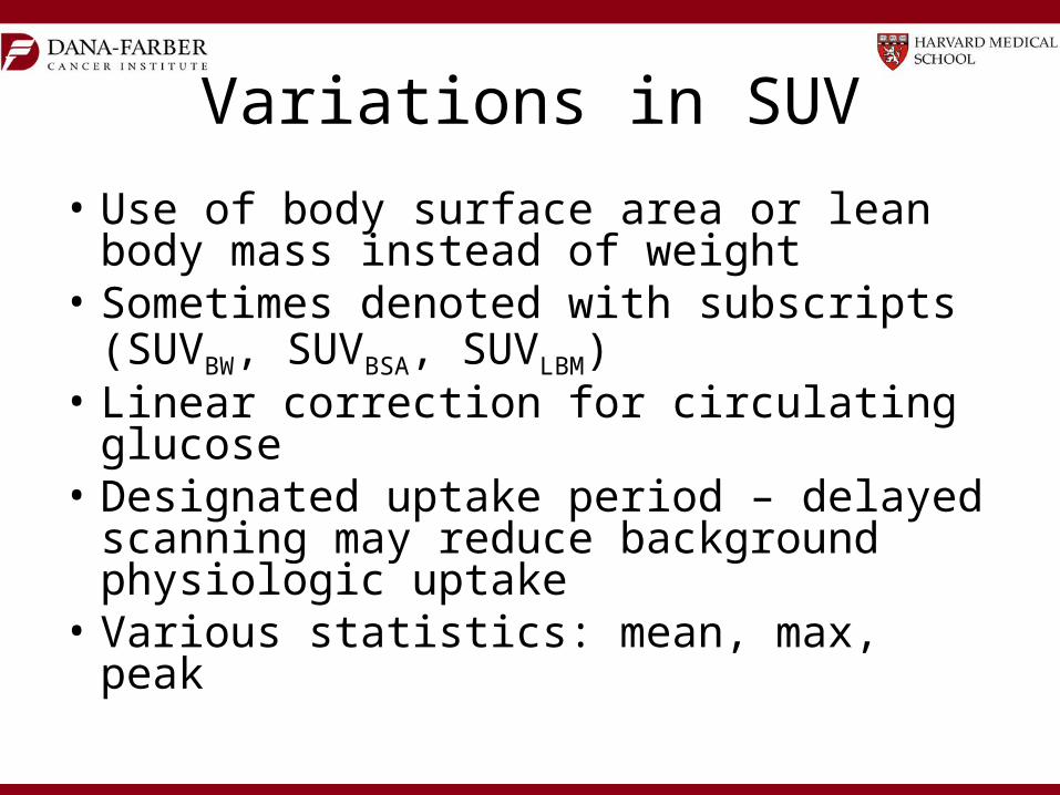
Variations in SUV
• Use of body surface area or lean body mass instead of weight
• Sometimes denoted with subscripts (SUVBW, SUVBSA, SUVLBM)
• Linear correction for circulating glucose• Designated uptake period – delayed scanning
may reduce background physiologic uptake• Various statistics: mean, max, peak
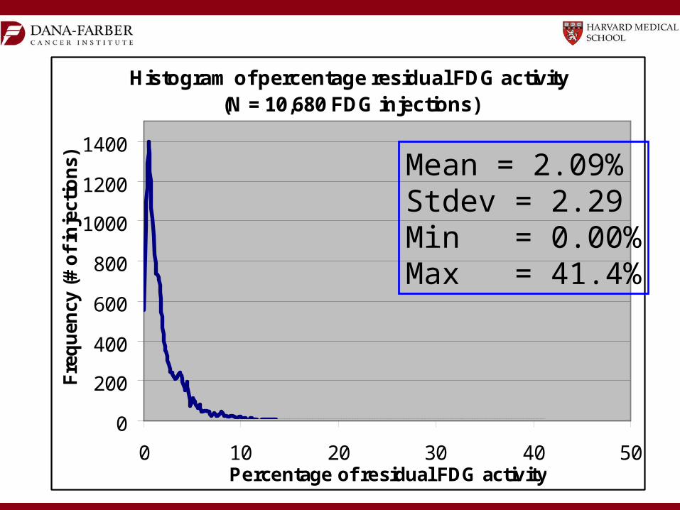
Histogram of percentage residual FDG activity(N = 10,680 FDG injections)
0
200
400
600
800
1000
1200
1400
0 10 20 30 40 50Percentage of residual FDG activity
Fre
qu
ency
(#
of
inje
ctio
ns) Mean = 2.09%
Stdev = 2.29Min = 0.00%Max = 41.4%
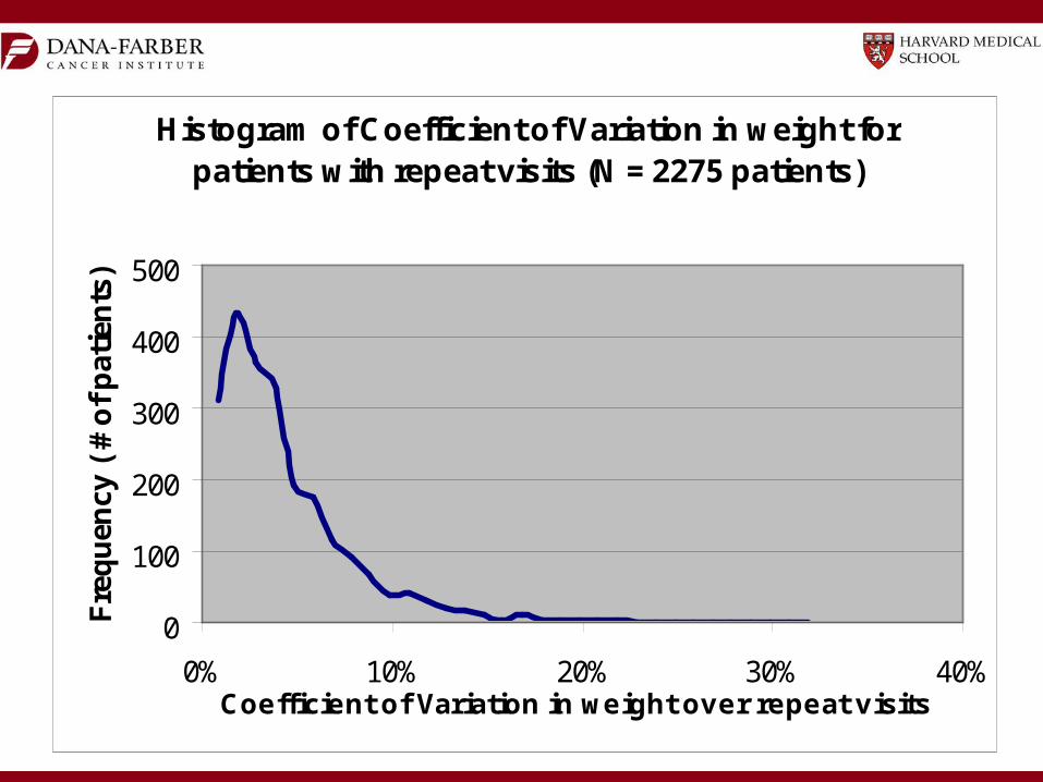
Histogram of Coefficient of Variation in weight for patients with repeat visits (N = 2275 patients)
0
100
200
300
400
500
0% 10% 20% 30% 40%Coefficient of Variation in weight over repeat visits
Fre
qu
ency
( #
of
pat
ien
ts)
The Good News
• Most differences in scanner hardware and reconstruction software cancel out in longitudinal studies of the same patient
• Although there is no universal cutoff, the SUV can help differentiate malignancy from normal tissue
• Changes in SUV after therapy have been shown to correlate with clinical outcome (e.g. survival)