Embed Size (px)
Citation preview

Introduction to Neuroimaging
Aaron S. Field, MD, PhD
Neuroradiology
University of Wisconsin–Madison
SPINE
Updated 6/13/06

Anatomy


Radiographic Anatomy
ML Richardson, Univ. Of Washington

ML Richardson, Univ. Of Washington
Cervical Spine – AP View

ML Richardson, Univ. Of Washington
Cervical Spine – Lateral View

ML Richardson, Univ. Of Washington
Cervical Spine – Oblique View

ML Richardson, Univ. Of Washington
Cervical Spine – Open-Mouth (Dens) View
ML Richardson, Univ. Of Washington
Lumbar Spine – AP View

ML Richardson, Univ. Of Washington
Lumbar Spine – Lateral View

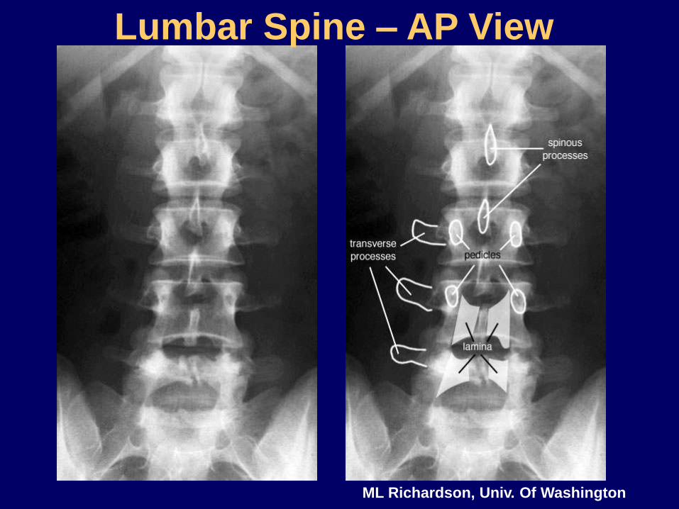
MRI Anatomy
Source: CW Kerber and JR Hesselink, Spine Anatomy, UCSD Neuroradiology


Source: CW Kerber and JR Hesselink, Spine Anatomy, UCSD Neuroradiology


Spine Pathology
• Trauma
• Degenerative disease
• Tumors and other masses
• Inflammation and infection
• Vascular disorders
• Congenital anomalies

Trauma

Evaluating Trauma
• Fracture – plain film / CT
• Dislocation – plain film / CT
• Ligamentous injury – MRI
• Cord injury – MRI
• Nerve root avulsion – MRI

Plain film findings may be
very subtle or absent!
Anterolisthesis of
C6 on C7
(Why??)

CT
Fractures of C6 left
pedicle and lamina

CT – 2D Reconstructions
Acquire images axially…
…reconstruct sagittal / coronal

26M MVA

Vertebral body burst fx
with retropulsion into
spinal canal
2D Reformats


Vertebral Artery Dissection/Occlusion
Secondary to C6 Fracture

Hyperflexion fx with
ligamentous disruption and
cord contusion

Nerve root avulsion
Axial Coronal Sagittal

Degenerative Disease

Degenerative Disc (and Facet Joint) Disease
Foraminal
stenosis
Thickening/Buckling
of Ligamentum
Flavum

Degenerative Disc (and Facet Joint) Disease

Degenerative Disc (and Facet Joint) Disease


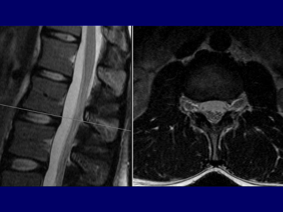
Lumbar Spinal Stenosis

Lumbar Spinal Stenosis

Lumbar Spinal Stenosis

Lumbar Spinal Stenosis

Lumbar Spinal Stenosis
Lumbar Spinal Stenosis
Disc bulge, facet hypertrophy and flaval ligament thickening
frequently combine to cause central spinal stenosis
Note the trefoil shape of stenotic spinal canal


Lumbar Spinal Stenosis
Disc bulge, facet hypertrophy and flaval ligament thickening
frequently combine to cause central spinal stenosis
Note the trefoil shape of stenotic spinal canal
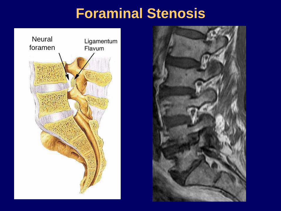
Foraminal Stenosis
Neural
foramen

Cervical Spinal Stenosis

MRI - Degenerative Disc Disease
• 20-40 36% have degenerated disc
• 50 85-95% have degenerated disc
• 60-80 98% have degenerated disc
• <60 20% have asymptomatic disc
herniation
Age:
Conclusion: Abnormal findings on MRI frequently DO NOT
relate to symptoms (and vice versa) !!

MRI – Herniated Disc Levels
• 85-95% at L4-L5, L5-S1
• 5-8% at L3-L4
• 2% at L2-L3
• 1% at L1-L2, T12-L1
• Cervical: most common C4-C7
• Thoracic: 15% in asymptomatic pts. at
multiple levels, not often symptomatic

Annular

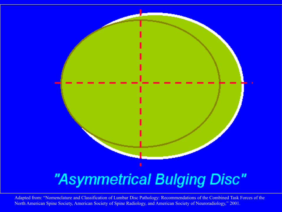
Adapted from: “Nomenclature and Classification of Lumbar Disc Pathology: Recommendations of the Combined Task Forces of the
North American Spine Society, American Society of Spine Radiology, and American Society of Neuroradiology,” 2001.
Adapted from: “Nomenclature and Classification of Lumbar Disc Pathology: Recommendations of the Combined Task Forces of the
North American Spine Society, American Society of Spine Radiology, and American Society of Neuroradiology,” 2001.

Adapted from: “Nomenclature and Classification of Lumbar Disc Pathology: Recommendations of the Combined Task Forces of the
North American Spine Society, American Society of Spine Radiology, and American Society of Neuroradiology,” 2001.

Adapted from: “Nomenclature and Classification of Lumbar Disc Pathology: Recommendations of the Combined Task Forces of the
North American Spine Society, American Society of Spine Radiology, and American Society of Neuroradiology,” 2001.

Adapted from: “Nomenclature and Classification of Lumbar Disc Pathology: Recommendations of the Combined Task Forces of the
North American Spine Society, American Society of Spine Radiology, and American Society of Neuroradiology,” 2001.

Protrusion Extrusion Extrusion
Adapted from: “Nomenclature and Classification of Lumbar Disc Pathology: Recommendations of the Combined Task Forces of the
North American Spine Society, American Society of Spine Radiology, and American Society of Neuroradiology,” 2001.

Protrusion Protrusion w/
migration
Protrusion w/
migration +
sequestration
Adapted from: “Nomenclature and Classification of Lumbar Disc Pathology: Recommendations of the Combined Task Forces of the
North American Spine Society, American Society of Spine Radiology, and American Society of Neuroradiology,” 2001.

Abnormal Disc
Bulge
Symmetric Asymmetric
Herniation
Broad-based Focal
Extrusion Protrusion
Sequestered Migrated Neither
> 180º< 180º
< 90º90º–180º
No waistWaist*
Adapted from: “Nomenclature and Classification of Lumbar Disc Pathology: Recommendations of the Combined Task Forces of the
North American Spine Society, American Society of Spine Radiology, and American Society of Neuroradiology,” 2001.
*(In any plane)

Central Disc Protrusion

L5-S1 Disc Extrusion Into Lateral Recess
with Impingement of R S1 Nerve Root
L-S1DiscR-S1
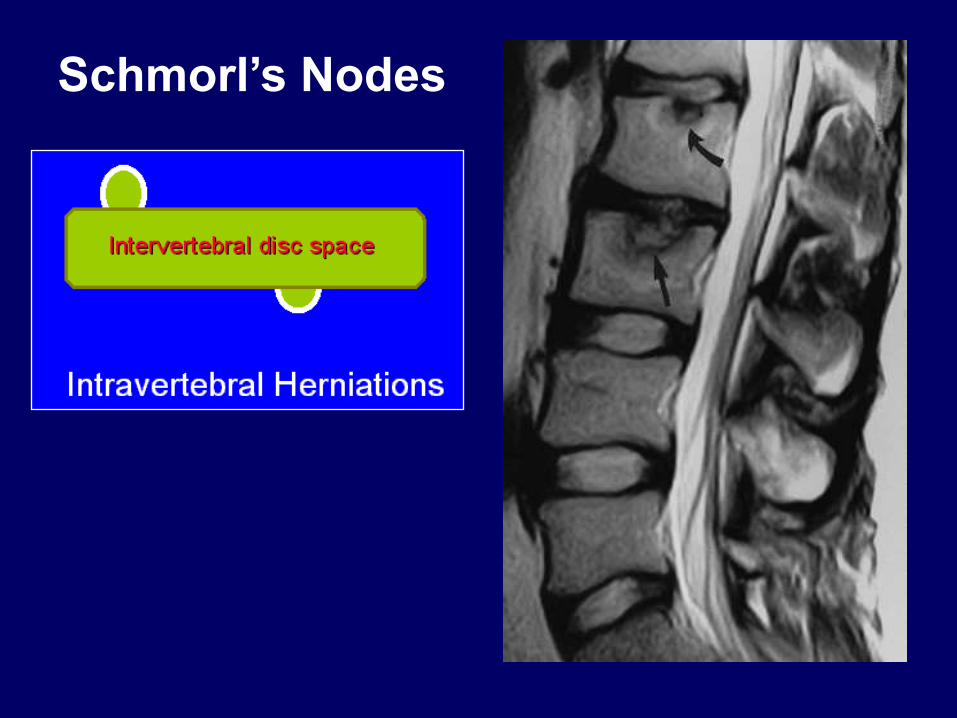
Schmorl’s Nodes

Cervical Radiculopathy

Lumbosacral Radiculopathy (Sciatica)
Important:
A herniated disc at (e.g.) L4-5 may impinge either the L4 or L5 nerve roots!
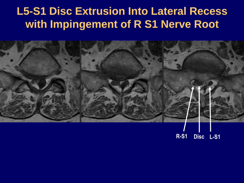
L5-S1 Disc Extrusion Into Lateral Recess
with Impingement of R S1 Nerve Root
L-S1DiscR-S1
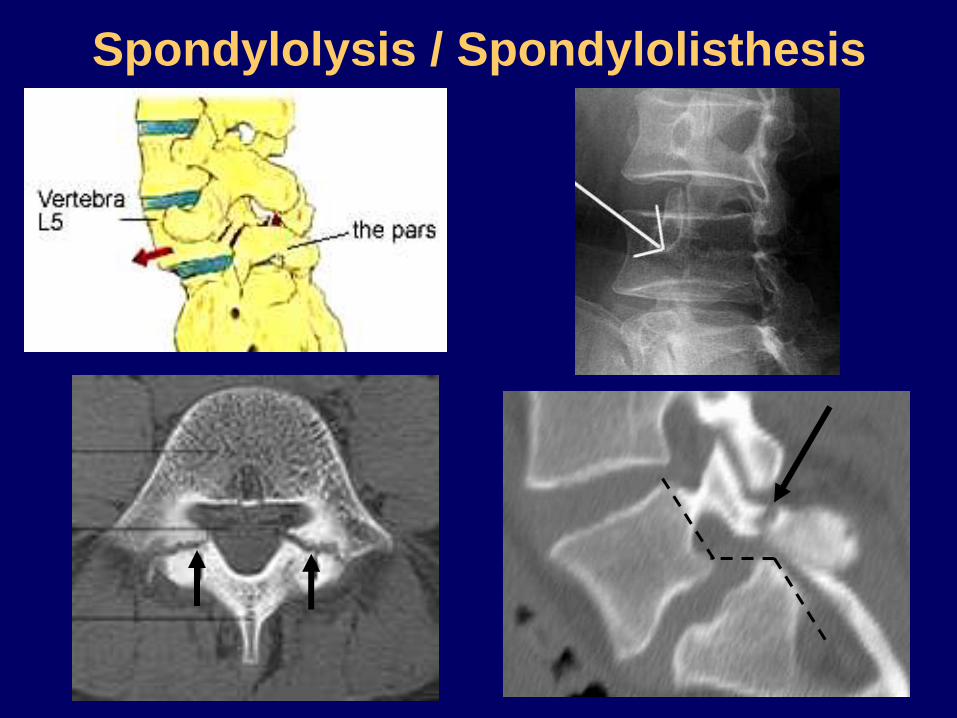
Spondylolysis / Spondylolisthesis
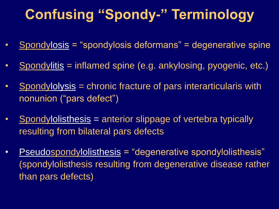
Confusing “Spondy-” Terminology
• Spondylosis = “spondylosis deformans” = degenerative spine
• Spondylitis = inflamed spine (e.g. ankylosing, pyogenic, etc.)
• Spondylolysis = chronic fracture of pars interarticularis with
nonunion (“pars defect”)
• Spondylolisthesis = anterior slippage of vertebra typically
resulting from bilateral pars defects
• Pseudospondylolisthesis = “degenerative spondylolisthesis”
(spondylolisthesis resulting from degenerative disease rather
than pars defects)
Tumors and Other Masses

• Extradural = outside the thecal sac (including vertebral bone lesions)
• Intradural / extramedullary = within thecal sac but outside cord
• Intramedullary = within cord
Classification of Spinal Lesions

• Herniated disc
• Vertebral hemangioma
• Vertebral metastasis
• Epidural abscess or hematoma
• Synovial cyst
• Nerve sheath tumor (also intradural/extramedullary)
Neurofibroma
Schwannoma
Common Extradural Lesions

• Nerve sheath tumor (also extradural)
Neurofibroma
Schwannoma
• Meningioma
• Drop Metastasis
Common Intradural
Extramedullary Lesions

• Astrocytoma
• Ependymoma
• Hemangioblastoma
• Cavernoma
• Syrinx
• Demyelinating lesion (MS)
• Myelitis
Common Intramedullary Lesions

Classification of Spinal Lesions
Extradural IntramedullaryIntradural
ExtramedullaryDuraCord

Extradural: Vertebral Body Tumor

Extradural: Vertebral Metastases
T2 (Fat Suppressed) T1 T1+C (fat suppressed)

Extradural: Vertebral Metastases
T2 (Fat Suppressed) T1 T1+C (fat suppressed)
?

Vertebral Metastases vs. Hemangiomas
Hemangiomas (Benign, usually asymptomatic, commonly incidental):
Bright on T1 and T2 (but dark with fat suppression)
Enhancement variable
Metastases:
Dark on T1, Bright on T2 (even with fat suppression)
Enhancement

Vertebral Hemangiomas

Diffusely T1-hypointense marrow
signal may represent widespread
vertebral metastases as in this
patient with prostate Ca
This can also be seen in the setting
of anemia, myeloproliferative
disease, and various other chronic
disease states
Extradural: Vertebral Metastases

Extradural: Epidural Abscess
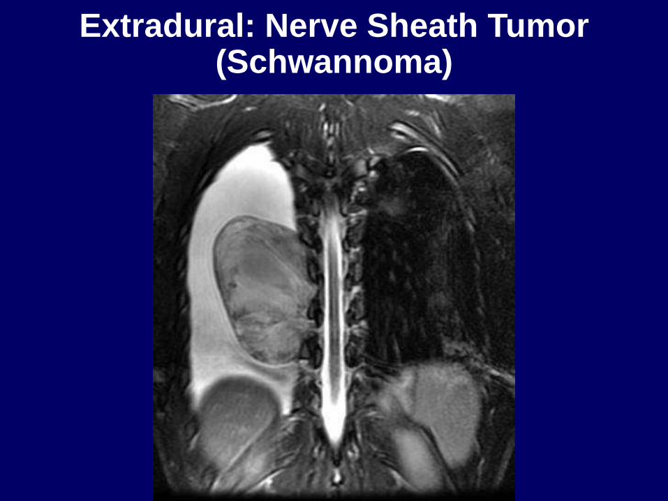
Extradural: Nerve Sheath Tumor(Schwannoma)

Intradural Extramedullary: Meningioma

Intradural Extramedullary: Meningioma
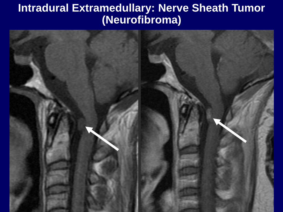
Intradural Extramedullary: Nerve Sheath Tumor(Neurofibroma)

Intradural Extramedullary: “Drop Mets”
T2 T1 T1+C

Intradural Extramedullary: “Drop Mets”

Intradural Extramedullary: Arachnoid Cyst
T2 T1

Intramedullary: Astrocytoma

Intramedullary: Astrocytoma

Intramedullary: Cavernoma

Intramedullary: Ependymoma

Seen with:
• congenital lesions
• Chiari I & II
• tethered cord
• acquired lesions
• trauma
• tumors
• arachnoiditis
• idiopathic
Intramedullary: Syringohydromyelia

Seen with:
• congenital lesions
• Chiari I & II
• tethered cord
• acquired lesions
• trauma
• tumors
• arachnoiditis
• idiopathic
Intramedullary: Syringohydromyelia

Confusing “Syrinx” Terminology
• Hydromyelia: Fluid accumulation/dilatation within central
canal, therefore lined by ependyma
• Syringomyelia: Cavitary lesion within cord parenchyma, of
any cause (there are many). Located adjacent to central
canal, therefore not lined by ependyma
• Syringohydromyelia: Term used for either of the above, since
the two may overlap and cannot be discriminated on imaging
• Hydrosyringomyelia: Same as syringohydromyelia
• Syrinx: Common term for the cavity in all of the above

Infection and Inflammation

Infectious Spondylitis / Diskitis
Common chain of events (bacterial spondylitis):
1. Hematogenous seeding of subchondral VB
2. Spread to disc and adjacent VB
3. Spread into epidural space epidural abscess
4. Spread into paraspinal tissues psoas abscess
5. May lead to cord abscess

Infectious Spondylitis / Diskitis
T2 T1 T1+C T1+C
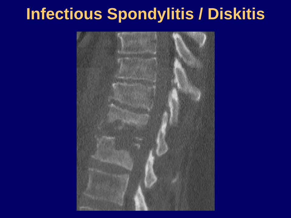
Infectious Spondylitis / Diskitis

Pyogenic Spondylitis / Diskitis
with Epidural Abscess


T1
T2
T1 + C
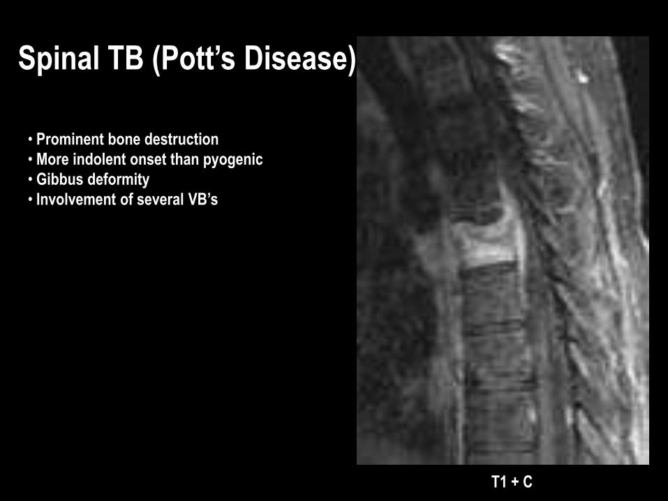
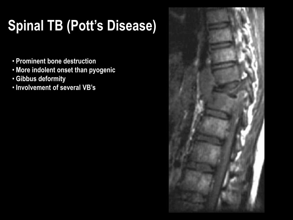
Spinal TB (Pott’s Disease)
• Prominent bone destruction
• More indolent onset than pyogenic
• Gibbus deformity
• Involvement of several VB’s
Spinal TB (Pott’s Disease)
• Prominent bone destruction
• More indolent onset than pyogenic
• Gibbus deformity
• Involvement of several VB’s

Transverse Myelitis
Inflamed cord of uncertain cause
Viral infections
Immune reactions
Idiopathic
Myelopathy progressing over hours to weeks
DDX: MS, glioma, infarction

Multiple Sclerosis
Inflammatory demyelination eventually
leading to gliosis and axonal loss
T2-hyperintense lesion(s) in cord
parenchyma
Typically no cord expansion (vs. tumor);
chronic lesion may show atrophy

Multiple Sclerosis
Inflammatory demyelination eventually
leading to gliosis and axonal loss
T2-hyperintense lesion(s) in cord
parenchyma
Typically no cord expansion (vs. tumor);
chronic lesion may show atrophy

Cord Edema
As in the brain, may be secondary to
ischemia (e.g. embolus to spinal artery)
or
venous hypertension (e.g. AV fistula)

Spine Imaging Guidelines
1. Uncomplicated LBP usually self-limited, requires no imaging
2. Consider imaging if:
• Trauma
• Cancer
• Immunocompromise / suspected infection
• Elderly / osteoporosis
• Significant neurologic signs / symptoms
3. Back pain with signs / symptoms of spinal stenosis or radiculopathy, no trauma:
Start with MRI; use CT if:
• Question regarding bones or surgical (fusion) hardware
• Resolve questions / solve problems on MRI (typically use CT myelography)
• MRI contraindicated

4. Begin with plain films for trauma; CT to solve problems or to detail known
fractures; MRI to evaluate soft-tissue injury (ligament disruption, cord contusion)
5. MRI for sx of radiculopathy, cauda equina syn, cord compression, myelopathy
6. Fusion hardware is safe for MRI but may degrade image quality; still worth a try
7. Indications for IV contrast in MRI:
• Tumor, infection, inflammation (myelitis), any cord lesion
• Post-op L-spine (discriminate residual/recurrent disk herniation from scar)
8. Emergent or scheduled? Emergent only if immediate surgical or radiation therapy
decision needed (e.g. cord compression, cauda equina syndrome)
9. Difficult to image entire spine in detail; target study to likely level of pathology
10. CT chest/abdomen/pelvis includes T-L spine (no need to rescan trauma pts*)
* If image data still on scanner (24-48 hours)
Spine Imaging Guidelines (cont.)

Introduction to Neuroimaging
Aaron S. Field, MD, PhD
Neuroradiology
University of Wisconsin–Madison
SPINE


![[PPT]PowerPoint Presentation - MIDAG - Medical Image …midag.cs.unc.edu/.../Tutorial/NeuroAnatomyReview.ppt · Web viewAnatomy for Neuroimaging J. Keith Smith, M.D., Ph.D. Neuroradiology](https://img.pdfslide.us/doc/110x75/5b29e20c7f8b9a9c1a8b5937/pptpowerpoint-presentation-midag-medical-image-midagcsuncedututorial.jpg)