Embed Size (px)
Citation preview
Introduction to NeuropathologyCharles G Eberhart MD PhD

• Cellular components of the CNS• Pathology of Neurons• Pathology of Glia• Microscopic appearance of common CNS disease processes
• Introduction to CNS development
OUTLINE
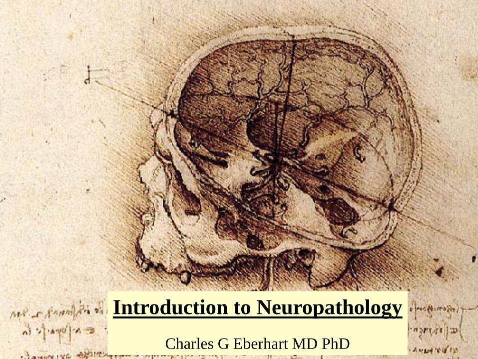
Cellular components of the CNS
•Meninges•Neurons• Glia
AstrocytesOligodendrogliaEpendymal Cells
• Choroid Plexus• Microglia
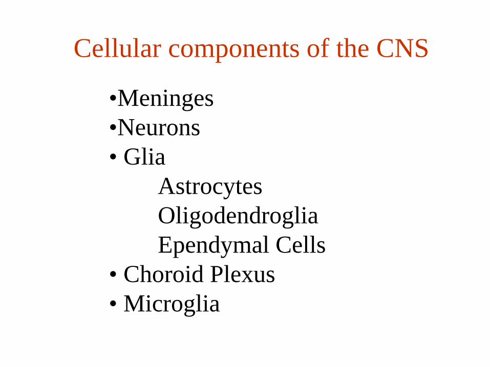
An Axial SectionOf Human Cortex

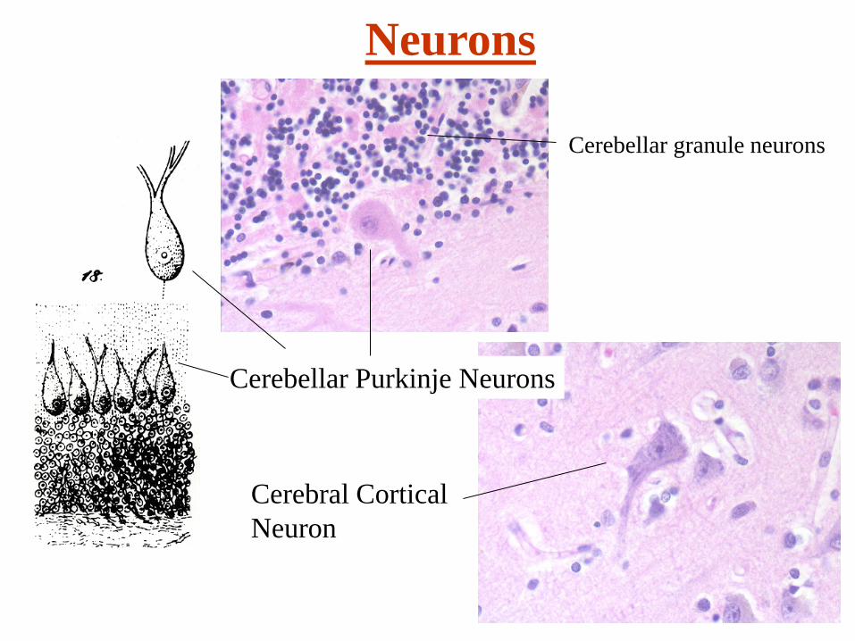
Neurons
Neurons Drawn by Franz Nissl1860-1919
• About 1011 neurons in the CNS• Great variation in size and shape• All have dendrites, soma and axon• Generally have abundant cytoplasm and prominent nucleolus (“fried egg”)
• Nissl substance composed of RER• Can be organized in groups (nuclei, ganglia) or in layers
• Selective vulnerability of some types
DendriticTree
Neurons
Cerebellar granule neurons
Cerebellar Purkinje Neurons
Cerebral Cortical Neuron
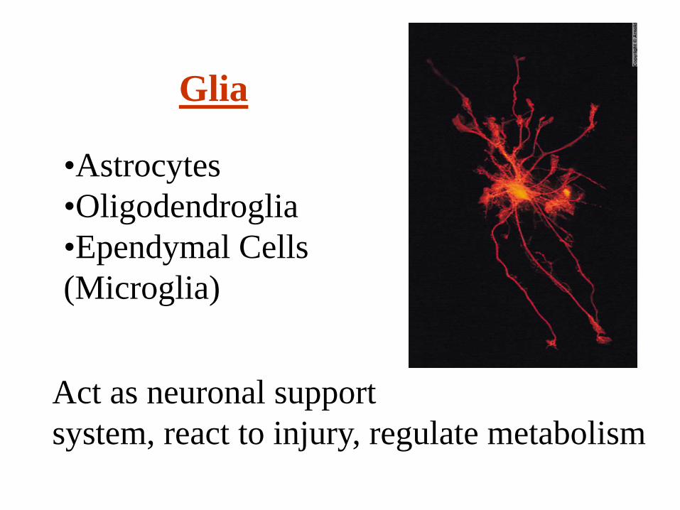
Glia
•Astrocytes•Oligodendroglia•Ependymal Cells(Microglia)
Act as neuronal supportsystem, react to injury, regulate metabolism

Glia - Oligodendrocytes• Common in white matter• Cytoplasmic processes of oligodendrocytes wrap around and insulate axons.
• Small, round, lymphocyte-likenuclei with dense chromatin
• Can have clear “halos” around cells
Oligos

Glia - Astrocytes• Branched cells found in both white and grey matter
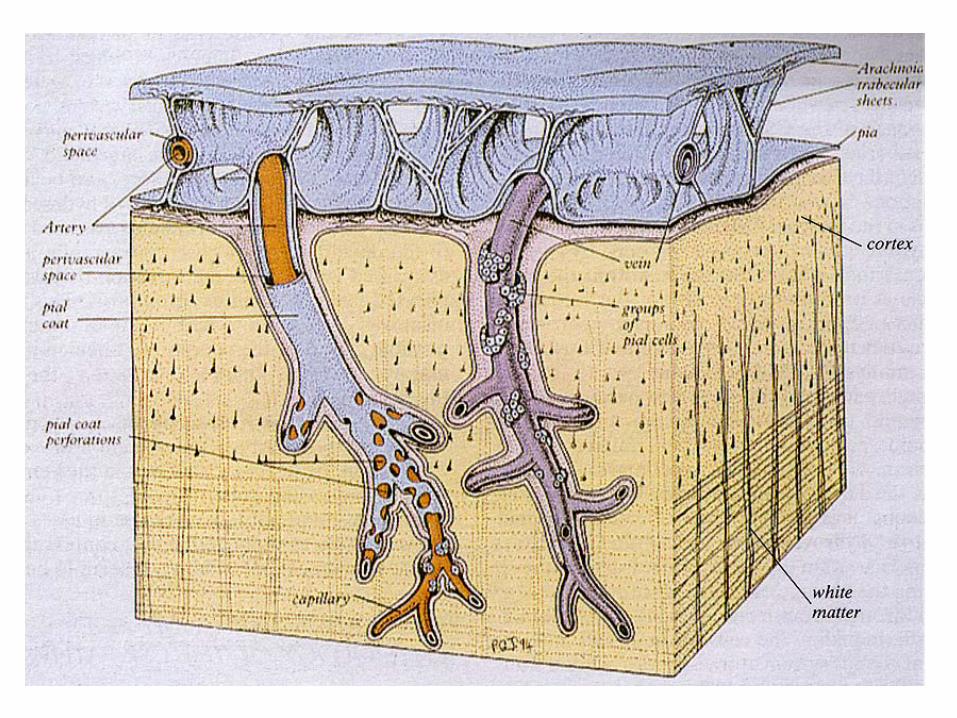
• Astrocytic processes abut neurons,vessels, the pia and ependyma (glia limitans)
• Act as metabolic buffers, detoxifiers,suppliers of nutrients, and physical barriers
• Astrocytic nuclei are roundto oval and slightly largerthan those of oligodendrocytes• Major cell in CNS repairAstrocyte
Neuropil = “nerve felt”
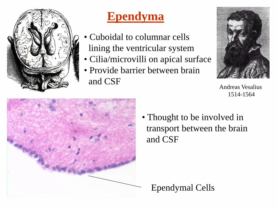
Ependyma
Andreas Vesalius 1514-1564
• Cuboidal to columnar cellslining the ventricular system
• Cilia/microvilli on apical surface• Provide barrier between brainand CSF
• Thought to be involved in transport between the brain and CSF
Ependymal Cells
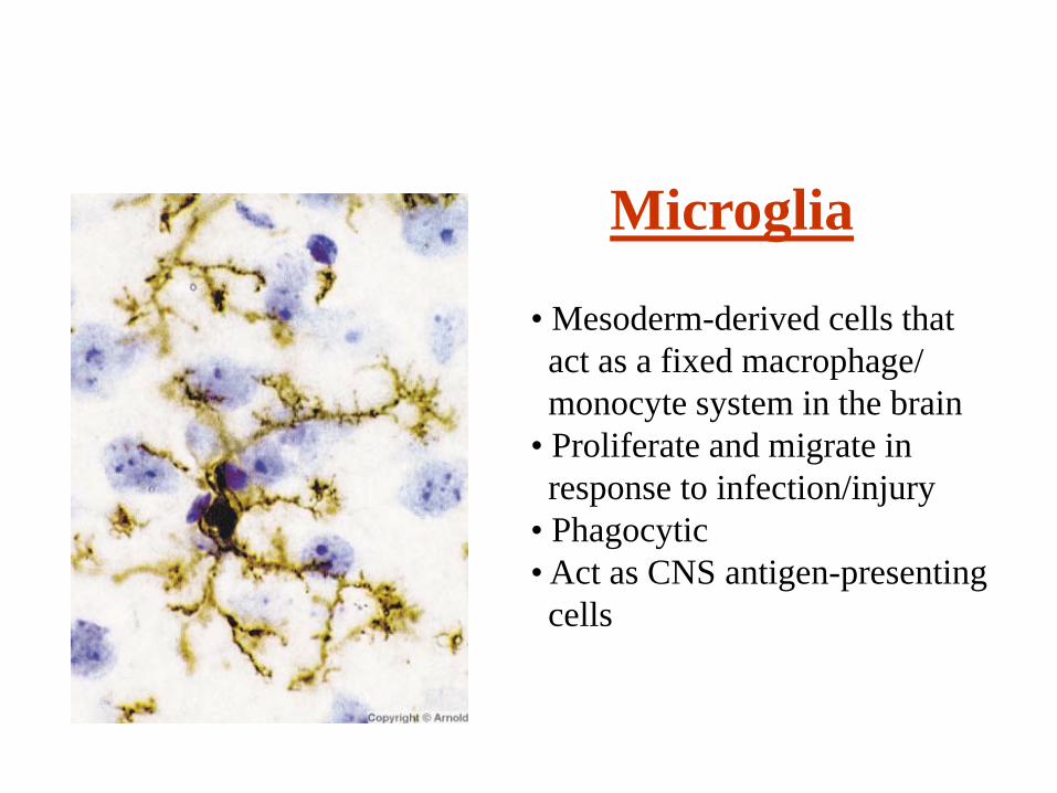
Microglia
• Mesoderm-derived cells thatact as a fixed macrophage/monocyte system in the brain
• Proliferate and migrate in response to infection/injury
• Phagocytic• Act as CNS antigen-presenting cells
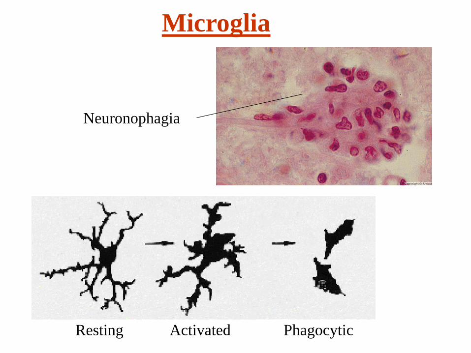
Microglia
Resting Activated Phagocytic
Neuronophagia

Choroid Plexus• Specialized cells derived from the ependyma that secrete CSF• Papillary fronds of cuboidalepithelium covering vascular cores• Tight junctions maintain blood-CSF barrier
• About 20ml of CSF produced per hour
• Normal CSF volume is ~140ml• ~25ml in ventricles, the rest in the subarachnoid space
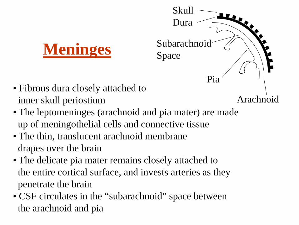
Meninges
SkullDura
SubarachnoidSpace
Pia
Arachnoid• Fibrous dura closely attached toinner skull periostium
• The leptomeninges (arachnoid and pia mater) are madeup of meningothelial cells and connective tissue
• The thin, translucent arachnoid membranedrapes over the brain
• The delicate pia mater remains closely attached to the entire cortical surface, and invests arteries as they penetrate the brain
• CSF circulates in the “subarachnoid” space betweenthe arachnoid and pia
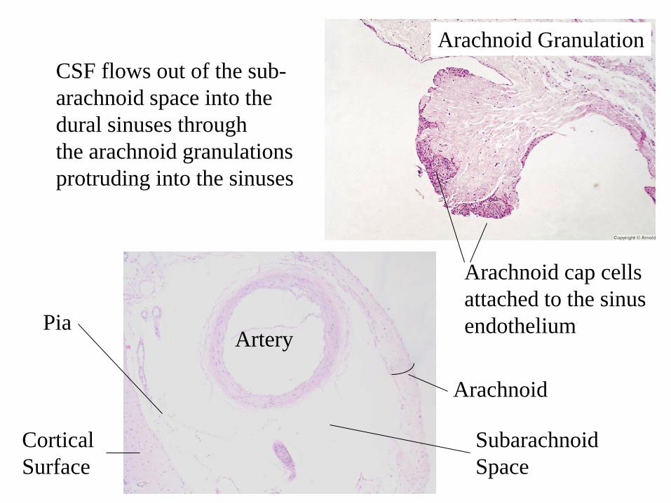
CorticalSurface
Arachnoid
Artery
SubarachnoidSpace
Pia
CSF flows out of the sub-arachnoid space into the dural sinuses throughthe arachnoid granulations protruding into the sinuses
Arachnoid Granulation
Arachnoid cap cellsattached to the sinusendothelium
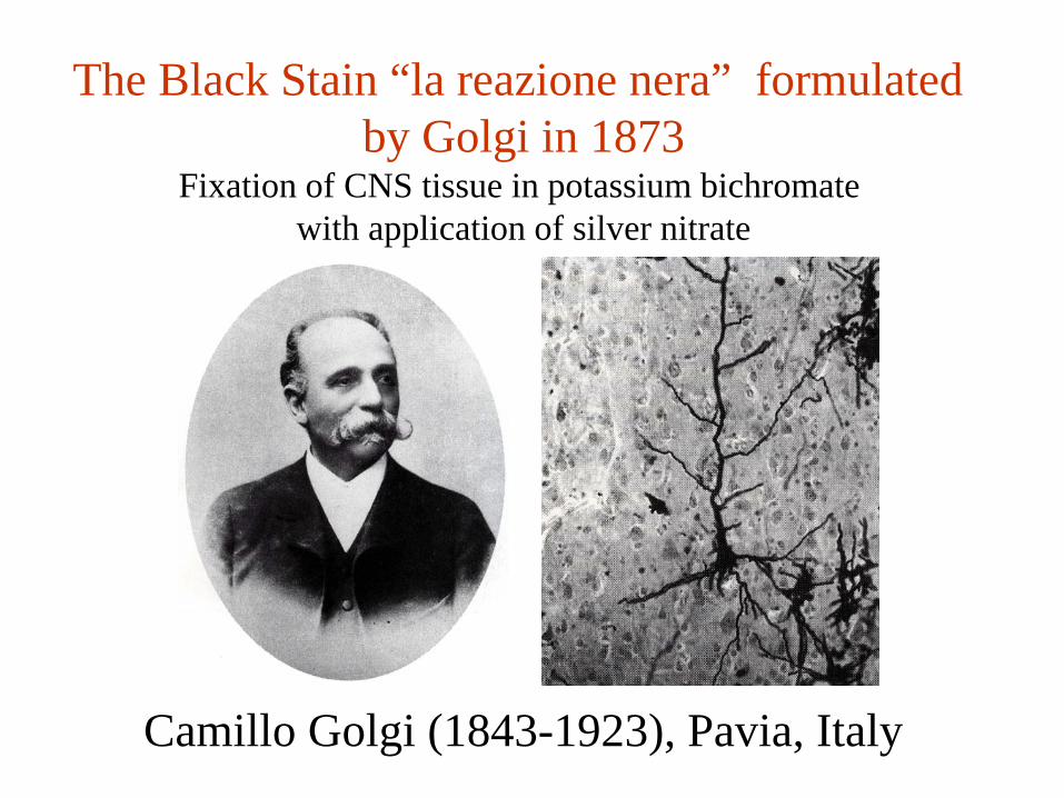
Camillo Golgi (1843-1923), Pavia, Italy
The Black Stain “la reazione nera” formulated by Golgi in 1873
Fixation of CNS tissue in potassium bichromate with application of silver nitrate
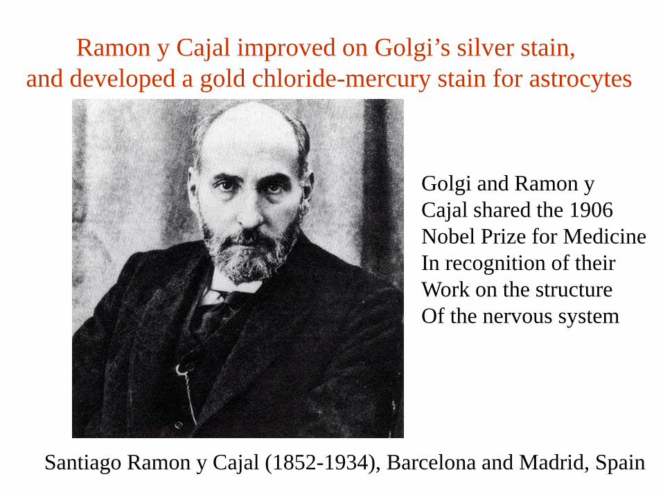
Santiago Ramon y Cajal (1852-1934), Barcelona and Madrid, Spain
Ramon y Cajal improved on Golgi’s silver stain, and developed a gold chloride-mercury stain for astrocytes
Golgi and Ramon yCajal shared the 1906Nobel Prize for Medicine In recognition of theirWork on the structureOf the nervous system
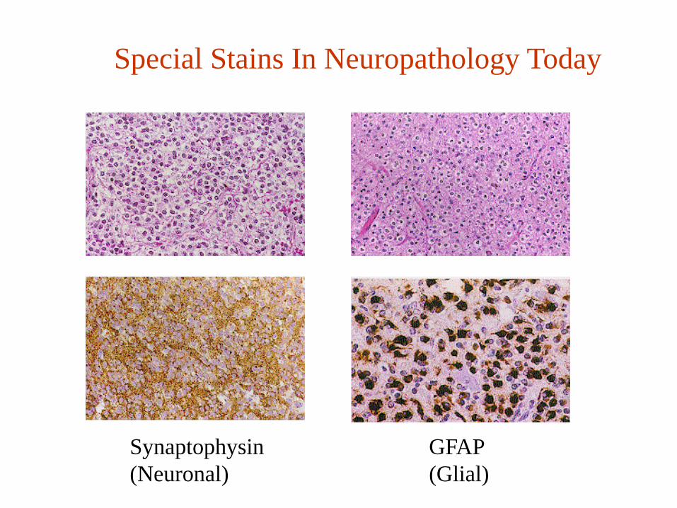
Special Stains In Neuropathology Today
Synaptophysin GFAP(Neuronal) (Glial)
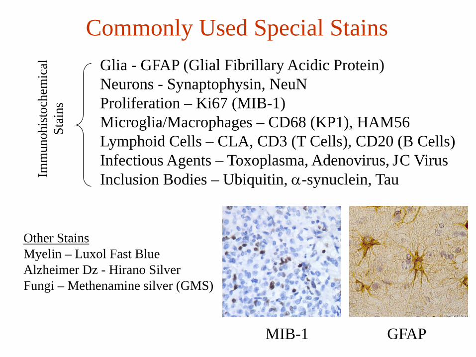
Commonly Used Special StainsGlia - GFAP (Glial Fibrillary Acidic Protein)Neurons - Synaptophysin, NeuNProliferation – Ki67 (MIB-1)Microglia/Macrophages – CD68 (KP1), HAM56Lymphoid Cells – CLA, CD3 (T Cells), CD20 (B Cells)Infectious Agents – Toxoplasma, Adenovirus, JC VirusInclusion Bodies – Ubiquitin, α-synuclein, TauIm
mun
ohis
toch
emic
alSt
ains
Other StainsMyelin – Luxol Fast BlueAlzheimer Dz - Hirano SilverFungi – Methenamine silver (GMS)
MIB-1 GFAP
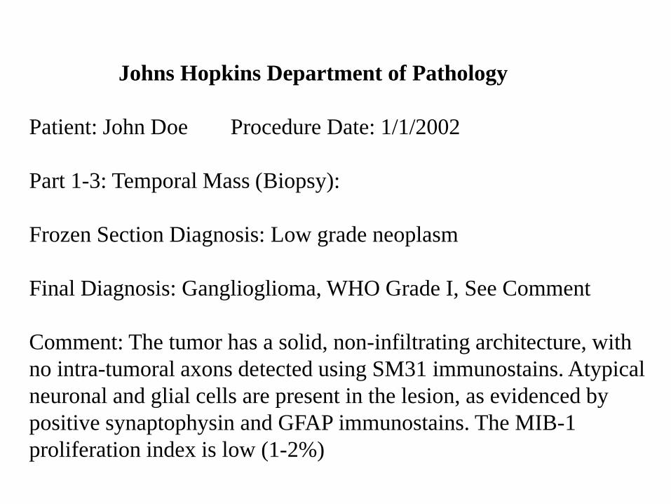
Johns Hopkins Department of Pathology
Patient: John Doe Procedure Date: 1/1/2002
Part 1-3: Temporal Mass (Biopsy):
Frozen Section Diagnosis: Low grade neoplasm
Final Diagnosis: Ganglioglioma, WHO Grade I, See Comment
Comment: The tumor has a solid, non-infiltrating architecture, with no intra-tumoral axons detected using SM31 immunostains. Atypical neuronal and glial cells are present in the lesion, as evidenced by positive synaptophysin and GFAP immunostains. The MIB-1 proliferation index is low (1-2%)
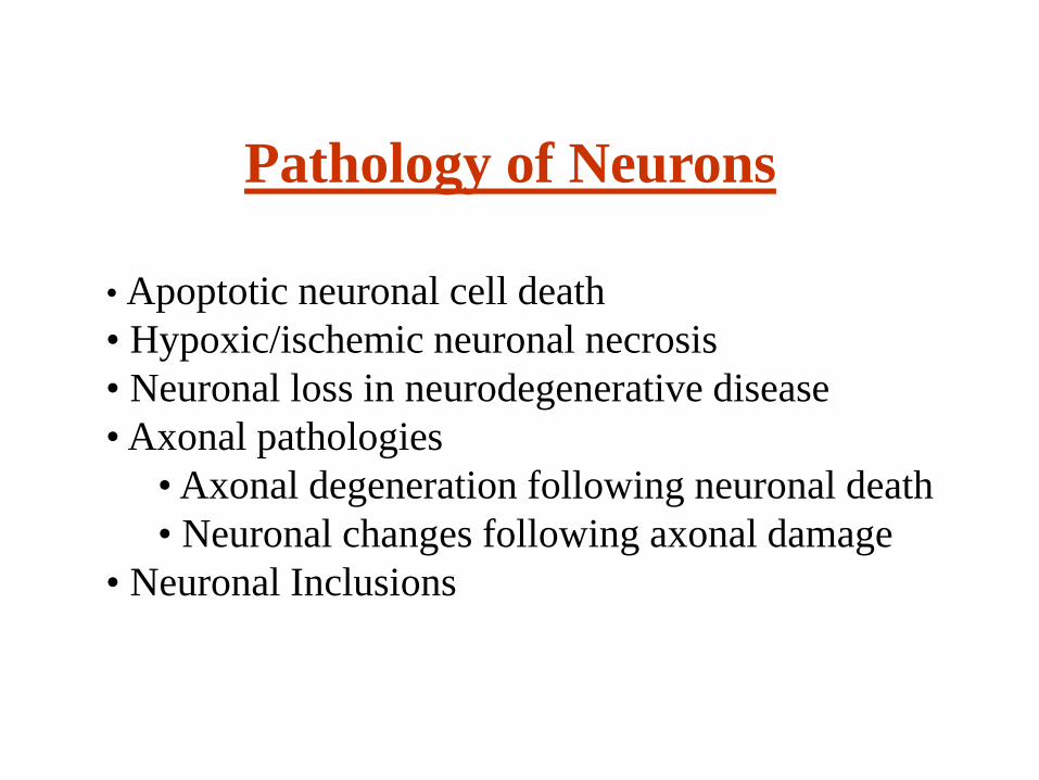
Pathology of Neurons
• Apoptotic neuronal cell death• Hypoxic/ischemic neuronal necrosis• Neuronal loss in neurodegenerative disease• Axonal pathologies
• Axonal degeneration following neuronal death• Neuronal changes following axonal damage
• Neuronal Inclusions
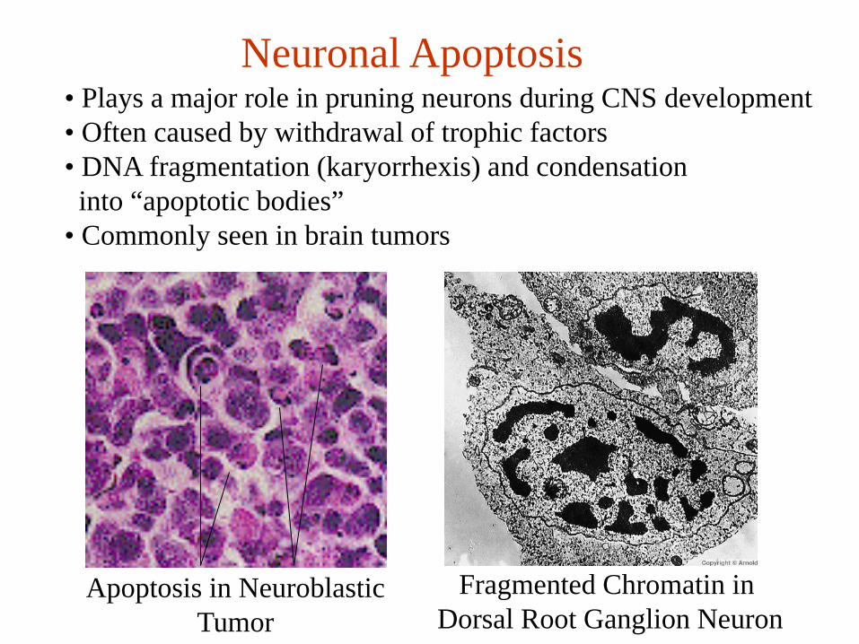
Neuronal Apoptosis
Apoptosis in NeuroblasticTumor
Fragmented Chromatin in Dorsal Root Ganglion Neuron
• Plays a major role in pruning neurons during CNS development• Often caused by withdrawal of trophic factors• DNA fragmentation (karyorrhexis) and condensation into “apoptotic bodies”
• Commonly seen in brain tumors
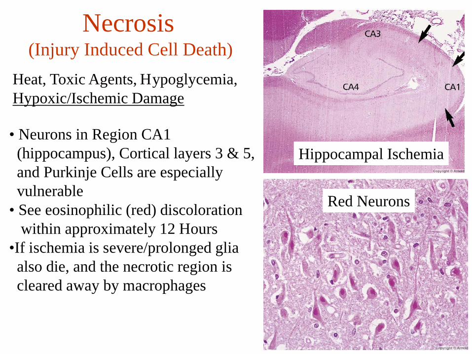
Heat, Toxic Agents, Hypoglycemia, Hypoxic/Ischemic Damage
Hippocampal Ischemia
Red Neurons
• Neurons in Region CA1 (hippocampus), Cortical layers 3 & 5, and Purkinje Cells are especially vulnerable
• See eosinophilic (red) discolorationwithin approximately 12 Hours
•If ischemia is severe/prolonged gliaalso die, and the necrotic region is cleared away by macrophages
Necrosis(Injury Induced Cell Death)
LFB CD68
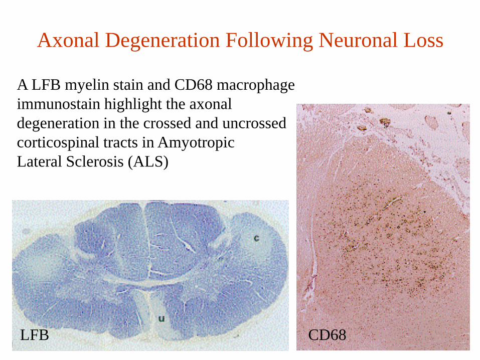
Axonal Degeneration Following Neuronal Loss
A LFB myelin stain and CD68 macrophageimmunostain highlight the axonal degeneration in the crossed and uncrossedcorticospinal tracts in Amyotropic Lateral Sclerosis (ALS)
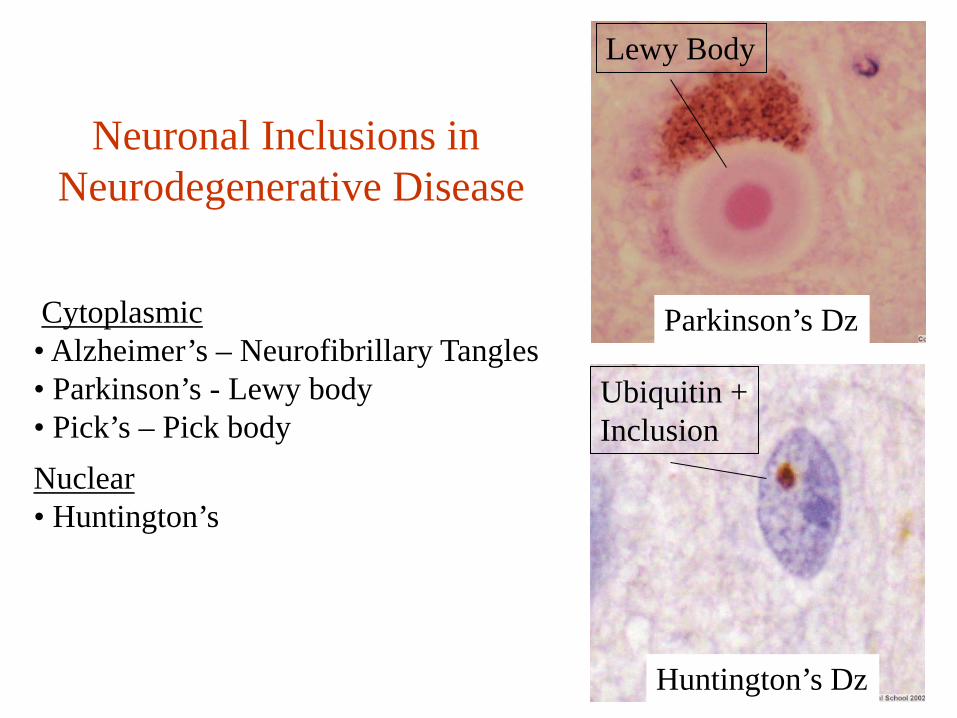
Huntington’s Dz
Parkinson’s Dz
Lewy Body
Ubiquitin +Inclusion
Neuronal Inclusions in Neurodegenerative Disease
Cytoplasmic• Alzheimer’s – Neurofibrillary Tangles• Parkinson’s - Lewy body• Pick’s – Pick bodyNuclear• Huntington’s

Pathology of Glia
Reactive AstrocytosisA non-specific reaction to infection, seizures, autoimmune disease,infarction, etcFibrillary GliosisProliferation of reactive astrocytesPiloid GliosisSeen around spinal cord cavities (syrinx)And other long-standing reactive gliosisIn cerebellum and hypothalamus. AlsoIn Alexander’s disease
Reactive Astrocytosis
Piloid Gliosis
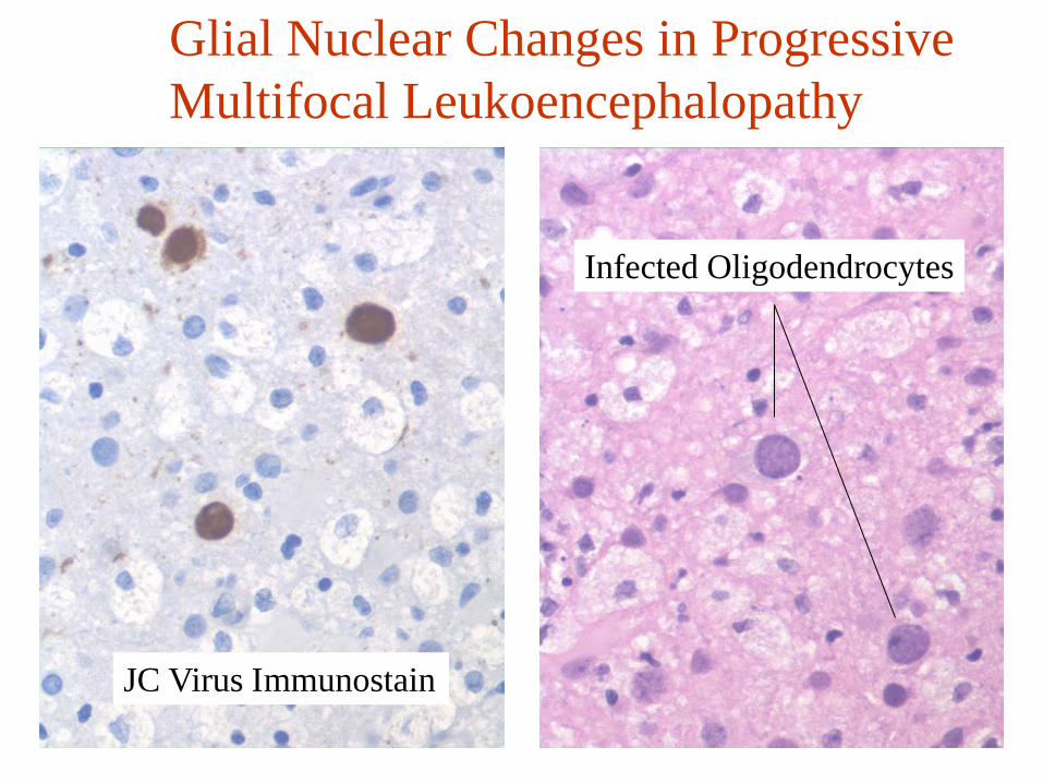
Glial Nuclear Changes in ProgressiveMultifocal Leukoencephalopathy
JC Virus Immunostain
Infected Oligodendrocytes
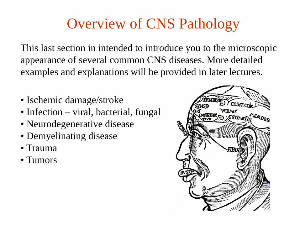
Overview of CNS PathologyThis last section in intended to introduce you to the microscopicappearance of several common CNS diseases. More detailed examples and explanations will be provided in later lectures.
• Ischemic damage/stroke• Infection – viral, bacterial, fungal• Neurodegenerative disease• Demyelinating disease• Trauma• Tumors
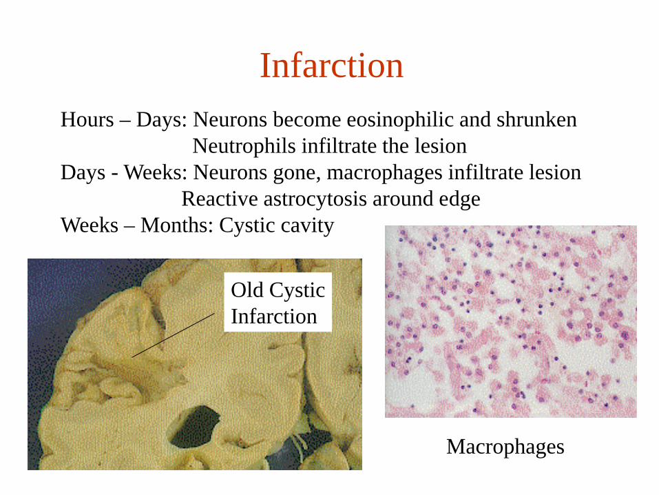
InfarctionHours – Days: Neurons become eosinophilic and shrunken
Neutrophils infiltrate the lesionDays - Weeks: Neurons gone, macrophages infiltrate lesion
Reactive astrocytosis around edgeWeeks – Months: Cystic cavity
Macrophages
Old CysticInfarction

Bacterial Infection
Meningitis Abcess
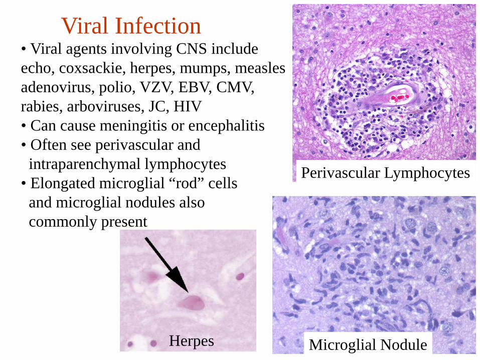
Viral Infection
Perivascular Lymphocytes
Microglial Nodule
• Viral agents involving CNS includeecho, coxsackie, herpes, mumps, measlesadenovirus, polio, VZV, EBV, CMV,rabies, arboviruses, JC, HIV• Can cause meningitis or encephalitis• Often see perivascular andintraparenchymal lymphocytes
• Elongated microglial “rod” cellsand microglial nodules also commonly present
Herpes
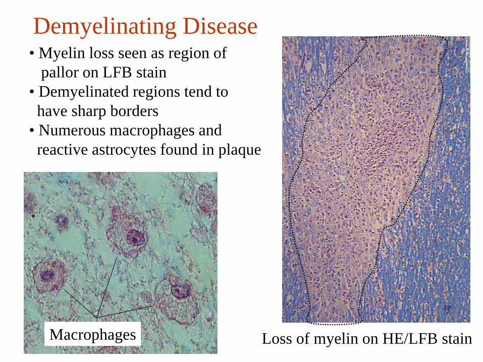
Demyelinating Disease
Macrophages Loss of myelin on HE/LFB stain
• Myelin loss seen as region of pallor on LFB stain
• Demyelinated regions tend to have sharp borders
• Numerous macrophages and reactive astrocytes found in plaque
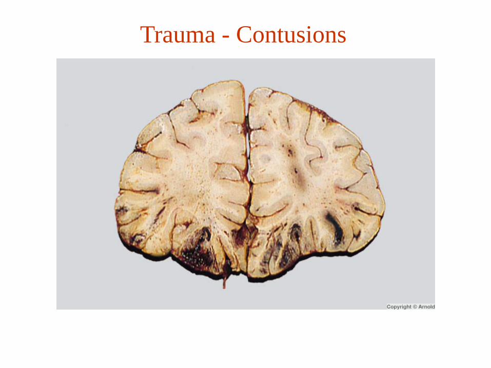
Trauma - Contusions
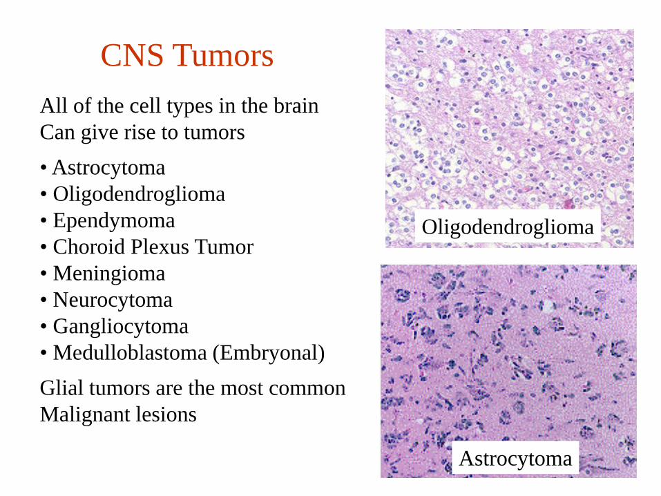
CNS TumorsAll of the cell types in the brain Can give rise to tumors• Astrocytoma• Oligodendroglioma• Ependymoma• Choroid Plexus Tumor• Meningioma• Neurocytoma• Gangliocytoma• Medulloblastoma (Embryonal)Glial tumors are the most commonMalignant lesions
Oligodendroglioma
Astrocytoma
Shifting Gears….A very brief introduction to CNS
development and imaging
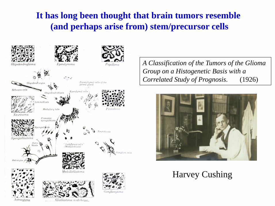
It has long been thought that brain tumors resemble (and perhaps arise from) stem/precursor cells
A Classification of the Tumors of the Glioma Group on a Histogenetic Basis with a Correlated Study of Prognosis. (1926)
Harvey Cushing
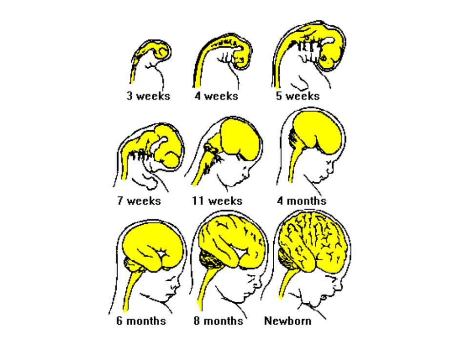

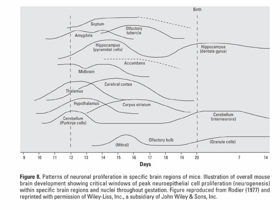
Cerebellar AnlageVZ

Conventions & TerminologyBody Planes
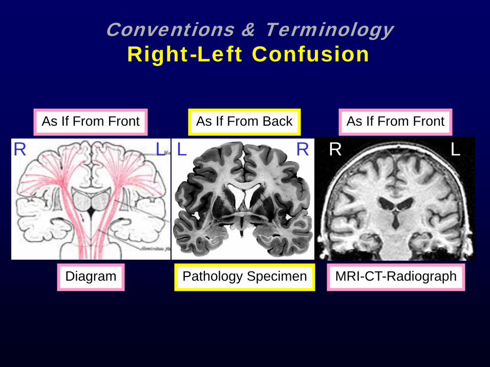
Conventions & TerminologyRight-Left Confusion
R LAs If From Front As If From Front
RL LRAs If From Back
Diagram MRI-CT-RadiographPathology Specimen

Conventions & TerminologyMore Right-Left Confusion
R L
MRI As If From Bottom Of Feet
L R
ODOS
Visual Field PathwaysAs If From Top Of Head
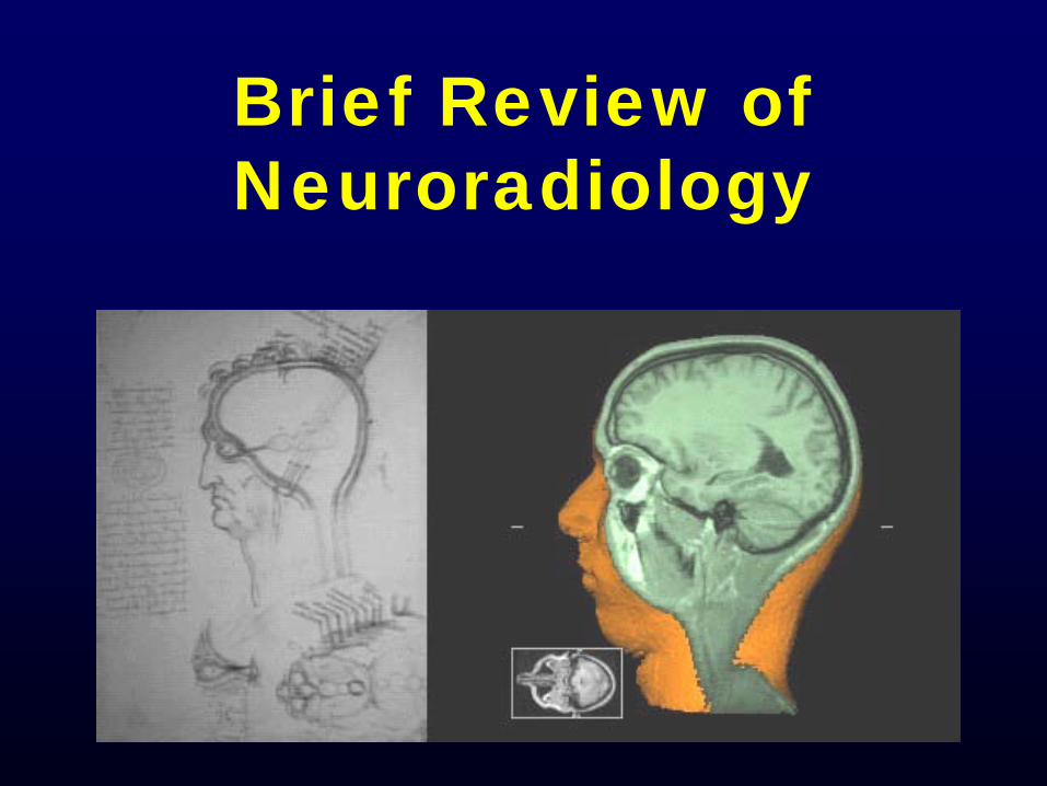
Brief Review of Neuroradiology

IMAGING:
MRI Brain with intraparenchymal hemorrhage from mycotic aneurysm
Elucidates…
1) pathoanatomy, 2) pathology,3) pathophysiology4) clinical risk
CLOT
MIDLINE SHIFT
EDEMA

Non contrast or plain imaging appearances
Scan Uses CSF Lesion Blood Bone
CT Rapid screen
Dark Dark White White
T1 MRI
Anatomy Dark Dark White Dark
T2 MRI
Lesion ident.
White White Varies with age of bleed
Dark
FLAIR Lesion ident.
Dark White Varies with age of bleed
Dark
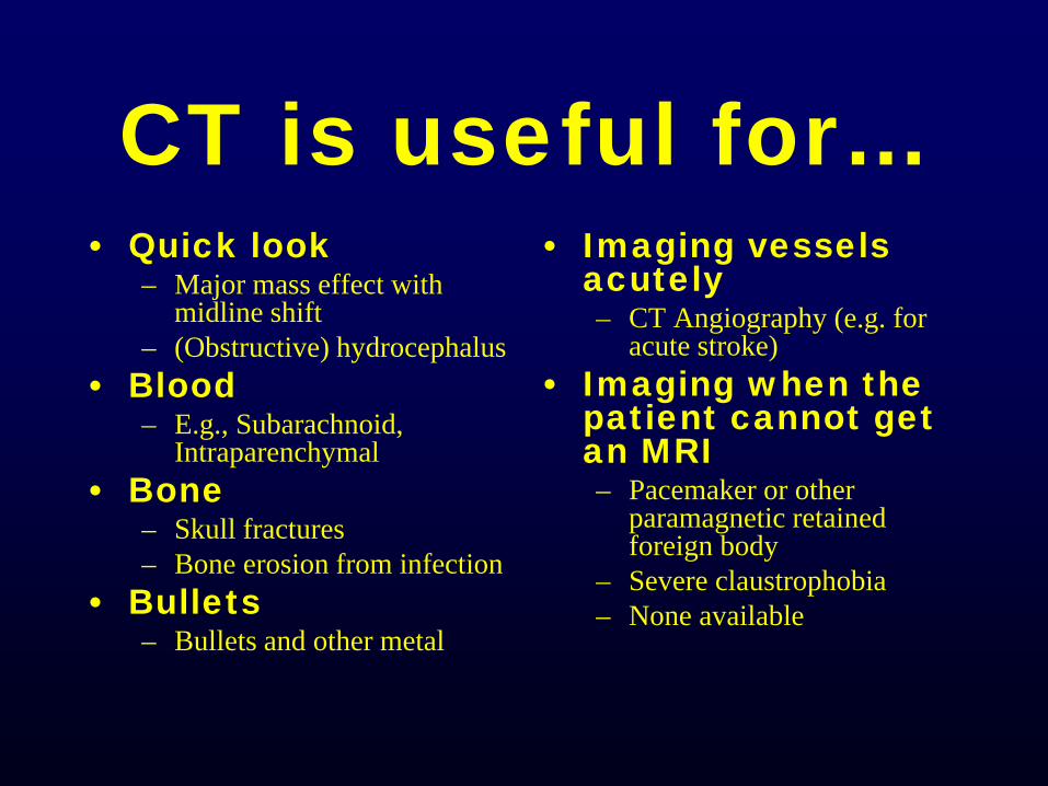
CT is useful for…• Quick look
– Major mass effect with midline shift
– (Obstructive) hydrocephalus• Blood
– E.g., Subarachnoid, Intraparenchymal
• Bone– Skull fractures– Bone erosion from infection
• Bullets– Bullets and other metal
• Imaging vessels acutely– CT Angiography (e.g. for
acute stroke)• Imaging when the
patient cannot get an MRI– Pacemaker or other
paramagnetic retained foreign body
– Severe claustrophobia– None available
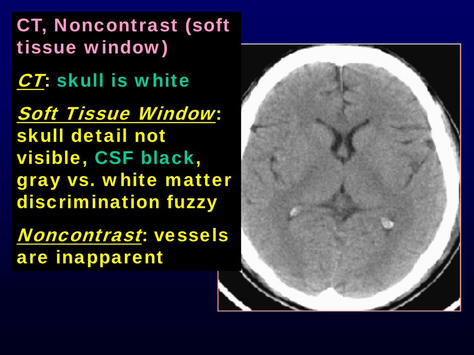
CT, Noncontrast (soft tissue window)
CT: skull is white
Soft Tissue Window: skull detail not visible, CSF black, gray vs. white matter discrimination fuzzy
Noncontrast: vessels are inapparent
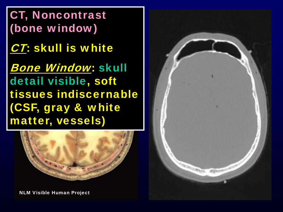
NLM Visible Human Project
CT, Noncontrast (bone window)
CT: skull is white
Bone Window: skull detail visible, soft tissues indiscernable (CSF, gray & white matter, vessels)
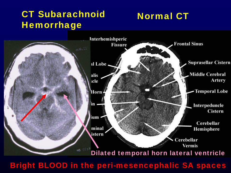
CT SubarachnoidHemorrhage
Normal CT
Bright BLOOD in the peri-mesencephalic SA spaces Dilated temporal horn lateral ventricle
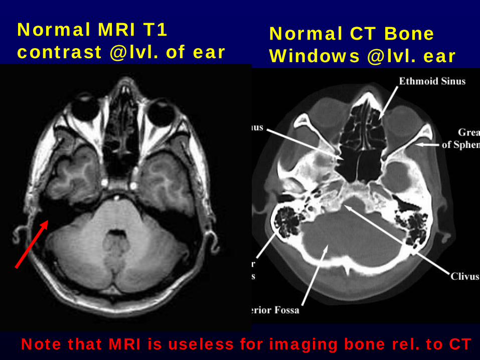
Normal CT BoneWindows @ lvl. ear
Note that MRI is useless for imaging bone rel. to CT
Normal MRI T1contrast @ lvl. of ear

MRI is useful for…• Anatomic detail• Subtle or small
pathology– including lesions
without large mass effect (esp. white matter disease)
• Posterior fossa lesions
• Acute stroke (Diffusion Weighted Imaging [DWI])
• Imaging Vessels (MR angiogram or venogram [MRA/V])
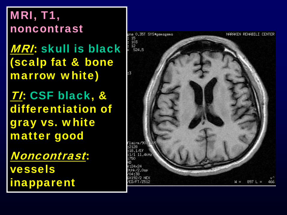
MRI, T1, noncontrast
MRI: skull is black(scalp fat & bone marrow white)
TI: CSF black, & differentiation of gray vs. white matter good
Noncontrast: vessels inapparent
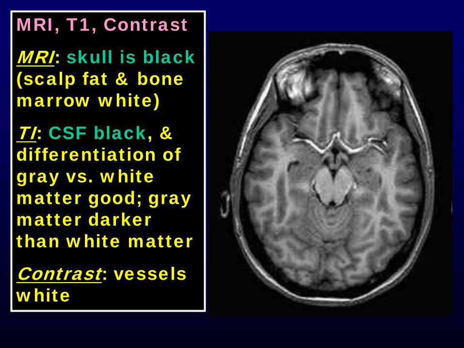
MRI, T1, Contrast
MRI: skull is black(scalp fat & bone marrow white)
TI: CSF black, & differentiation of gray vs. white matter good; gray matter darker than white matter
Contrast: vessels white
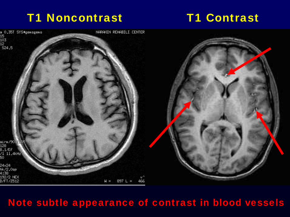
T1 Noncontrast T1 Contrast
Note subtle appearance of contrast in blood vessels
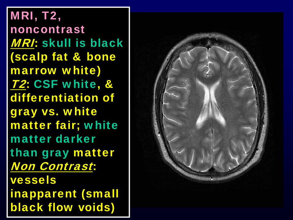
MRI, T2, noncontrastMRI: skull is black(scalp fat & bone marrow white)T2: CSF white, & differentiation of gray vs. white matter fair; white matter darker than gray matterNon Contrast: vessels inapparent (small black flow voids)
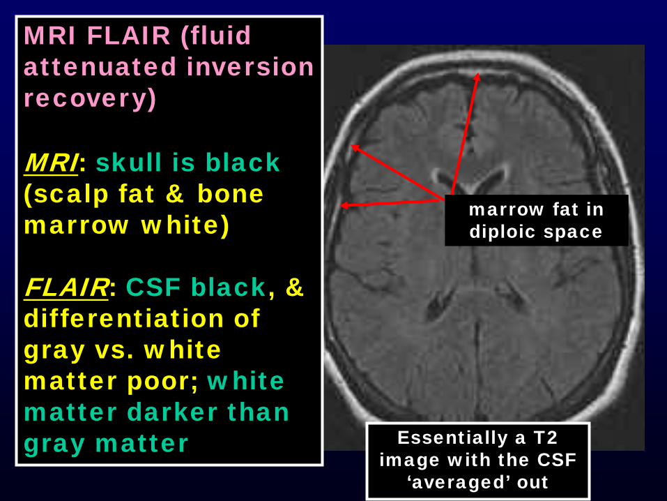
MRI FLAIR (fluid attenuated inversion recovery)
MRI: skull is black(scalp fat & bone marrow white)
FLAIR: CSF black, & differentiation of gray vs. white matter poor; white matter darker than gray matter
marrow fat in diploic space
Essentially a T2 image with the CSF
‘averaged’ out
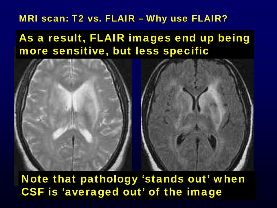
MRI scan: T2 vs. FLAIR – Why use FLAIR?
Note that pathology ‘stands out’ when CSF is ‘averaged out’ of the image
As a result, FLAIR images end up being more sensitive, but less specific