Embed Size (px)
DESCRIPTION
Introduction to High Frequency Ventilation. Michael Haines, MPH, RRT-NPS, AE-C. Objectives. TYPES OF HFV There are four basic types of HFV: High frequency jet ventilation High frequency oscillatory ventilation High frequency percussive ventilation - PowerPoint PPT Presentation
Citation preview
Introduction to High Frequency
Ventilation
Michael Haines, MPH, RRT-NPS, AE-C
Objectives
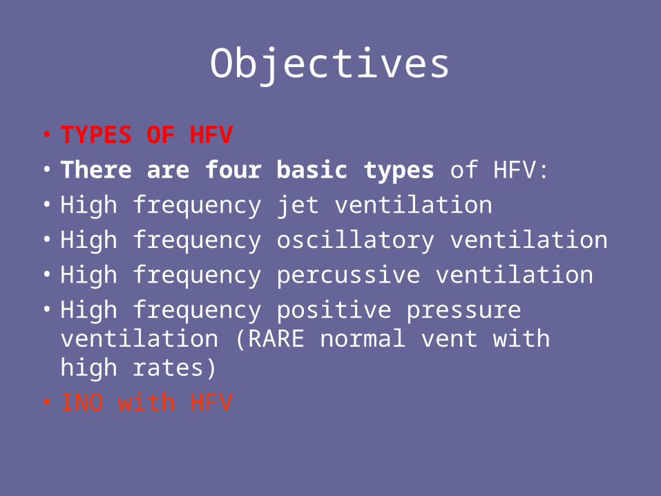
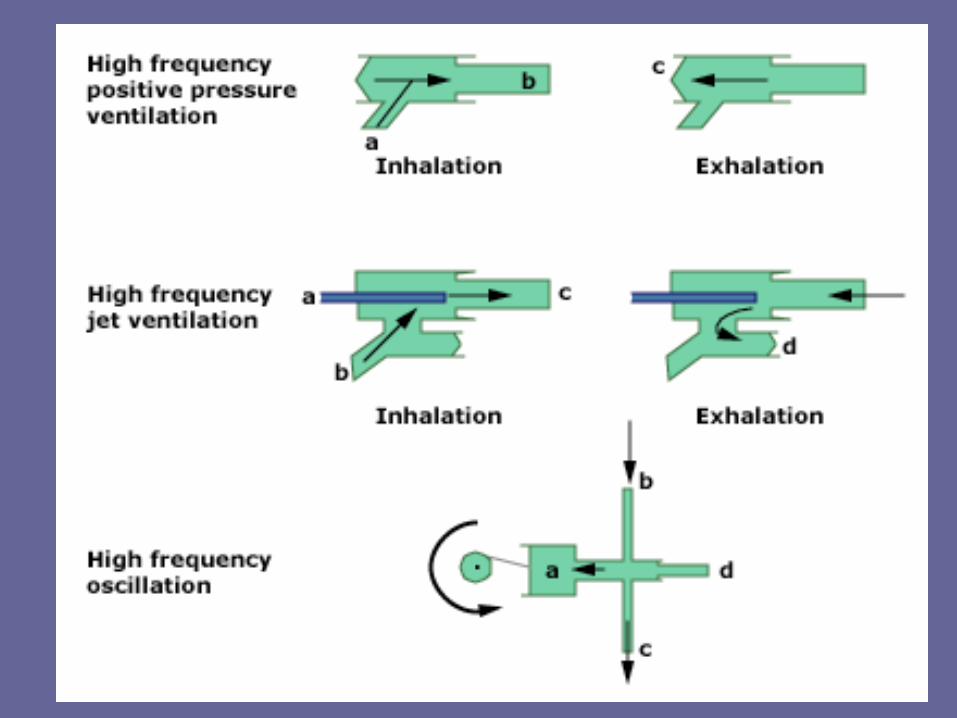
• TYPES OF HFV • There are four basic types of HFV:• High frequency jet ventilation• High frequency oscillatory ventilation• High frequency percussive ventilation • High frequency positive pressure ventilation
(RARE normal vent with high rates)• INO with HFV
Introduction
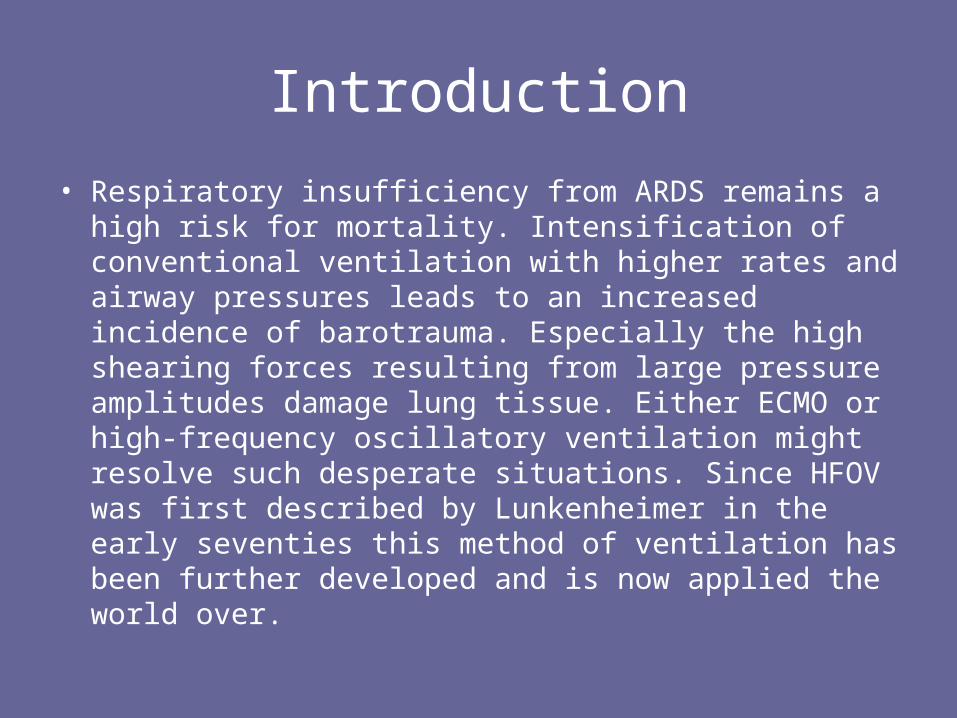
• Respiratory insufficiency from ARDS remains a high risk for mortality. Intensification of conventional ventilation with higher rates and airway pressures leads to an increased incidence of barotrauma. Especially the high shearing forces resulting from large pressure amplitudes damage lung tissue. Either ECMO or high-frequency oscillatory ventilation might resolve such desperate situations. Since HFOV was first described by Lunkenheimer in the early seventies this method of ventilation has been further developed and is now applied the world over.

High Frequency Jet Ventilation
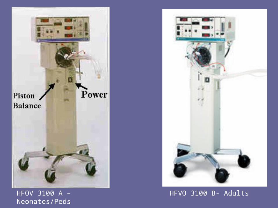
HFOV 3100 A – Neonates/Peds
HFVO 3100 B- Adults
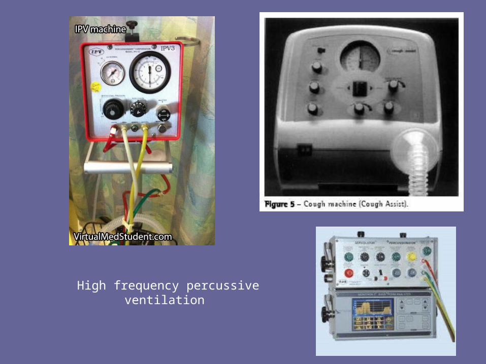
High frequency percussive ventilation
Introduction
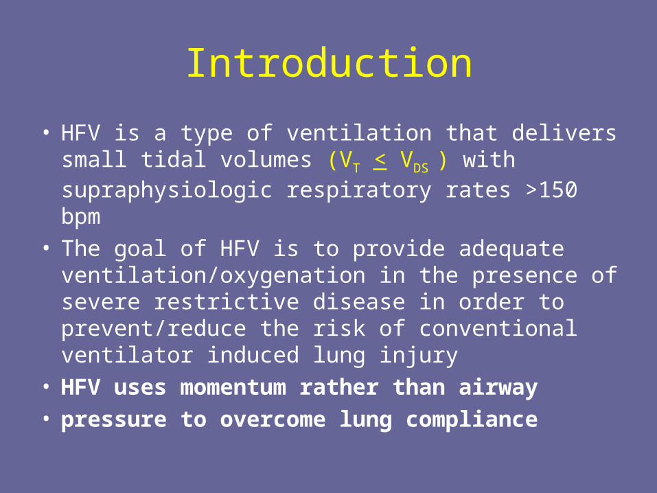
• HFV is a type of ventilation that delivers small tidal volumes (VT < VDS ) with supraphysiologic respiratory rates >150 bpm
• The goal of HFV is to provide adequate ventilation/oxygenation in the presence of severe restrictive disease in order to prevent/reduce the risk of conventional ventilator induced lung injury
• HFV uses momentum rather than airway• pressure to overcome lung compliance
Introduction
• High-frequency ventilation was first introduced 30 years ago as a method for reducing intrathoracic pressure during thoracic and laryngeal surgery. High-frequency oscillation was developed in the 1970's for the treatment of lung disease of prematurity but is now used for acute hypoxemic respiratory failure in all ages.
Introduction
• The key difference from CMV is the usage of high rates and low tidal volumes. Because the tidal volume is smaller than the dead space, the gas transport during HFV cannot be explained by bulk flow theory as in CMV.
• HFV is not uncommon in nature and in fact occurs in:– Humming birds– Panting dogs
Introduction
• Traditional wisdom: VA = f (VT - VDS)
• Physiologic (effective) dead space can become < anatomic dead space
»Animal studies of HFV: adequate ventilation can be achieved with VT as low as 1 ml/kg
» Flow streaming reduces effective dead space - only portions of the anatomic DS are used
» Fresh gas penetrates some alveoli
Introduction
• During conventional ventilation direct alveolar ventilation accomplishes pulmonary gas exchange. According to the classic concept of pulmonary ventilation the amount of gas reaching the alveoli equals the applied tidal volume minus the deadspace volume.
• At tidal volumes below the size of the anatomical deadspace this model fails to explain gas exchange. Instead, considerable mixing of fresh and exhaled gas in the airways and lungs is believed to be the key to the success of HFV in ventilating the lung at such very low tidal volumes.
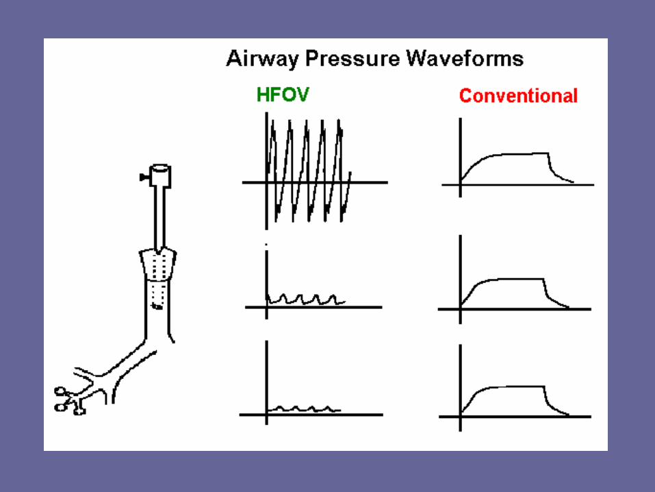
Amplitude and Hertz
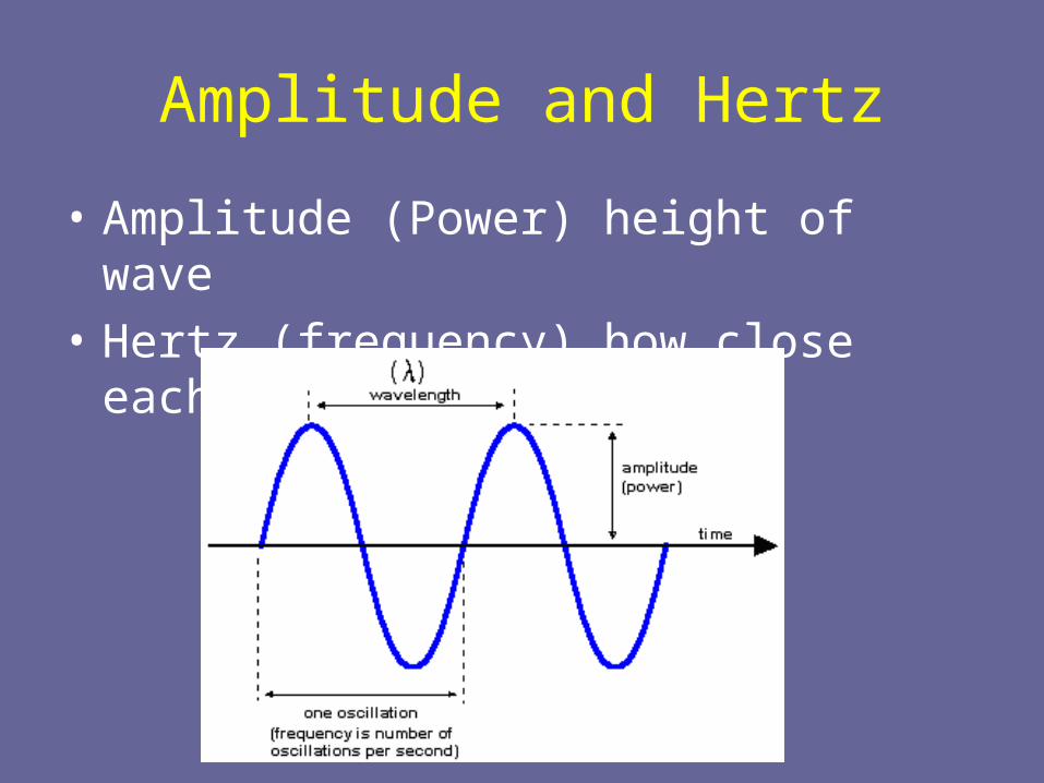
• Amplitude (Power) height of wave
• Hertz (frequency) how close each wave is
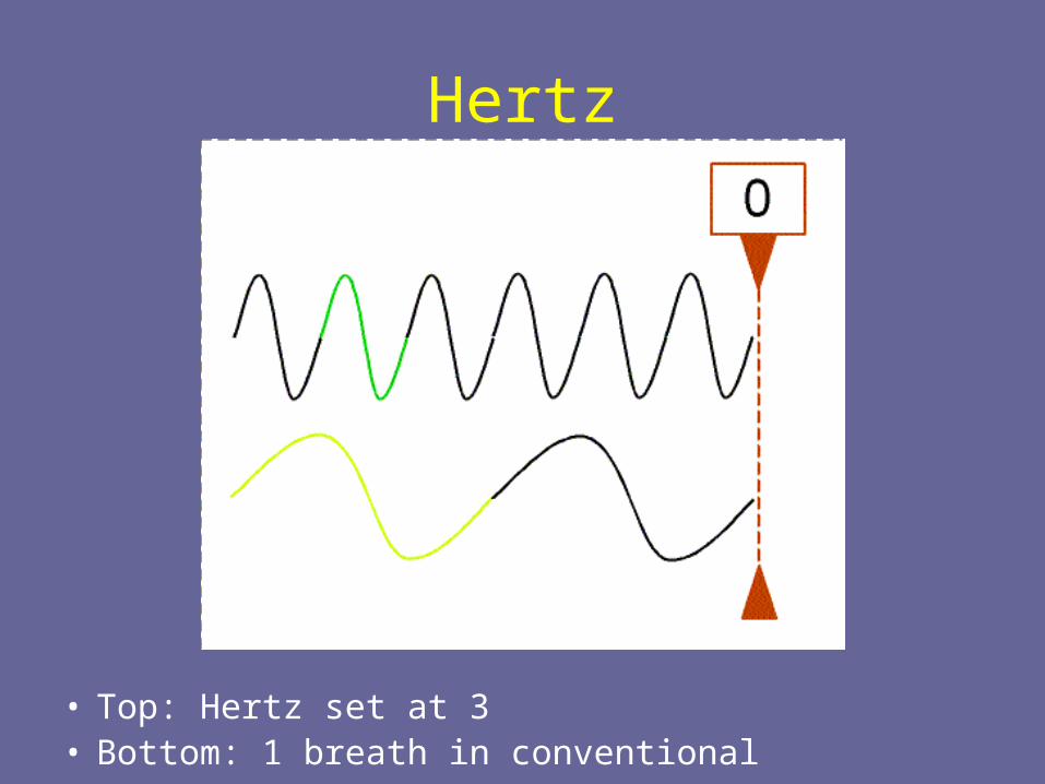
Hertz
• Top: Hertz set at 3• Bottom: 1 breath in conventional
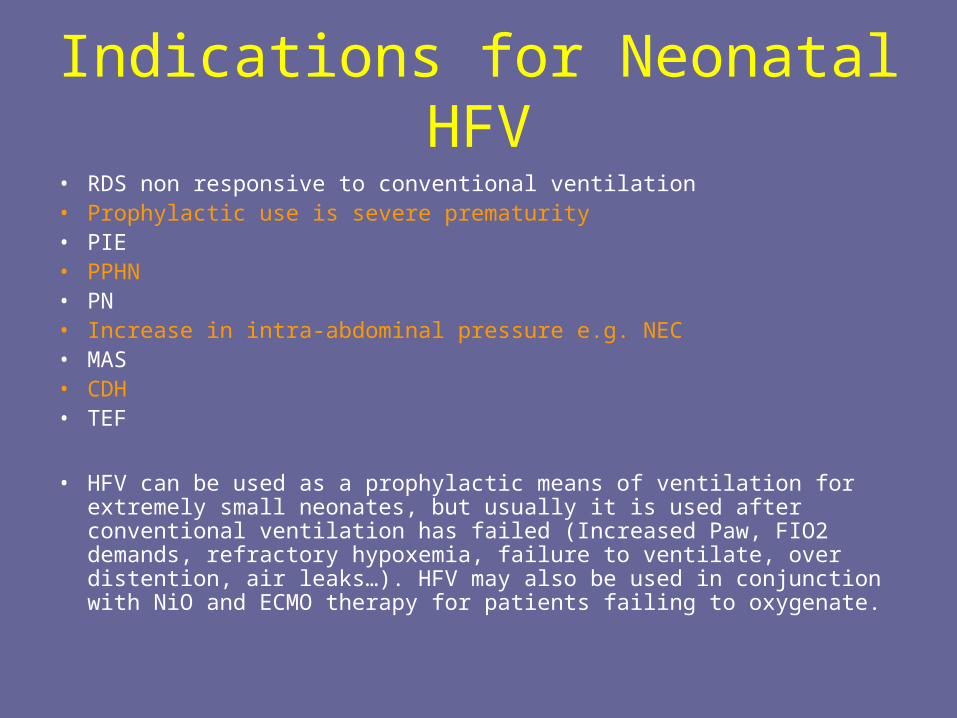
Indications for Neonatal HFV• RDS non responsive to conventional ventilation• Prophylactic use is severe prematurity• PIE• PPHN• PN• Increase in intra-abdominal pressure e.g. NEC• MAS• CDH• TEF
• HFV can be used as a prophylactic means of ventilation for extremely small neonates, but usually it is used after conventional ventilation has failed (Increased Paw, FIO2 demands, refractory hypoxemia, failure to ventilate, over distention, air leaks…). HFV may also be used in conjunction with NiO and ECMO therapy for patients failing to oxygenate.
Indications for Adult HFV
• ARDS/ALI (only disease process that has been currently studied for use)
• Possibly massive airleak and BP fistula
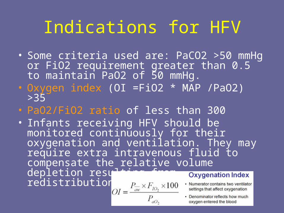
Indications for HFV
• Some criteria used are: PaCO2 >50 mmHg or FiO2 requirement greater than 0.5 to maintain PaO2 of 50 mmHg.
• Oxygen index (OI =FiO2 * MAP /PaO2) >35• PaO2/FiO2 ratio of less than 300 • Infants receiving HFV should be monitored
continuously for their oxygenation and ventilation. They may require extra intravenous fluid to compensate the relative volume depletion resulting from redistribution of blood flow
Why we use HFV
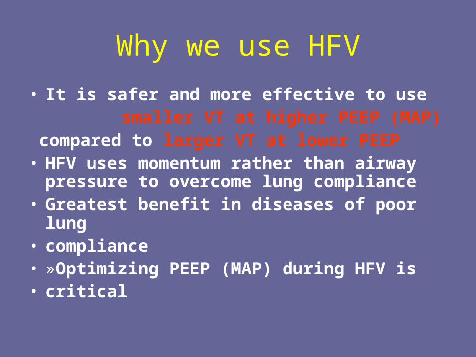
• It is safer and more effective to use smaller VT at higher PEEP (MAP) compared to larger VT at lower PEEP• HFV uses momentum rather than airway
pressure to overcome lung compliance• Greatest benefit in diseases of poor lung• compliance• »Optimizing PEEP (MAP) during HFV is• critical
Why we use HFV
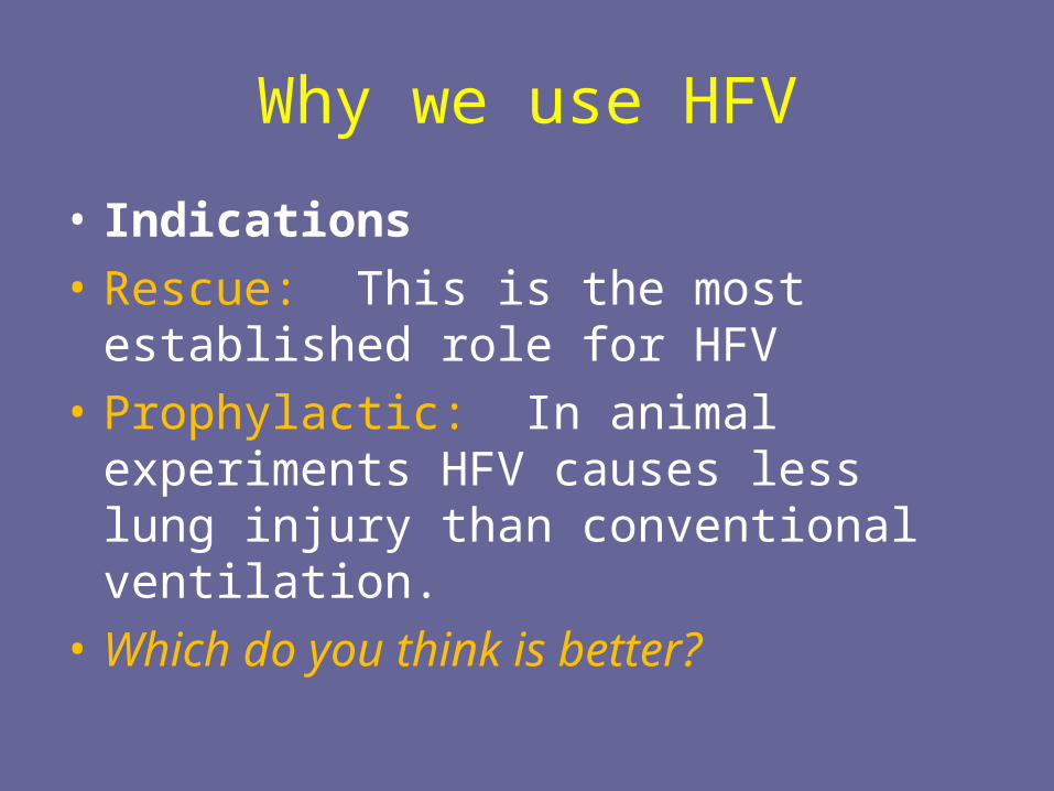
• Indications
• Rescue: This is the most established role for HFV
• Prophylactic: In animal experiments HFV causes less lung injury than conventional ventilation.
• Which do you think is better?
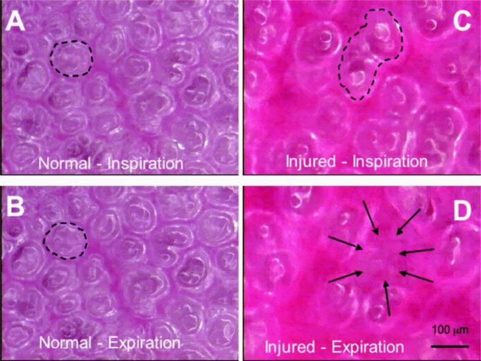
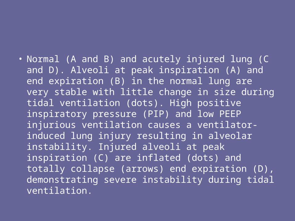
• Normal (A and B) and acutely injured lung (C and D). Alveoli at peak inspiration (A) and end expiration (B) in the normal lung are very stable with little change in size during tidal ventilation (dots). High positive inspiratory pressure (PIP) and low PEEP injurious ventilation causes a ventilator-induced lung injury resulting in alveolar instability. Injured alveoli at peak inspiration (C) are inflated (dots) and totally collapse (arrows) end expiration (D), demonstrating severe instability during tidal ventilation.
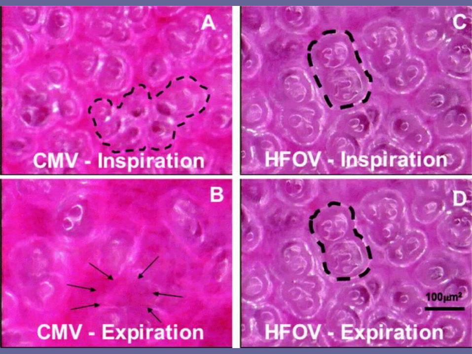
• In vivo photomicrographs of subpleural alveoli in the rat after lung injury by saline lavage ventilated with either conventional mechanical ventilation (CMV) or high-frequency oscillatory ventilation (HFOV) using a 2.5-internal diameter tracheal tube. With CMV, a group of alveoli are seen inflated during inspiration (dots) but collapse with expiration (arrows). Alveoli are very stable with HFOV during ventilation. The same alveolus is seen with HFOV at inflation and exhalation (dots).
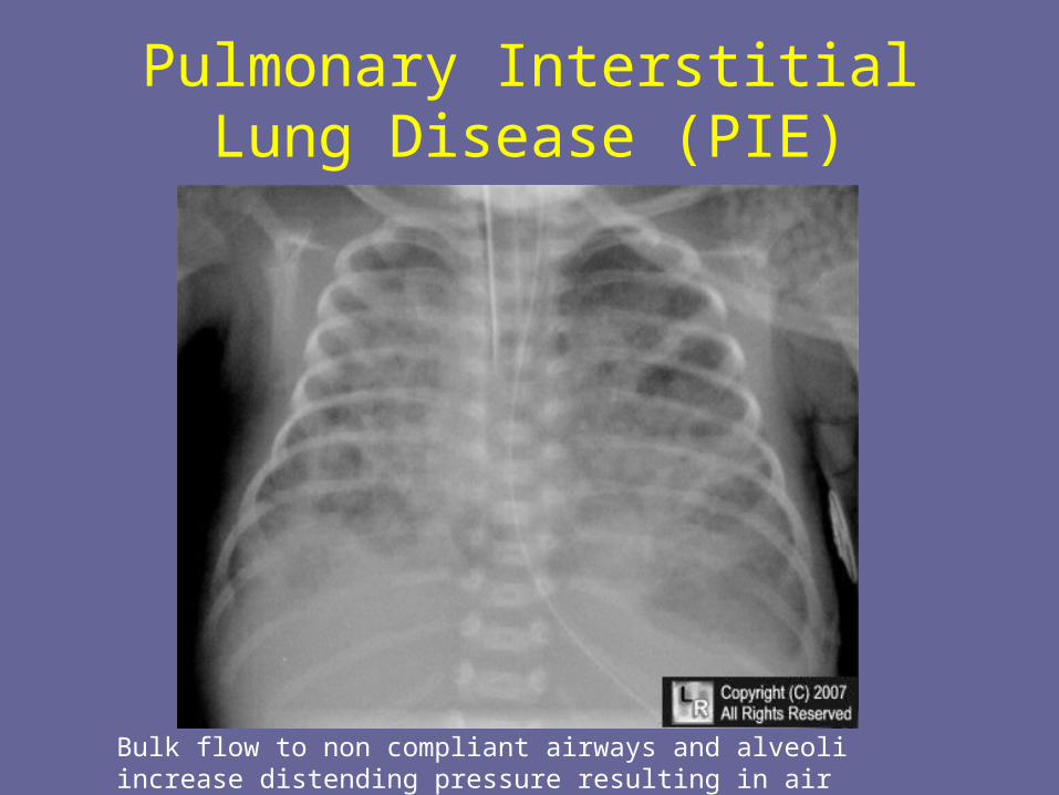
Pulmonary Interstitial Lung Disease (PIE)
Bulk flow to non compliant airways and alveoli increase distending pressure resulting in air leaks
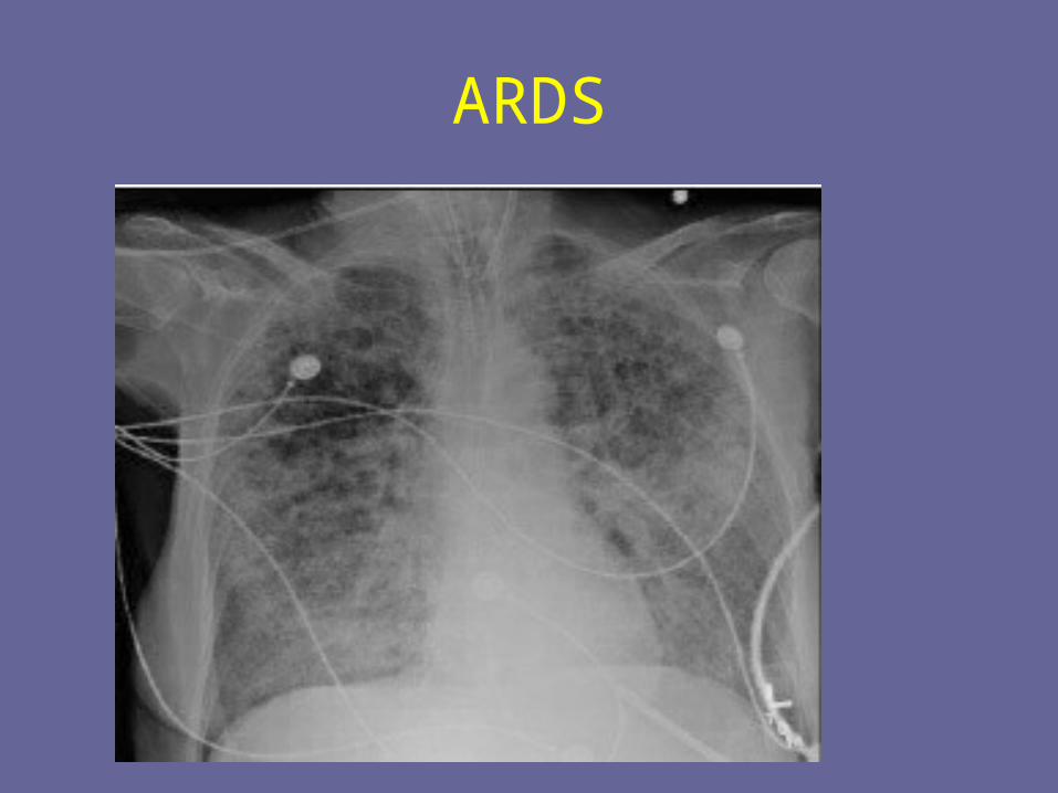
ARDS
Air leaks from the over stretching of non compliant alveoli can cause:
• Subcutaneous emphysema• Pneumothorax• P.I.E.• Pneumomediastinum• Pneumo-pericardium• Pneumo-retroperitoneum• pneuomoperitoneum
HFV is a method of mechanical ventilation that employs supra-physiological breathing rates and tidal volumes frequently less than dead space. Because conventional ventilation relies on the production of large pressure changes to induce mass flow of gas in and out of the lungs, it may be associated with deleterious consequences of volume and pressure changes at alveolar level. These include air leaks, such as PIE and pneumothorax, and bronchiolar-alveolar injury leading to chronic lung disease.
How does HFV work?
• HFV enhances both bulk flow (convection) and diffusion of respiratory gases»Abundant fresh gas “washes out” expired gas from the airways»Decreased pCO2 at the gas exchange boundaryincreases diffusion
• Linear relationship between ventilator rate• and CO2 elimination is no longer valid• »CV: CO2 = f x VT; HFV: CO2 = f x VT
2
• »Changes in rate have less impact on gas• exchange than similar changes made during CMV
Venegas and Fredberg: Crit Care Med 22 (suppl):S49, 1994
How does gas exchange occur with HFV
• Direct ventilation of most proximal alveoli units by bulk convection
• Direct Bulk Flow Some alveoli situated in the proximal tracheobronchial tree receive a direct flow of inspired air. This leads to gas exchange by traditional mechanisms of convective or bulk flow.
How does gas exchange occur with HFV
• Pendalluft effect – asynchronous flow among alveoli due to asymmetries in airflow impedance. This causes gas to re-circulate among lung units and improve gas exchange. In healthy and, more so, in diseased lungs, the mechanics of air flow vary among lung regions and units within regions. Variation in regional airway resistance and compliance cause some regions to fill and empty more rapidly than others. Some gas may flow between regions if these characteristics vary among regions that are in close proximity.
• Turbulence in the large airways causing enhanced gas mixing
How does gas exchange occur with HFV
• Taylor dispersion – Turbulent eddies and secondary swirling motions occur when convective flow is superimposed on diffusion. Some fresh gas may mix with gas from alveoli, increasing the amount of gas exchange that would occur from simple bulk flow.
• Collateral ventilation through non-airway connections between neighboring alveoli
How does gas exchange occur with HFV
• Cardiogenic Mixing• Mechanical agitation from the contracting
heart contributes to gas mixing, especially in peripheral lung units in close proximity to the heart.
• Molecular Diffusion• As in other modes of ventilation, this
mechanism may play an important role in mixing of air in the smallest bronchioles and alveoli, near the alveolocapillary membranes.
How does gas exchange occur with HFV
• Asymmetric velocity – convective gas transport is enhanced by asymmetry between inspiratory and expiratory velocity profiles that occur at branch points in the airways. The velocity profile of air moving through an airway under laminar flow conditions is parabolic. Air closest to the tracheobronchial wall has a lower velocity than air in the center of the airway lumen. This parabolic velocity profile is usually more pronounced during the inspiratory phase of respiration because of differences in flow rates. With repeated respiratory cycles, gas in the center of the airway lumen advances further into the lung while gas on the margin (close to the airway wall) moves out toward the mouth
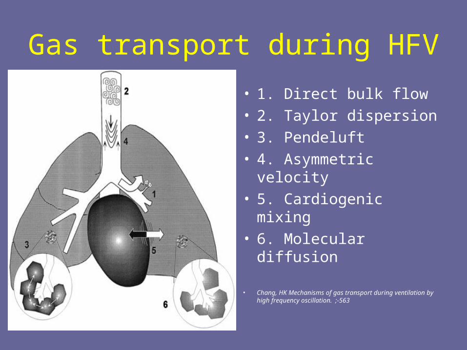
Gas transport during HFV
• 1. Direct bulk flow• 2. Taylor dispersion• 3. Pendeluft• 4. Asymmetric
velocity• 5. Cardiogenic mixing• 6. Molecular diffusion
• Chang, HK Mechanisms of gas transport during ventilation by high frequency oscillation. ;-563
Oxygenation with HFV
• Oxygenation is determined by lung volume (affected by MAP) and FiO2. It is important to maintain adequate lung volume to prevent atelectasis and to preserve surfactant function to achieve adequate oxygenation.
• Adequate MAP should be used to recruit alveoli and maintain lung volume above functional residual capacity (FRC).
• In contrast to CMV, lung volume is maintained at a relatively constant level during HFV.
Oxygenation with HFV
• The ventilation/perfusion matching would improve as a result of alveolar recruitment when lung volume increases. The near constant lung volumes in HFV results in better gas distribution and avoids the development of regional atelectasis in less compliant lung units, hence resulting in better ventilation/perfusion matching.
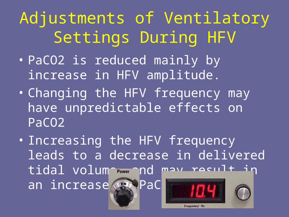
Adjustments of Ventilatory Settings During HFV
• PaCO2 is reduced mainly by increase in HFV amplitude.
• Changing the HFV frequency may have unpredictable effects on PaCO2
• Increasing the HFV frequency leads to a decrease in delivered tidal volume, and may result in an increase in PaCO2.
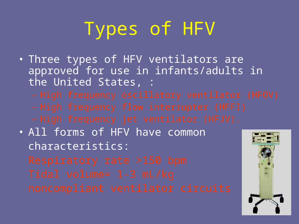
Types of HFV
• Three types of HFV ventilators are approved for use in infants/adults in the United States, : – High frequency oscillatory ventilator (HFOV)– High frequency flow interrupter (HFFI)– High frequency jet ventilator (HFJV).
• All forms of HFV have common characteristics:
Respiratory rate >150 bpmTidal volume= 1-3 mL/kgnoncompliant ventilator circuits
HFOV
• HFOV employs either a piston or diaphragm to oscillate a bias flow of gas to generate both positive and negative pressure fluctuations termed as amplitude.
• The adjustable parameters include:– Power– IT%– Mean airway pressure (MAP) – Bias flow– Frequency (Hz)– Amplitude
• Frequency is usually fixed for a particular patient group.• The recommended range is 10-15 Hz for premature
infants and 8-10 Hz for term infants, around 5 Hz for adults
HFOV• Characterized by high respiratory rates between 3.5 to 15
hertz (210 - 900 breaths per minute), where 1 Hz=1 breath per second.
• The rates used vary widely depending upon patient size, age, and disease process. Typically the smaller the patient the higher the Hz and vise versa.
• Pressure oscillates around the constant distending pressure (equivalent to Mean Airway Pressure), which is usually set slightly higher than the MAP on conventional ventilation.
• Gas is pushed into the lung during inspiration, and then pulled out during expiration (active exhalation).
HFOV
• HFOV generates very low tidal volumes that are generally less than the dead space of the lung (watch for atelectasis and sudden drop in vital signs during implementation).
• Tidal volume is dependent on endotracheal tube size, power and frequency.
HFOV
• Different mechanisms of gas transfer are believed to come into play in HFOV compared to normal mechanical ventilation.
• Often used in patients who have refractory hypoxemia that cannot be corrected by normal mechanical ventilation such as is the case in the following disease processes: severe ARDS, ALI and other oxygenation diffusion issues. In some neonatal patients HFOV may be used as the first-line ventilator due to the high susceptibility of the premature infant to lung injury from conventional ventilation.
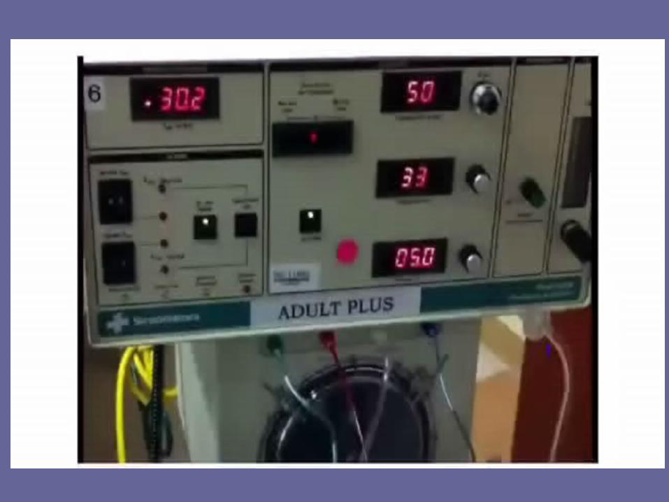
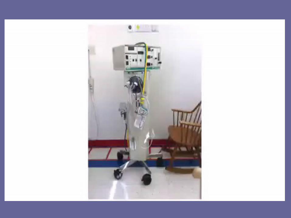
HFOVSensormedics 3100A
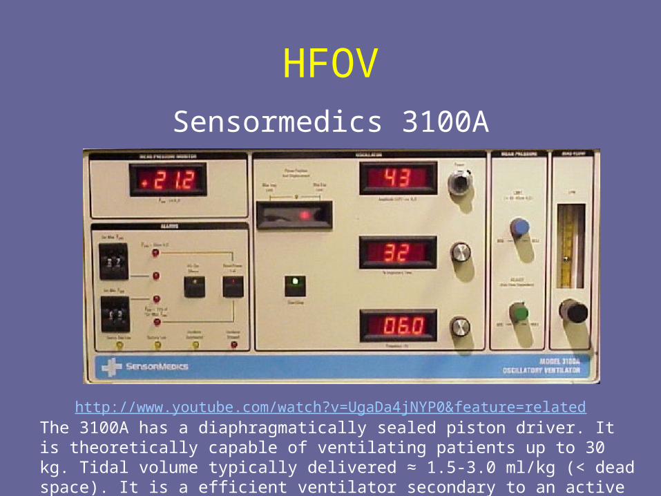
http://www.youtube.com/watch?v=UgaDa4jNYP0&feature=related
The 3100A has a diaphragmatically sealed piston driver. It is theoretically capable of ventilating patients up to 30 kg. Tidal volume typically delivered ≈ 1.5-3.0 ml/kg (< dead space). It is a efficient ventilator secondary to an active expiratory phase, but it is not capable of delivering sigh breaths for alveolar recruitment.
SensorMedics 3100A Oscillatory Ventilator
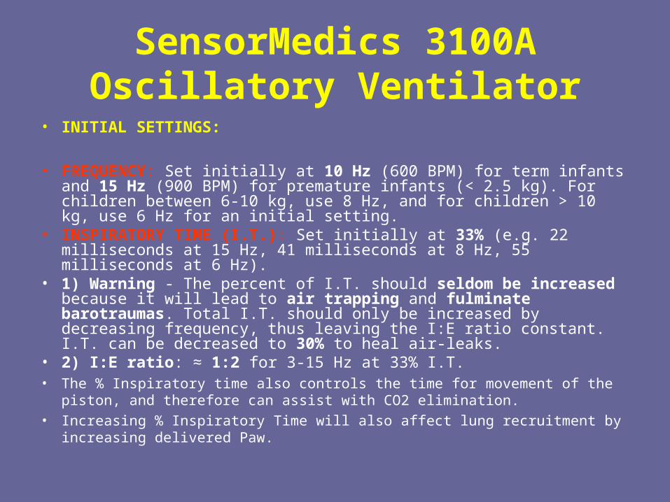
• INITIAL SETTINGS:
• FREQUENCY: Set initially at 10 Hz (600 BPM) for term infants and 15 Hz (900 BPM) for premature infants (< 2.5 kg). For children between 6-10 kg, use 8 Hz, and for children > 10 kg, use 6 Hz for an initial setting.
• INSPIRATORY TIME (I.T.): Set initially at 33% (e.g. 22 milliseconds at 15 Hz, 41 milliseconds at 8 Hz, 55 milliseconds at 6 Hz).
• 1) Warning - The percent of I.T. should seldom be increased because it will lead to air trapping and fulminate barotraumas. Total I.T. should only be increased by decreasing frequency, thus leaving the I:E ratio constant. I.T. can be decreased to 30% to heal air-leaks.
• 2) I:E ratio: ≈ 1:2 for 3-15 Hz at 33% I.T.• The % Inspiratory time also controls the time for movement of the piston, and
therefore can assist with CO2 elimination.• Increasing % Inspiratory Time will also affect lung recruitment by increasing
delivered Paw.
SensorMedics 3100A Oscillatory Ventilator
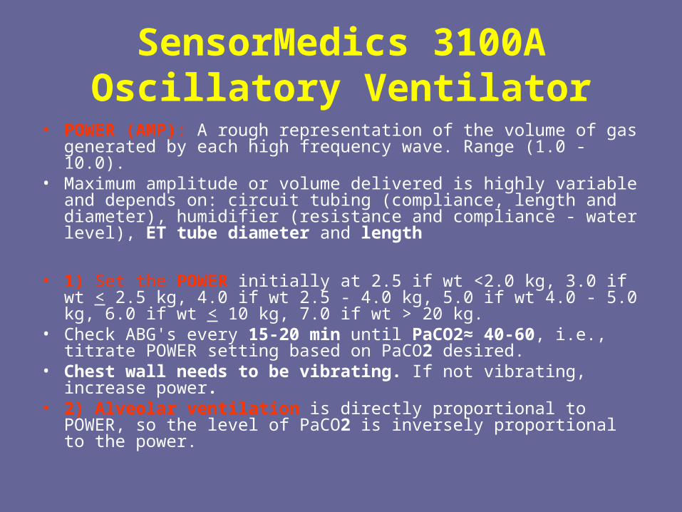
• POWER (AMP): A rough representation of the volume of gas generated by each high frequency wave. Range (1.0 - 10.0).
• Maximum amplitude or volume delivered is highly variable and depends on: circuit tubing (compliance, length and diameter), humidifier (resistance and compliance - water level), ET tube diameter and length
• 1) Set the POWER initially at 2.5 if wt <2.0 kg, 3.0 if wt < 2.5 kg, 4.0 if wt 2.5 - 4.0 kg, 5.0 if wt 4.0 - 5.0 kg, 6.0 if wt < 10 kg, 7.0 if wt > 20 kg.
• Check ABG's every 15-20 min until PaCO2≈ 40-60, i.e., titrate POWER setting based on PaCO2 desired.
• Chest wall needs to be vibrating. If not vibrating, increase power. • 2) Alveolar ventilation is directly proportional to POWER, so the
level of PaCO2 is inversely proportional to the power.
SensorMedics 3100A Oscillatory Ventilator
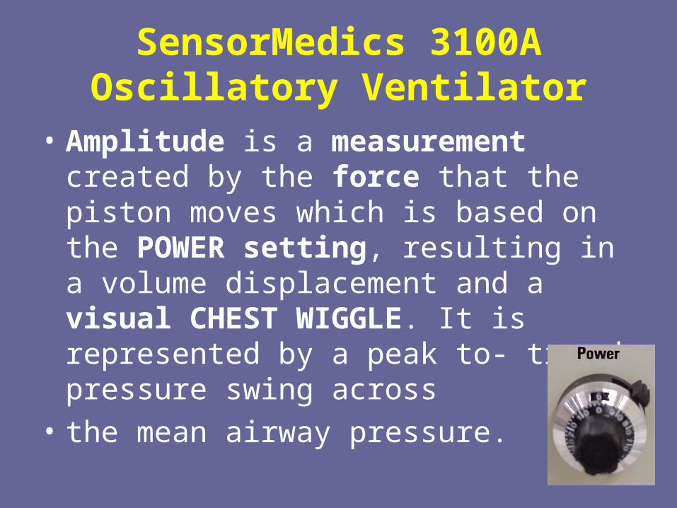
• Amplitude is a measurement created by the force that the piston moves which is based on the POWER setting, resulting in a volume displacement and a visual CHEST WIGGLE. It is represented by a peak to- trough pressure swing across
• the mean airway pressure.
SensorMedics 3100A Oscillatory Ventilator
• MAP: Initial MAP 4 cm above MAP while on CMV• Oxygenation on HFOV is directly proportional to MAP, which is
similar to CMV, however with the SensorMedics HFOV the MAP is generated by PEEP. Thus during HFOV: MAP = PEEP.
• 1. Initial MAP Settings:• a) Neonates - Initial MAP should be 2-4 cm above the MAP on CMV.• b) Infants/Children - Initial MAP should be 4-8 cm above the MAP on
CMV.• c) If starting immediately on HFOV - use a MAP of ≈ 8-10 cm in
neonates and 15-18 cm in infants/children.
• Check CXR 2 hrs after converting to HFOV, then adjust MAP to achieve optimal lung volume (9-10 ribs expanded).
• If not oxygenating, increase MAP by 2 cm every hour until oxygenation improves. Adjust Power to keep PaCO2 45-55.
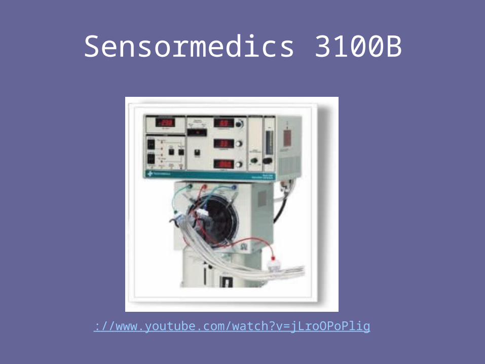
Sensormedics 3100B
://www.youtube.com/watch?v=jLroOPoPlig
SensorMedics 3100B Oscillatory Ventilator
• Initial Settings and Suggestions for Larger Patient Use On 3100 B
• Patients with ARDS >35 Kg• Set Paw 5 cmH20 above CV Paw• FiO2 100%• Set Hertz at 5-6• Power 4.0, adjust for good chest wiggle• I time % at 33%• Set Bias Flow at >25 lpm, may need to go higher
SensorMedics 3100B Oscillatory Ventilator
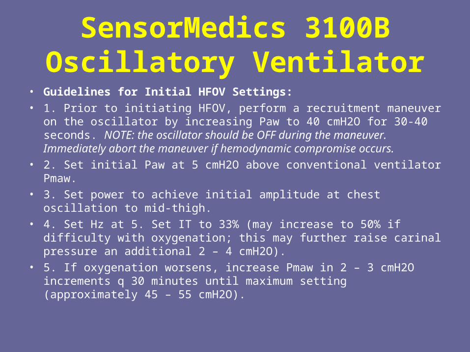
• Guidelines for Initial HFOV Settings: • 1. Prior to initiating HFOV, perform a recruitment maneuver on the
oscillator by increasing Paw to 40 cmH2O for 30-40 seconds. NOTE: the oscillator should be OFF during the maneuver. Immediately abort the maneuver if hemodynamic compromise occurs.
• 2. Set initial Paw at 5 cmH2O above conventional ventilator Pmaw. • 3. Set power to achieve initial amplitude at chest oscillation to mid-thigh. • 4. Set Hz at 5. Set IT to 33% (may increase to 50% if difficulty with
oxygenation; this may further raise carinal pressure an additional 2 – 4 cmH2O).
• 5. If oxygenation worsens, increase Pmaw in 2 – 3 cmH2O increments q 30 minutes until maximum setting (approximately 45 – 55 cmH2O).
SensorMedics 3100B Oscillatory Ventilator
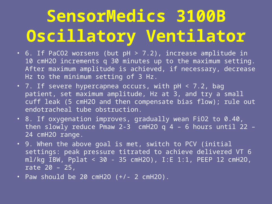
• 6. If PaCO2 worsens (but pH > 7.2), increase amplitude in 10 cmH2O increments q 30 minutes up to the maximum setting. After maximum amplitude is achieved, if necessary, decrease Hz to the minimum setting of 3 Hz.
• 7. If severe hypercapnea occurs, with pH < 7.2, bag patient, set maximum amplitude, Hz at 3, and try a small cuff leak (5 cmH2O and then compensate bias flow); rule out endotracheal tube obstruction.
• 8. If oxygenation improves, gradually wean FiO2 to 0.40, then slowly reduce Pmaw 2-3 cmH2O q 4 – 6 hours until 22 – 24 cmH2O range.
• 9. When the above goal is met, switch to PCV (initial settings: peak pressure titrated to achieve delivered VT 6 ml/kg IBW, Pplat < 30 - 35 cmH2O), I:E 1:1, PEEP 12 cmH2O, rate 20 – 25,
• Paw should be 20 cmH2O (+/- 2 cmH2O).
SensorMedics 3100B Oscillatory Ventilator
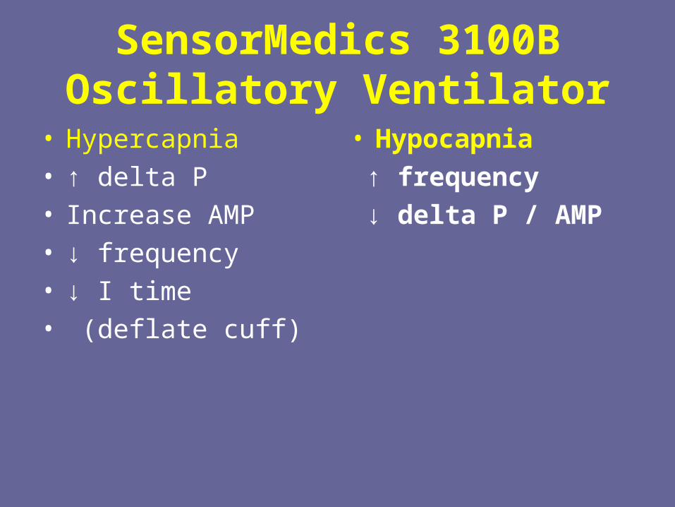
• Hypercapnia• ↑ delta P• Increase AMP• ↓ frequency• ↓ I time• (deflate cuff)
• Hypocapnia
↑ frequency
↓ delta P / AMP
SensorMedics 3100B Oscillatory Ventilator
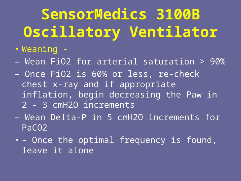
• Weaning -
– Wean FiO2 for arterial saturation > 90%
– Once FiO2 is 60% or less, re-check chest x-ray and if appropriate inflation, begin decreasing the Paw in 2 - 3 cmH2O increments
– Wean Delta-P in 5 cmH2O increments for PaCO2
• – Once the optimal frequency is found, leave it alone
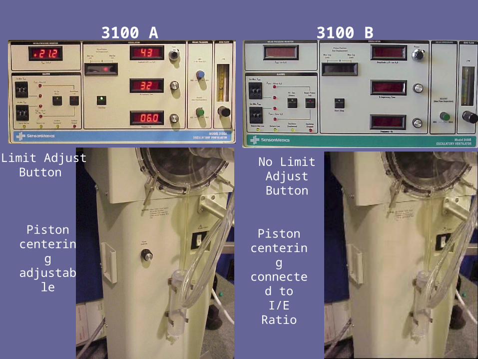
3100 A 3100 B
Limit AdjustButton
Piston centeringadjustable
No Limit AdjustButton
Piston centeringconnected
toI/E Ratio
High Frequency Jet Ventilation
• This is the modification of a technique initially developed to provide respiratory support during bronchoscopy. Gas from a high pressure source is delivered in short bursts down a fine cannula, the tip of which is lying within the endotracheal tube and pointing down towards the periphery of the lung
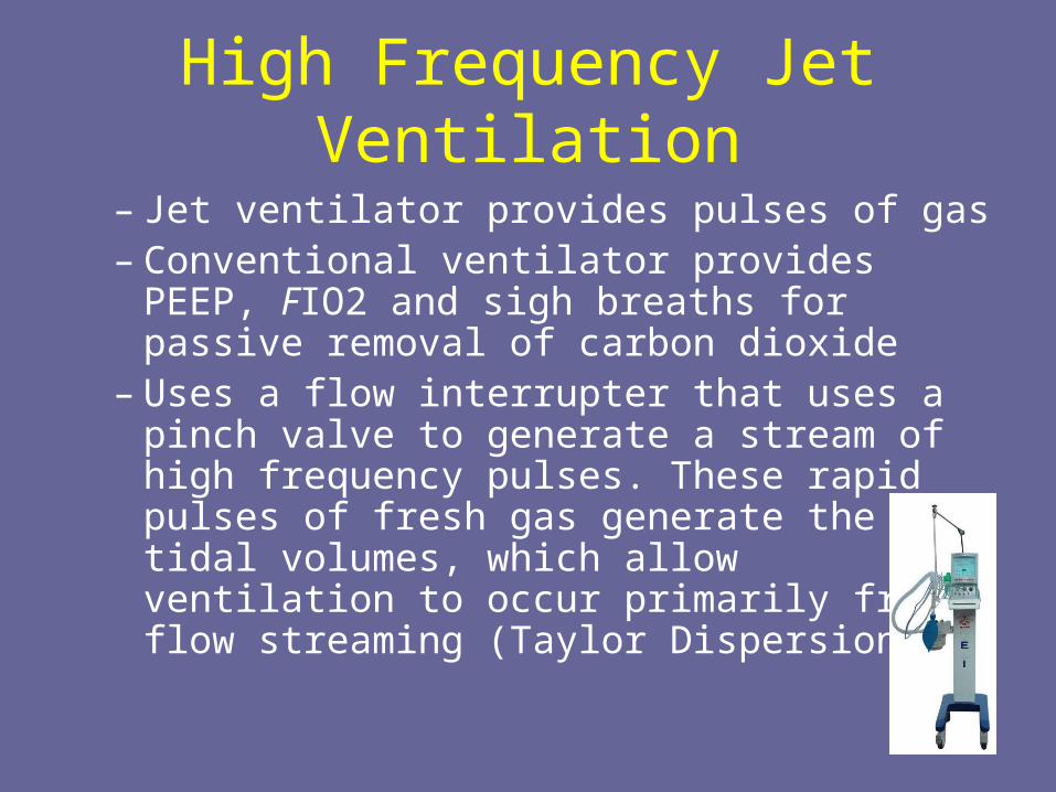
High Frequency Jet Ventilation
– Jet ventilator provides pulses of gas– Conventional ventilator provides PEEP, FIO2
and sigh breaths for passive removal of carbon dioxide
– Uses a flow interrupter that uses a pinch valve to generate a stream of high frequency pulses. These rapid pulses of fresh gas generate the tidal volumes, which allow ventilation to occur primarily from flow streaming (Taylor Dispersion)
High Frequency Jet Ventilation• A high pressure ‘’jet’’ of gas flows out of the
adaptor and into the airway. This jet of gas occurs for a very brief duration, about 0.02 seconds, and at high frequency: 4-11 hertz. Tidal volumes ≤ 1 ml/Kg are used during HFJV. This combination of small tidal volumes delivered for very short periods of time create the lowest possible distal airway and alveolar pressures produced by a mechanical ventilator. Exhalation is passive
High Frequency Jet Ventilation
• Jet ventilators utilize various I:E ratios--between 1:1.1 and 1:12-- to help achieve optimal exhalation. Conventional mechanical breaths are sometimes used to aid in reinflating the lung. Optimal PEEP is used to maintain alveolar inflation and promote ventilation-to-perfusion matching. Jet ventilation has been shown to reduce ventilator induced lung injury by as much as 20%.
High Frequency Jet Ventilation
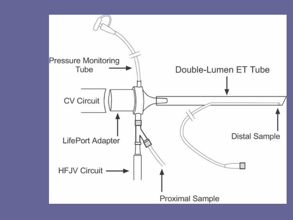
• A conventional ventilator is always run in tandem with the jet to generate the PEEP and sigh breaths. Expiration on HFJV is passive from elastic recoil. A special ET adapter is used during HFJV. This adapter has a jet port through which the High Frequency Jet pulses are introduced and a pressure monitoring port for determining the delivered pressures.
• Expiration is passive, air-trapping can occur
High Frequency Jet Ventilation• Aerosolized saline solution in the inspiratory
circuit is used to humidify the inspired air. Some additional gas is entrained during inspiration from a side port in the circuit. This form of HFV generally delivers a V of 2 to 5 mL/kg at a f of 100 to 200 breaths/min. The jet pressure (which determines the velocity of air jets) and the duration of the inspiratory jet (and, thus, the inspiratory/expiratory ratio [I/E]) are controlled by the operator.
INITIAL HFJV SETTINGS
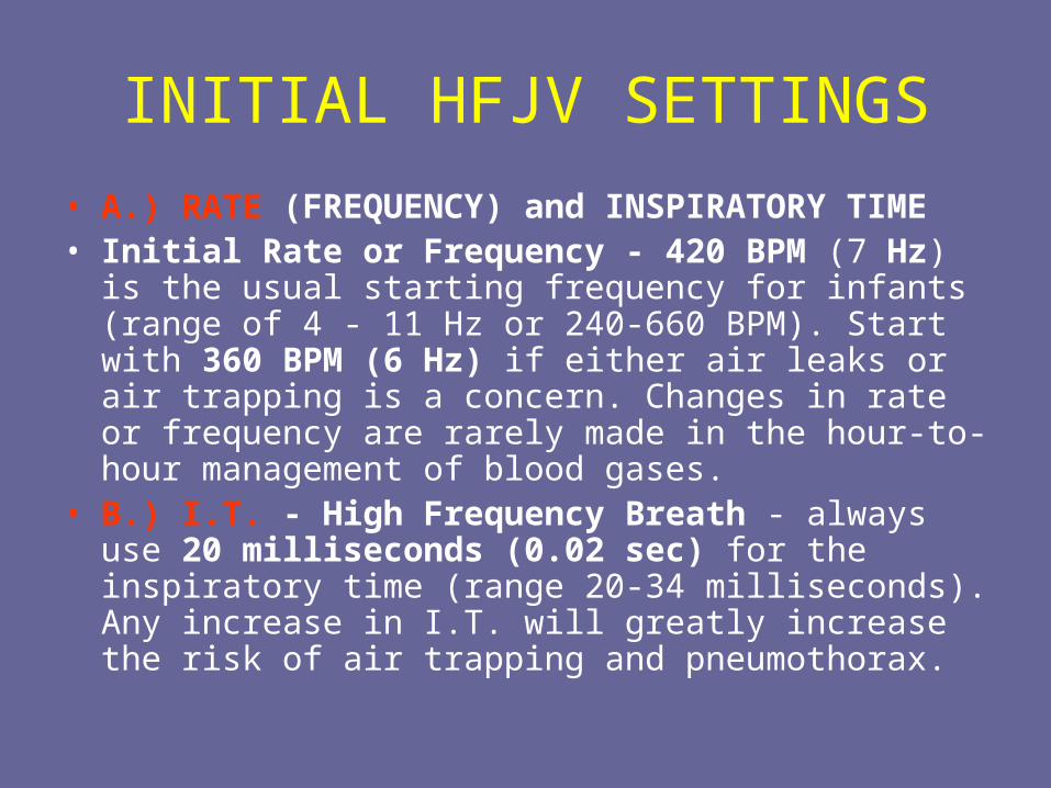
• A.) RATE (FREQUENCY) and INSPIRATORY TIME• Initial Rate or Frequency - 420 BPM (7 Hz) is the usual
starting frequency for infants (range of 4 - 11 Hz or 240-660 BPM). Start with 360 BPM (6 Hz) if either air leaks or air trapping is a concern. Changes in rate or frequency are rarely made in the hour-to-hour management of blood gases.
• B.) I.T. - High Frequency Breath - always use 20 milliseconds (0.02 sec) for the inspiratory time (range 20-34 milliseconds). Any increase in I.T. will greatly increase the risk of air trapping and pneumothorax.
INITIAL HFJV SETTINGS
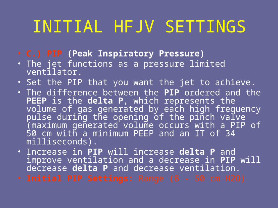
• C.) PIP (Peak Inspiratory Pressure)• The jet functions as a pressure limited ventilator. • Set the PIP that you want the jet to achieve. • The difference between the PIP ordered and the PEEP
is the delta P, which represents the volume of gas generated by each high frequency pulse during the opening of the pinch valve (maximum generated volume occurs with a PIP of 50 cm with a minimum PEEP and an IT of 34 milliseconds).
• Increase in PIP will increase delta P and improve ventilation and a decrease in PIP will decrease delta P and decrease ventilation.
• Initial PIP Settings: Range (8 - 50 cm H2O)
INITIAL HFJV SETTINGS
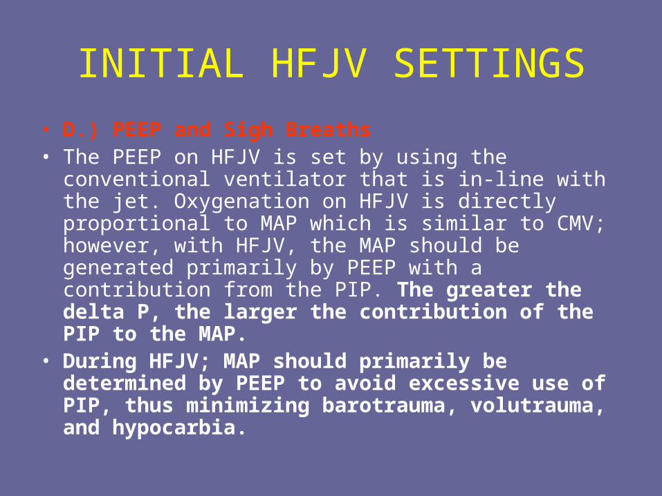
• D.) PEEP and Sigh Breaths• The PEEP on HFJV is set by using the conventional
ventilator that is in-line with the jet. Oxygenation on HFJV is directly proportional to MAP which is similar to CMV; however, with HFJV, the MAP should be generated primarily by PEEP with a contribution from the PIP. The greater the delta P, the larger the contribution of the PIP to the MAP.
• During HFJV; MAP should primarily be determined by PEEP to avoid excessive use of PIP, thus minimizing barotrauma, volutrauma, and hypocarbia.
Adjusting HFJV
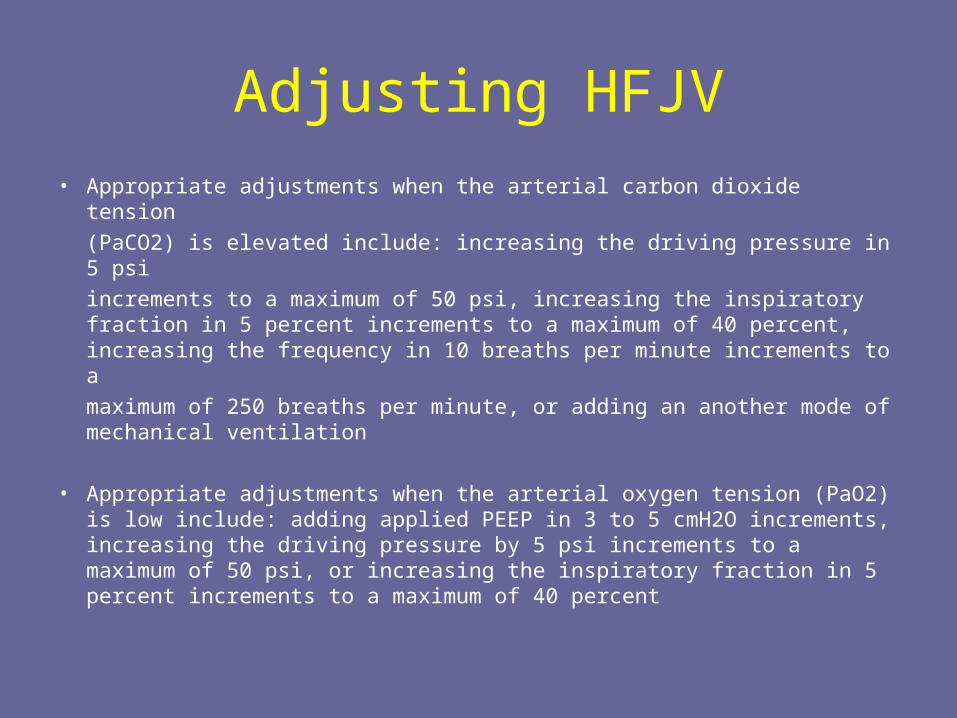
• Appropriate adjustments when the arterial carbon dioxide tension
(PaCO2) is elevated include: increasing the driving pressure in 5 psi
increments to a maximum of 50 psi, increasing the inspiratory fraction in 5 percent increments to a maximum of 40 percent, increasing the frequency in 10 breaths per minute increments to a
maximum of 250 breaths per minute, or adding an another mode of mechanical ventilation
• Appropriate adjustments when the arterial oxygen tension (PaO2) is low include: adding applied PEEP in 3 to 5 cmH2O increments, increasing the driving pressure by 5 psi increments to a maximum of 50 psi, or increasing the inspiratory fraction in 5 percent increments to a maximum of 40 percent
HFJV
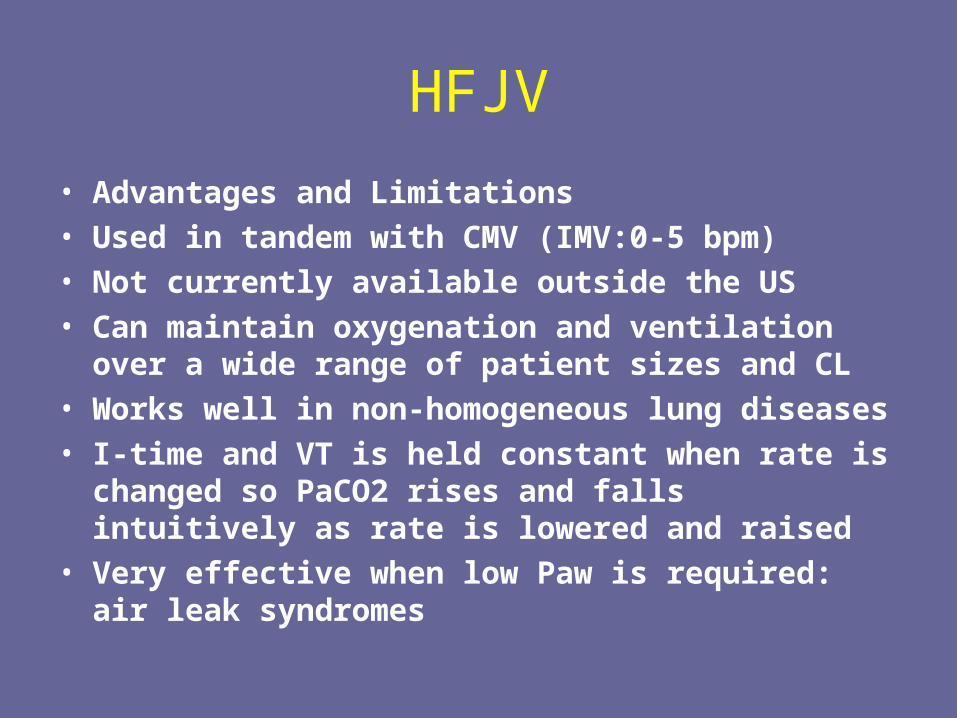
• Advantages and Limitations• Used in tandem with CMV (IMV:0-5 bpm)• Not currently available outside the US• Can maintain oxygenation and ventilation over a
wide range of patient sizes and CL• Works well in non-homogeneous lung diseases• I-time and VT is held constant when rate is changed
so PaCO2 rises and falls intuitively as rate is lowered and raised
• Very effective when low Paw is required: air leak syndromes
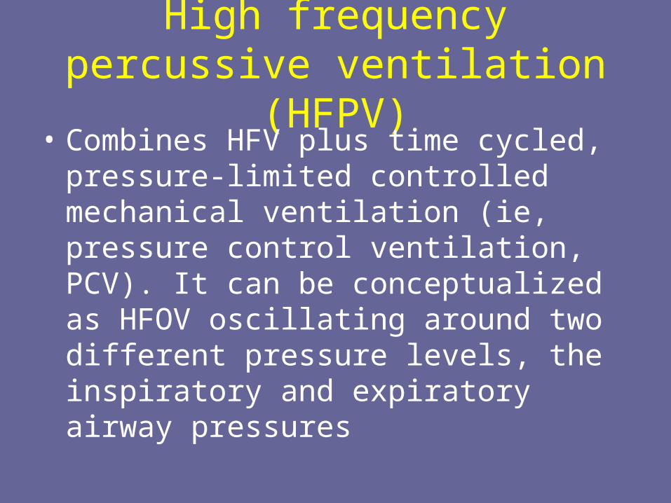
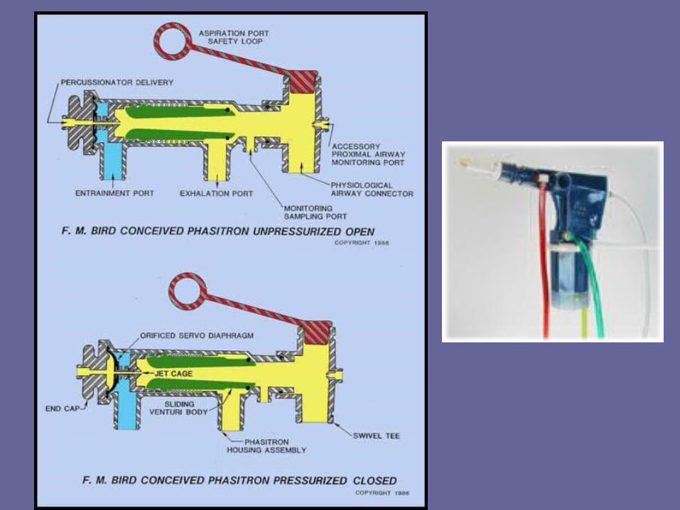
High frequency percussive ventilation (HFPV)
• Combines HFV plus time cycled, pressure-limited controlled mechanical ventilation (ie, pressure control ventilation, PCV). It can be conceptualized as HFOV oscillating around two different pressure levels, the inspiratory and expiratory airway pressures
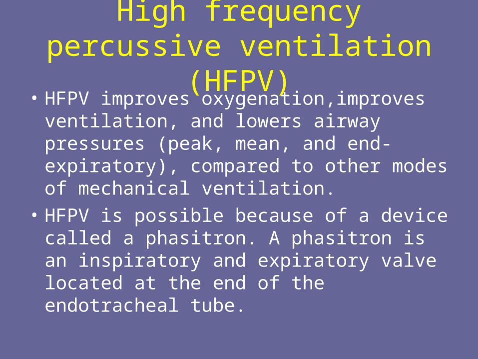
High frequency percussive ventilation (HFPV)
• HFPV improves oxygenation,improves ventilation, and lowers airway pressures (peak, mean, and end-expiratory), compared to other modes of mechanical ventilation.
• HFPV is possible because of a device called a phasitron. A phasitron is an inspiratory and expiratory valve located at the end of the endotracheal tube.
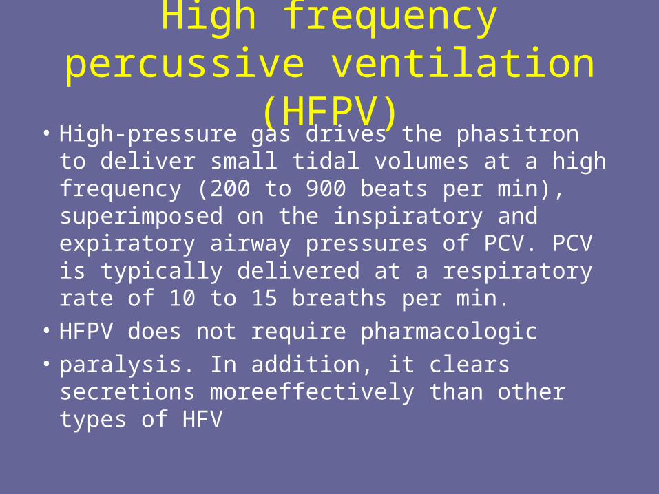
High frequency percussive ventilation (HFPV)
• High-pressure gas drives the phasitron to deliver small tidal volumes at a high frequency (200 to 900 beats per min), superimposed on the inspiratory and expiratory airway pressures of PCV. PCV is typically delivered at a respiratory rate of 10 to 15 breaths per min.
• HFPV does not require pharmacologic
• paralysis. In addition, it clears secretions moreeffectively than other types of HFV
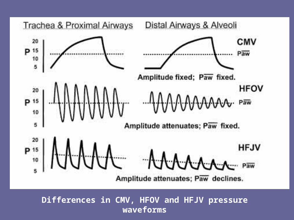
Differences in CMV, HFOV and HFJV pressure waveforms
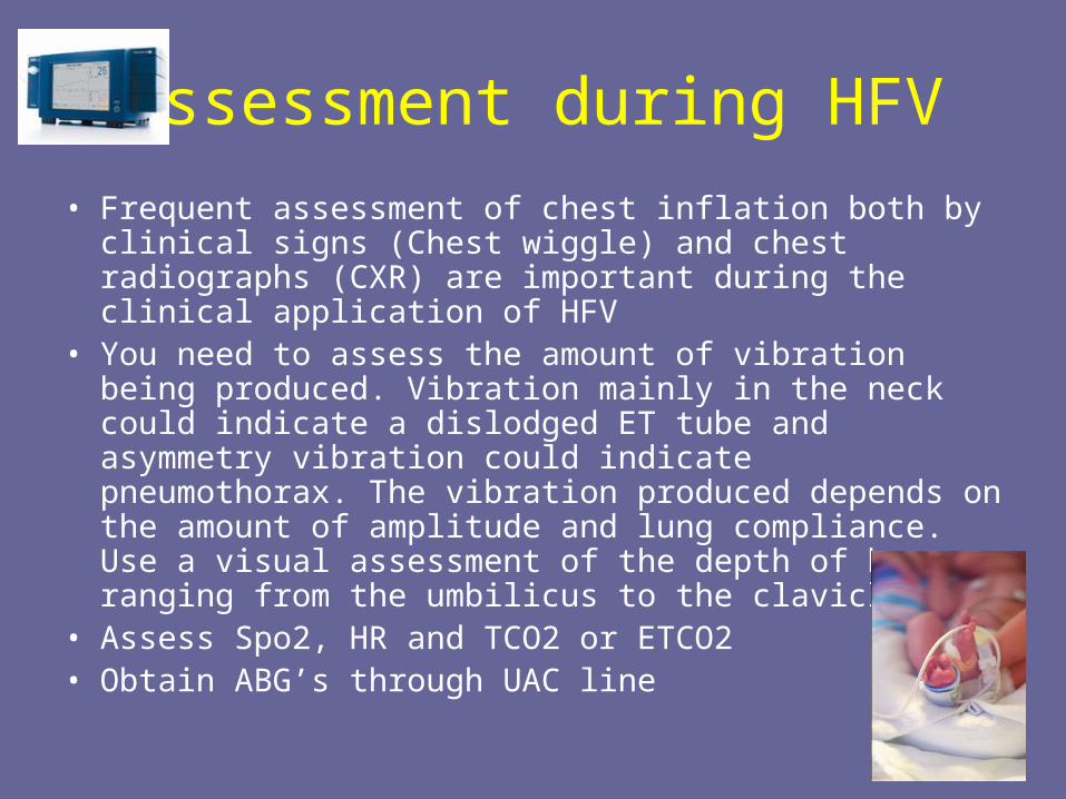
Assessment during HFV
• Frequent assessment of chest inflation both by clinical signs (Chest wiggle) and chest radiographs (CXR) are important during the clinical application of HFV
• You need to assess the amount of vibration being produced. Vibration mainly in the neck could indicate a dislodged ET tube and asymmetry vibration could indicate pneumothorax. The vibration produced depends on the amount of amplitude and lung compliance. Use a visual assessment of the depth of bounce ranging from the umbilicus to the clavicle.
• Assess Spo2, HR and TCO2 or ETCO2 • Obtain ABG’s through UAC line
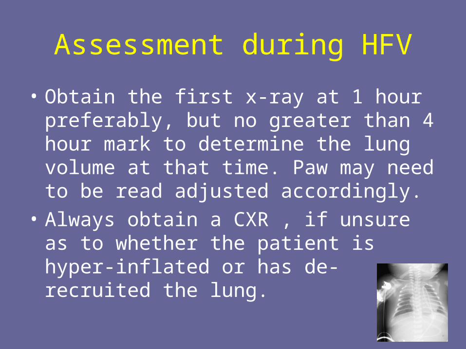
Assessment during HFV
• Obtain the first x-ray at 1 hour preferably, but no greater than 4 hour mark to determine the lung volume at that time. Paw may need to be read adjusted accordingly.
• Always obtain a CXR , if unsure as to whether the patient is hyper-inflated or has de-recruited the lung.
Assessment during HFV
• Chest wiggle that is absent or becomes diminished is a
• clinical sign that the airway or ET tube is obstructed.
• CW present on one side only is an indication that the ET tube has slipped down a primary bronchus or a pneumothorax has occurred.
• Check the position of the ET tube or obtain a CXR.
• Reassess CW following any position change.
Assessment during HFV
• Monitoring of infant’s heart rate may be problematic via ECG electrodes. Heart rate can be monitored as a ‘pulse’ through the UAC . Evaluation for heart murmurs may require a temporary pause in HFOV therapy.
• Blood Pressure. Be prepared for a potential blood pressure drop; this is due to the increased intra-thoracic pressure that oscillation can cause, resulting in decreased venous return.
Assessment during HFV
• Auscultation:Listening to breath sounds in infants ventilated on HFOV may be helpful, as the sounds (friction sounds) become reduced in the affected side when the endotracheal tube is low and ventilates only 1 lung or when a pneumothorax is present. These changes may occur before the infant becomes symptomatic. Thus auscultation should be performed at the time of routine assessment or if there is clinical deterioration
• Adventitious/Vesicular BS are not easily auscultated
Assessment during HFV
• Suctioning • Keep to a minimum and us In-line suction catheter • Press Stop button briefly on SensorMedics while briefly
inserting and withdrawing catheter. PAW is maintained throughout. You may increase Paw/FIO2 slightly– Rationale for pausing – The oscillator causes a pressure pulse
in the airways. When suctioning if the sensormedics isn’t turned off the secretions get pushed back down because of this pulse pressure. So you are having ineffective clearance of secretions. There is also the potential of air trapping with active piston movement
– Push Reset button anytime ventilator becomes disconnected and suddenly stops
Hazards to HFV
- Air-trapping causing lung over-inflation- Improper sedation/fighting of ventilator- Pulmonary interstitial emphysema- Intraventricular haemorrhage &
periventricular leukomalacia- Tracheal damage- Sudden disconnect and alveolar collapse- Noise pollution
Key points to starting HFV
• Choosing the RIGHT patient. HFV is NOT for every patient
• When to Start?
Early application provides protection and reduces incidence of further lung damage
• Rescue may or may not improve mortality chances. The later HFV is started the less chance of survival
Key Points
• Retrospective studies suggest that HFV improves gas exchange in infants with severe respiratory failure and hence reduces the need for ECMO. The response rate to HFV appears to be disease-specific. Infants who have homogeneous lung diseases, such as RDS or pneumonia, are more likely to respond more favorably to HFV than those who have more heterogeneous lung disease such as ARDS, meconium aspiration pneumonia.
Key points to starting HFV
• Sedation/Paralytic may be required during application of HFV
• Watch for sudden deterioration in vital signs• Watch for Atelectasis and pneumothorax• ALLOW FOR PERMISSIVE HYPERCAPNIA
Case Study:• http://www.viasyshealthcare.com/prod_serv/dow
nloads/HFOVCaseStudy5_I.pdf
Nitric Oxide
• Nitric Oxide (NO) Therapy is used to relax smooth muscle to improve blood flow to alveoli to improve ventilation/perfusion mismatch, decrease pulmonary vascular resistance, decrease pulmonary pressures, improve oxygenation and reduce need for ECMO
• NO acts as a specific pulmonary vasodilator reducing PVR and pulmonary artery pressure
• Inhaled NO provides selective vasodilation of the pulmonary arterioles without systemic effect.
Nitric Oxide
Nitric Oxide (NO), not to be confused with the anesthetic nitrous oxideselective vasodilation properties.
NO is the active metabolite of a number of other vasodilators, including sodium nitroprusside and nitroglycerin. Produced in all human organ systems, including the nasopharynx and lungs.
In high concentration, NO is profoundly toxic and causes disease identical to Acute Respiratory Distress Syndrome (ARDS). In presence of oxygen, NO is broken down to form nitrogen dioxide (NO2). In the blood NO interacts with hemoglobin. The byproduct of this reaction produces increased levels of methemoglobin. Methemoglobin will not carry oxygen, and therefore, its level must be closely monitored during NO therapy.
Nitric Oxide
• Nitric oxide is approved by the US Food and Drug Administration for hypoxic respiratory failure of the term and near-term newborn
• Hypoxic respiratory failure in neonates born at or near term may be caused by such conditions as primary persistent pulmonary hypertension, respiratory distress syndrome, aspiration syndromes, pneumonia or sepsis, and congenital diaphragmatic hernia.

Nitric Oxide
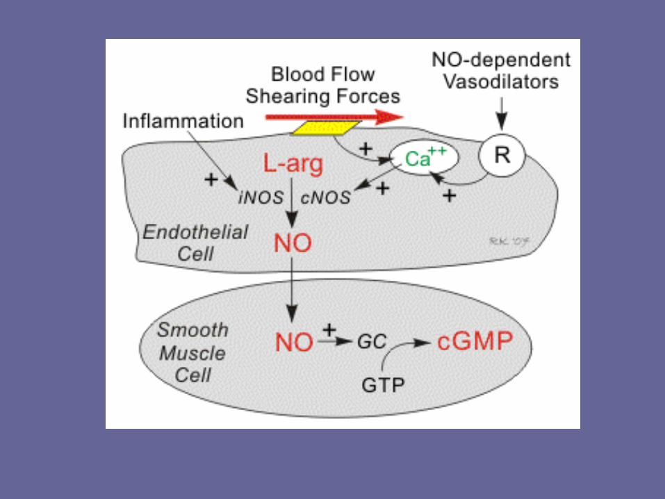
• Inhaled nitric oxide (iNO) is a selective pulmonary vasodilator for which the mechanism of action involves guanylyl cyclase activation leading to production of cyclic guanosine monophosphate and subsequent smooth muscle relaxation
Nitric Oxide
• Inhaled nitric oxide (INO) is indicated for:• Hypoxic respiratory failure • Newborns older than 34 weeks gestational age
who have either: – Persistent pulmonary hypertension of the neonate
(PPHN) – Congenital diaphragmatic hernia (CDH) – Oxygenation index (OI) > 25
• A special ventilator injects NO into the ventilator circuit. Key clinical information about INO appears below.

Nitric Oxide
• NO diffuses into the vascular smooth muscle cells adjacent to the endothelium where it binds to and activates guanylyl cyclase. This enzyme catalyzes the dephosphorylation of GTP to cGMP, which serves as a second messenger for many important cellular functions, particularly for signaling smooth muscle relaxation.

Nitric Oxide
• Cyclic GMP induces smooth muscle relaxation by multiple mechanisms including – Increased intracellular cGMP, which inhibits calcium
entry into the cell, and decreases intracellular calcium concentrations
– Activates K+ channels, which leads to hyperpolarization and relaxation
– Stimulates a cGMP-dependent protein kinase that activates myosin light chain phosphatase, the enzyme that dephosphorylates myosin light chains, which leads to smooth muscle relaxation.

Nitric Oxide indications
• Inhaled nitric oxide (INO) is indicated for:• Hypoxic respiratory failure • Newborns older than 34 weeks gestational age
who have either: – Persistent pulmonary hypertension of the neonate
(PPHN) – Congenital diaphragmatic hernia (CDH) – Oxygenation index (OI) > 25
• A special ventilator injects NO into the ventilator circuit. Key clinical information about INO appears below.

Application of NO
• Dosing begins at 20 ppm• Wean by decreasing dose by ½ each time • Wean to 1 ppm • Increase FIO2 when discontinuing INO
– The PaO2 decreases when INO is discontinued because the body needs time to increase NO productionPotential problems
• NO + O2 → NO2; presence of nitrogen dioxide (NO2) is not a problem at therapeutic doses
• NO + red blood cell → metHb; monitor levels
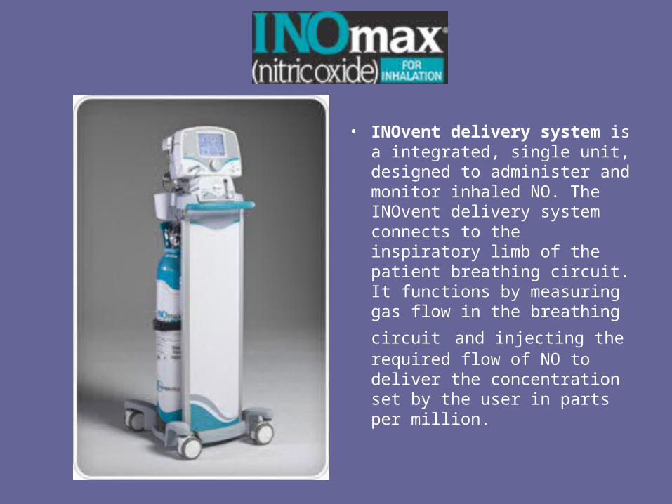
• INOvent delivery system is a integrated, single unit, designed to administer and monitor inhaled NO. The INOvent delivery system connects to the inspiratory limb of the patient breathing circuit. It functions by measuring gas
flow in the breathing circuit and injecting the required flow of NO to deliver the concentration set by the user in parts per million.
Contraindications
Absolute contraindications:
• Patients with congenital or acquired methemglobinemia reductase deficiency
Relative contraindications:
• Patients with a bleeding diathesis Intracranial hemorrhage
• Severe left ventricular failure
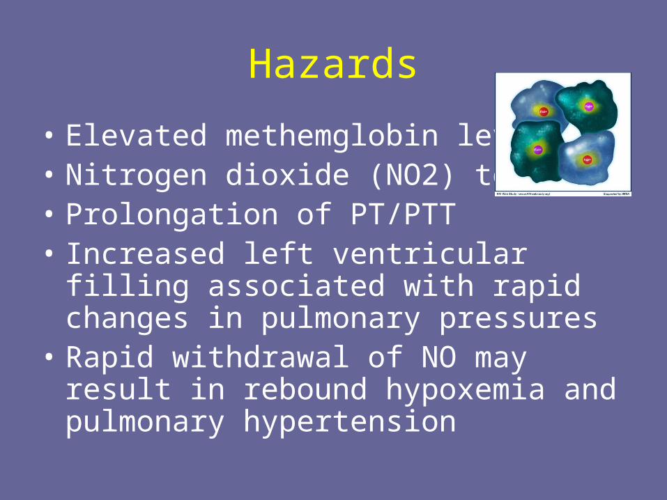
Hazards
• Elevated methemglobin levels • Nitrogen dioxide (NO2) toxicity • Prolongation of PT/PTT • Increased left ventricular filling associated
with rapid changes in pulmonary pressures
• Rapid withdrawal of NO may result in rebound hypoxemia and pulmonary hypertension
Precautions
• When given via mechanical ventilator, an increase in exhaled tidal volumes might be noted. This increase occurs as a result of additional gas flow from NO into the circuitry.
• Trigger sensitivity of the ventilator might be compromised, especially if the patient is on an assisted mode of ventilation.
Monitoring while using NO
• FiO2 • Tidal volume • Trigger sensitivity • Pulse oximetry • Arterial blood gases must be obtained at baseline and as follows: • Post initiation • Each hour or PRN as needed for six hours • 30 minutes after each NO concentration adjustment • At any time when clinically indicated • Pulmonary artery pressure • Platelet count • NO2 levels
http://inomax.com/View InoMax MOA


![Airway Humidification During High-Frequency Percussive ... · ventilation, high-frequency ventilation, airway humidification . [Respir Care 2009;54(3):350 358.] Introduction In general,](https://img.pdfslide.us/doc/110x75/5edb55e8ad6a402d66658116/airway-humidification-during-high-frequency-percussive-ventilation-high-frequency.jpg)