Embed Size (px)
Citation preview
Introduction to Clinical Anesthesia
This syllabus is designed to provide basic medical knowledge of anesthesia for junior
medical students from the University of Arkansas for Medical Sciences during one-week
anesthesiology clerkship at Arkansas Children’s Hospital.
More information including video lectures geared towards students can be found at
http://www.archildrens.org/video/anesthesiology.asp
OBJECTIVES
• Assess and evaluate preoperative risk factors for anesthesia and surgery.
• Basic Airway Management assessment.
• Describe the principles of applied physiology and pharmacology with regards to
Anesthesiology
• Basic management of fluids and electrolytes in patients undergoing anesthesia.
Introduction
The specialty of anesthesiology has advanced as a medical specialty in the past few
decades. As perioperative physicians, anesthesiologists are now actively involved in patient
care, working in preoperative clinics, operating rooms, post anesthesia care units and
intensive care units. Anesthesiologists are also involved in taking care of patients with acute
and chronic pain syndromes in pain management clinics, ambulatory care centers, surgical
centers, administration and research.
In United States the first organization of practicing anesthesiologists was formed in 1905,
was named "The Long Island Society of Anesthetists". The name was changed to New York
Society of Anesthetists in 1911.
The American Board of Anesthesiology (ABA) was formed in 1938 as an affiliate board of
the American Board of Surgery. ABA became an independent board in 1941.
The principal sub-disciplines of the practice of anesthesiology include, cardiothoracic
anesthesia, ambulatory anesthesia, obstetric anesthesia, neuroanesthesia, pediatric
anesthesia, regional anesthesia, critical care medicine and acute and chronic pain
management.
Preoperative evaluation
Preanesthesia evaluation is designed to provide patients an opportunity to discuss the
anesthetic plan before surgery with their anesthesiologist. In the preoperative clinic an
anesthesiologist will evaluate the medical condition of all patients, and in conjunction with
the patient, will formulate a plan for the Perioperative anesthetic care. Special emphasis is
given to airway evaluation, cardiopulmonary status, liver and kidney disease and any
necessary labs, chest X-rays and ECG are performed in the clinic. The patients’ old charts
are reviewed for any previous anesthesia related problems, and for comorbidities, including
hypertension, coronary artery disease, pulmonary disease, diabetes, renal and hepatic
disease, neurological disease, and among others social habits, Previously treated medical
conditions are reviewed thoroughly, as is the patients’ surgical history.
The Medical History:
The main objective of good medical history is to uncover and assess the severity of any
pathologic condition that would influence the selection of intraoperative techniques,
monitoring and anesthetics. The focus should be on the preanesthetic problem areas
pertaining to history, physical examination and the surgical condition. The medical problems
should be characterized by date and time of onset, severity, functional limitation due to the
medical condition and response to therapy. Other important areas of emphasis are,
allergies, the current medication list and the review of systems.
Cardiac History
Recent onset of chest pain, severity of chest pain, history of myocardial infarction,
exercise tolerance and response to treatment. History of hypertension (controlled or
uncontrolled), valvular heart disease, symptomatic dysrhythmias and patients’ functional
status is assessed according to the New York Heart Association classification.
New York Heart Association classification
Class I: Cardiac disease without limitation of physical activity
Class II: Slight limitation of physical activity
Ordinary physical activity results in angina or fatigue
Class III: Marked limitation of physical activity
Class IV: Angina at rest, increased with activity
Pulmonary History
History of shortness of breath, asthma, COPD, emphysema and smoking history.
Recent upper respiratory tract infections with fever and sputum production. The patients’
exercise tolerance and sleep pattern should be assessed.
Renal and liver disease
Organ dysfunction can affect the metabolism and clearance of certain intravenous as
well as inhalational anesthetics. Serious bleeding problem can occur with renal (platelet
dysfunction) or liver disease (deficient clotting factors).
GI reflux disease
If present, patients are prone to aspiration of gastric contents. Hiatal hernia is
believed to increase the risk of aspiration, as is diabetes, history of chronic narcotic
ingestion, obesity and pregnancy.
Diabetes
If present, close monitoring of blood glucose should be considered perioperatively.
Diabetes also affect gastric emptying, having a significant impact on preoperative
medication selection and management.
Rheumatoid arthritis especially if treated with steroids and ankylosing spondylosis with
involvement of C-spine (difficult airway, possible atlanto-axial subluxation etc).
Alcoholism and drug abuse: Increased tolerance to many sedative and narcotics
Family History: Specific history of previous anesthetic problems, history of malignant
hyperthermia, enzyme deficiency and other familial and inherited diseases.
Allergies: Antibiotics, anesthetics, analgesics, sedatives/ hypnotics.
Medications: Appropriate instructions must be given to the patient preoperatively regarding
their medication management.
Bleeding: Abnormal platelet function or hereditary deficiency of clotting factors, aspirin
therapy and kidney disease.
NPO status should be part of the checklist preoperatively so that proper anesthesia
induction technique can be planned.
PHYSICAL EXAMINATION
AIRWAY EVALUATION
The airway evaluation is an integral part of preanesthesia evaluation. The examination of
airway should always include
Overall appearance
Neck: stout or thin, long or short? Sunken cheeks and Presence of beard may make
mask fit difficult.
Mouth
Mouth opening (measured in cm or fingerbreadth)
Anterior displacement of mandible
Tongue size
Visibility of uvula
Protrusion of upper incisors
Loose or damaged teeth; prostheses,
Movement
Flexion/ extension of neck
Sniffing position
Palpation
Trachea in midline
Distance from mentum to hyoid
Nose
Both nares patent
Protuberant nose suggests poor mask fit and difficult mask ventilation.
There are three preoperative airway examinations that attempt to predict the ease of
endotracheal intubation.
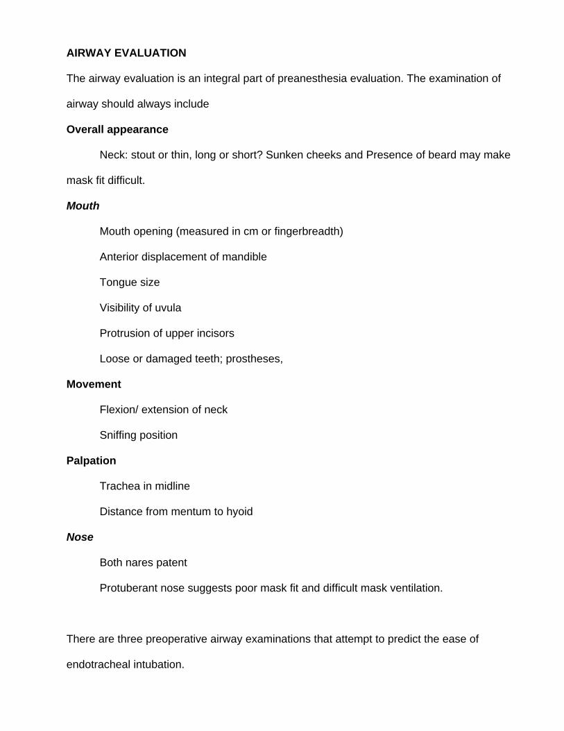
1: Size of tongue in relation to the size of oral cavity
MALAMPATI CLASSIFICATION (Modification)
Patient is asked to open mouth widely
Class 1: Soft palate, fauces, uvula, anterior and posterior faucial pilars can be seen.
Class 2: Soft palate, fauces, uvula can be seen. The tongue masks anterior and posterior
faucial pillars.
Class 3: Soft palate and the base of uvula can be seen only.
Class 4: Only hard palate is visible.
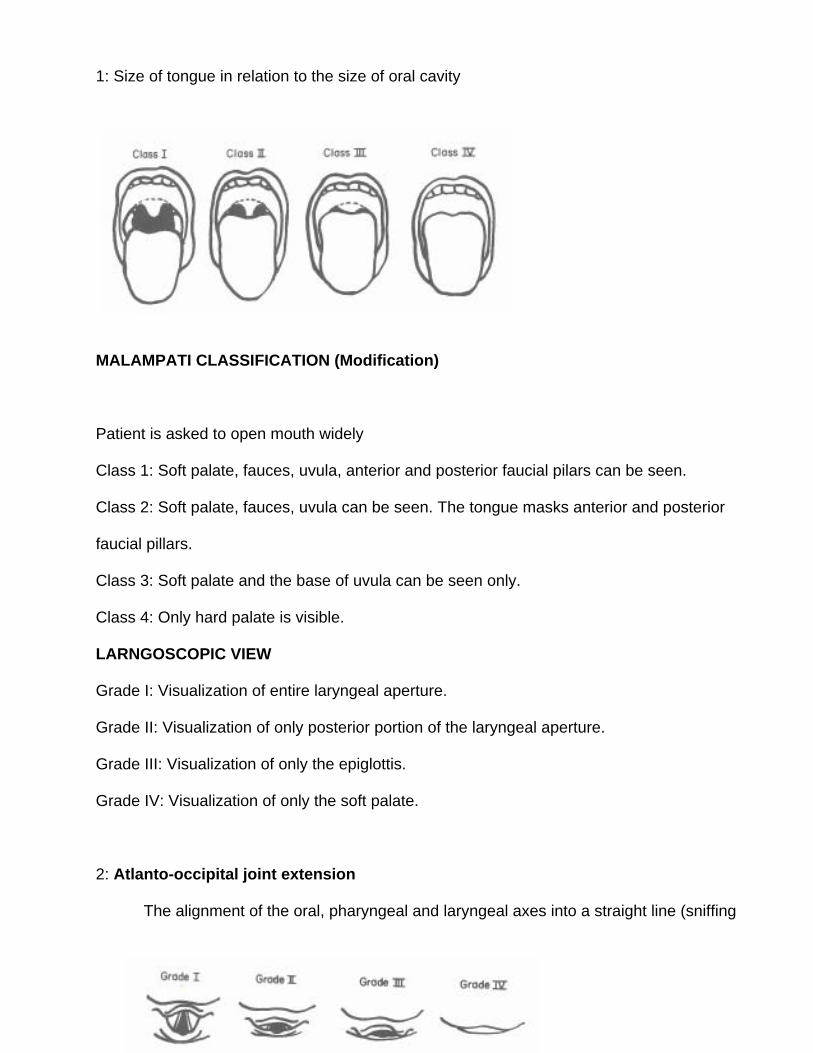
LARNGOSCOPIC VIEW
Grade I: Visualization of entire laryngeal aperture.
Grade II: Visualization of only posterior portion of the laryngeal aperture.
Grade III: Visualization of only the epiglottis.
Grade IV: Visualization of only the soft palate.
2: Atlanto-occipital joint extension
The alignment of the oral, pharyngeal and laryngeal axes into a straight line (sniffing
position). This will allow less of the tongue obscuring the laryngeal view and there will be
much less need for displacing the tongue anteriorly.
3: Thyro-mental distance:
The space anterior to the larynx determines how readily the laryngeal axis will fall in
line with the pharyngeal axis when the atlanto-occipital joint is extended.
When there is a large mandibular space, the tongue is easily contained within this
large compartment and does not have to be pulled maximally forward in order to reveal the
larynx. The distance between inside the mandible to hyoid bone should be greater than 6
cm or three fingerbreadths.
Prior surgical history and intubation record should always be available (when applicable).
Other medical conditions with involvement of airway and C-spine include:
Rheumatoid Arthritis involving, cervical spine, TMJ and Cricoarytenoid joint
TMJ Dysfunction (impedes mouth opening)
Acromegaly
Cancer of head and neck, particularly, involving upper airway and trachea.
History of prior radiation treatment of neck (for cancer treatment)
Obstructive sleep apnea
Prior airway surgery
Facial trauma with mandibular fracture (CSF rhinorrhea, etc)
NECK: Neck examination should be performed as part of airway evaluation. Presence of
carotid bruit, midline masses which can deviate or compress the trachea.
LUNGS: Presence of any abnormal lung sounds (wheezing, rales) merit further evaluation
of the patients' pulmonary status.
HEART: Assessment should include heart rate, rhythm and presence or absence of
murmur and distention of jugular (JVD).
Examination of the extremities and back is part of the preoperative evaluation.
ASA Physical Status Classification
Class 1: A normal healthy patient
Class 2: A patient with mild systemic disease that results in no functional limitation.
Class 3: A patient with severe systemic disease that results in functional limitation.
Class 4: A patient with severe systemic disease that is a constant threat to life.
Class 5: A moribund patient that is not expected to survive for 24 hours with or without the
operation.
Class 6: A declared brain-dead patient whose organs are being removed for donor
purposes.
The modification E is added to the ASA physical status classification to indicate that the
case is done emergently.
Preoperative labs tests are indicated either to confirm the findings on abnormal physical
examination or that will help the anesthesiologist to manage the patient's problems
perioperatively.
EKG: Male or female, 50 years of age and older with coexisting cardio-pulmonary risk
factors.
Chest X-rays: Chest x-rays are not indicated for any asymptomatic patient who is less than
75 years of age and has no cardio-pulmonary risk factors. Chest X-ray may be helpful in
diagnosing the existence of tracheal deviation, mediastinal mass, lung mass, aortic
aneurysm, pulmonary edema, pneumonia, atelectasis, fracture of clavicle and cardiomegaly.
THE MONITORS
The ASA (American Society of Anesthesiologists) requires that during all types of
anesthetics the patient’s oxygenation, ventilation, and circulation must be monitored
continuously. Temperature monitoring must be readily available. Therefore the minimum
monitors we must use is as follows.
• Oxygen analyzer
• Pulse oximetry
• Capnometry
• Stethoscope
• EKG
• Blood pressure cuff
• Temperature probe
How does the pulse oximeter works?
Pulse oximeter combines the principles of oximetry and plethysmography to noninvasively
measure oxygen saturation in arterial blood. The pulse oximeter probe contains two light
emitting diodes at wavelengths of 940nm and 660 nm. Oxygenated and reduced
hemoglobin differ in light absorption (940 and 660 nm respectively). Thus the change in light
absorption during arterial pulsation is the basis of oximetry determination. The ratio of the
absorption at the two wavelengths is analyzed by a microprocessor to record the oxygen
saturation.
What is Capnometry?
Capnometry is the measurement of end-tidal carbon dioxide tension. This provides valuable
information to the anesthesiologist. The presence of end tidal CO2 aids in confirming
endotracheal intubation. Alteration in the slope of the graph can give clues to the presence
of airway obstruction. A rapid fall in reading may signify extubation, air embolism or low
cardiac output with hypovolemia.
THE AIRWAY
The anatomic difference between adult and pediatric airway is an important aspect of airway
examination.
The larynx lies at C4/C5 in adults and at C3/C4 in pediatric age group. In children the cricoid
ring is the narrowest part as compared to glottic opening is the narrowest part in adults. The
epiglottis is crescent shaped in adults and is long and omega shaped in children.
Predictors of Difficult Airway
Short muscular neck
Prominent upper incisors
Protruding mandible
Receding mandible
Small mouth opening
Full beard
Large tongue
Limited neck mobility
Limited mouth opening due to TMJ
TECHNIQUE FOR OROTRACHEAL INTUBATION
Equipment
Properly sized endotracheal tube
Laryngoscope handle and blades (Miller and Macintosh)
Functioning suction catheter
Oxygen supply source and properly functioning equipment to mask ventilate before
intubation (anesthesia machine, Ambu-bag).
Oral and nasal airways
Drugs: induction agents and muscle relaxants
HEAD POSITIONING
(A): Successful exposure of the glottic opening by direct laryngoscopy requires alignment of
the oral, pharyngeal and laryngeal axes.
(B): Elevation of the patient’s head with pads under the occiput and shoulders remaining on
the table aligns the pharyngeal and laryngeal axes.
(C): Head extension at the atlanto-occipital joint creates the shortest distance and nearly
straight line from incisors teeth to the glottic opening.
Planning for Anesthesia
A good anesthetic begins with a good plan. There is no rigid format for planning anesthesia.
Rather, each plan is adapted to each case. The fundamental goal of anesthetic
management is to provide safety, comfort and convenience, first for the patient and second
for those caring for the patient.
After a good plan, a good preparation is required for a good anesthetic.
Before every anesthetic, every anesthesiologist should go through a checklist of necessary
items including, anesthesia machine, ventilator, oxygen and nitrous supply check, suction
device, monitors and anesthesia cart.
Before bringing the patient to the operating room, the proper verification of patient's identity,
the planned procedure and the site of the procedure should be carried out by the
anesthesiologist. All the preparations should be completed before the patient enters the
room including the placement of a working peripheral intravenous line.
INDUCTION OF GENERAL ANESTHESIA
1. ASA standard monitors (Pulse oximeter, NIBP cuff, ECG and temperature).
2. Preoxygenation with 100% Oxygen or Denitrogenation with proper fitting mask .
3. Inhalation or Intravenous induction of general anesthesia.
4. Endotracheal intubation and securing the ET tube.
5. Protection of the pressure points.
MAINTENANCE OF ANESTHESIA
Careful and continous vigilance of vital sings and depth of anesthesia is the integral part of
the maintenance phase. Pulse oximetry, End-tidal carbon di-oxide tension, patient's
temperature, ECG and blood pressure are continuously monitored during the maintenance
phase. End-tidal concentration of nitrous oxide and inhalation agents (isoflurane, halothane
etc) is continuously monitored for the proper depth of anesthesia (analgesia, amnesia,
sedation and muscle relaxation). It is important to keep track of the blood loss during the
case and should be replaced hourly with crystalloid. Fluid therapy should be guided by
monitoring hourly urine output (0.5 cc/Kg/Hr).
EMERGENCE FROM GENERAL ANESTHESIA
1. Reversal of muscle relaxation.
2. Turning off the inhalation agents and nitrous oxide
3. Meeting the extubation criteria
4. Extubation of trachea
5. Transfer of the patient to post anesthesia care unit.
Intravenous Anesthetics and Muscle Relaxants
1. Intravenous anesthetics
Mechanism of action: Usually inhibits the activity or activates the inhibitory, signaling
pathways in the brain. Facilitatory actions on GABA receptors appear most important,
although modulation of many other receptors and channels plays a role as well.
Uses:
• Induction of general anesthesia.
• Supplementation of general anesthetics intraoperatively.
• Maintainance of general anesthesia
• Maintainance of continous IV sedation in OR, ICU and other remote locations.
• Protection of the brain in patients’ with increased intracranial pressure.
Propofol (Diprivan)
Propofol is an alkylphenol, formulated as 1-% solution dissolved in 10% intralipid
(explaining the milky white color).
Site of action: GABAA receptors in CNS. GABAA is inhibitory neurotransmitter in CNS. Onset
of action is within 30 to 60sec after IV injection and duration of action (hypnosis) is between
3 to 10 minutes.
Uses:
• Induction and maintenance of general anesthesia.
Continous intravenous sedation in the ICU and Operating rooms for procedures done
under monitored anesthesia care.
Effects and side effects:
• Hypnotic and amnestic properties
• No analgesic properties
• Respiratory depression and bronchodilation
• Cardiovascular depression and hypotension due to peripheral vasodilation
• Antiemetic properties
• Pain on injection (pretreatment with lidocaine will attenuates the pain)
• Myoclonus (rare)
Contraindications:
· Cardiovascular instability and patients’ hypersensitivity to drug and its components.
Pharmacology:
• Metabolized in liver by conjugation to glucuronide and sulfate.
• Formulation: 10 mg/ml
• Dose: 1-2.5 mg/kg for induction; 50-150 mcg/kg/min. for maintenance of general
anesthesia
• 10 to 50 mcg/Kg/min. for sedation in the ICU and remote locations.
Barbiturates
Barbiturates are derivatives of barbituric acid. Site of action: primarily GABA receptors in
CNS. They enhance and mimic the activity of GABAA in CNS. They have hypnotic and
amnestic properties but are not analgesics.
Thiopental (Sodium Pentothal)
Uses:
• Intravenous induction agent with rapid onset and offset time.
• Attenuates acute rise in blood pressure (e.g. head pinning during neurosurgical
procedures).
• Acute perioperative seizure control.
• Provides brain protection by decreasing cerebral blood flow, cerebral oxygen
consumption and intracranial pressure (ICP).
Effects and side effects:
• Thiopental reversibly reduces cerebral electrical activity to the level of EEG silence,
with a significant reduction in cerebral metabolism.
• Decrease in blood pressure is mainly due to decreased peripheral vascular
resistance, which can be more pronounced in hypovolemic patients or those with
cardiovascular disease.
• Temporary depresses ventilation and decreases cerebral responsiveness to
increased CO2.
Contraindications:
• Hypovolemia
• Poor cardiac status
Pharmacology:
• Metabolized in liver
• Formulation: 25 mg/ml
• Induction dose: 3-5 mg/kg
• Onset: 30 seconds
• Duration of action: 5-10 minutes
Etomidate
Etomidate is an imidazole, supplied as a highly hyperosmotic solution (>4500 mOsm/l) in
propylene glycol. Site of action: GABAA receptors in CNS.
Uses:
Etomidate is a drug of choice for induction of general anesthesia in hemodynamically
unstable patients or in those patients with marginal cardiac reserve.
Effects and side effects:
• Cerebral effects: Decreased cerebral blood flow, decreased cerebral oxygen
consumption and decreased ICP.
• In contrast to thiopental, etomidate has minimal effects on the cardiovascular system.
• Respiratory effects are minimal but it reduces the cerebral response to increased
CO2 (hypercarbia).

• Dose-dependent, reversible suppression of adrenal gland by inhibiting 11-b-
hydroxylase, a key enzyme in steroid production.
• Myoclonus
• Pain on injection and thrombophlebitis
• Post-op nausea and vomiting
Pharmacology:
• Metabolized in liver by ester hydrolysis or by N-dealkylation
• Formulation: 2 mg/ml
• Dose: 0.2 to 0.6 mg/kg
• Onset: < 1 min.
• Peak effect 1 min
• Duration of action: 3-10 min.
Ketamine
Ketamine is a phencyclidine derivative (similar to PCP).
Site of action: Inhibition of signaling at the NMDA receptor, although multiple secondary
sites (opioid receptors, muscarinic acetylcholine receptors) exist.
Uses:
• Induction of anesthesia in children, by IM or IV route
• Induction of anesthesia in hypovolemic patients (etomidate is preferable).
• Supplementation of sedation during painful procedures due to its analgesic property.
• Ketamine increases cerebral blood flow and ICP; hence, it is contraindicated in
patients with increased ICP.
Effects and side effects:
• Potent analgesic
• "Dissociative" anesthesia
• Adrenergic activation
• Bronchodilator and maintains CO2 responsiveness
• Amnesia
• Nystagmus
• Induces salivation
• Dreaming and emergence reactions (less in children)
Contraindications:
• Increased ICP
• Open globe- eye injury
• Ischemic heart disease
• Psychological disease
Pharmacology:
• Formulation: Two concentrations: 10 mg/ml and 100 mg/ml. Careful!
• Dose: 0.5-2 mg/kg IV, 4-6 mg/kg IM for induction of general anesthesia.
• Onset: 1 min. IV, 5 min. IM
• Duration of action: 15 min.
Benzodiazepines
A large family of drugs, only midazolam currently used in the OR. Potent sedative and
amnestic action (anterograde).
Site of action: GABAA receptors.
Midazolam (Versed)
Uses:
• Sedative and hypnotic agent mainly used for sedation perioperatively. Occasionally it
is used for induction of anesthesia (cardiac surgery)
• Provides good amnesia in patients who do not tolerate any anesthetic (trauma
patients)
Effects and side effects:
• Sedation (especially for therapeutic procedures)
• Amnesia (anterograde)
• Modest respiratory depression (by decreasing tidal volume, not respiratory rate)
• Modest hemodynamic and respiratory effects when used in conjunction with
narcotics.
Contraindications:
• Elderly patients can exhibit paradoxical reactions (disinhibition)
• Patients with marginal respiratory function ( especially patients who have received
narcotics)
Pharmacology:
• Formulation: 1 mg/ml
• Dose: 0.5- 5 mg/hr for sedation
• Onset: 5 min.
• Duration of action: 45 min.
2. Opiates (Narcotics)
Opiates are derivatives of morphine and act at opiate receptors present at multiple sites.
Potent analgesics, some have mild sedative properties.
Uses:
• Supplementation of general anesthesia
• Pain relief (analgesia perioperatively)
• Induction of general anesthesia in cardiac patients (because of cardiac stability).
• Premedication (blunting of hemodynamic response to intubation). Narcotics should
not be given long before induction to pain free patients because of dysphoric
reactions.
Side effects:
• Respiratory depression (they decrease respiratory rate not tidal volume)
• Nausea and vomiting
• Muscle rigidity (especially if given rapidly in large doses such as during induction in
cardiac anesthesia).
• Urinary retention, pruritus, dysphoria
Contraindications:
· Increased ICP, neurological disease and respiratory failure.
Morphine
Use:
Post-operative pain relief; because of its relatively long duration of action, long term ICU
pain therapy.
Effects and side effects:
• Strong analgesic
• Histamine release (not seen with most of the other compounds), leading to:
Decrease in blood pressure (hypotension)
Contraindications:
· Morphine allergy
Pharmacology:
• Formulation: 10 mg/ml
• Dose: 0.01-0.1 mg/kg
• Onset: 10 min.
• Duration of action: 2 h
Synthetic narcotic analgesics: fentanyl, alfentanil, sufentanil and Remifentanil
Fentanyl is the "standard" narcotic for perioperative use.
Alfentanil is used primarily for relief of brief, intense pain (e.g. head pinning), or for
supplementation of anesthesia close to the end of a case. It is approximately 5 times less
potent than fentanyl.
Sufentanil is approximately 10 times as potent as fentanyl, which is not reflected in the
formulation!
Remifentanil is metabolized by plasma esterase, and therefore, short-acting. It is
administered by continuous infusion.
3. Inhaled Anesthetics
Inhalational anesthetics are commonly used in anesthesia practice worldwide due to their
ease of administration and rapid excretion. With the use of inhalation agents the depth of
anesthesia can be altered rapidly and measured readily.
PHARMACOLOGY
MINIMUM ALVEOLAR CONCENTRATION (MAC)
Is the concentration at which 50% of the patients do not move in response to skin incision at
one atmospheric pressure. The value of MAC for each inhalational agent is different and
listed in table below.
Isoflurane Enflurane Halothane Desflurane Sevofluran
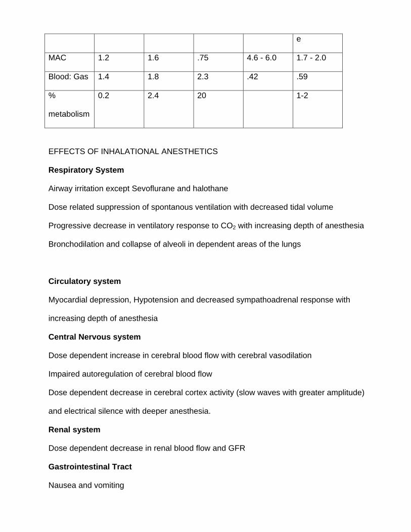
e
MAC 1.2 1.6 .75 4.6 - 6.0 1.7 - 2.0
Blood: Gas 1.4 1.8 2.3 .42 .59
%
metabolism
0.2 2.4 20 1-2
EFFECTS OF INHALATIONAL ANESTHETICS
Respiratory System
Airway irritation except Sevoflurane and halothane
Dose related suppression of spontanous ventilation with decreased tidal volume
Progressive decrease in ventilatory response to CO2 with increasing depth of anesthesia
Bronchodilation and collapse of alveoli in dependent areas of the lungs
Circulatory system
Myocardial depression, Hypotension and decreased sympathoadrenal response with
increasing depth of anesthesia
Central Nervous system
Dose dependent increase in cerebral blood flow with cerebral vasodilation
Impaired autoregulation of cerebral blood flow
Dose dependent decrease in cerebral cortex activity (slow waves with greater amplitude)
and electrical silence with deeper anesthesia.
Renal system
Dose dependent decrease in renal blood flow and GFR
Gastrointestinal Tract
Nausea and vomiting
Skeletal Muscles
Potent inhalational agents produce modest skeletal muscle relaxation by central depression
and enhancement of muscle relaxation produced by non-depolarizing muscle relaxants.
4. Muscle relaxants
Muscle relaxants block the nicotinic acetylcholine receptors at the muscle endplate, thereby
inhibiting neuromuscular transmission and inducing muscle flaccidity. Inactivation of the
receptor can be attained in two ways: by depolarizing the receptor continuously, which leads
to a complex form of desensitization (depolarizing muscle relaxants); or by competitively
antagonizing the receptor (non-depolarizing muscle relaxants).
The degree of relaxation can be assessed using a twitch monitor. The two standard modes
of testing are the train-of-four (four pulses at 0.5 sec intervals) and tetanus (usually at 50 Hz
for 5 seconds).
Depolarizing muscle relaxants
The only depolarizer in clinical use is succinylcholine. It is the muscle relaxant with the
briefest duration of action, because of its rapid metabolism by butyrylcholinesterase
("pseudocholinesterase") in plasma. The rapid onset (30 to 60 seconds) is dose dependent
and minimizes the time for rapid sequence intubation. The rapid degradation allows patients
to manage their own airway quickly again after an unsuccessful endotracheal intubation.
Succinylcholine administration results in a parallel decrease in height of all twitches on the
train-of-four (no "fade"). After administration of high doses and or repeated administration of
succinylcholine, patient can develop phase II block, a pattern similar to that seen with non-
depolarizing drugs.
Side effects:
• The initial depolarization of muscles causes fasciculations due to . These are
associated with muscle pain postoperatively, and can largely be prevented by
administration of a small dose of non-depolarizing relaxant (curare 3 mg) one minute
prior to succinylcholine. In that case the dose of succinylcholine should be increased
by 50% to compensate for the antagonism of succinylcholine by the non-depolarizing
drug. Intravenous lidocaine, benzodiazepines, Ca+2 channel blockers, etc. also
appear to prevent myalgia.
• The muscle depolarization also results in release of K+ from myocyte. In patients with
upregulated nicotinic receptors (burns, major trauma, paralyzed limbs, head trauma,
neuromuscular disease), the use of succinylcholine can lead to cardiac arrest.
• Succinylcholine can induce malignant hyperthermia in susceptible patients.
• Prolonged paralysis occurs in case of butyrylcholinesterase abnormalities (as
succinylcholine is not metabolized)
• Increases in intra-ocular pressure
• Increases in intracranial pressure (modest)
• Bradycardia, particularly in children, after administration of a second dose (pre-treat
with atropine)
Contraindications:
• The difficult airway
• Documented or suspected susceptibility to malignant hyperthermia
• Up-regulated nicotinic receptors (burn patients)
• Children <5 yr. (Controversial).
• Patients with open eye injury (controversial)
• Patients with increased ICP (a relative contraindication)
• Hyperkalemia and renal disease
• Patients with history of atrial or ventricular arrhythmias.
Pharmacology:
• Formulation: 20 mg/ml
• Dose: 1 to 1.5 mg/kg; in children 2 mg/kg
• Onset: 30 sec
• Duration of action: 3 to 15 min.
Non-depolarizing muscle relaxants
There are two main types of non-depolarizing muscle relaxants.
1. Steroidal compounds
Short acting (Rapacurium and mivacurium
Intermediate acting (cis-atracurium, Vecuronium, Rocuronium)
Long Acting (Pancuronium, Pipecuronium, Doxacurium)
They competitively bind to the nicotinic receptors at the neuromuscular junction, preventing
depolarization.
Side effects
• Histamine release
• Vagolytic effect (muscarinic inhibition) ( steroidal compounds)
• Sympathomimetic effect (autonomic ganglia stimulation)
Contraindications:
Few. Non-depolarizing muscle relaxants are very safe drugs, as long as the airway is
adequately protected.
Caution using pancuronium with Demerol.
Hist Vago Gangl Formulati
on
Dose Onset Duration Cost
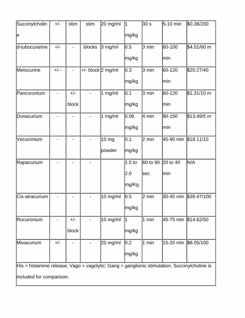
Succinylcholin
e
+/- stim stim 20 mg/ml 1
mg/kg
30 s 5-10 min $0.36/200
d-tubocurarine +/- - blocks 3 mg/ml 0.5
mg/kg
3 min 60-100
min
$4.51/60 m
Metocurine +/-- - +/- block 2 mg/ml 0.3
mg/kg
3 min 60-120
min
$20.27/40
Pancuronium - +/-
block
- 1 mg/ml 0.1
mg/kg
3 min 60-120
min
$1.31/10 m
Doxacurium - - - 1 mg/ml 0.06
mg/kg
4 min 90-150
min
$13.49/5 m
Vecuronium - - - 10 mg
powder
0.1
mg/kg
2 min 45-90 min $18.11/10
Rapacurium - - - 1.5 to
2.0
mg/Kg
60 to 90
sec
20 to 40
min
N/A
Cis-atracurium - - - 10 mg/ml 0.5
mg/kg
2 min 30-45 min $39.47/100
Rocuronium - +/-
block
- 10 mg/ml 1
mg/kg
1 min 45-75 min $14.62/50
Mivacurium +/- - - 20 mg/ml 0.2
mg/kg
1 min 15-20 min $8.05/100
His = histamine release; Vago = vagolytic; Gang = ganglionic stimulation. Succinylcholine is
included for comparison.
Reversal
Inhibitors of plasma cholinesterases, induce increased availability of acetylcholine at the
neuromuscular junction, which competitively reverses the neuromuscular blockade.
Side effects:
• Bradycardia from cardiac muscarinic stimulation
• Bronchoconstriction
These side effects can be (partially) attenuated by administration of a muscarinic antagonist,
which is usually given at the same time as the reversal drug. Two cholinesterase inhibitors
are used clinically: neostigmine and edrophonium.
Neostigmine
Neostigmine is slower in onset than edrophonium, but it forms covalent (strong) bond with
plasma cholinesterase, thus it can reverse a deeper neuromuscular block. The muscarinic
antagonist glycopyrrolate (7-15 mcg/kg), which has a longer duration of action and a longer
time to onset than atropine, is often used with neostigmine to minimize cardiovascular
changes and other unwanted nicotinic effects.
Pharmacology:
• Formulation: 1 mg/ml
• Dose: 0.04-0.07 mg/kg
• Onset: full reversal is attained in approximately 10 – 15 min.
• Duration of action: 1.5 h
Edrophonium
Edrophonium, when given intravenously, has rapid onset of action than neostigmine. At
equivalent doses neostigmine and edrophonium has similar duration of action. Atropine (7-
10 mcg/Kg) is often used in combination with edrophonium to block muscarinic effects. The
degree of block that edrophonium is able to antagonize, however, is much less profound, as
it forms ionic (weaker) bond with actelycholinesterase.
Pharmacology:
• Formulation: 10 mg/kg
• Dose: 0.5-1 mg/kg
• Onset: full reversal is attained in approximately 5 min.
• Duration of action: 1 to 1.5 hr. (a long-acting relaxants may outlast edrophonium)
CRYSTALLOIDS AND COLLOIDS
Part One
Intravascular Volume Assessment and Fluid Replacement
Total body water is approximately 60 % of body weight in males and 55 % of body weight in
females.
(40% is intracellular and 20 % is extracellular). Intravascular volume or plasma volume is
1/4th of extracellular fluid volume. Estimated total blood volume in a 70-kg patient is
approximately 4900 cc
(70 Kgx70cc/Kg).
I. Goals of Fluid Resuscitation
The primary objective of perioperative fluid management is maintenance of adequate tissue
perfusion. In a simple term,
DO2 = CaO2 x CO x 10
Where, DO2 is (delivery of oxygen), CaO2 (total oxygen content in blood) and CO (cardiac
output)
Normally, DO2 is regulated by local and systemic factors with changes in vascular tone in
response to changes in regional and systemic oxygen consumption (VO2). In normal
situations, VO2 becomes DO2-dependent when DO2 is reduced to critical levels, <330 ml
O2/min/m2. This is when oxygen extraction reaches a maximum. Further reductions in DO2
result in a reduction in VO2.
In certain conditions such as ARDS, sepsis, and severe burns, VO2 will be dependent on
DO2 at levels that are higher than the normal DO2. In these high-risk patients, survival has
been shown to be associated with supernormal DO2, >600 ml O2/min/m2. Studies suggest
that treatment of the components of DO2 may improve survival.
CaO2 = (Hb x SaO2 x 1.34) + (0.003 x PaO2)]
DO2 = Cardiac Index x CaO2 x 10
Adequacy of Perfusion can be assessed by clinically and laboratory investigations:
Mental status changes
Poor Capillary refill
Skin color
Skin Temperature
Fast pulse rate
Low Urine Output
Low urinary sodium level
Acid-base status (Metabolic acidosis or alkalosis)
High Serum Lactate Levels
High Oxygen consumption
Low Mixed Venous Oxygen Saturation
In approaching the surgical patient who exhibits signs of low perfusion, such as oliguria or
hypotension and tachycardia, the most common etiology is insufficient intravascular volume.
II. Assessment of crystalloid requirement
a. Determining Preoperative Fluid Deficits
Intra operative maintenance fluid requirement can be assessed by maintenance fluid
requirements per hour in ml and is always replaced by isotonic saline solution,
1-10 Kg 4 ml/Kg /hr
11-20 Kg 2 ml/Kg/hr
21+ 1 ml/Kg/hr
b. Determination of intra op losses
Usual Intra-operative Fluid requirement in addition to maintenance fluids for minor to
major procedures
Minimal Trauma 4 ml/kg/hr
Moderate Trauma 6 ml/kg/hr
Severe Trauma 8 ml/kg/hr
Example: A 70-kg patient, NPO for 6 hours, undergoing one-hour hernia repair.
Deficit = 40 cc/hr for 1st 10 kg BW + 20 cc/hr for 2nd 10 kg BW and 50 cc/hr for rest of 50
kg =110 cc/hr
6 hours of NPO X 110 cc/hr = 660 pre-op deficit
Intra-op losses = 4 cc/kg/hr X 70 kg X 1 hr = 280 cc plus blood loss
C .To understand the distribution of crystalloid infused intravenously it is important to have
good knowledge of distribution of water in normal individuals. Normal serum sodium and
albumin concentration plays an important role in the distribution of water between
intracellular and extracellular space.
Volume infused = PV increment × distribution volume / Normal PV
For example, assume that acute blood loss of 1000 cc is to be replaced with D5W, which
contains no sodium. After cellular uptake of glucose, the remaining water would distribute
throughout total body water (TBW).
1000 = volume of D5W infused x 42/3
Volume infused to replace 1000 cc blood loss = 14 liters
Another example is using colloids, assuming membrane permeability is normal. Each gram
of albumin holds about 14-15 cc of water in the intravascular space
Each gram of starch holds about 16-17 cc of water in the intravascular space
Therefore, 500 cc of 5% albumin, (25 Gm of albumin), would expand the PV by 375 cc,
three quarters of the infused volume. 25 % human serum albumin will expand plasma
volume by 400 ml for each 100 ml of 25 % albumin infused.
III. CHOICE OF FLUID
a. Isotonic Crystalloid
Normal Saline (0.9%) (Na 154 meq/L, Cl 154 meq/L, osmolality 308 mosms/L)
Lactate Ringer's (Hartman's solution) (Na 130 meq/L, Cl 109 meq/L,
K 4 meq/L, Lactate 28 meq/L, Ca 3 meq/L, osmolality 273 mosms/L)
Normosol-R (Na 140 meq/L, Cl 98 meq/L, K 5 meq/L, Mg 3 meq/L)
Plasmalyte (Na 140 meq/L, Cl 98 meq/L, K 5 meq/L, Mg 3 meq/L, gluconate 23 and
osmolality 294 mosms/L)
b. Isotonic Colloid
5% Albumin (Na 145 meq/L and Cl 145 meq/L)
25% Albumin (Na 145 meq/L and Cl 145 meq/L)
6% Hetastarch (Na 154 meq/L and Cl 154 meq/L) (hydroxyethyl starch, Hespan)
c. Hypertonic Saline and hypertonic solutions with colloids
Hypertonic Solutions( 3% and 7.5% NaCl ) has been extensively investigated in humans.
These solutions are inexpensive, are known to promote urine flow, improve microvascular
blood flow, improve cardiac output and Blood pressure in hemorrhage patients following
trauma but they rapidly decline after discontinuation of therapy. Hypertonic solutions restore
regional cerebral blood flow and reduce brain water contents as compared to isotonic and
hypotonic solutions.
Blood Products
Indications for Blood transfusion
To increase oxygen carrying capacity in patients with depleted intravascular volume.
Patients with hematocrit values greater than 30% rarely require blood transfusion whereas
patients with acute anemia or hematocrit value less than 21% frequently require blood
transfusions. The ultimate determination of that hematocrit or hemoglobin value at which
blood should be given will have to be a clinical judgment based on many factors, such as
cardiovascular status, age, anticipated additional blood loss, arterial oxygen contents, mixed
venous oxygen saturation (svo2), cardiac output and blood volume.
ASA (American Society of Anesthesiologists) Recommendations for Blood
Transfusion
The task force concluded that Transfusion of RBC's is rarely indicated when the
hemoglobin concentration is greater than 10g/dl and is almost always indicted when
it is less than 6g/dl especially when the anemia is acute.
1. The determination of whether intermediate hemoglobin concentration (6-10g/dl)
justify or require RBC transfusion should be based on the patient's risk for
complications of inadequate oxygenation.
2. The use of single hemoglobin "trigger" for all patients and other approaches that fail
to consider all-important physiologic and surgical factors affecting oxygenation are
not recommended.
3. When appropriate, preoperative autologus blood donation, intraoperative and
postoperative blood recovery, acute normovolemic hemodilution, and measures to
decrease blood loss (deliberate hypotension and pharmacologic agents) may be
beneficial.
4. The indications for transfusion of autologus RBC's may be more liberal than for
allogenic RBC's because of the lower risks associated with the former.
Emergency Transfusion
For situations that do not allow time for complete type, screen and cross matching, the
preferred order for the selection of partially cross-match blood should be as follows
• Type-specific partially cross-matched blood
• Type-specific uncross matched blood
• Type O Rh negative uncross matched blood
• Type O Rh positive blood
Complications
Change in oxygen transport (shifting of oxyhemoglobin dissociation curve to left)
Dilutional Thrombocytopenia
Low factor V and factor VIII levels (labile factors)
Disseminated intravascular coagulation
Hemolytic transfusion reactions
Febrile reactions
Citrate intoxication
Hyperkalemia and other electrolyte abnormalities
Hypothermia
Acid-base abnormalities
Infections (hepatitis, HIV, CMV, HTLV)
Platelets
Platelets are prepared from whole blood within eight hours of collection. Each unit of
platelets contains approximately 5.5 x 10 platelets in 50 to 70 ml of plasma. As many as
10 units of platelets may be pooled together in a single component bag. Platelets are stored
at room temperature with continuous gentle agitation to prevent aggregation.
10
Platelets should be administered to correct a deficiency in number or platelet function. One
unit of platelets generally increases platelets count by approximately 5-10 x 10 /L. patients
with microvascular bleeding and thrombocytopenia usually require platelet therapy.
9
Platelets transfusion should be performed in patients with
• Anticipated and actual blood loss
• Microvascular bleeding with low platelets count
• Anticipated platelets dysfunction with microvascular bleeding (Uremia, aspirin
therapy)
The prophylactic administration of platelets is not recommended in patients with low
platelets count due to some chronic cause.
Fresh Frozen Plasma
After removal of red blood cells and platelets, plasma is stored within eight hours of blood
donation. Frozen FFP (-18°C) can be stored for up to one year. Prior to administration, FFP
must be thawed and stored at 1 to 6°C and must be used within 24 hours.
Indications
• FFP is indicated for the treatment of microvascular bleeding due to congenital or
acquired coagulopathy.
• Warfarin effects reversal in case of emergent situation. 5 to 8 ml/Kg is typically
adequate to urgently reverse warfarin.
• DIC (Disseminated intravascular coagulation).
• FFP is not indicated for volume expansion in case of massive hemorrhage or used as
a source of nutrition.
• When FFP is indicated, 10 to 15 ml/Kg of FFP is needed to achieve a minimum of
30% of the plasma factor concentration.