Embed Size (px)
Citation preview

2
3
Introduction
Welcome to the 2nd annual RamanFest Conference. RamanFest, an international two day event, offers presentations and discussions on the current state of advanced applied Raman spectroscopy. This year’s event is being held in conjunction with Harvard University, and is being chaired by: Professor Sunney Xie of Harvard University Dr. Dan Fu of Harvard University Dr. Andrew Whitley of HORIBA Scientific We are holding a gala dinner at the Sheraton Commander Hotel on Thursday evening. Our Guest Speaker is Professor Mildred Dresselhaus of MIT.
RamanFest Book of Abstracts Agenda……………………………………………………………………………………………………………..Pages 4 - 5 Poster Titles………………………………………………………………………………………………………Page 6 Presentation Abstracts…………………………………………………………………………………….. Pages 7 - 22 Poster Abstracts (Posters will be available all day)…………………………………………..……….. Pages 23 - 35 Speaker Bios…………………………………………………………………………………………………….. Pages 36 - 47 A tour of Professor Sunney Xie’s laboratory at Harvard University is scheduled for Friday, June 13th, at the conclusion of RamanFest.
4
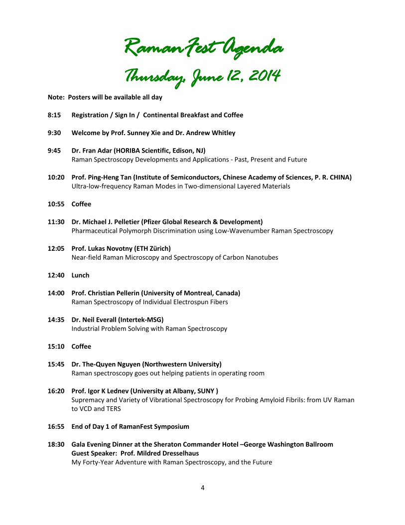
RamanFest Agenda Thursday, June 12, 2014
Note: Posters will be available all day 8:15 Registration / Sign In / Continental Breakfast and Coffee 9:30 Welcome by Prof. Sunney Xie and Dr. Andrew Whitley 9:45 Dr. Fran Adar (HORIBA Scientific, Edison, NJ) Raman Spectroscopy Developments and Applications - Past, Present and Future 10:20 Prof. Ping-Heng Tan (Institute of Semiconductors, Chinese Academy of Sciences, P. R. CHINA) Ultra-low-frequency Raman Modes in Two-dimensional Layered Materials 10:55 Coffee 11:30 Dr. Michael J. Pelletier (Pfizer Global Research & Development) Pharmaceutical Polymorph Discrimination using Low-Wavenumber Raman Spectroscopy 12:05 Prof. Lukas Novotny (ETH Zürich) Near-field Raman Microscopy and Spectroscopy of Carbon Nanotubes 12:40 Lunch 14:00 Prof. Christian Pellerin (University of Montreal, Canada) Raman Spectroscopy of Individual Electrospun Fibers 14:35 Dr. Neil Everall (Intertek-MSG) Industrial Problem Solving with Raman Spectroscopy 15:10 Coffee 15:45 Dr. The-Quyen Nguyen (Northwestern University) Raman spectroscopy goes out helping patients in operating room 16:20 Prof. Igor K Lednev (University at Albany, SUNY ) Supremacy and Variety of Vibrational Spectroscopy for Probing Amyloid Fibrils: from UV Raman to VCD and TERS 16:55 End of Day 1 of RamanFest Symposium 18:30 Gala Evening Dinner at the Sheraton Commander Hotel –George Washington Ballroom Guest Speaker: Prof. Mildred Dresselhaus My Forty-Year Adventure with Raman Spectroscopy, and the Future
5
Friday June 13, 2014 Note: Posters will be available all day 8:15 Continental Breakfast and Coffee 9:15 Prof. Sanford A. Asher (University of Pittsburgh) UV Raman Studies of Protein and Peptide Structure and Folding Studies 9:50 Prof. Igor Chourpa (University of Tours) SERS and Fluorescence as Analytical Tools to Study Theranostic Nanosystems 10:25 Prof. Ji-Xin Cheng (Purdue University) Microsecond Time Scale Spectroscopic Imaging for In vivo Molecular Analysis 11:00 Coffee 11:35 Prof. Paul Champion (Northeastern University) Coherent Low-frequency Vibrational Motion in Proteins and Biomolecules 12:10 Prof. Lawrence D. Ziegler (Boston University) In Vitro Cellular Activity Probed by SERS: Applications for Diagnostics and Forensics 12:45 Lunch 14:15 Prof. Wei Min (Columbia University) Bioorthogonal Nonlinear Vibrational Imaging 14:50 Prof. Sunney Xie (Harvard University) Label Free Vibrational Imaging for Medicine 15:25 Round Table and Concluding Remarks 16:00 Visit to Prof. Sunney Xie Laboratories
6
Poster Titles 1. Characterization of Renal Ischemia Metabolites in the Mammalian Kidney using Raman
Spectroscopy Fran Adar1, Neal Patel2, Joseph Barone2 and Ephrem O. Olweny2
2. Surface Enhanced Raman Spectroscopy using a Novel Substrate for Environmental Applications Mark Creelman, Aran Paulus, Robert Chebi, Iuliu Blaga, Ezra Van Gelder, Richard A. Mathies, and Fanqing Frank Chen
3. UV Resonance Raman and Fluorescence Spectroscopies with QM/MD Calculations Characterize Trp Protein Environment Azaria S. Einsenberg and Laura J. Juszczak
4. Study of Stresses in Hard Materials using Raman and Photoluminescence Spectroscopy Rudolph M. Erasmus 1,2, Maxwell Vhareta 1,2, J. Darrell Comins 1,2
5. Wafer-scale Aluminum Nano-plasmonics
M. George, S. Nielson, and E. Gardner
6. Simple and Accurate New Method for Orientation Quantification using Polarized Raman Spectroscopy Marie Richard-Lacroix and Christian Pellerin
7. Raman Spectroscopy for Electrochemical Energy Storage and Conversion Nir Pour, Wesley T. Hong, Milind J. Gadre, Yueh-Lin Lee, Dane Morgan, Yang Shao-Horn
8. Surface-Enhanced Raman Scattering of Antibody Functionalized Gold Nanoparticles for the Detection of Cardiovascular Biomarkers In Vitro Marinella Sandros
9. Confocal Raman Micro-spectroscopy Discriminates Live Human Metastatic Melanoma and Skin Fibroblast Tells Andrew C. Terentis*, Sara A. Fox, Samantha J. Friedman, and Emily S. Spencer
10. Vertical Integration of Raman Spectroscopy into the Chemistry Curriculum Evonne Rezler, Andrew Terentis, Jerome Haky
11. Polyaniline-Gold Substrates for EC-SERS Biosensing Applications Ryan West, NIST
12. Toward a high-throughput and Depth-Accurate 3-D characterization of Carbon Nanotube-Polymer Composites using Confocal Raman Microscopy
Minhua Zhao1,3,*, Doyoung Moon2, Kalman Migler2, J. Alexander Liddle1 and Rachel J. Cannara1
13. Use of Digilab’s Raman Plate Reader “IDENTITY” for HTS Applications in Chemistry and Biology Igor Zlatkin, Digilab Inc.
7
Abstracts Dr. Fran Adar HORIBA Scientific Raman Spectroscopy: The Synergism between the Instrumentation Evolution and the Emerging Applications
Dr. John Chalmers and Dr. Fran Adar
The evolution of the instrumentation used to detect the Raman effect, from the initial instrument used by CV Raman himself, through to the current use of multichannel detectors on grating-based spectrographs with microscope sampling devices, will be reviewed, while indicating what fields of science have been explored during these time periods. In the earliest period we will indicate how Raman spectra were originally used as an aid to determine molecular structure. During the 1960’s it was successfully used to study the physics of semiconducting materials and devices. The introduction of the microscope in ~1974 as a sampling device first simplified experimental conditions, but also provided information on a scale commensurate with many questions of microstructure and with problems of manufacturing defects. Curiously, while the original concept of the microscope in the early 1970’s was focused on Raman imaging, technological limitations prevented its practical implementation. As the ability to acquire high quality map data over a region of interest improved, development of multivariate statistical means of treating the data has provided high quality Raman images that are now yielding solutions to problems.
8
Prof. Ping-Heng Tan Institute of Semiconductors, Chinese Academy of Sciences, P. R. CHINA Ultra-Low-Frequency Raman modes in two-dimensional layered materials
The fast progress of graphene research, fuelled by the unique properties of this two dimensional (2D) material, paved the way to experiments on other 2D layered materials (LM).[1] Atoms within each layer in 2D LMs are held together by covalent bonds, while van der Waals interactions keep the layers together. The interlayer van der Waals (vdW) coupling and low frequency phonon modes, and how they evolve with the number of layers, are important for both the mechanical and electrical properties of 2D layered materials.
We will address the recent advance on the experimental micro-Raman technique to access the ultra-low phonon mode in 2D LMs.[2,3] The research progress on the rigid-layer vibrations both for shear and layer breathing modes in various 2D LMs from graphenes to transition metal dichalcogenides are reviewed. Their scaling rule with layer number can be modeled by an atomic linear chain model, with general applicability to any layered material, allowing a reliable diagnostic of their thickness.
We also uncover the shear mode for MLGs and show that it provides a direct measurement of the interlayer coupling[3]. The corresponding shear modes can be well-fitted with a Breit-Wagner-Fano lineshape, which arises as quantum interference between the shear mode and a continuum of Raman-active electronic transitions. This makes it a probe for the quasiparticles near the Dirac point by quantum interference. By measuring the interlayer shear modes[4,5] using Raman spectroscopy, we also probe the coupling at the interface between two artificially stacked few-layer graphenes, rotated with respect to each other.
References
[1] F. Bonaccorso, P.H. Tan and A. C. Ferrari, ACS Nano, 7:3 (2013) 1838-1844.
[2] X. Zhang, P. H. Tan, et al., Phys. Rev. B, 87 (2013) 115413.
[3] P. H. Tan et al., Nature Materials, 11:4 (2012)294-300.
[4] P. H. Tan, et al., Phys. Rev. B, 2014, submitted.
[5] J. B. Wu, P. H. Tan, et al., Nature Communications, 2014, submitted.
9
Dr. Michael J. Pelletier Pfizer Global Research and Development Low-Wavenumber Stokes and Anti-Stokes Raman Microscopy for Pharmaceutical Tablet Characterization Discrimination between polymorphs of an active pharmaceutical ingredient (API) in a pharmaceutical product is important because an undesired API crystal form may have different bioavailability or stability characteristics than the desired form. Micro-Raman mapping has proven useful for nondestructive, spatially resolved identification of API polymorphs even at low API levels in the drug product. Raman spectral polymorph discrimination is usually based on small band shifts in the fingerprint region (400-1800 cm-1). These shifts result from functional groups, such as a carbonyl group, experiencing different microenvironments in the different crystal forms. Low-wavenumber Raman bands (-200 to 200 cm-1) result from larger scale motions, such as deformation of the molecular skeleton or even the whole unit cell. Since low-wavenumber Raman bands are more directly related to the entire crystal structure than vibrational bands from small functional groups, they often improve API polymorph discrimination. The anti-Stokes segment of this spectral region provides additional Raman intensity, though no additional spectroscopic information, that can contribute to improved accuracy and spectral artifact detection. Volume holographic filters for laser intensity rejection allow simultaneous acquisition of the Stokes/anti-Stokes low-wavenumber region along with the fingerprint region of Raman spectra. Using multivariate analysis, we demonstrate and evaluate polymorph identification from combined low-wavenumber and fingerprint Raman spectra of pharmaceutical tablet image pixels. Multivariate discrimination between polymorphs is surprisingly robust to spectral variation due to crystal orientation. The strong and polymorph-selective API bands typical of the low wavenumber Raman spectral region allow polymorph-specific mapping at rates as high as 50 spectra per second. Michael J. Pelletier1, Shawn Mehrens1, Christine C. Pelletier2 1. Pfizer Worldwide R&D 2. Gales Ferry, CT
10
Prof. Lukas Novotny ETH Zürich, Photonics Laboratory, Switzerland Near-field Raman Microscopy and Spectroscopy of Nanocarbon Materials
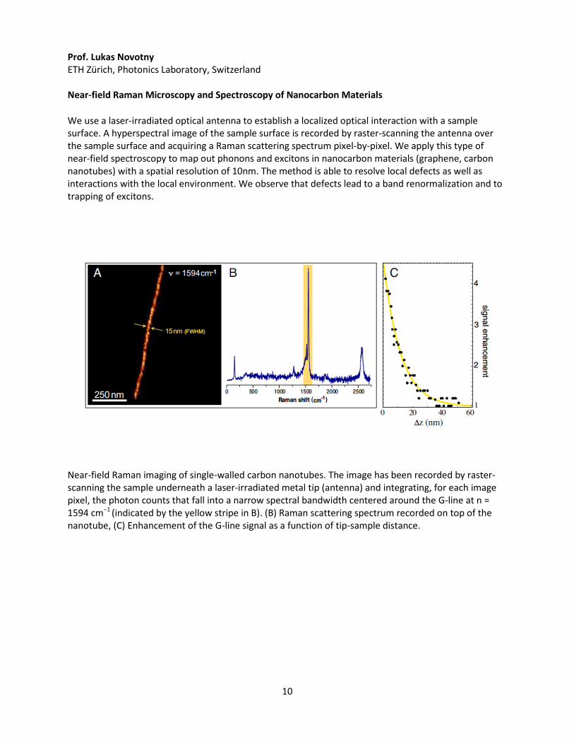
We use a laser-irradiated optical antenna to establish a localized optical interaction with a sample surface. A hyperspectral image of the sample surface is recorded by raster-scanning the antenna over the sample surface and acquiring a Raman scattering spectrum pixel-by-pixel. We apply this type of near-field spectroscopy to map out phonons and excitons in nanocarbon materials (graphene, carbon nanotubes) with a spatial resolution of 10nm. The method is able to resolve local defects as well as interactions with the local environment. We observe that defects lead to a band renormalization and to trapping of excitons.
Near-field Raman imaging of single-walled carbon nanotubes. The image has been recorded by raster-scanning the sample underneath a laser-irradiated metal tip (antenna) and integrating, for each image pixel, the photon counts that fall into a narrow spectral bandwidth centered around the G-line at n = 1594 cm−1 (indicated by the yellow stripe in B). (B) Raman scattering spectrum recorded on top of the nanotube, (C) Enhancement of the G-line signal as a function of tip-sample distance.
11
Prof. Christian Pellerin and Dr. Marie Richard-Lacroix Department of Chemistry, University of Montreal
Raman Spectroscopy of Individual Electrospun Fibers
Electrospinning is widely used for producing nanofibers that may be used in tissue engineering, selective filtration, etc. However, the control of their properties is limited by the fact that most characterization techniques require bundles of fibers to obtain information about their structure and orientation. We have shown that polarized confocal Raman microscopy is a powerful tool for characterizing individual electrospun nanofibers using poly(ethylene terephthalate) and polystyrene as model systems. Highly reproducible polarized spectra with good signal-to-noise ratio allow quantifying molecular orientation, crystallinity, and conformation.
In conducting this study, we were faced with a limitation of the conventional orientation quantification procedure for polymers: the need to measure the depolarization ratio of an isotropic sample and to assume that it remains fixed in the oriented samples. The error associated with this can be very significant. We have recently proposed a new procedure to quantify orientation of polymers by Raman spectroscopy based on the most probable orientation distribution function. Its concept and practical application will be described.
12
Dr. Neil Everall Intertek-Wilton, UK Industrial Problem Solving with Raman Spectroscopy Raman spectroscopy occupies an interesting position in the gamut of techniques available to the industrial scientist. On the one hand, it may not be the best possible technique for obtaining a particular piece of information when working in ideal circumstances. For example, if one needs to solve the structure of a soluble, volatile molecule, then one would probably turn first to NMR and mass spectrometry, if available. If one needs to image morphology with <<1 m resolution
microscopy would be the first choice. Fundamental studies of crystalline phases would probably begin with X-ray diffraction, not Raman. However, in the real world, samples present themselves in awkward, non-ideal circumstances. They may be stubbornly insoluble and non-volatile. We might need to image chemistry with high spatial resolution or analyse tiny defects in situ in larger objects. We might want to look inside a reactor in real time, not having the luxury to remove a sample for NMR or mass spec. Perhaps we have to study crystalline domains with a spatial resolution beyond that which is routinely achievable by XRD, or we might want to analyse samples non-destructively, perhaps in-vivo. In all these cases, and more, we can find that Raman offers a better alternative to the “gold standard” technique, and in fact, Raman is often the only viable tool to solve a particular problem. In this talk, real case histories are drawn from the workload of a lab that supports the R&D, production and technical service activities of a wide range of industries. The examples are chosen to illustrate Raman spectroscopy’s unique offering in an industrial environment, focusing particularly on the use of confocal Raman microscopy. This includes Raman imaging of the structures of blends, coatings and laminates, analysis of defects in composites, mapping of cure in UV-cured coatings, and studies of polymer crystallinity on the micron scale. All of these examples would have been difficult or impossible to achieve using other techniques.
13
Prof. The-Quyen Nguyen Northwestern University Raman Spectroscopy goes out Helping Patients in the Operating Room
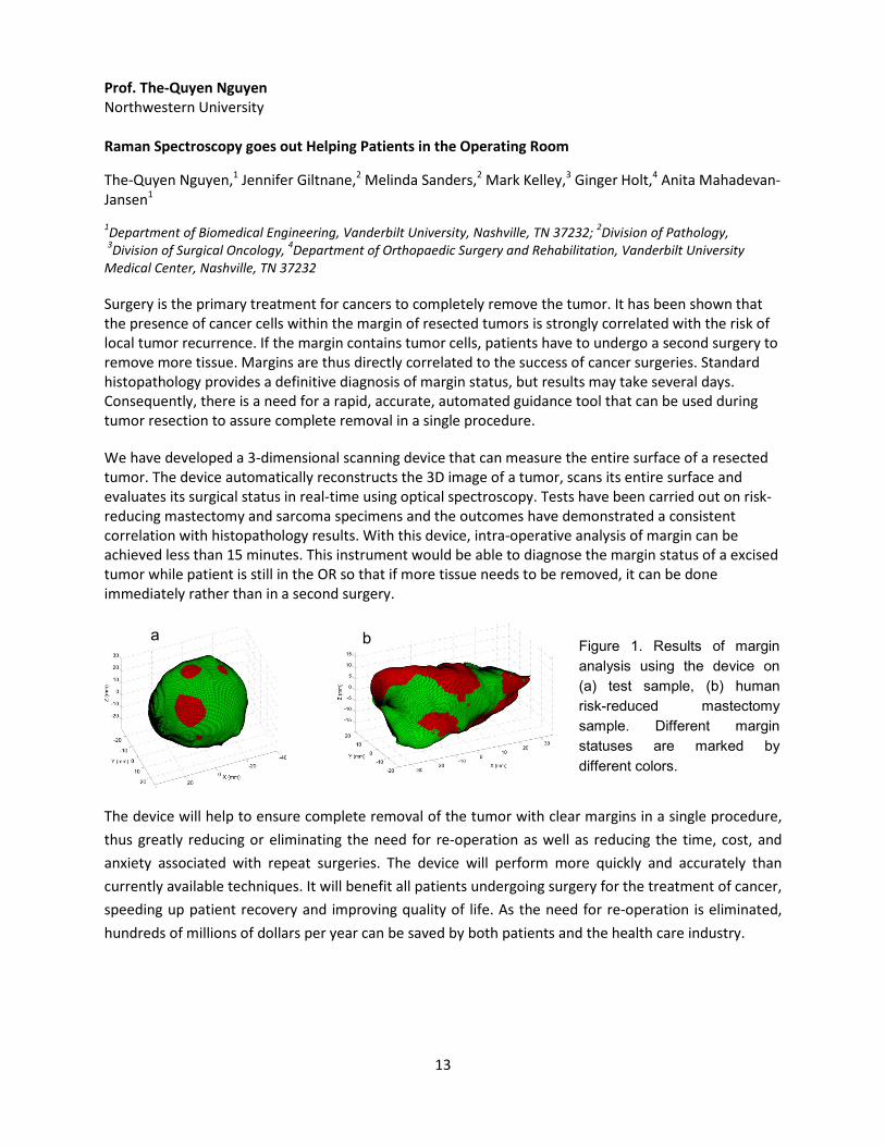
The-Quyen Nguyen,1 Jennifer Giltnane,2 Melinda Sanders,2 Mark Kelley,3 Ginger Holt,4 Anita Mahadevan-Jansen1 1Department of Biomedical Engineering, Vanderbilt University, Nashville, TN 37232; 2Division of Pathology, 3Division of Surgical Oncology, 4Department of Orthopaedic Surgery and Rehabilitation, Vanderbilt University Medical Center, Nashville, TN 37232 Surgery is the primary treatment for cancers to completely remove the tumor. It has been shown that the presence of cancer cells within the margin of resected tumors is strongly correlated with the risk of local tumor recurrence. If the margin contains tumor cells, patients have to undergo a second surgery to remove more tissue. Margins are thus directly correlated to the success of cancer surgeries. Standard histopathology provides a definitive diagnosis of margin status, but results may take several days. Consequently, there is a need for a rapid, accurate, automated guidance tool that can be used during tumor resection to assure complete removal in a single procedure. We have developed a 3-dimensional scanning device that can measure the entire surface of a resected tumor. The device automatically reconstructs the 3D image of a tumor, scans its entire surface and evaluates its surgical status in real-time using optical spectroscopy. Tests have been carried out on risk-reducing mastectomy and sarcoma specimens and the outcomes have demonstrated a consistent correlation with histopathology results. With this device, intra-operative analysis of margin can be achieved less than 15 minutes. This instrument would be able to diagnose the margin status of a excised tumor while patient is still in the OR so that if more tissue needs to be removed, it can be done immediately rather than in a second surgery.
The device will help to ensure complete removal of the tumor with clear margins in a single procedure, thus greatly reducing or eliminating the need for re-operation as well as reducing the time, cost, and anxiety associated with repeat surgeries. The device will perform more quickly and accurately than currently available techniques. It will benefit all patients undergoing surgery for the treatment of cancer, speeding up patient recovery and improving quality of life. As the need for re-operation is eliminated, hundreds of millions of dollars per year can be saved by both patients and the health care industry.
a b Figure 1. Results of margin analysis using the device on (a) test sample, (b) human risk-reduced mastectomy sample. Different margin statuses are marked by different colors.
14
Prof. Igor K. Lednev University at Albany, State University of New York Supremacy and Variety of Vibrational Spectroscopy for Probing Amyloid Fibrils: From UV Raman to VCD and TERS
In spite of the key medical importance of amyloid fibrils, the molecular mechanism of fibrillation is not fully understood. At least in part this is because amyloid fibrils are non-crystalline and insoluble, and thus are not amenable to conventional X-ray crystallography and solution NMR, the classical tools of structural biology. Together with our collaborators we have developed and applied novel experimental approaches based on advanced vibrational spectroscopy for characterizing structure and dynamics of amyloid fibril during the last decade. These include deep ultraviolet resonance Raman (DUVRR) spectroscopy, vibrational circular dichroism (VCD) and tip-enhanced Raman spectroscopy (TERS). In addition to hardware, we developed advanced statistical methods for analyzing spectroscopic data including two dimensional correlation spectroscopy (2DCoS). The application of these complimentary methods for amyloid fibril characterization will be discussed.
We established a detail fibrillation mechanism by detecting the structural intermediates at early stages of fibrillation and determining the sequential order of their appearance through 2DCoS analysis of DUVRR data. DUVRR spectroscopy combined with hydrogen-deuterium allowed us for characterizing the fibril core structure for various fibril polymorphs. A new protein folding-aggregation phenomenon, spontaneous refolding of one fibril polymorph to another was discovered. Fibril polymorphs prepared from the same protein under slightly different pH conditions exhibit opposite chirality according to VSD measurements. Overwhelming majority of structural information accumulated so far about amyloid fibrils are limited to its bulk or core properties. However, the fibril surface determines the biological activity and associated toxicity. TERS offers a unique opportunity to characterize the surface structure of an individual fibril due to a high depth and lateral spatial resolution of the method in the nanometer range. We utilized TERS for characterizing the secondary structure and amino acid residue composition of the fibril surface. It was found that the surface is strongly heterogeneous and consists of clusters with various protein conformations. The propensity of various amino acids on the fibril surface and specific surface secondary structure elements were evaluated.
15
Gala Dinner Guest Speaker: Prof. Mildred Dresselhaus MIT My Forty-Year Adventure with Raman Spectroscopy, and the Future Graphene has been known to the science community since the pioneering work of Wallace in 1947. In this talk I share my over-40-year adventure with Raman spectroscopic studies of nanocarbons. In the 1970s and 1980s we studied single and few graphene layers coming from graphite intercalation compounds. From Raman studies of fullerenes in the 1980s and early 1990s, we moved to spectroscopic studies of carbon nanotubes in the 1990s and then back to graphene in the last decade after Geim and Novoselov started the recent explosion of interest in graphene. For the past 40 years there has been a remarkable growth in nanocarbon research and Raman spectroscopy has played a major role in increasing our understanding of the physics and chemistry of nanocarbons during this long period of time. This overview will address the past, present, and look to the future of graphene research and what lies beyond graphene.
16
Prof. Sanford A. Asher University of Pittsburgh UV Raman Studies of Protein and Peptide Structure and Folding Studies UV Raman excitation into the ~200 nm peptide bond electronic transitions enhance peptide bond amide vibrations of the backbone. A particular band (the amide III3) reports on the Ramachandran psi angle and peptide bond hydrogen bonding. This band is Raman scattered independently by each peptide bond with insignificant coupling between adjacent peptide bonds. Isotope editing of a peptide bond (by replacing the Calpha- H with Calpha- D) allows us to determine the frequency of individual peptide bonds within a peptide or protein to yield their psi angles. Consideration of the Boltzmann equilibria allows us to determine the psi angle Gibbs free energy landscape along the psi (un)folding coordinate that connects secondary structure conformations. The psi angle coordinate is the most important reaction coordinate necessary to understand mechanism(s) of protein folding.
We examine the details of peptide folding conformation dynamics with laser T-jumps where the water temperature is elevated by an 1.9 mM IR nsec laser pulse and we monitor the ~200 nm UV Raman spectrum as a function of time. These spectra show the time evolution of conformation. We will discuss the role of salts on stabilizing conformations in solution.
17
Prof. Igor Chourpa Université de Tours François Rabelais, France SERS and Fluorescence as Analytical Tools to Study Theranostic Nanosystems
Surface-enhanced Raman scattering (SERS) spectroscopy and fluorescence spectroscopy are analytical techniques well-recognised in several scientific domains, from chemistry to biology. This is due to the strength of these techniques, namely high molecular selectifity and sensitivity enabling single-molecule detection. In the present communication, we will describe how the combination or coupling of SERS with fluorescence can increase even more their potential in analytical and diagnostic applications. In particular, we will focus on complementary use of SERS- and fluorescence- based approaches in the pluridisciplinary research on novel biocompatible nanosystems developed for theranosis (therapy and diagnosis) of cancers[1, 2]. The applications presented will concern several aspects: from nanosystem characterization[3, 4] and study of drug loading/release in suspension[4-6], drug delivery in live cancer cells[7] to development of multimodal biomedical imaging concepts.
References
1. Gautier J, Allard-Vannier E, Munnier E, Soucé M, Chourpa I. Recent advances in theranostic nanocarriers of doxorubicin based on iron oxide and gold nanoparticles. J Control Release. 2013 Jul 10;169(1-2):48-61. 2. Gautier J, Allard-Vannier E, Hervé-Aubert K, Soucé M, Chourpa I. Design strategies of hybrid metallic nanoparticles for theragnostic applications. Nanotechnology. 2013 Nov 1;24(43):432002. 3. Kaaki K, Hervé-Aubert K, Chiper M, Shkilnyy A, Soucé M, Benoit R, Paillard A, Dubois P, Saboungi ML, Chourpa I. Magnetic nanocarriers of doxorubicin coated with poly(ethylene glycol) and folic acid: relation between coating structure, surface properties, colloidal stability, and cancer cell targeting. Langmuir. 2012 Jan 17;28(2):1496-505. 4. Chiper M, Hervé Aubert K, Augé A, Fouquenet JF, Soucé M, Chourpa I. Colloidal stability and thermo-responsive properties of iron oxide nanoparticles coated with polymers: advantages of Pluronic® F68-PEG mixture. Nanotechnology. 2013 Oct 4;24(39):395605. 5. Gautier J, Munnier E, Douziech-Eyrolles L, Paillard A, Dubois P, Chourpa I. SERS spectroscopic approach to study doxorubicin complexes with Fe(2+) ions and drug release from SPION-based nanocarriers. Analyst. 2013 Nov 12;138(24):7354-61. 6. Šimáková P, Gautier J, Procházka M, Hervé-Aubert K, Chourpa I. Polyethylene-glycol-Stabilized Ag Nanoparticles for Surface-Enhanced Raman Scattering Spectroscopy: Ag Surface Accessibility Studied Using Metalation of Free-Base Porphyrins. J. Phys. Chem. C, Article ASAP.DOI: 10.1021/jp5005709. Publication Date (Web): March 17, 2014. 7. Chourpa I, Lei FH, Dubois P, Manfait M, Sockalingum GD. Intracellular applications of analytical SERS spectroscopy and multispectral imaging. Chem. Soc. Rev., 2008, 37: 993 - 1000.
18
Prof. Ji-xin Cheng Purdue University Microsecond Time Scale Spectroscopic Imaging for In Vivo Molecular Analysis
Raman spectroscopic imaging of highly dynamic systems was inhibited by relatively long spectral acquisition time. Recently developed multiplex coherent anti-Stokes Raman scattering (CARS) microscopy reduced the acquisition time to tens of millisecond. The CARS signal is, however, mixed with a pixel-dependent nonresonant background, which makes quantitative analysis difficult. Here, we report a novel spectroscopic imaging scheme based on parallel lock-in free detection of spectrally dispersed stimulated Raman scattering signal using a homebuilt tuned amplifier array. Our method reduced the spectral acquisition time to 30 microseconds per pixel, which is faster than multiplex CARS by three orders of magnitude. Aided by multivariate curve resolution analysis, we have monitored molecular penetration into skin tissue in situ and in real time. Fast spectroscopic imaging opens a new window for in situ analysis of target molecules in highly dynamic environment such as live cells. The reported technique also holds the potential for direct visualization of chemistry that occurs at microsecond time scale.
19
Prof. Paul Champion Physics Department, Northeastern University
Coherent Low-Frequency Vibrational Motion in Proteins and Biomolecules
Recent studies have demonstrated how either static or transient distortions along specific normal modes of the heme group can help to activate coherent motions induced by ultrashort laser pulses. These coherent motions are associated with heme out-of-plane normal modes that would otherwise be forbidden by symmetry. The distortion-induced enhancement mechanism is most relevant for low-frequency modes, which are particularly susceptible to distortion by the protein environment because of their weak force constants. The protein can evidently “tune” or distort the heme to perform a multitude of different tasks and these distortions can be monitored by measuring the low frequency Raman-active vibrational coherences. Results on several different biomolecular systems demonstrate that vibrational coherence spectroscopy is a sensitive probe of thermally accessible and functionally relevant distortions of the active site heme chromophore. The low frequency motions (ħω < kBT∼300K∼200 cm-1) associated with these distortions are able to extract energy from the thermal bath and utilize it for barrier crossing. These low frequency modes are prime candidates to serve as biochemical reaction coordinates and their ability to mix with other delocalized low-frequency modes of the protein, or with binding partners, offers a potential control mechanism.

20
Prof. Lawrence D. Ziegler Boston University In Vitro Cellular Activity Probed by SERS: Applications for Diagnostics and Forensics Surface enhanced Raman spectroscopy (SERS) excited at 785 nm is found to be a sensitive probe of the metabolic products of bacterial and human cells. Cells removed from the human body undergo characteristic in vitro robust biological activity whose detection can be exploited for biomedical, diagnostic and forensic applications. In particular, the degradation products resulting from energy depletion in bacterial cells provides a unique SERS signature that can be both species and strain specific. This methodology is being developed for diagnosing blood and urinary tract infections with antibiotic specificity. 785 nm excited SERS spectra of human blood and red blood cells (RBCs) are due to blood plasma and hemoglobin, respectively and may be exploited for several biomedical purposes including blood aging monitoring and malria detection. The SERS spectrum of stored whole human blood changes dramatically over ~ 24 hours becoming nearly dominated by hypoxanthine, a metabolite of purine degradation, over this period of time due to its release into blood serum from white blood cells. Tumor cells are well-known to exhibit high metabolic rates compared to normal, non-pathogenic cells. Again, characteristic SERS vibrational signatures due to molecules like adenine, hypoxanthine and NADH appear over the course of several hours from single cancer cells. Thus SERS may provide a procedure for in vitro single cell cancer detection as well as fundamental studies of the effects of genetic or proteomic manipulation for cancer therapy efficacy evaluation. Finally, the use of SERS for trace detection and identification of human body fluids such as blood, semen, vaginal fluid and saliva will be described and demonstrates that SERS can serve as a novel methodology for ultrasensitive forensic identification at crime scenes.
21
Prof. Wei Min Columbia University Bioorthogonal Nonlinear Vibrational Imaging
Innovations in spectroscopy principles and microscopy technology have significantly impacted modern biology and medicine. Here we will discuss an emerging chemical imaging platform, stimulated Raman scattering (SRS) microscopy, which can enhance the feeble spontaneous Raman transition by virtue of stimulated emission. When coupled with stable isotopes such as deuterium or exogenous chemical moieties such as alkynes, the resulting bioorthogonal nonlinear vibrational imaging is well suited for probing metabolism of living systems in vivo at microscopic level. Physical principle of the underlying optical spectroscopy and emerging biomedical applications such as imaging lipid metabolism, protein synthesis, protein degradation, DNA replication, RNA synthesis, glucose uptake, and drug tracking will be presented.
22
Prof. Sunney Xie Harvard University, Department of Chemistry and Chemical Biology
Label Free Vibrational Imaging for Medicine
Stimulated Raman scattering microscopy is a label-free and noninvasive imaging technique using vibrational spectroscopy as the contrast. Recent advances have allowed significant improvements in sensitivity, selectivity, robustness, cost; opening a wide range of biomedical applications.
23
Poster Abstracts 1. Characterization of Renal Ischemia Metabolites in the Mammalian Kidney using Raman
Spectroscopy
Fran Adar1, Neal Patel2, Joseph Barone2 and Ephrem O. Olweny2
1) HORIBA Scientific, Edison, NJ, USA; 2) Rutgers-Robert Wood Johnson Medical School, New Brunswick, NJ, USA A number of disease processes are known to cause kidney dysfunction via generation of ischemia. Additionally, controlled ischemia is often implemented during renal surgery so as to minimize hemorrhage, which can result in short or long term renal dysfunction. Current understanding of the impact of ischemia on the involved kidney in humans is limited, with conflicting data reported in the literature. The ability to assess the impact of ischemia in real time could help tailor surgical technique in the future. Raman spectroscopy is uniquely suited to study renal ischemia given its high molecular specificity and rapid spectral acquisition time. In an experimental porcine model, we modeled surgical ischemia by cross-clamping the renal vascular pedicle of one kidney while leaving the opposite kidney unclamped as a control. Wedge biopsies were obtained from each kidney at time 0 (pre-clamping), at hourly intervals up to 3 hours, and also after reperfusion of the ischemic kidney. Tissues were flash-frozen, sectioned to 20 microns, and studied using Raman microscopy. The abundant spectrum observed in ischemic kidneys was the resonance-enhanced signal of whole mitochondria in the reduced state, with control kidneys exhibiting much weaker spectra for the same. The conclusion that mitochondria were present in the reduced state came from comparison with published spectra of mitochondria and their fractions at similar excitation wavelengths, comparing both the sharp Raman bands and the unquenched luminescence that has been documented to accompany the Raman bands. From these results we infer that the ischemic damage to the kidney results in decoupling of the mitochondria and build up of reducing equivalents, producing the reduced-state cytochromes.
24
2. Surface Enhanced Raman Spectroscopy Using a Novel Substrate for Environmental Applications Mark Creelman, Aran Paulus, Robert Chebi, Iuliu Blaga, Ezra Van Gelder, Richard A. Mathies, and Fanqing Frank Chen Optokey, Inc. , 4062 Fabian Way, Palo Alto, CA 94303 Surface Enhanced Raman Scattering (SERS) is an effective vibrational technique for the identification of small molecular analytes due to its ability to spectrally fingerprint chemical species with high sensitivity, and with no need for additional labeling. Harnessing the SERS effect requires plasmonically active nanostructures, typically with 10 to 100 nm features separated by 10 to 100 µm and a metallic layer of gold or silver. While SERS offers increased sensitivity, the first implementation to form nanofeatures used colloids of 50 to 100 nm. These suffered from poor uniformity and repeatability making quantification and repeatability of Raman data difficult. In order to increase the robustness of SERS as an analytical method, semiconductor nanofabrication techniques have been used to produce nanostructures with precise size, aspect ratio, location and distance control. Optokey is developing SERS substrates with gold nanofeatures to be used with a 633 nm laser for environmental applications. Although the second and third generation of pesticides, chlorinated organics such as DDT and 2,4D, carbamates and organophosphorous compounds, have either been banned or replaced by other more specific and selective chemicals, these compounds accumulate in soils, water and the food chain, making their continuous monitoring necessary. SERS is capable of selectively detecting the carbon-chlorine, carbon- nitrogen and phosphorous-oxygen bonds at ppm concentrations. In this poster we will present initial results demonstrating sensitivity for chlorinated organics.
25
3. Trp-Gly/GlyTrp Dipeptide W7 Mode Linked to Fluorescence Emission Energy via QM-MM Calculations
Azaria S. Einsenberg and Laura J. Juszczak
Dept. of Chemistry, Brooklyn College/The City University of New York, 2900 Bedford Avenue , Brooklyn, NY 11210
ABSTRACT. Tryptophan is a useful amino acid for the study of proteins because its indole residue is fluorescent. Moreover, the fluorescence emission maximum, lifetime and quantum yield are all sensitive to the tryptophan environment. On the other hand, fluorescence spectra do not yield the atomic level details that UV resonance Raman (UVRR) spectroscopy can. The rich variety of fluorescence data can be correlated to UVRR spectra, yielding a better understanding of fluorescence spectra changes, and therefore, the protein environment that caused them. In an effort to elucidate the relationship between fluorescence spectra and tryptophan local environment, both resonance Raman spectra and theoretical calculations (molecular dynamics as well as quantum mechanics) on simpler dipeptide systems have been employed with some success. The Raman spectra and molecular dynamics simulations can explain a relationship between molecular conformation and fluorescence maxima shift, and electrostatic isosurfaces generated by quantum mechanical calculations can be used to correlate aromaticity of the tryptophan to the experimental quantum yields and fluorescence lifetimes. An overview of our recent results for several tryptophan dipeptides will be presented.
26
4. Study of Stresses in Hard Materials using Raman and Photoluminescence Spectroscopy
Rudolph M. Erasmus 1,2, Maxwell Vhareta 1,2, J. Darrell Comins 1,2
University of the Witwatersrand, Johannesburg, South Africa
1. DST-NRF Centre of Excellence in Strong Materials, University of the Witwatersrand, Johannesburg, South Africa; 2. School of Physics, University of the Witwatersrand, Private Bag 3, WITS 2050, Johannesburg, South Africa
Raman and photoluminescence (PL) spectroscopy techniques are both optical means of determining residual stresses, crystallinity and defect characteristics in a wide range of materials. Here these techniques are applied to single crystals of diamond and polycrystalline diamond (PCD) tools. In single crystal diamond, plastic deformation associated with the rosette pattern of an impression created at high temperature (1400oC) on a {001} face of a type Ib HPHT synthetic diamond was studied. Raman spectroscopy was used to map the residual stresses and degree of plastic deformation in a 3D volume associated with the impression, while PL spectroscopy was used to map the distribution of the nitrogen-vacancy defect centres that were generated during the impression process. The “arms” of the pattern are in compression (~1.5 GPa) while the centre of the impression is in tension (~1 GPa). Raman spectroscopy was also applied to the PCD layer of a PCD/Co-WC drilling toolbit. The toolbits consist of a PCD layer sintered onto a tungsten carbide substrate. The different thermal expansion coefficients of the substrate and the PCD result in residual compressive stresses in the diamond layer and hence resistance to crack formation. Results are presented showing that thermal annealing at 800oC for 30 minutes negatively affects the toolbit properties, and that the stress distribution between the top of the PCD table and the PCD-WC interface can be readily determined.
Fatigue-type processes in PCD were studied using a ball on three balls fatigue arrangement. PCD discs detached from the WC substrate were cyclically loaded under constant amplitude load control at a frequency of 10 Hz in order to fatigue the samples. The Raman stress measurements were carried out at room temperature using a 36 point mapping array in area close to the size of the PCD samples. The calculated average surface stress for the different samples shows a general compressive stress for a relatively low number of fatigue cycles but enters a tensile regime with exposure to a sufficiently large number of cycles. Conditions where an average surface tensile stress exists promote crack initiation and provide an explanation of catastrophic failure of tool-bits in drilling operations.
Raman spectroscopy is thus a very valuable technique in the characterisation of the nature and distribution of stresses in hard materials.
Related papers:
1) Erasmus RM, Comins JD, Mofokeng V, Martin Z 2011 Diam Relat Mater 20 907-911
2) Erasmus RM, Daniel RD and Comins JD 2011 J Appl Phys 109 013527
3) Vhareta M, Erasmus RM, Comins JD 2014 Diam Relat Mater 45 34-425.
27
5. Wafer-scale Aluminum Nano-Plasmonics
M. George, S. Nielson, and E. Gardner Moxtek, Inc., 452 West 1260 North, Orem, USA Obtaining a reliable, cost effective, yet flexible manufacturing source for periodic micro- and nano-structures has often been a challenge. Many groups perform their own lithography and etching or use a foundry to generate such structures, but this typically limits the substrate size to a few square inches or can have large up-front costs for masks and other tooling. In addition, the periodicities of structures readily available from commercial sources are typically limited to several hundred nanometers and larger, which can limit device usefulness in the visible spectrum. Moxtek has addressed some of these challenges by leveraging existing capabilities in wafer-scale patterning of sub-wavelength wire grid polarizers into the fabrication of 1D and 2D periodic plasmonic structures from aluminum.
This presentation will review recent advances in aluminum plasmonics and discuss experimental and optical modeling results for aluminum 2D nano-hole arrays, with potential applications in surface plasmon resonance sensing, SERS, and surface-enhanced fluorescence spectroscopy (SEFS). Potential markets include micro-arrays for bio-related assays and trace level chemical detection. In addition, the presentation will review commercial results on narrow-band, cloaked wire grid polarizers composed of nano-stacked metal and dielectric layers patterned over 200 mm diameter wafers for projection display applications.
28
6. Simple and Accurate New Method for Orientation Quantification using Polarized Raman Spectroscopy
Marie Richard-Lacroix and Christian Pellerin, University of Montreal, Canada Molecular orientation has a significant impact on numerous physical properties of materials and is thus a critical parameter for their characterization. In recent years, confocal Raman spectroscopy, with its submicron resolution, has emerged as an indispensable tool for this purpose. However, strict quantification of the order parameters, ⟨P_2 ⟩ and ⟨P_4 ⟩, using this technique remains a challenging task. Indeed, the standard method used for orientation quantification is experimentally complex because it necessitates having access to an isotropic sample of the same chemical and phase composition as the sample of interest before any quantitative information can be extracted. This condition is often difficult, if not impossible, to meet, limiting greatly the applicability of Raman spectroscopy to characterize a wide variety of materials. We have established a new method for orientation quantification that is based on the most probable distribution and that eliminates this restrictive requirement. Its wide experimental applicability has been demonstrated using common polymers, such as high density polyethylene (PE), poly(ethylene terephthalate) (PET) and polystyrene (PS), that show vastly different levels of orientation. Our results unambiguously show that this new method is experimentally simpler and that it provides improved accuracy for quantifying the level of molecular orientation when compared to the standard method. We expect that this method will greatly extend the accessibility of Raman spectroscopy for molecular orientation studies of polymer systems. Richard-Lacroix, M.; Pellerin, C. Appl. Spectrosc., 2013, 67, 409. Richard-Lacroix, M.; Pellerin, C. Macromolecules, 2013, 46, 14, 5561
29
7. Raman spectroscopy for Electrochemical Energy Storage and Conversion
Nir Pour, Wesley T. Hong, Milind J. Gadre, Yueh-Lin Lee, Dane Morgan, Yang Shao-Horn
Massachusetts Institute of Technology - Electrochemical Energy Lab
Electrochemical energy storage and conversion is a significant scientific challenge for realizing renewable energy technologies. The storage and conversion of energy is obtained by the forming and breaking of chemical bonds. Raman spectroscopy can provide valuable insights of these bonds through its symmetry-selectivity and vibrational sensitivity. We apply Raman spectroscopy to a wide variety of electrochemical applications in the solid state, aqueous solution, and non-aqueous environments. For solid state systems, in situ Raman spectroscopy provides a useful means of probing bond strengths of the system under different operating conditions. This is particularly insightful for high-temperature (500-800ºC) solid oxide fuel cells, for which the bond strength plays an important role in the conversion efficiency. Through ex situ and in situ measurements, we can also observe the redox species in solution and electrode stability during charge and discharge of energy storage devices, such as vanadium flow batteries. From this, we can monitor the state of charge and obtain deeper understanding of the reaction mechanism of these systems.
30
8. Surface-Enhanced Raman Scattering of Antibody Functionalized Gold Nanoparticles for the Detection of Cardiovascular Biomarkers In Vitro
K. Bowey1, J.F. Tanguay2, M. Tabrizian1,3, M.G. Sandros4 1. Biomedical Engineering Department, McGill University, Montréal, Québec, Canada 2. Montréal Heart Institute, Montréal, Québec, Canada 3. Faculty of Dentistry, McGill University, Montréal, Québec, Canada 4. Joint School of Nanoscience and Nanoengineering, University of North Carolina at Greensboro, Greensboro, North Carolina, USA Accurate detection and localization of inflammatory biomarkers, such as vascular adhesion molecule-1 (VCAM-1), is crucial for early diagnosis and prevention of atherosclerotic disease. Surface-enhanced Raman scattering (SERS) is an analytical tool well suited for this goal, since it can provide concurrent target identification and imaging when combined with confocal microscopy. Most SERS nanoprobes are synthesized via conventional adsorption; however this approach is often lengthy and can result in particulate instability and aggregation. In the present work, we report the construction and characterization of a robust SERS nanoprobe with strong scattering signal via microwave technology and suitable stability to support antibody functionalization. The nanoprobe was successfully employed to map the expression of VCAM-1 in human coronary artery endothelial (HCAE) cells. Results demonstrated that microwave technology is a viable option for the construction of SERS probes to detect inflammatory biomarker expression in vitro.
31
9. Confocal Raman Micro-Spectroscopy Discriminates Live Human Metastatic Melanoma and Skin Fibroblast Cells
Andrew C. Terentis*, Sara A. Fox, Samantha J. Friedman, and Emily S. Spencer Department of Chemistry and Biochemistry, Florida Atlantic University, Boca Raton, FL 33431 Confocal Raman micro-spectroscopy (CRM) continues to develop as a promising technique with possible clinical applications for the diagnosis and treatment of skin cancers. CRM studies of single cells can provide information on the biochemical content of cancer cells in situ, potentially providing new biochemical fingerprints or markers of cancer cells. Here we report a CRM study of single, living human metastatic melanoma cells (SK-Mel-2) and normal skin fibroblast cells (BJ) cultured and examined under identical experimental conditions. A total of almost 1200 Raman spectra were measured from more than 120 BJ and SK-Mel-2 cells using an inverted microscope with 647 nm laser excitation. Raman spectra were measured from within three distinct intracellular regions of the cells – cytoplasm, nucleoplasm and nucleolus. When Raman spectra from each cell type were compared using Principal Components Analysis (PCA) and Linear Discriminant Analysis with leave-one-dish-out cross-validation (LDA-CV), the two cell types were discriminated with 93% (cytoplasm), 98% (nucleolus), and 96% (nucleoplasm) accuracy. The main biochemical differences identified between the two cell types were higher RNA levels in the nucleoli of BJ cells and high amounts of lipid and collagen in the cytoplasm of SK-Mel-2 cells. For both cell types, higher levels of RNA were detected in the nucleoli versus the nucleoplasm. PCA with LDA-CV was 98% (cytoplasm), 93% (nucleoplasm), and 73% (nucleolus) accurate in identifying the intracellular region based on the Raman spectra from both cell types. No significant trend was observed when the data were analyzed with respect to cell passage number. Thus, CRM with PCA and LDA-CV successfully discriminated two skin-cancer-relevant cell lines while detecting different amounts of nucleic acids, lipids, and proteins in distinct intracellular regions, further underscoring its potential as a clinical diagnostic tool.
32
10. Vertical Integration of Raman Spectroscopy into the Chemistry Curriculum
Evonne Rezler, Andrew Terentis, Jerome Haky
Department of Chemistry and Biochemistry, Charles E. Schmidt College of Science, Florida Atlantic University
We are vertically integrating Raman spectroscopy experiments and experiences into the undergraduate chemistry curriculum that address key course-specific student learning outcomes (SLOs) and more fundamental anchoring chemical concepts or “big ideas”. To date, we have acquired a sophisticated research-grade Raman spectrometer. We are now in the process of developing and testing experiments to enable us to employ the Raman spectrometer to introduce and reinforce increasingly more complex chemistry concepts as part of a restructured chemistry curriculum implementing a “spectroscopy-to-learn” approach. In this approach, we present Raman spectroscopy as a practical tool to teach concepts such as molecular structure, quantitative analysis, elucidation of reaction mechanism and applications of spectroscopy for research.
We are evaluating the effectiveness of this innovation for improving student understanding of chemical principles and attitudes towards learning. To accomplish this we are comparing examination scores, assignment grades, survey results, and long-term performance of students who complete the modified courses with those of similar demographics who completed the original curriculum. We are establishing a website for students and faculty throughout the region and the country, who are interested in implementing this approach in their curricula. We will report on the progress of this project: the challenges of instituting our assessment, developing course-specific Raman spectroscopy experiments, and changing the culture of chemistry majors at our institution. This work is funded through a National Science Foundation (NSF) Transforming Undergraduate Education in Science, Technology, Engineering and Mathematics (TUES) Type I grant (#1244807).
33
11. Polyaniline-Gold Substrates for EC-SERS Biosensing Applications
Ryan West
NIST
Development of robust biosensors for on-line/in-line detection would enable continuous monitoring and control of process parameters in pharmaceutical manufacturing, and new approaches for medical diagnostics and bioterrorism security. We are developing a bi-modal approach to biosensing, utilizing simultaneous electrochemical (EC) and surface-enhanced Raman Spectroscopy (SERS) techniques, to enhance a sensor’s figures of merit and its reliability in critical applications. When used in tandem, these two techniques can provide information about electrochemical activity and molecule-specific vibrational signatures. As a first step towards realizing this goal, we are optimizing the growth and deposition of Au nanoparticles (Au-NPs) on electropolymerized polyaniline (PANI) films leading to PANI/Au-NP composites with tunable electrochemical and SERS properties. PANI was chosen because of its rich EC behavior and its ability to spontaneously bind gold salts and reduce them to metallic gold clusters.
The film deposition and ongoing optimization of EC-SERS responses are carried out with in-house fabricated electrode arrays and flow-cell for precise control over solution composition and electrical potential. The electrode arrays consist of 16 individually-addressable platinum disc electrodes with diameter of 1 mm. Studies with the array have allowed us to efficiently explore the dependence of film thickness, morphology, and nanoparticle size and density on deposition parameters such as KAuCl4 concentration, applied electrode potential, deposition time, etc. In particular, the Au NP size and spacing (density) is of prime interest for optimizing the SERS response. Using electron microscopy the film morphology, NP size and NP density is correlated with SERS response.
34
12. Toward a High-Throughput and Depth-Accurate 3-D Characterization of Carbon Nanotube-Polymer Composites Using Confocal Raman microscopy
Minhua Zhao1,3,*, Doyoung Moon2, Kalman Migler2, J. Alexander Liddle1 and Rachel J. Cannara1
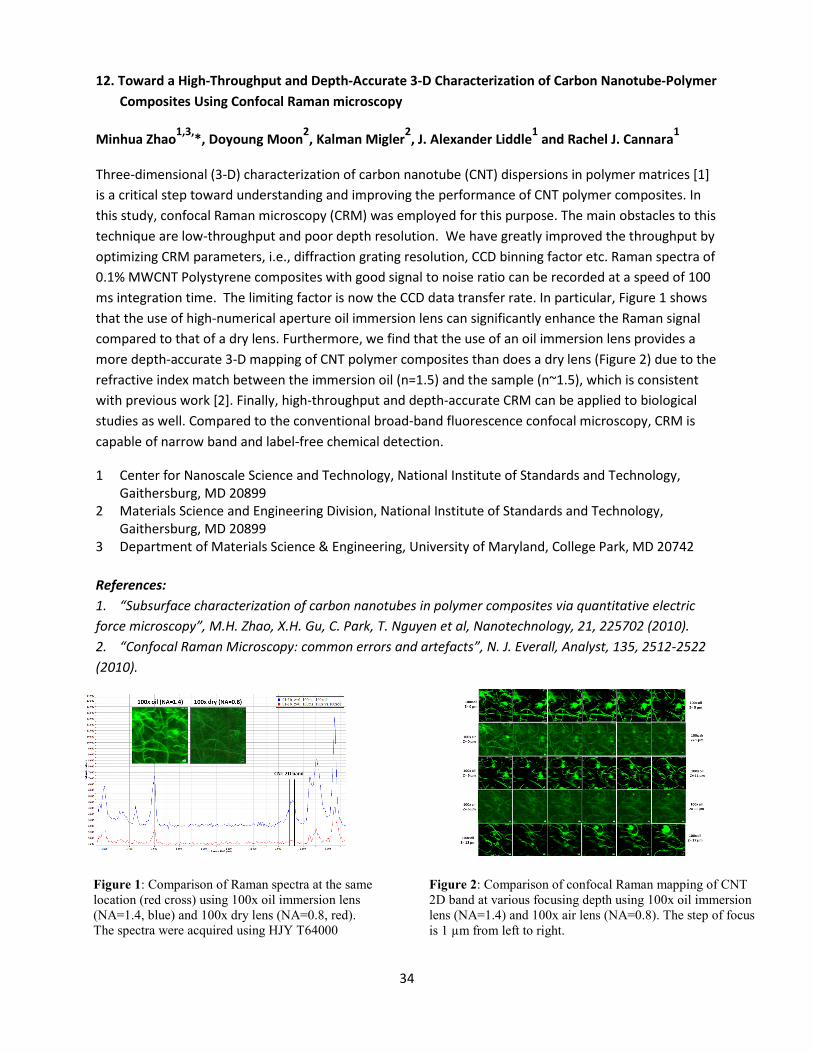
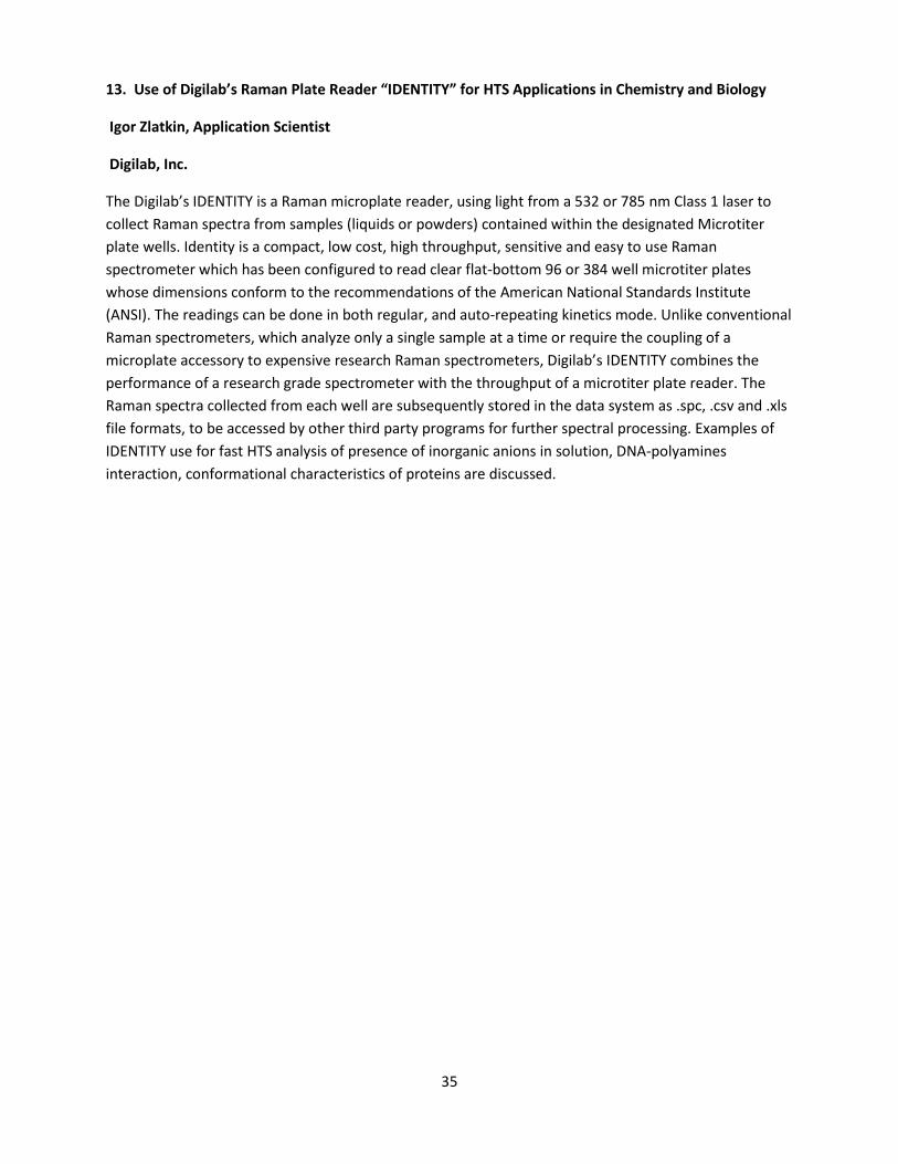
Three-dimensional (3-D) characterization of carbon nanotube (CNT) dispersions in polymer matrices [1] is a critical step toward understanding and improving the performance of CNT polymer composites. In this study, confocal Raman microscopy (CRM) was employed for this purpose. The main obstacles to this technique are low-throughput and poor depth resolution. We have greatly improved the throughput by optimizing CRM parameters, i.e., diffraction grating resolution, CCD binning factor etc. Raman spectra of 0.1% MWCNT Polystyrene composites with good signal to noise ratio can be recorded at a speed of 100 ms integration time. The limiting factor is now the CCD data transfer rate. In particular, Figure 1 shows that the use of high-numerical aperture oil immersion lens can significantly enhance the Raman signal compared to that of a dry lens. Furthermore, we find that the use of an oil immersion lens provides a more depth-accurate 3-D mapping of CNT polymer composites than does a dry lens (Figure 2) due to the refractive index match between the immersion oil (n=1.5) and the sample (n~1.5), which is consistent with previous work [2]. Finally, high-throughput and depth-accurate CRM can be applied to biological studies as well. Compared to the conventional broad-band fluorescence confocal microscopy, CRM is capable of narrow band and label-free chemical detection.
1 Center for Nanoscale Science and Technology, National Institute of Standards and Technology, Gaithersburg, MD 20899 2 Materials Science and Engineering Division, National Institute of Standards and Technology, Gaithersburg, MD 20899 3 Department of Materials Science & Engineering, University of Maryland, College Park, MD 20742 References: 1. “Subsurface characterization of carbon nanotubes in polymer composites via quantitative electric force microscopy”, M.H. Zhao, X.H. Gu, C. Park, T. Nguyen et al, Nanotechnology, 21, 225702 (2010). 2. “Confocal Raman Microscopy: common errors and artefacts”, N. J. Everall, Analyst, 135, 2512-2522 (2010).
Figure 1: Comparison of Raman spectra at the same location (red cross) using 100x oil immersion lens (NA=1.4, blue) and 100x dry lens (NA=0.8, red). The spectra were acquired using HJY T64000
Figure 2: Comparison of confocal Raman mapping of CNT 2D band at various focusing depth using 100x oil immersion lens (NA=1.4) and 100x air lens (NA=0.8). The step of focus is 1 µm from left to right.
35
13. Use of Digilab’s Raman Plate Reader “IDENTITY” for HTS Applications in Chemistry and Biology
Igor Zlatkin, Application Scientist
Digilab, Inc.
The Digilab’s IDENTITY is a Raman microplate reader, using light from a 532 or 785 nm Class 1 laser to collect Raman spectra from samples (liquids or powders) contained within the designated Microtiter plate wells. Identity is a compact, low cost, high throughput, sensitive and easy to use Raman spectrometer which has been configured to read clear flat-bottom 96 or 384 well microtiter plates whose dimensions conform to the recommendations of the American National Standards Institute (ANSI). The readings can be done in both regular, and auto-repeating kinetics mode. Unlike conventional Raman spectrometers, which analyze only a single sample at a time or require the coupling of a microplate accessory to expensive research Raman spectrometers, Digilab’s IDENTITY combines the performance of a research grade spectrometer with the throughput of a microtiter plate reader. The Raman spectra collected from each well are subsequently stored in the data system as .spc, .csv and .xls file formats, to be accessed by other third party programs for further spectral processing. Examples of IDENTITY use for fast HTS analysis of presence of inorganic anions in solution, DNA-polyamines interaction, conformational characteristics of proteins are discussed.
36
Speaker Bios Dr. Fran Adar HORIBA Scientific [email protected] Fran Adar is the Raman Applications Scientist/Manager/Principle Scientist at HORIBA Scientific. Taking advantage of education and experience in Physics and Biophysics, Dr. Adar has developed applications of the Raman Microscope. Applications are in areas as widespread as semiconductors, ceramics, contaminant identification, polymer morphology, catalysts, metal oxides, pharmaceuticals. She
has received awards from the local Microbeam Society (Irene Dion Payne), the Federation of Analytical Chemistry and Spectroscopy Societies (Charles Mann Award), from the Coblentz Society (William-Wright Award) and delivered an address at the prestigious Waters Symposium at the Pittsburg Conference on the history of the development of Raman instrumentation. In 2012 she was invited to be a fellow of the Society for Applied Spectroscopy. Since 2007, Dr. Adar has been writing a column for Spectroscopy whose goals are to point out where Raman spectroscopy and microscopy are having an impact on evolving technologies, and to guide new users into the field. Dr. Adar continues to work with new and experienced Raman users developing applications, and pushing instrumentation developments to accommodate new applications enabled by evolving technologies.
• BS (1966), MS (1968) and Ph.D. (1972) in Physics – University of Pennsylvania • Post-doctoral Fellow and Assistant Professor – Johnson Foundation, Department of Biophysics,
University of Pennsylvania (1972-1978) • Raman Applications Scientist/Manager/Principle Scientist – Jobin Yvon/HORIBA Scientific (1978
to present)
37
Professor Sanford Asher University of Pittsburgh [email protected] Sanford Asher, Distinguished Professor of Chemistry, University of Pittsburgh received his B.A./Chemistry at the University of Missouri, St. Louis, 1971; Ph.D./Chemistry at the University of California, Berkeley, 1977; continuing as Research Fellow, Harvard University, 1977-1980. Dr. Asher’s academic career began in 1980 as Assistant Professor in Pitt’s Chemistry department. Dr. Asher’s research at Pitt involves development of new materials and new spectroscopic techniques. His group developed UVRR spectroscopy as a new technique for
fundamental and applied structural and trace studies of molecules in complex matrices. The Asher group uses UVRR to examine the first stages in protein folding, while investigating the use of UVRR for detection of explosive molecules, especially stand-off detection. The Asher group develops new photonic crystal optical devices and chemical sensing devices from self-assembling colloidal particles.
Dr. Asher pioneered the development of smart hydrogel materials for chemical sensing. He is recipient of many awards, recently 2011 Charles E. Kaufman Award; 2008 Pittsburgh Spectroscopy Award; 2002 Ellis R. Lippincott Award; 1999 Bomen-Michelson Award. Professor Asher served as Co-Director of Pitt’s Materials Research Center and several scientific advisory boards. Professor Asher consults for PPG Industries, ChemImage Corp., ThermoFisher and is author of 280 publications and inventor of 29 patents in the photonic crystals area.
38
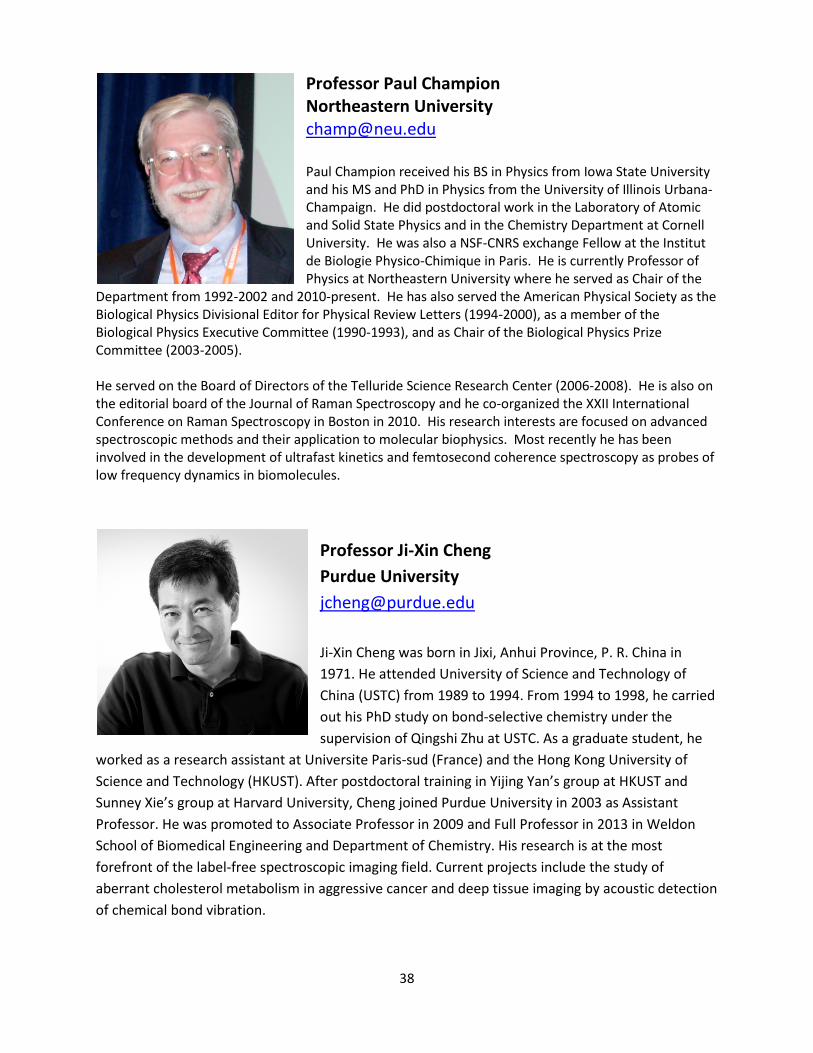
Professor Paul Champion Northeastern University [email protected] Paul Champion received his BS in Physics from Iowa State University and his MS and PhD in Physics from the University of Illinois Urbana-Champaign. He did postdoctoral work in the Laboratory of Atomic and Solid State Physics and in the Chemistry Department at Cornell University. He was also a NSF-CNRS exchange Fellow at the Institut de Biologie Physico-Chimique in Paris. He is currently Professor of Physics at Northeastern University where he served as Chair of the
Department from 1992-2002 and 2010-present. He has also served the American Physical Society as the Biological Physics Divisional Editor for Physical Review Letters (1994-2000), as a member of the Biological Physics Executive Committee (1990-1993), and as Chair of the Biological Physics Prize Committee (2003-2005). He served on the Board of Directors of the Telluride Science Research Center (2006-2008). He is also on the editorial board of the Journal of Raman Spectroscopy and he co-organized the XXII International Conference on Raman Spectroscopy in Boston in 2010. His research interests are focused on advanced spectroscopic methods and their application to molecular biophysics. Most recently he has been involved in the development of ultrafast kinetics and femtosecond coherence spectroscopy as probes of low frequency dynamics in biomolecules.
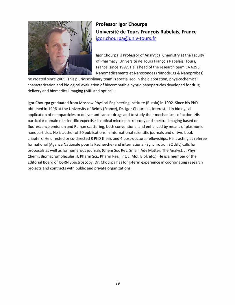
Professor Ji-Xin Cheng Purdue University [email protected] Ji-Xin Cheng was born in Jixi, Anhui Province, P. R. China in 1971. He attended University of Science and Technology of China (USTC) from 1989 to 1994. From 1994 to 1998, he carried out his PhD study on bond-selective chemistry under the supervision of Qingshi Zhu at USTC. As a graduate student, he
worked as a research assistant at Universite Paris-sud (France) and the Hong Kong University of Science and Technology (HKUST). After postdoctoral training in Yijing Yan’s group at HKUST and Sunney Xie’s group at Harvard University, Cheng joined Purdue University in 2003 as Assistant Professor. He was promoted to Associate Professor in 2009 and Full Professor in 2013 in Weldon School of Biomedical Engineering and Department of Chemistry. His research is at the most forefront of the label-free spectroscopic imaging field. Current projects include the study of aberrant cholesterol metabolism in aggressive cancer and deep tissue imaging by acoustic detection of chemical bond vibration.
39
Professor Igor Chourpa
Université de Tours François Rabelais, France [email protected] Igor Chourpa is Professor of Analytical Chemistry at the Faculty of Pharmacy, Université de Tours François Rabelais, Tours, France, since 1997. He is head of the research team EA 6295 Nanomédicaments et Nanosondes (Nanodrugs & Nanoprobes)
he created since 2005. This pluridisciplinary team is specialized in the elaboration, physicochemical characterization and biological evaluation of biocompatible hybrid nanoparticles developed for drug delivery and biomedical imaging (MRI and optical). Igor Chourpa graduated from Moscow Physical Engineering Institute (Russia) in 1992. Since his PhD obtained in 1996 at the University of Reims (France), Dr. Igor Chourpa is interested in biological application of nanoparticles to deliver anticancer drugs and to study their mechanisms of action. His particular domain of scientific expertise is optical microspectroscopy and spectral imaging based on fluorescence emission and Raman scattering, both conventional and enhanced by means of plasmonic nanoparticles. He is author of 50 publications in international scientific journals and of two book chapters. He directed or co-directed 8 PhD thesis and 4 post-doctoral fellowships. He is acting as referee for national (Agence Nationale pour la Recherche) and international (Synchrotron SOLEIL) calls for proposals as well as for numerous journals (Chem Soc Rev, Small, Adv Matter, The Analyst, J. Phys. Chem., Biomacromolecules, J. Pharm Sci., Pharm Res., Int. J. Mol. Biol, etc.). He is a member of the Editorial Board of ISSRN Spectroscopy. Dr. Chourpa has long-term experience in coordinating research projects and contracts with public and private organizations.
40
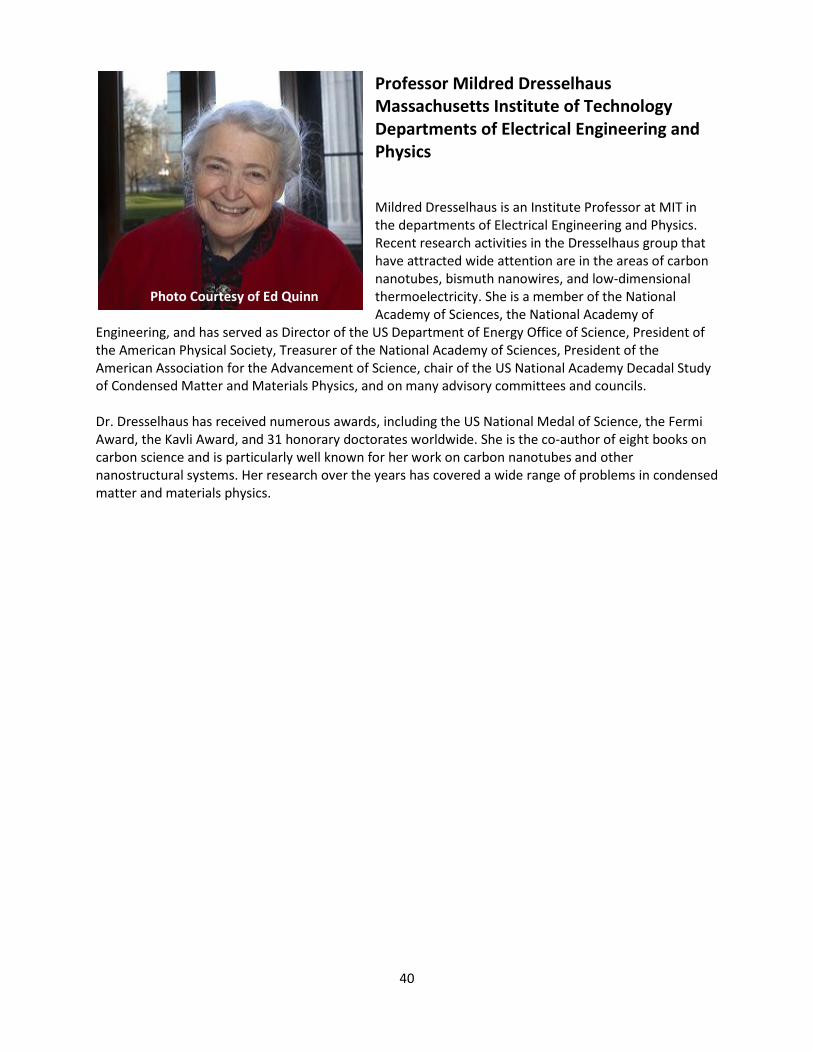
Professor Mildred Dresselhaus Massachusetts Institute of Technology Departments of Electrical Engineering and Physics Mildred Dresselhaus is an Institute Professor at MIT in the departments of Electrical Engineering and Physics. Recent research activities in the Dresselhaus group that have attracted wide attention are in the areas of carbon nanotubes, bismuth nanowires, and low-dimensional thermoelectricity. She is a member of the National Academy of Sciences, the National Academy of
Engineering, and has served as Director of the US Department of Energy Office of Science, President of the American Physical Society, Treasurer of the National Academy of Sciences, President of the American Association for the Advancement of Science, chair of the US National Academy Decadal Study of Condensed Matter and Materials Physics, and on many advisory committees and councils. Dr. Dresselhaus has received numerous awards, including the US National Medal of Science, the Fermi Award, the Kavli Award, and 31 honorary doctorates worldwide. She is the co-author of eight books on carbon science and is particularly well known for her work on carbon nanotubes and other nanostructural systems. Her research over the years has covered a wide range of problems in condensed matter and materials physics.
Photo Courtesy of Ed Quinn
41
Dr. Neil Everall Intertek [email protected] Neil Everall gained his BSc in Chemistry in 1981 from the University of York, UK, and his PhD in 1986 from the University of Durham, UK, researching ultrafast Raman Spectroscopy for fluorescence rejection. In 1988 he joined ICI plc, where he led the company’s Vibrational Spectroscopy activity for more than 13 years and eventually became ICI’s senior measurement scientist. He is now employed by Intertek-MSG, where he develops infrared
and Raman spectroscopic techniques to solve research, production and application problems for the chemicals, materials and life-science industries. He also carries out fundamental research on vibrational spectroscopy, for example on confocal Raman microscopy and Raman photon migration. He has authored ~90 journal articles and numerous book chapters, he was an Associate Editor of Wiley’s “Handbook of Vibrational Spectroscopy”, and co-edited Wiley’s “Vibrational Spectroscopy of Polymers: Principles and Practice” (2007). Neil’s research has been recognized by the Society for Applied Spectroscopy Meggers Award (2002 & 2006), the Coblentz Society Williams-Wright Award (2003), and the 2007 Mann Award for Applied Raman Spectroscopy. He is a Fellow of the Royal Society of Chemistry and he was elected a Fellow of the Society of Applied Spectroscopy in 2009. Neil served as European Associate Editor for the journal “Applied Spectroscopy” from 2000-2012.
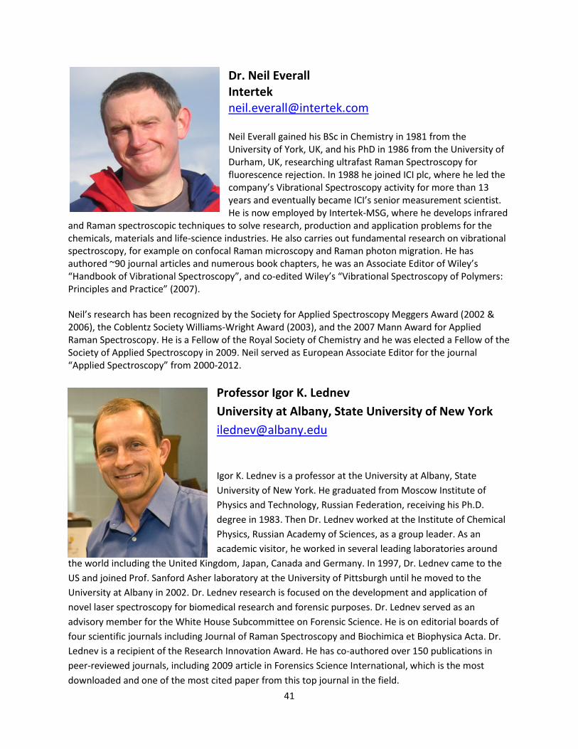
Professor Igor K. Lednev University at Albany, State University of New York [email protected] Igor K. Lednev is a professor at the University at Albany, State University of New York. He graduated from Moscow Institute of Physics and Technology, Russian Federation, receiving his Ph.D. degree in 1983. Then Dr. Lednev worked at the Institute of Chemical Physics, Russian Academy of Sciences, as a group leader. As an academic visitor, he worked in several leading laboratories around
the world including the United Kingdom, Japan, Canada and Germany. In 1997, Dr. Lednev came to the US and joined Prof. Sanford Asher laboratory at the University of Pittsburgh until he moved to the University at Albany in 2002. Dr. Lednev research is focused on the development and application of novel laser spectroscopy for biomedical research and forensic purposes. Dr. Lednev served as an advisory member for the White House Subcommittee on Forensic Science. He is on editorial boards of four scientific journals including Journal of Raman Spectroscopy and Biochimica et Biophysica Acta. Dr. Lednev is a recipient of the Research Innovation Award. He has co-authored over 150 publications in peer-reviewed journals, including 2009 article in Forensics Science International, which is the most downloaded and one of the most cited paper from this top journal in the field.
42
Professor Wei Min Columbia University [email protected] Dr. Wei Min graduated from Peking University, China, with a Bachelor's degree in 2003. He received his Ph.D. in Chemistry from Harvard University in 2008 studying single-molecule biophysics with Prof. Sunney Xie. After continuing his postdoctoral work in Xie group, Dr. Min joined the faculty of Department of Chemistry at Columbia University in July of 2010. Dr. Min's current research interests focus
on developing novel optical spectroscopy and microscopy technology to address complex problems in biology and medicine. His contribution has been recognized by a number of honors, including Alfred P. Sloan Research Fellowship (2013), NIH Director's New Innovator Award (2012) and Faculty Finalist of Blavatnik Awards for Young Scientists of the New York Academy of Sciences (2012).
Dr. The-Quyen Nguyen Northwestern University
[email protected] Dr. The-Quyen Nguyen received his Ph.D. in Physical Chemistry from Ecole Central de Paris in 2007. He has developed a new generation of Raman spectrometers using a monochromator, a digital micromirror device as light modulator and a photomultiplier tube as detector. His Ph.D. thesis was recognized with the "Instrument Award" by the
French Society of Physics and the French Society of Chemistry, and also received the Medal of the French Academy of Agriculture for excellence. In 2008, Dr. Nguyen went to work for Horiba Jobin-Yvon and then decided to devote his energies to biomedical research and the development of innovative optical instruments for clinical use. He was recently honored with the 2014 SPIE Translational Research Award for his use of Raman spectroscopy in the operating room for surgical margin evaluation. This SPIE award recognizes the outstanding contribution in the field of biomedial optics with the potential to transform clinical practice and improve the lives of patients. Dr. Nguyen is currently a Research Assistant Professor at the Biomedical Engineering Department of Northwestern University, where he develops breakthrough optical spectral and imaging techniques for non-invasive screening, diagnosis and detection of cancers as well as optical instrumentations for large-scale/multi-sites clinical trials
43
Professor Lukas Novotny ETH Zürich, Photonics Laboratory, Switzerland [email protected] Lukas Novotny is Professor of Photonics at ETH Zürich, Switzerland. He received his PhD in 1996 from the same institution. His doctoral work was in collaboration with the IBM Research Laboratories and dealt with theoretical problems in near-field optics. From 1996-99 he was a postdoctoral fellow at the Pacific Northwest National Laboratory, working on new schemes of single molecule detection and nonlinear spectroscopy. In 1999 he joined the faculty of the Institute of Optics where he started one of the first research
programs with focus on nano-optics. Novotny is a co-author of the textbook 'Principles of Nano-Optics', which is currently in its second edition. He is a Fellow of the Optical Society of America and the American Association for the Advancement of Science.
Professor Christian Pellerin University of Montreal [email protected] Christian Pellerin is an associate professor at the Department of chemistry of Université de Montréal. He received his B.Sc. in chemistry in 1997 from Université du Québec à Trois-Rivières and his Ph.D. in chemistry in 2002 from Université Laval under the
supervision of Michel Pézolet and Robert E. Prud'homme. He joined the faculty at Université de Montréal in 2005 after a postdoctoral fellowship with John F. Rabolt and D. Bruce Chase at the University of Delaware. His research interests include the structure and properties of electrospun nanofibers, molecular glasses, supramolecular polymer complexes, and mussel-based biomaterials, in addition to applying novel infrared and Raman spectroscopy techniques.
44
Dr. Michael J. Pelletier Pfizer Global Research and Development [email protected] Mike is an Associate Research Fellow in the QbD Methods Development group at Pfizer. His work involves the identification and development of new spectroscopic technologies, applied Raman and NIR spectroscopy, and PAT for process understanding. Prior to that he was a Principle Engineer at NASA's Jet Propulsion Laboratory where he won
funding for, and led, in-situ analysis projects involving microfluidics and Raman spectroscopy. He was awarded the Williams Wright Award and the Charles Mann Award for his research in applied spectroscopy. Mike has over 50 peer-reviewed publications (15 single-author) including 6 patents, several book chapters, and a book on Raman spectroscopy. Mike is a Fellow of the Society for Applied Spectroscopy and a Senior Member of the Optical Society of America.
Professor Ping-Heng Tan Chinese Academy of Sciences [email protected] Ping-Heng Tan, received the BS (1996) from Peking University, and Ph. D in Graduate University of Chinese Academy of Sciences, 2001. After finishing his PhD he became a postdoctoral fellow at the Walter Schottky Institute at the Technical University of Munich, Germany. From 2003, he was an associate professor and then professor at the institute of Semiconductors, Chinese Academy of Sciences. He had been awarded as
KC Wong Royal Society Fellow to work at University of Cambridge for one year from 2006 to 2007. Tan has authored about 90 peer-reviewed papers in scientific journals with around 2000 citations to his papers and H index 24. He had received National One-hundred Excellent Doctoral Dissertations Awards, Lu JiaXi Awards for excellent young scientists and National Science Fund for Distinguished Young Scholars. In 2013, he was elected as the Secretary-General of the Professional Committee on Light Scattering of Chinese Physical Society.
45
Professor Lawrence D. Ziegler Boston University [email protected] Professor Ziegler received his Ph.D. in Chemistry from Cornell University in 1978 (advisor: A. C. Albrecht) where he carried out Raman experimental and theoretical studies. After an NIH Postdoctoral Fellowship (advisor: Bruce Hudson) at the University of Oregon, and an NRC Research Associateship at NRL, he held appointments of Assistant Professor and Professor in Chemistry at
Northeastern University. In 1991 he moved to Boston University where he is currently Professor and Chair of the Chemistry Department and a member of the BU Photonics Center. He pioneered the development of UV resonance Raman, resonance hyper-Raman, and resonance rotational Raman scattering for applications including the study of short-time chemical reaction dynamics. Subsequent research interests include the characterization of ultrafast responses of transparent materials, solvation dynamics in dense fluids and supercritical fluids, ultrafast electronic relaxation in novel semiconducting materials (e.g. carbon nanotubes and WBG semiconductors), and ultrafast IR studies of biological waters.
More recently, his lab has explored the use of surface enhanced Raman spectroscopy for bioanalytical applications including rapid infectious disease diagnostics, blood aging, cancer detection and forensics. He was co-Organizer of the 22nd International Conference on Raman Spectroscopy (Boston, August 2010) and is currently Senior Associate Editor for the Journal of Raman Spectroscopy.
46
Professor Sunney Xie Harvard University [email protected] Xiaoliang Sunney Xie received a B.S. from Peking University in 1984, and his Ph.D. from the University of California at San Diego in 1990, followed by a short postdoctoral experience at the University of Chicago. In 1992, Xie joined Pacific Northwest National Laboratory, where he later became a Chief Scientist. In 1999, he was appointed Professor of Chemistry at Harvard University. He is now the Mallinckrodt Professor of Chemistry and Chemical Biology at Harvard, and the Cheung Kong Visiting Professor at Peking University, Biodynamics Optical Imaging Center (BIOPIC).
Xie has made major contributions to the emergence of the field of single-molecule biophysical chemistry and its application to biology. His team also pioneered the development of coherent Raman scattering microscopy and single cell whole genome sequencing. His honors include the Harrison Howe Award, Biophysical Society Founders Award, E.O. Lawrence Award in Chemistry, Leibinger Innovation Prize, the NIH Director's Pioneer Award, the Sackler Prize for Physical Sciences. Xie is a fellow of the American Academy of Arts and Sciences and a member of the National Academy of Sciences.
47
Dr. Dan Fu Harvard University [email protected] Dan Fu is a Postdoctoral Fellow, in the Department of Chemistry and Chemical Biology at Harvard University. Dr. Dan Fu was born in Hubei, China. He received his bachelor's degree from Peking University in China (2003). In 2009, he completed his Ph.D study at Princeton University under the supervision of Professor Warren Warren, working on the development of novel label-free multiphoton absorption microscopy methods. After that, he worked as a postdoctoral associate at the
G.R.Harrison Spectroscopy Lab led by Professor Michael Feld at Massachusetts Institute of Technology, where he investigated quantitative phase microscopy and its applications to live cell imaging.
In 2010, he moved to Harvard University to work with Professor Sunney Xie as a postdoctral fellow, where he developed multiplex stimulated Raman scattering microscopy and hyperspectral stimulated Raman scattering microscopy. Currently his main research interests are the application of stimulated Raman scattering microscopy to various biological and biomedical problems such as cancer diagnosis and staging, studying lipid metabolism and drug-cell interaction.
Dr. Andrew Whitley HORIBA Scientific [email protected]
Andrew Whitley has concentrated his career in vibrational spectroscopy product development and sales. He is currently the Vice President of Sales at HORIBA Scientific in New Jersey and remains the Director of Raman Spectroscopy, which he has been doing since 2000.
Prior to this, he was the US Raman Product Manager at Renishaw. He also worked for Renishaw in the UK and prior to that he was at Bruker during the FT-Raman breakthroughs of the early 1990’s. Many thanks to everyone at Harvard University and HORIBA Scientific for all their efforts in putting this RamanFest symposium together.
And a special thanks to Larry Valles of Harvard University, and Li Yan and Joanne Lowy of HORIBA Scientific for their patience, perseverance and dedication to ensuring a successful meeting.