Embed Size (px)
Citation preview

[Expand]
Historic drawing of human oocyte and spermatozoa
Cell division - 3 types
Lecture - FertilizationFrom Embryology
Embryology - 18 Aug 2015 Translate
Introduction
This lecture and the associated laboratory will cover male and female gametogenesis andfertilisation.
Development is 1 embryonic cell producing about 1013 (100,000,000,000,000) cells inthe adult at any one time (over time with cell death and ongoing replacement this issubstantially more).
This is where the first embryonic cell begins! Fertilization is the fusion of haploidgametes, egg (oocyte) and sperm (spermatozoa), to form the diploid zygote. Note thoughthere can be subtle differences in the fertilization process which occurs naturally withinthe body or through reproductive technologies outside the body, the overall product inboth cases is a diplod zygote.
Some Recent Research - Meiosis Podcast Biosights 18 March 2013 - Breaking eggsymmetry (http://jcb.rupress.org/content/200/5/567/suppl/DC2) | JCB 16 June 16 2014How sperm get into the zona (http://jcb.rupress.org/content/205/6/754.2.full)
Lecture - Print PDF
Lecture Archive: 2014 Lecture PDF | 2013 | 2012 (http://embryology.med.unsw.edu.au/embryology/index.php?title=Lecture_-_Fertilization&oldid=96435) | 2011 (http://embryology.med.unsw.edu.au/embryology/index.php?title=Lecture_-_Fertilization&oldid=60496)| 2010 | 2009
Lecture Objectives1. Broad understanding of reproductive cycles.2. Understand the key features of gametogenesis.3. Understand the differences in male and female gametogenesis.4. Brief understanding of the differences between mitosis and meiosis.5. Understanding of the events in fertilization.
Lecture Resources

[Collapse]
[Collapse]
[Expand]
Movies
Oocyte MeiosisPage | Play
OvulationPage | Play | Audio
OvulationPage | Play
Oocyte TransportPage | Play
SpermatozoaPage | Play
Spermatozoa MotilityPage | Play
Human FertilizationPage | Play
Human FertilizationPage | Play
FertilizationPage | Play
Pronuclear FusionPage | Play
Ectopic Polar BodyPage | Play
Male MitochondriaPage | Play
References
Hill, M.A. (2015). UNSW Embryology(15th ed.) Retrieved August 18, 2015,fromhttps://embryology.med.unsw.edu.au
Cell Division Links: Meiosis | Mitosis | Lecture - Cell Division andFertilization | Spermatozoa Development | Oocyte Development | Fertilization |Zygote
Menstrual Cycle | Oocyte | Zona pellucida | Spermatozoa | Meiosis | Fertilization| Mitosis | Week 1
Moore, K.L., Persaud, T.V.N. & Torchia,M.G. (2011). The developing human:clinically oriented embryology (9th ed.).Philadelphia: Saunders.
The following chapter links only work with a UNSW connection.
First Week of Human Development(http://www.unsw.eblib.com.wwwproxy0.library.unsw.edu.au/patron/Read.aspx?p=1430154&pg=35)Second Week of Human Development(http://www.unsw.eblib.com.wwwproxy0.library.unsw.edu.au/patron/Read.aspx?p=1430154&pg=63)
Schoenwolf, G.C., Bleyl, S.B., Brauer,P.R., Francis-West, P.H. & Philippa H.(2015). Larsen's human embryology (5thed.). New York; Edinburgh: ChurchillLivingstone.
The following chapter links only work with a UNSW connection.
Chapter 1 - Gametogenesis, Fertilization, and First Week(https://login.wwwproxy0.library.unsw.edu.au/login?url=http://www.unsw.eblib.com/patron/FullRecord.aspx?p=2074524)
ECHO360 Recording
Human Reproductive CycleSexual reproduction in most species is regulated by regular endocrine changes, or cycles, in the female. These cycles begin postnatally,function for variable times and can then decrease or cease entirely.
Human reproduction is regulated in females by the menstrual cycle, a regular cyclic hormonal change which coordinate changes in theovary and internal reproductive tract. This cycle commences at puberty and ends at menopause.Non-primates (rats, mice, horses, pig) reproduction is regulated in females by the estrous cycle (British spelling, oestrous).
Female Male
Menstrual Cycle a regular cycle of reproduction (28 days)begins at puberty, release of 1 egg (oocyte) every cycleEndocrine controlled (HPG axis) Hypothalamus - Pituitary -Gonad
continuous production of sperm (spermatozoa)begins at puberty, release millions of spermatozoaEndocrine controlled (HPG axis) Hypothalamus - Pituitary -Gonad

GametogenesisMeiosis in the gonad (ovary or testis) produces the haploid gametes, oocyte and spermatozoa (egg and sperm). Meiosis time course and finalgamete number differs between female and male.
Male - Spermatogenesis
Human spermatozoa (electron microscope)
Mouse spermatozoa (electron microscope)

The testes have two functions.
1. produce the male gametes or spermatozoa2. produce male sexual hormone, testosterone (internal and external genitalia, sex characteristics)
Historic testis drawing
Child Seminiferoustubule
Adult Seminiferoustubule
Seminiferous tubulecross-section andsupporting cells
Human spermatozoa
Human Spermatozoa
Human spermatozoa take about 48 days from entering meiosis until morphologically mature spermatozoa.
Spermatogonia - are the diploid firstcells of spermatogenesisPrimary spermatocytes - large, enterthe prophase of the first meiotic divisionSecondary spermatocytes - small,complete the second meiotic divisionSpermatid - immature spermatozoaSpermatozoa - differentiated gamete
Spermatozoa development:primordial germ cell -spermatogonia - primaryspermatocyte - secondaryspermatocytes - spermatid -spermatozoa
Sertoli cells (support cells)
Interstitial cells or Leydig cells (producehormone)
Female - Oogenesis
The ovaries have two functions.
1. produce the female gametes or oocytes2. produce female hormones, estrogen and progesterone (secondary sex characteristics, menstrual cycle)

[Expand]
Male gametogenesis
infant ovary
overview of ovary
three stages of follicledevelopment
primary follicle
tertiary follicle
In an adult human female the development of a primordial follicle containing an oocyte to a preovulatory follicle takes in excess of 120 days.
Human Follicle Development
Human Ovulation
Human ovary follicle development
Ovarian Follicle Stages: primordial follicle - primary follicle - secondary follicle - tertiary follicle - preovulatory follicle
Follicle cells (support cells) Theca cells (produce hormone)
Ovulation Movie
Meiosis Differences
Male Meiosis
Meiosis initiated continuously in a mitotically dividing stem cell population4 gametes produced / meiosisMeiosis completed in days or weeksMeiosis and differentiation proceed continuously without cell cycle arrestDifferentiation of gamete occurs while haploid after meiosis endsSex chromosomes excluded from recombination and transcription during firstmeiotic prophase
MBoC - Figure 20-27. The stages of spermatogenesis(http://www.ncbi.nlm.nih.gov/books/bv.fcgi?&rid=mboc4.figgrp.3734)
Female Meiosis
Meiosis initiated once in a finite population of cells1 gamete produced / meiosisCompletion of meiosis delayed for months or years

[Expand]
[Expand]
[Expand]
Female gametogenesis
Early zygote showing polar bodies
Human uterine tube ciliated epithelium
Meiosis arrested at 1st meiotic prophase and reinitiated in a smaller population of cellsDifferentiation of gamete occurs while diploid in first meiotic prophaseAll chromosomes exhibit equivalent transcription and recombination duringmeiotic prophase
The Cell - Figure 14.37. Meiosis of vertebrate oocytes(http://www.ncbi.nlm.nih.gov/books/bv.fcgi?&rid=cooper.figgrp.2492)
Polar Bodies
In female gametogenesis only a single (1) haploid egg is produced from meiosis. Inmale gametogenesis four (4) haploid sperm are produced from meiosis. So whathappens to all the extra DNA in producing this single egg?
In Meiosis 1 the "extra" DNA is excluded to the periphery as a 1st polarbody, which encloses the extra DNA.In Meiosis 2 the "extra" DNA is once again excluded as a 2nd polar body.The first polar body may also under go meiosis 2 producing a 3rd polar body.
These polar bodies are not gametes.Polar bodies appear to have no other function other than to dispose of the extraDNA in oogenesis.
Recent research in mice suggest that the position of oocyte polar body mayinfluence fertilization site.
Meiosis Polar Body Movie
FertilizationGamete formation, menstrual cycle and fertilisation will also be covered in detail in thisweek's Laboratory. Fertilization is the complete process resulting in the fusion of haploidgametes, egg and sperm, to form the diploid zygote. The recent development of aidedfertilization is described as in vitro fertilization (in vitro = "in glass", outside the body,IVF). Clinically, all these aided fertilization techniques are grouped as AssistedReproductive Technologies or ART.
Oogenesis - 1 gamete produced/meiosis + 3 polar bodies, meiosis is slow, 1 egg produced and released at ovulationSpermatogenesis - 4 gametes produced/meiosis, meiosis is fast, 200-600 million sperm released at ejaculation
Fertilization Movies
Fertilization Site
Fertilization resulting in embryo development usually occurs in first 1/3 of uterinetube (oviduct, Fallopian tube)The majority of fertilized oocytes do not go on to form an embryoFertilization can also occur outside uterine tube associated with AssistedReproductive Technologies (IVF, GIFT, ZIFT...) and ectopic pregnancyOocyte ovulation - release from the ovary with associated cells, into peritonealcavity, uterine tube fimbria then into uterine tube (oviduct, uterine horn, fallopiantube) and epithelial cilia mediated movement.Spermatozoa ejaculation - deposited in vagina, movement of tail to "swim" inuterine secretions through cervix, uterine body and into uterine tube, haveapproximately 24-48h to fertilize oocyte.
Prior to the fertilization process commencing both the gametes complete of a number ofbiological processes.
Oocyte Meiosis - completes Meiosis 1 and commences Meiosis 2 (arrests atMetaphase II).Spermatozoa Capacitation - following release (ejaculation) and mixing with other glandular secretions, activates motility andacrosome preparation.Migration - both oocyte and spermatozoa.
Endocrinology - Diagram of the comparative anatomy of the male and female reproductive tracts(http://www.ncbi.nlm.nih.gov/bookshelf/br.fcgi?book=endocrin&part=A972&rendertype=box&id=A1230)
Gamete Movement Movies
Fertilization - Male

Human spermatozoa (light microscope)
Human MII oocyte cortical granules
Spermatozoa: Ejaculation - Capacitation - Spermatozoa motility - Chemotaxis - Bindingto zona pellucida - Acrosome reaction - Membrane fusion
Ejaculation
about 3.5 ml, containing 200 - 600 million spermatozoaby volume less than 10 % spermatozoaaccessory glands contribute majority of volume (60 % seminal vesicle, 10 %bulbourethral, 30 % prostate)
Male Infertility
Oligospermia (Low Sperm Count) - less than 20 million sperm after 72 hourabstinence from sexAzoospermia (Absent Sperm) - blockage of duct networkImmotile Cilia Syndrome - lack of sperm motility
Capacitation
spermatozoa activation process - removal of glycoprotein coat and seminal proteinsand alteration of sperm mitochondria
Spermatozoa motility
tail of spermatozoa provide movement by microtubulesenergy for this movement is provided by mitochondria in tail initial segment
Chemotaxis
oocyte cumulus cells release progesterone (may also be other oocyte and follicular fluid factors)
Spermatozoa Binding
Zona pellucida protein ZP2 acts as receptor for spermatozoa binding (species specific)
Acrosome Reaction
exocytosis of acrosome contents (calcium mediated) MBoC - Figure 20-31. The acrosome reaction that occurs when a mammaliansperm fertilizes an egg (http://www.ncbi.nlm.nih.gov/books/bv.fcgi?rid=mboc4.figgrp.3741)enzymes to digest the zona pellucidaexposes sperm surface proteins to bind ZP2
Membrane fusion
between spermatozoa and oocyte cell membranes, allows sperm nuclei passage into egg cytoplasmmembrane fusion also initiates oocyte processes to block polyspermy
Fertilization - Oocyte
Oocyte: Membrane depolarization - Cortical reaction - Meiosis 2 completion
Membrane Depolarization
caused by spermatozoa membrane fusion, acts as primary block to polyspermy(fertilisation by more than one spermatozoa)
Cortical Reaction
Inositol triphosphate (IP3) pathway elevates intracellular calcium, exocytosis ofcortical granulesenzyme alters ZP2 so it will no longer bind sperm plasma membraneMBoC - Figure 20-32. How the cortical reaction in a mouse egg is thought toprevent additional sperm from entering the egg(http://www.ncbi.nlm.nih.gov/books/bv.fcgi?rid=mboc4.figgrp.3743)
Meiosis 2
completion of 2nd meiotic divisionforms second polar body (third polar body may be formed by meiotic division of the first polar body)
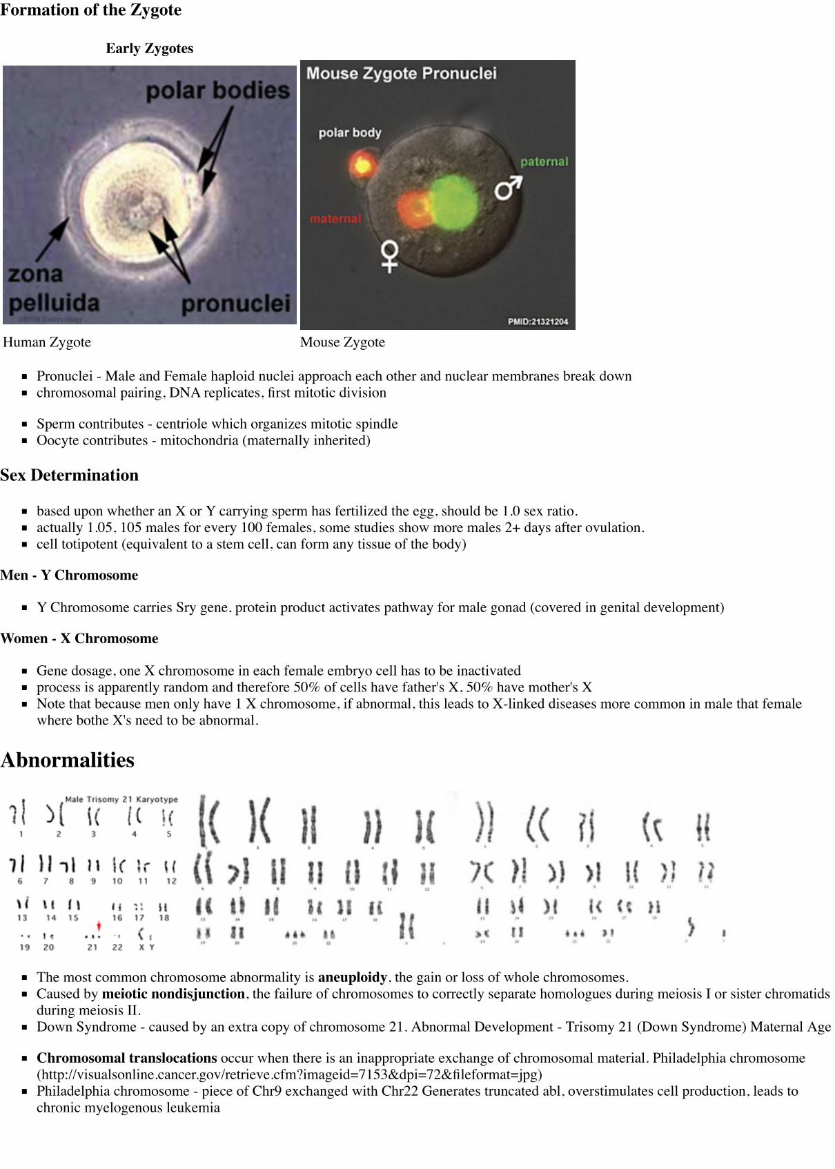
Formation of the Zygote
Early Zygotes
Human Zygote Mouse Zygote
Pronuclei - Male and Female haploid nuclei approach each other and nuclear membranes break downchromosomal pairing, DNA replicates, first mitotic division
Sperm contributes - centriole which organizes mitotic spindleOocyte contributes - mitochondria (maternally inherited)
Sex Determination
based upon whether an X or Y carrying sperm has fertilized the egg, should be 1.0 sex ratio.actually 1.05, 105 males for every 100 females, some studies show more males 2+ days after ovulation.cell totipotent (equivalent to a stem cell, can form any tissue of the body)
Men - Y Chromosome
Y Chromosome carries Sry gene, protein product activates pathway for male gonad (covered in genital development)
Women - X Chromosome
Gene dosage, one X chromosome in each female embryo cell has to be inactivatedprocess is apparently random and therefore 50% of cells have father's X, 50% have mother's XNote that because men only have 1 X chromosome, if abnormal, this leads to X-linked diseases more common in male that femalewhere bothe X's need to be abnormal.
Abnormalities
The most common chromosome abnormality is aneuploidy, the gain or loss of whole chromosomes.Caused by meiotic nondisjunction, the failure of chromosomes to correctly separate homologues during meiosis I or sister chromatidsduring meiosis II.Down Syndrome - caused by an extra copy of chromosome 21. Abnormal Development - Trisomy 21 (Down Syndrome) Maternal Age
Chromosomal translocations occur when there is an inappropriate exchange of chromosomal material. Philadelphia chromosome(http://visualsonline.cancer.gov/retrieve.cfm?imageid=7153&dpi=72&fileformat=jpg)Philadelphia chromosome - piece of Chr9 exchanged with Chr22 Generates truncated abl, overstimulates cell production, leads tochronic myelogenous leukemia

Hydatidiform Mole
Complete Mole - Only paternal chromosomes.Partial Mole - 3 sets of chromosomes ( (triploidy) instead of the usual 2.
NextLab 1 - Gametogenesis and Fertilisation
Homework
Beginning your online work - Working Online in this course
1. Make your own page.1. Log-in to the embryology website using your student ID and Zpass.2. Click your student number (shown in red at the top right of the screen following log-in)3. Create page using the tab at the top of the page, and save.
2. How would you identify your Type in a group and add to your page.3. What was the most interesting thing you learnt in today's lecture?
If you have done the above correctly your ZID should be blue and not red on this page ANAT2341 2015 Students.
UNSW Embryology LinksSpermatozoa Development Oocyte Development Fertilization Trisomy 21 (Down Syndrome)2012 Lecture (http://embryology.med.unsw.edu.au/embryology/index.php?title=Lecture_-_Fertilization&oldid=96435)
Cell Division Links: Meiosis | Mitosis | Lecture - Cell Division and Fertilization | Spermatozoa Development | Oocyte Development| Fertilization | Zygote
References
Online Textbooks
Developmental Biology by Gilbert, Scott F. Sunderland (MA): Sinauer Associates, Inc.; c2000Figure 2.9. Summary of meiosis(http://www.ncbi.nlm.nih.gov/books/bv.fcgi?highlight=meiosis&rid=dbio.figgrp.200) | fusion of egg and sperm plasma membranes(http://www.ncbi.nlm.nih.gov/books/bv.fcgi?&rid=dbio.figgrp.1360)Molecular Biology of the Cell 4th ed. Alberts, Bruce; Johnson, Alexander; Lewis, Julian; Raff, Martin; Roberts, Keith; Walter, PeterNew York and London: Garland Science; c2002 - IV. Internal Organization of the Cell Chapter 17. The Cell Cycle and ProgrammedCell Death Programmed Cell Death (http://www.ncbi.nlm.nih.gov:80/books/bv.fcgi?db=Books&rid=mboc4.chapter.3167) | AnOverview of the Cell Cycle (http://www.ncbi.nlm.nih.gov:80/books/bv.fcgi?db=Books&rid=mboc4.section.3169) | Figure 17-1. Thecell cycle (http://www.ncbi.nlm.nih.gov:80/books/bv.fcgi?db=Books&rid=mboc4.figgrp.3168) | Fertilization(http://www.ncbi.nlm.nih.gov/books/bv.fcgi?&rid=mboc4.section.3738)Molecular Cell Biology by Lodish, Harvey; Berk, Arnold; Zipursky, S. Lawrence; Matsudaira, Paul; Baltimore, David; Darnell, JamesE. New York: W. H. Freeman & Co.; c1999 Chapter 13. Regulation of the Eukaryotic Cell Cycle Regulation of the Eukaryotic Cell

Cycle (http://www.ncbi.nlm.nih.gov:80/books/bv.fcgi?db=Books&rid=mcb.chapter.3432) | Overview of the Cell Cycle and Its Control(http://www.ncbi.nlm.nih.gov:80/books/bv.fcgi?db=Books&rid=mcb.section.3463) | Figure 13-2. Current model for regulation of theeukaryotic cell cycle (http://www.ncbi.nlm.nih.gov:80/books/bv.fcgi?db=Books&rid=mcb.figgrp.3467) | Movies Proposed alternativemechanisms for chromosome congression. (http://www.ncbi.nlm.nih.gov/books/bv.fcgi?&rid=mcb.figgrp.5522) | Centromericattachment of microtubules. (http://www.ncbi.nlm.nih.gov/books/bv.fcgi?&rid=mcb.figgrp.5509) | The stages of mitosis andcytokinesis in an animal cell. (http://www.ncbi.nlm.nih.gov/books/bv.fcgi?&rid=mcb.figgrp.5500)The Cell - A Molecular Approach by Cooper, Geoffrey M. Sunderland (MA): Sinauer Associates, Inc.; c2000- IV. Cell RegulationChapter 14. The Cell Cycle The Eukaryotic Cell Cycle (http://www.ncbi.nlm.nih.gov:80/books/bv.fcgi?db=Books&rid=cooper.section.2433) | Figure 14.1. Phases of the cell cycle (http://www.ncbi.nlm.nih.gov:80/books/bv.fcgi?db=Books&rid=cooper.figgrp.2435) | Figure 14.32. Comparison of meiosis and mitosis (http://www.ncbi.nlm.nih.gov/books/bv.fcgi?&rid=cooper.figgrp.2486) | Figure 14.37. Meiosis of vertebrate oocytes (http://www.ncbi.nlm.nih.gov/books/bv.fcgi?&rid=cooper.figgrp.2492)HSTAT - In Vitro Fertilization As A Medical Treatment For Male or Female Infertility (http://www.ncbi.nlm.nih.gov/books/bv.fcgi?&rid=hstat6.section.1395#1396)
MBoC MBoC - Figure 20-18. Influence of Sry on gonad development (http://www.ncbi.nlm.nih.gov/books/bv.fcgi?rid=mboc4.figgrp.3716)Endocrinology Endocrinology - Comparative anatomy of male and female reproductive tracts(http://www.ncbi.nlm.nih.gov/bookshelf/br.fcgi?book=endocrin&part=A972&rendertype=box&id=A1230)
Search
Bookshelf cell division (http://www.ncbi.nlm.nih.gov/sites/entrez?db=Books&cmd=search&term=cell+division) | mitosis(http://www.ncbi.nlm.nih.gov/sites/entrez?db=Books&cmd=search&term=mitosis) | meiosis (http://www.ncbi.nlm.nih.gov/sites/entrez?db=Books&cmd=search&term=meiosis) | fertilization (http://www.ncbi.nlm.nih.gov/sites/entrez?db=Books&cmd=search&term=fertilization)
Pubmed cell division (http://www.ncbi.nlm.nih.gov/sites/gquery?itool=toolbar&cmd=search&term=cell+division) | mitosis(http://www.ncbi.nlm.nih.gov/sites/gquery?itool=toolbar&cmd=search&term=mitosis) | meiosis(http://www.ncbi.nlm.nih.gov/sites/gquery?itool=toolbar&cmd=search&term=meiosis) | fertilization(http://www.ncbi.nlm.nih.gov/sites/gquery?itool=toolbar&cmd=search&term=fertilization)
Reviews
Dennis W Stacey, Masahiro Hitomi Cell cycle studies based upon quantitative image analysis. Cytometry A: 2008, 73(4);270-8[PubMed:18163464]
Christoph Schorl, John M Sedivy Analysis of cell cycle phases and progression in cultured mammalian cells. Methods: 2007, 41(2);143-50 [PubMed:17189856]
Termsasthenozoospermia - (asthenospermia) Term for reduced sperm motility and can be the cause of male infertility.blood-testis barrier - (BTB) Formed by tight junctions, basal ectoplasmic specializations, desmosome-like junctions and gap junctionsbetween adjacent Sertoli cells near the basement membrane of the seminiferous epithelium.diploid - (Greek, di = double + ploion = vessel) Having two sets of chromosomes, the normal state for all cells other than the gametes.haploid - (Greek, haploos = single) Having a single set of chromosomes as in mature germ/sex cells (oocyte, spermatozoa) followingreductive cell division by meiosis. Normally cells are diploid, containing 2 sets of chromosomes.Leydig cell - (interstitial cell) Male gonad (testis) cell which secrete the androgen testosterone, beginning in the fetus. These cells arenamed after Franz von Leydig (1821 - 1908) a German scientist who histologically described these cells.meiosis - The cell division that occurs only in production of germ cells where there is a reduction in the number of chromosomes(diploid to haploid) which is the basis of sexual reproduction. All other non-germ cells in the body divide by mitosis.mitosis - The normal division of all cells, except germ cells, where chromosome number is maintained (diploid). In germ cell division(oocyte, spermatozoa) meiosis is a modified form of this division resulting in reduction in genetic content (haploid). Mitosis, divisionof the nucleus, is followed by cytokinesis the division of the cell cytoplasm and the cytoplasmic contents. cytokinesis overlaps withtelophase.sperm annulus - (Jensen's ring; Latin, annulus = ring) A region of the mammalian sperm flagellum connecting the midpiece and theprincipal piece. The annulus is a septin-based structure formed from SEPT1, 4, 6, 7 and 12. Septins are polymerizing GTPases that canact as a scaffold forming hetero-oligomeric filaments required for cytokinesis and other cell cycle roles.spermatogenesis - (Greek, genesis = origin, creation, generation) The term used to describe the process of diploid spermatagoniadivision and differentiation to form haploid spermatazoa within the testis (male gonad). The process includes the following cellularchanges: meiosis, reoorganization of DNA, reduction in DNA content, reorganization of cellular organelles, morphological changes(cell shape). The final process of change in cell shape is also called spermiogenesis.spermatogenesis - (Greek, genesis = origin, creation, generation) The maturation process of the already haploid spermatazoa into themature sperm shape and organization. This process involves reorganization of cellular organelles (endoplasmic reticulum, golgiapparatus, mitochondria), cytoskeletal changes (microtubule organization) and morphological changes (cell shape, acrosome and tailformation).
spermatogonia - The cells located in the seminiferous tubule adjacent to the basal membrane that either divide and separate to renewthe stem cell population, or they divide and stay together as a pair (Apr spermatogonia) connected by an intercellular cytoplasmicbridge to differentiate and eventually form spermatazoa.spermatozoa head - Following spermiogenesis, the first region of the spermatozoa containing the haploid nucleus and acrosome. Inhumans, it is a flattened structure (5 µm long by 3 µm wide) with the posterior part of nuclear membrane forming the basal plateregion. The human spermatozoa is about 60 µm long, actively motile and divided into 3 main regions (head, neck and tail).spermatozoa neck - Following spermiogenesis, the second region of the spermatozoa attached to basal plate, transverse orientedcentriole, contains nine segmented columns of fibrous material, continue as outer dense fibres in tail. In humans, it forms a shortstructure (1 µm). The human spermatozoa is about 60 µm long, actively motile and divided into 3 main regions (head, neck and tail).spermatozoa tail - Following spermiogenesis, the third region of the spermatozoa that has a (head, neck and tail). The tail is alsodivided into 3 structural regions a middle piece, a principal piece and an end piece. In humans: the middle piece (5 µm long) is formedby axonema and dense fibres surrounded by mitochondria; the principal piece (45 µm long) fibrous sheath interconnected by regularlyspaced circumferential hoops; the final end piece (5 µm long) has an axonema surrounded by small amount of cytoplasm and plasmamembrane.spermatogonial stem cells - (SSCs) The spermatagonia cells located beside the seminiferous tubule basal membrane that either divideand separate to renew the stem cell population, or they divide and stay together as a pair (Apr spermatogonia) connected by anintercellular cytoplasmic bridge to differentiate and eventually form spermatazoa.sperm protein 56 - A component of the spermatozoa acrosomal matrix released to the sperm surface during capacitation.
External LinksExternal Links Notice - The dynamic nature of the internet may mean that some of these listed links may no longer function. If the link nolonger works search the web with the link text or name.
JCB - Movie Collection Mitosis (http://jcb.rupress.org/misc/annotatedvideo.shtml#Mitosis) | Cytokinesis(http://jcb.rupress.org/misc/annotatedvideo.shtml#Cytokinesis)2008 ANAT3231 Lecture PDF lecture14-15 1 slide/page (view) (http://cellbiology.med.unsw.edu.au/units/pdf/08L14-15s1.pdf) | 4slides/page (print) (http://cellbiology.med.unsw.edu.au/units/pdf/08L14-15s4.pdf) | 6 slides/page (print)(http://cellbiology.med.unsw.edu.au/units/pdf/08L14-15s6.pdf) | text (print) (http://cellbiology.med.unsw.edu.au/units/pdf/08L14-15txt.pdf)McGraw-Hill Animation comparing Mitosis and Meiosis (http://highered.mcgraw-hill.com/olcweb/cgi/pluginpop.cgi?it=swf::535::535::/sites/dl/free/0072437316/120074/bio17.swf::Comparison%20of%20Meiosis%20and%20Mitosis)Salmon Lab Mitosis Movies (http://www.bio.unc.edu/faculty/salmon/lab/moviesmitosis.html)
2015 Course: Week 2 Lecture 1 Lecture 2 Lab 1 | Week 3 Lecture 3 Lecture 4 Lab 2 | Week 4 Lecture 5 Lecture 6 Lab 3 | Week 5Lecture 7 Lecture 8 Lab 4 | Week 6 Lecture 9 Lecture 10 Lab 5 | Week 7 Lecture 11 Lecture 12 Lab 6 | Week 8 Lecture 13 Lecture 14Lab 7 | Week 9 Lecture 15 Lecture 16 Lab 8 | Week 10 Lecture 17 Lecture 18 Lab 9 | Week 11 Lecture 19 Lecture 20 Lab 10 | Week 12Lecture 21 Lecture 22 Lab 11 | Week 13 Lecture 23 Lecture 24 Lab 12 | Projects: Group 1 | Group 2 | Group 3 | Group 4 | Group 5 |Group 6 | Students | Student Sharing | Moodle page (http://moodle.telt.unsw.edu.au/course/view.php?id=15814)
Glossary LinksA | B | C | D | E | F | G | H | I | J | K | L | M | N | O | P | Q | R | S | T | U | V | W | X | Y | Z | Numbers | Symbols
Cite this page: Hill, M.A. (2015) Embryology Lecture - Fertilization. Retrieved August 18, 2015, fromhttps://embryology.med.unsw.edu.au/embryology/index.php/Lecture_-_Fertilization
What Links Here? (http://php.med.unsw.edu.au/embryology/index.php?title=Special:WhatLinksHere/Lecture_-_Fertilization)
© Dr Mark Hill 2015, UNSW Embryology ISBN: 978 0 7334 2609 4 - UNSW CRICOS Provider Code No. 00098G
Retrieved from ‘https://embryology.med.unsw.edu.au/embryology/index.php?title=Lecture_-_Fertilization&oldid=194006’
Categories: 2015 Science-Undergraduate Human Embryo Week 1 Fertilization Oocyte Spermatozoa In Vitro FertilizationAssisted Reproductive Technology
This page was last modified on 19 August 2015, at 08:23.This page has been accessed 13,392 times.