Embed Size (px)
Citation preview
CentralBringing Excellence in Open Access
Annals of Orthopedics & Rheumatology
Cite this article: Pavei B (2018) Vascular Injury Following Harvesting of Hamstring Tendon Graft for Anterior Cruciate Ligament Reconstruction. Ann Orthop Rheumatol 6(1): 1086.
*Corresponding authorBruno Pavei, Hospital de Clínicas de Porto Alegre, Porto Alegre, Rio Grande do Sul, Brazil, Email:
Submitted: 27 February 2018
Accepted: 10 March 2018
Published: 12 March 2018
Copyright© 2018 Pavei
OPEN ACCESS
Case Report
Vascular Injury Following Harvesting of Hamstring Tendon Graft for Anterior Cruciate Ligament ReconstructionBruno Pavei*Hospital de Clínicas de Porto Alegre, Porto Alegre, Rio Grande do Sul, Brazil
Keywords•Knee injuries•Anterior cruciate ligament•Vascular injury
Abstract
Objectives: To describe a rare case of significant vascular injury that occurred following the harvest of hamstring tendon graft for anterior cruciate ligament reconstruction, with the need of repair.
Case report: A 30-year-old male soccer player presented with significant swelling and acute pain two hours after anterior cruciate ligament (ACL) reconstruction. Clinical examination and MRI scan showed bleeding, and a large hematoma in the middle of the left thigh. Hemorrhagic contention was only possible after a secondary repair, where a vessel of the sartorius muscle was bleeding profusely 12 hours after the primary surgery.
Discussion: The presence of a large number of vessels on the medial side of the knee associated with blind flexor tendons graft hamstring is cause of vascular injury during removal of the hamstring graft.
Conclusion: Although uncommon, graft hamstring of flexor tendons can have devastating consequences if some hemostasis procedures are not correctly applied.
INTRODUCTIONAnterior cruciate ligament (ACL) is the most commonly
injured ligament of the knee. The arthroscopic reconstruction has become the current gold standard [1]. Currently, there are several options for grafts to replace the torn ACL; e.g. the hamstring tendons (semitendinosus and gracilis), patellar tendon, quadriceps tendon, and allografts from tissue banks [2].
The hamstring (sartorius, semitendinosus and gracilis) insert into the anteromedial aspect of the tibia and have been named pes anserinus [3]. Gracilis and semitendinosus lie between first and second layers of the medial structures of the knee. The sartorius fascia must be incised in order to expose the underlying semitendinosus and gracilis tendons. After dissection, an extractor isolates the tendons for subsequent joint replacement [4]. The insertion of the extractor may damage the structures adjacent to the pes anserinus tendons, including vessels and nerves, since this procedure is not done under direct visualization [4,5].
Injuries to the infrapatellar branch of the saphenous nerve are common (30-59%), but there are no report of vascular complications during hamstring tendon graft harvest [6]. We describe here a rare case of significant vascular injury that occurred following harvesting of hamstring tendon graft for ACL reconstruction.
CASE REPORT A 30-year-old male soccer player presented with an ACL
tear in his left knee. During the match, the athlete sustained an ACL tear after an external rotatory movement on a valgus knee. Analgesic (Tylenol 500mg every 6 hours and tramadol 50mg every 6 hours as needed) and anti-inflammatory (ketoprofen 100mg every 12 hours) drugs were prescribed for. 15 days later he was submitted to anamnesis and physical examination and was diagnosed with knee ligament instability (Table 1). MRI confirmed the ACL injury.
Surgery for ACL reconstruction was the treatment of choice. Preoperative laboratory tests showed hemoglobin of 15g/L, International Normalized Ratio (PT/INR) of 1.0, Activated Partial Thromboplastin Time (aPTT) of 33 seconds, 330 billion/L of platelets. The patient did not use any medication nor did antithrombotic prophylaxis.
The extraction of flexor tendons was performed using an inflated tourniquet during surgery. An anteromedial vertical incision was made, and the tissue dissected until the tendon area, small vessels were cauterized. Flexor tendons were located and a special stripper was used to harvest the graft. Arthroscopic anteromedial and anteromedial portals were made, the patient
CentralBringing Excellence in Open Access
Pavei (2018)Email:
Ann Orthop Rheumatol 6(1): 1086 (2018) 2/3
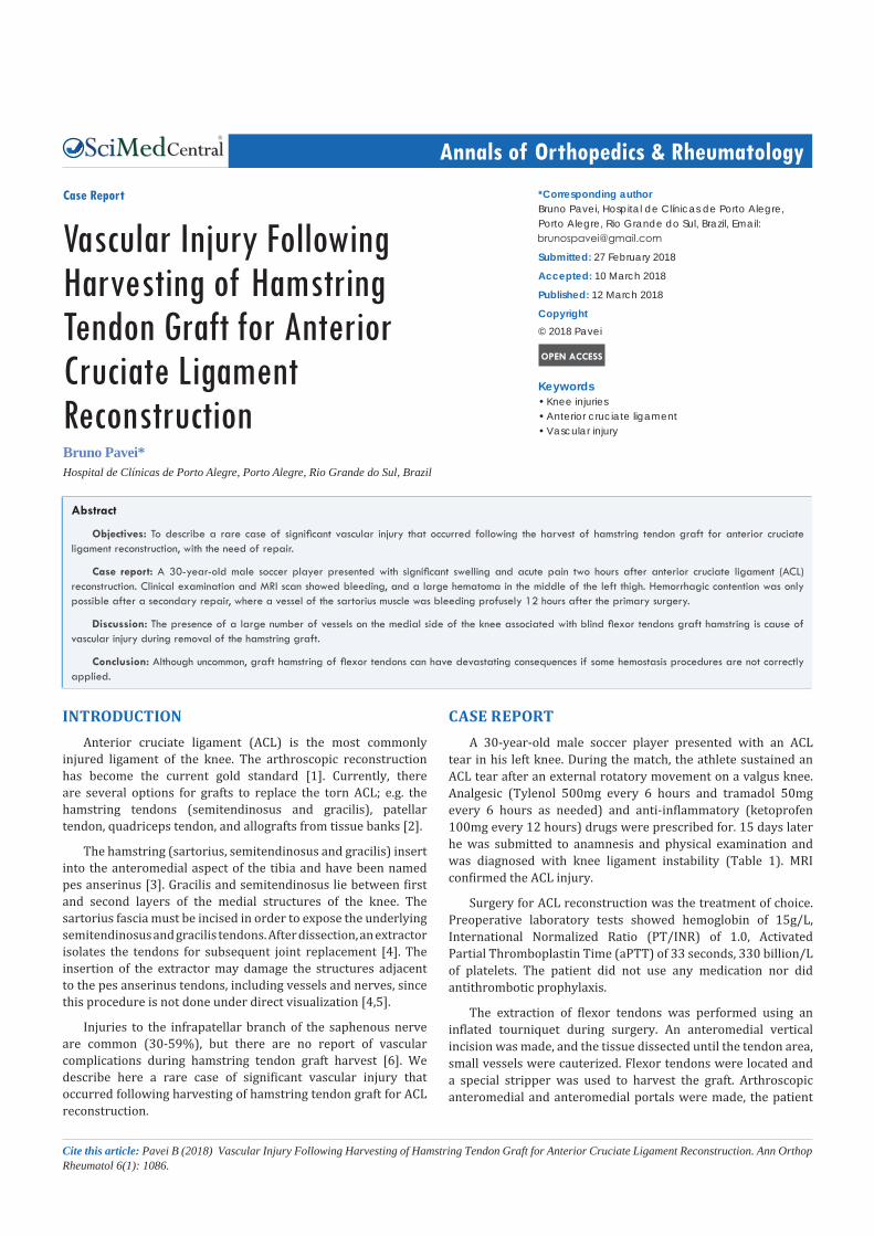
presented complete rupture of the anterior cruciate ligament without meniscal or chondral lesions. Injured ligament was removed; tibial and femoral tunnels were drilled for anatomic ligament reconstruction. The graft was fixed on the femur with Tight Rope (Arthrex®) and fixed on the tibia with an interference screw (Arthrex ®). After the surgery that lasted an hour and a half, the tourniquet was deflated. No compression bandages were applied. Two hours after the procedure, the patient remained lying in the recovery bed and presented important edema at the surgery site, remaining under observation in the hospital. Vital signs were normal. The pulse of the dorsalis pedis and posterior tibialis arteries was equivalent to the contralateral extremity. Swelling and pain worsened, the skin was tense, and the range of motion was limited 6 hours after surgery (Figure1).
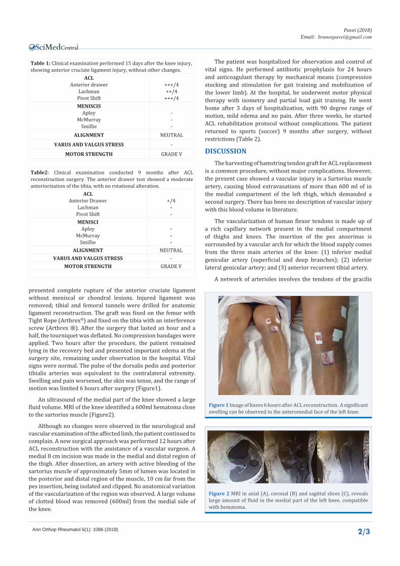
An ultrasound of the medial part of the knee showed a large fluid volume. MRI of the knee identified a 600ml hematoma close to the sartorius muscle (Figure2).
Although no changes were observed in the neurological and vascular examination of the affected limb, the patient continued to complain. A new surgical approach was performed 12 hours after ACL reconstruction with the assistance of a vascular surgeon. A medial 8 cm incision was made in the medial and distal region of the thigh. After dissection, an artery with active bleeding of the sartorius muscle of approximately 5mm of lumen was located in the posterior and distal region of the muscle, 10 cm far from the pes insertion, being isolated and clipped. No anatomical variation of the vascularization of the region was observed. A large volume of clotted blood was removed (600ml) from the medial side of the knee.
The patient was hospitalized for observation and control of vital signs. He performed antibiotic prophylaxis for 24 hours and anticoagulant therapy by mechanical means (compression stocking and stimulation for gait training and mobilization of the lower limb). At the hospital, he underwent motor physical therapy with isometry and partial load gait training. He went home after 3 days of hospitalization, with 90 degree range of motion, mild edema and no pain. After three weeks, he started ACL rehabilitation protocol without complications. The patient returned to sports (soccer) 9 months after surgery, without restrictions (Table 2).
DISCUSSIONThe harvesting of hamstring tendon graft for ACL replacement
is a common procedure, without major complications. However, the present case showed a vascular injury in a Sartorius muscle artery, causing blood extravasations of more than 600 ml of in the medial compartment of the left thigh, which demanded a second surgery. There has been no description of vascular injury with this blood volume in literature.
The vascularization of human flexor tendons is made up of a rich capillary network present in the medial compartment of thighs and knees. The insertion of the pes anserinus is surrounded by a vascular arch for which the blood supply comes from the three main arteries of the knee: (1) inferior medial genicular artery (superficial and deep branches); (2) inferior lateral genicular artery; and (3) anterior recurrent tibial artery.
A network of arterioles involves the tendons of the gracilis
Table 1: Clinical examination performed 15 days after the knee injury, showing anterior cruciate ligament injury, without other changes.
ACLAnterior drawer
LachmanPivot Shift
+++/4++/4
+++/4MENISCIS
ApleyMcMurray
Smillie
---
ALIGNMENT NEUTRAL
VARUS AND VALGUS STRESS -
MOTOR STRENGTH GRADE V
Table2: Clinical examination conducted 9 months after ACL reconstruction surgery. The anterior drawer test showed a moderate anteriorization of the tibia, with no rotational alteration.
ACLAnterior Drawer
LachmanPivot Shift
+/4--
MENISCIApley
McMurraySmillie
---
ALIGNMENT NEUTRALVARUS AND VALGUS STRESS -
MOTOR STRENGTH GRADE V
Figure 1 Image of knees 6 hours after ACL reconstruction. A significant swelling can be observed in the anteromedial face of the left knee.
Figure 2 MRI in axial (A), coronal (B) and sagittal slices (C), reveals large amount of fluid in the medial part of the left knee, compatible with hematoma.
CentralBringing Excellence in Open Access
Pavei (2018)Email:
Ann Orthop Rheumatol 6(1): 1086 (2018) 3/3
Pavei B (2018) Vascular Injury Following Harvesting of Hamstring Tendon Graft for Anterior Cruciate Ligament Reconstruction. Ann Orthop Rheumatol 6(1): 1086.
Cite this article
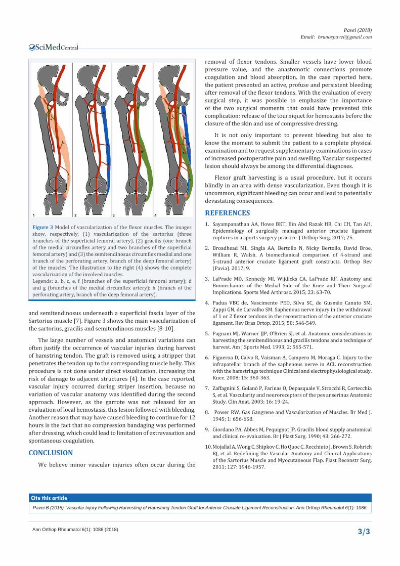
and semitendinosus underneath a superficial fascia layer of the Sartorius muscle [7]. Figure 3 shows the main vascularization of the sartorius, gracilis and semitendinous muscles [8-10].
The large number of vessels and anatomical variations can often justify the occurrence of vascular injuries during harvest of hamstring tendon. The graft is removed using a stripper that penetrates the tendon up to the corresponding muscle belly. This procedure is not done under direct visualization, increasing the risk of damage to adjacent structures [4]. In the case reported, vascular injury occurred during striper insertion, because no variation of vascular anatomy was identified during the second approach. However, as the garrote was not released for an evaluation of local hemostasis, this lesion followed with bleeding. Another reason that may have caused bleeding to continue for 12 hours is the fact that no compression bandaging was performed after dressing, which could lead to limitation of extravasation and spontaneous coagulation.
CONCLUSIONWe believe minor vascular injuries often occur during the
Figure 3 Model of vascularization of the flexor muscles. The images show, respectively, (1) vascularization of the sartorius (three branches of the superficial femoral artery), (2) gracilis (one branch of the medial circumflex artery and two branches of the superficial femoral artery) and (3) the semitendinosus circumflex medial and one branch of the perforating artery, branch of the deep femoral artery) of the muscles. The illustration to the right (4) shows the complete vascularization of the involved muscles.Legends: a, b, c, e, f (branches of the superficial femoral artery); d and g (branches of the medial circumflex artery); h (branch of the perforating artery, branch of the deep femoral artery).
removal of flexor tendons. Smaller vessels have lower blood pressure value, and the anastomotic connections promote coagulation and blood absorption. In the case reported here, the patient presented an active, profuse and persistent bleeding after removal of the flexor tendons. With the evaluation of every surgical step, it was possible to emphasize the importance of the two surgical moments that could have prevented this complication: release of the tourniquet for hemostasis before the closure of the skin and use of compressive dressing.
It is not only important to prevent bleeding but also to know the moment to submit the patient to a complete physical examination and to request supplementary examinations in cases of increased postoperative pain and swelling. Vascular suspected lesion should always be among the differential diagnoses.
Flexor graft harvesting is a usual procedure, but it occurs blindly in an area with dense vascularization. Even though it is uncommon, significant bleeding can occur and lead to potentially devastating consequences.
REFERENCES1. Sayampanathan AA, Howe BKT, Bin Abd Razak HR, Chi CH, Tan AH.
Epidemiology of surgically managed anterior cruciate ligament ruptures in a sports surgery practice. J Orthop Surg. 2017; 25.
2. Broadhead ML, Singla AA, Bertollo N, Nicky Bertollo, David Broe, William R. Walsh. A biomechanical comparison of 4-strand and 5-strand anterior cruciate ligament graft constructs. Orthop Rev (Pavia). 2017; 9.
3. LaPrade MD, Kennedy MI, Wijdicks CA, LaPrade RF. Anatomy and Biomechanics of the Medial Side of the Knee and Their Surgical Implications. Sports Med Arthrosc. 2015; 23: 63-70.
4. Padua VBC de, Nascimento PED, Silva SC, de Gusmão Canuto SM, Zuppi GN, de Carvalho SM. Saphenous nerve injury in the withdrawal of 1 or 2 flexor tendons in the reconstruction of the anterior cruciate ligament. Rev Bras Ortop. 2015; 50: 546-549.
5. Pagnani MJ, Warner JJP, O’Brien SJ, et al. Anatomic considerations in harvesting the semitendinosus and gracilis tendons and a technique of harvest. Am J Sports Med. 1993; 2: 565-571.
6. Figueroa D, Calvo R, Vaisman A, Campero M, Moraga C. Injury to the infrapatellar branch of the saphenous nerve in ACL reconstruction with the hamstrings technique Clinical and electrophysiological study. Knee. 2008; 15: 360-363.
7. Zaffagnini S, Golanò P, Farinas O, Depasquale V, Strocchi R, Cortecchia S, et al. Vascularity and neuroreceptors of the pes anserinus Anatomic Study. Clin Anat. 2003; 16: 19-24.
8. Power RW. Gas Gangrene and Vascularization of Muscles. Br Med J. 1945; 1: 656-658.
9. Giordano PA, Abbes M, Pequignot JP. Gracilis blood supply anatomical and clinical re-evaluation. Br J Plast Surg. 1990; 43: 266-272.
10. Mojallal A, Wong C, Shipkov C, Ho Quoc C, Recchiuto J, Brown S, Rohrich RJ, et al. Redefining the Vascular Anatomy and Clinical Applications of the Sartorius Muscle and Myocutaneous Flap. Plast Reconstr Surg. 2011; 127: 1946-1957.