Embed Size (px)
Citation preview
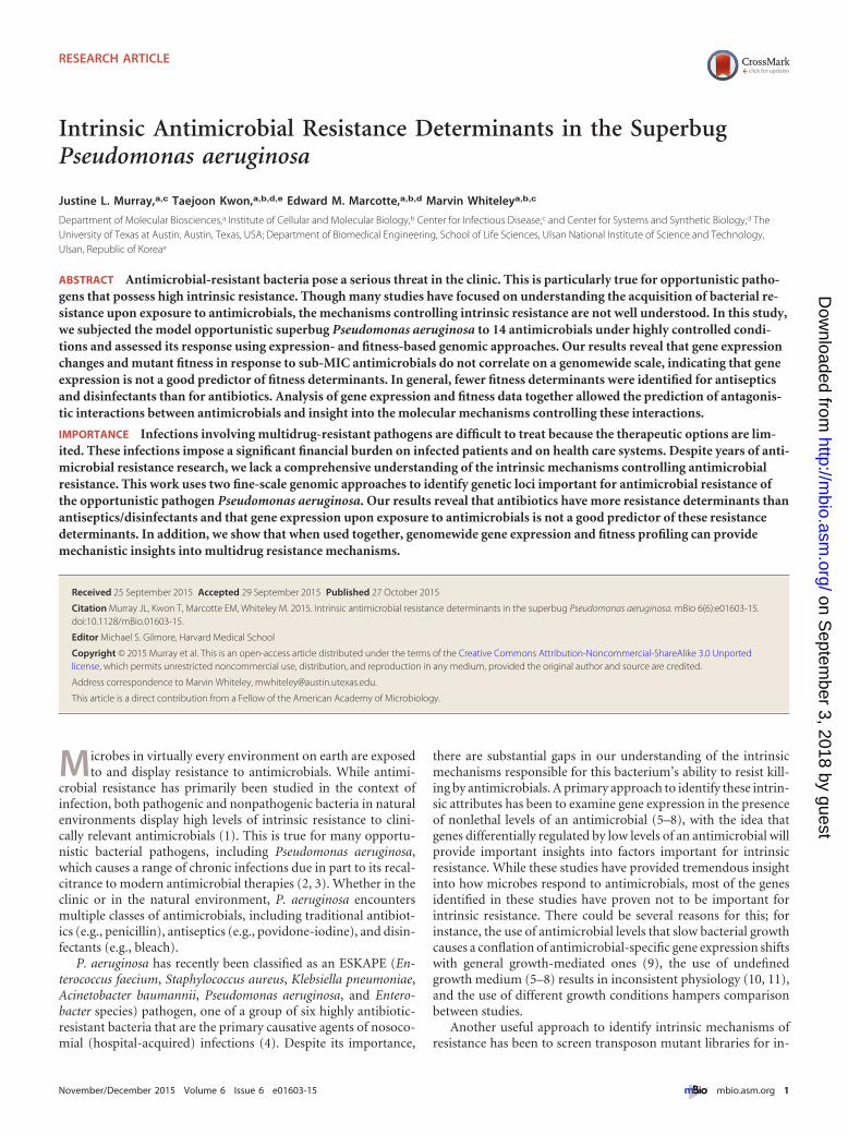
Intrinsic Antimicrobial Resistance Determinants in the SuperbugPseudomonas aeruginosa
Justine L. Murray,a,c Taejoon Kwon,a,b,d,e Edward M. Marcotte,a,b,d Marvin Whiteleya,b,c
Department of Molecular Biosciences,a Institute of Cellular and Molecular Biology,b Center for Infectious Disease,c and Center for Systems and Synthetic Biology,d TheUniversity of Texas at Austin, Austin, Texas, USA; Department of Biomedical Engineering, School of Life Sciences, Ulsan National Institute of Science and Technology,Ulsan, Republic of Koreae
ABSTRACT Antimicrobial-resistant bacteria pose a serious threat in the clinic. This is particularly true for opportunistic patho-gens that possess high intrinsic resistance. Though many studies have focused on understanding the acquisition of bacterial re-sistance upon exposure to antimicrobials, the mechanisms controlling intrinsic resistance are not well understood. In this study,we subjected the model opportunistic superbug Pseudomonas aeruginosa to 14 antimicrobials under highly controlled condi-tions and assessed its response using expression- and fitness-based genomic approaches. Our results reveal that gene expressionchanges and mutant fitness in response to sub-MIC antimicrobials do not correlate on a genomewide scale, indicating that geneexpression is not a good predictor of fitness determinants. In general, fewer fitness determinants were identified for antisepticsand disinfectants than for antibiotics. Analysis of gene expression and fitness data together allowed the prediction of antagonis-tic interactions between antimicrobials and insight into the molecular mechanisms controlling these interactions.
IMPORTANCE Infections involving multidrug-resistant pathogens are difficult to treat because the therapeutic options are lim-ited. These infections impose a significant financial burden on infected patients and on health care systems. Despite years of anti-microbial resistance research, we lack a comprehensive understanding of the intrinsic mechanisms controlling antimicrobialresistance. This work uses two fine-scale genomic approaches to identify genetic loci important for antimicrobial resistance ofthe opportunistic pathogen Pseudomonas aeruginosa. Our results reveal that antibiotics have more resistance determinants thanantiseptics/disinfectants and that gene expression upon exposure to antimicrobials is not a good predictor of these resistancedeterminants. In addition, we show that when used together, genomewide gene expression and fitness profiling can providemechanistic insights into multidrug resistance mechanisms.
Received 25 September 2015 Accepted 29 September 2015 Published 27 October 2015
Citation Murray JL, Kwon T, Marcotte EM, Whiteley M. 2015. Intrinsic antimicrobial resistance determinants in the superbug Pseudomonas aeruginosa. mBio 6(6):e01603-15.doi:10.1128/mBio.01603-15.
Editor Michael S. Gilmore, Harvard Medical School
Copyright © 2015 Murray et al. This is an open-access article distributed under the terms of the Creative Commons Attribution-Noncommercial-ShareAlike 3.0 Unportedlicense, which permits unrestricted noncommercial use, distribution, and reproduction in any medium, provided the original author and source are credited.
Address correspondence to Marvin Whiteley, [email protected].
This article is a direct contribution from a Fellow of the American Academy of Microbiology.
Microbes in virtually every environment on earth are exposedto and display resistance to antimicrobials. While antimi-
crobial resistance has primarily been studied in the context ofinfection, both pathogenic and nonpathogenic bacteria in naturalenvironments display high levels of intrinsic resistance to clini-cally relevant antimicrobials (1). This is true for many opportu-nistic bacterial pathogens, including Pseudomonas aeruginosa,which causes a range of chronic infections due in part to its recal-citrance to modern antimicrobial therapies (2, 3). Whether in theclinic or in the natural environment, P. aeruginosa encountersmultiple classes of antimicrobials, including traditional antibiot-ics (e.g., penicillin), antiseptics (e.g., povidone-iodine), and disin-fectants (e.g., bleach).
P. aeruginosa has recently been classified as an ESKAPE (En-terococcus faecium, Staphylococcus aureus, Klebsiella pneumoniae,Acinetobacter baumannii, Pseudomonas aeruginosa, and Entero-bacter species) pathogen, one of a group of six highly antibiotic-resistant bacteria that are the primary causative agents of nosoco-mial (hospital-acquired) infections (4). Despite its importance,
there are substantial gaps in our understanding of the intrinsicmechanisms responsible for this bacterium’s ability to resist kill-ing by antimicrobials. A primary approach to identify these intrin-sic attributes has been to examine gene expression in the presenceof nonlethal levels of an antimicrobial (5–8), with the idea thatgenes differentially regulated by low levels of an antimicrobial willprovide important insights into factors important for intrinsicresistance. While these studies have provided tremendous insightinto how microbes respond to antimicrobials, most of the genesidentified in these studies have proven not to be important forintrinsic resistance. There could be several reasons for this; forinstance, the use of antimicrobial levels that slow bacterial growthcauses a conflation of antimicrobial-specific gene expression shiftswith general growth-mediated ones (9), the use of undefinedgrowth medium (5–8) results in inconsistent physiology (10, 11),and the use of different growth conditions hampers comparisonbetween studies.
Another useful approach to identify intrinsic mechanisms ofresistance has been to screen transposon mutant libraries for in-
RESEARCH ARTICLE crossmark
November/December 2015 Volume 6 Issue 6 e01603-15 ® mbio.asm.org 1
on Septem
ber 3, 2018 by guesthttp://m
bio.asm.org/
Dow
nloaded from
creased or decreased susceptibility to sub-MIC antimicrobials(12–17). These studies have revealed novel resistance determi-nants; however, these experiments have been performed with lim-ited numbers of antimicrobials and are difficult to compare due todifferences in experimental design. In this study, we combinedgene expression analysis under highly controlled conditions withhigh-throughput fitness profiling to elucidate intrinsic resistancemechanisms to 14 antimicrobials in P. aeruginosa.
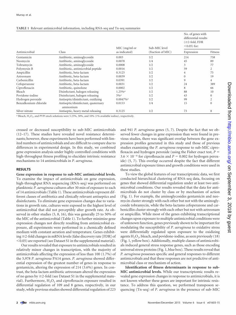
RESULTSGene expression in response to sub-MIC antimicrobial levels.To examine the impact of antimicrobials on gene expression,high-throughput RNA sequencing (RNA-seq) was performed onplanktonic P. aeruginosa cultures after 30 min of exposure to eachof 14 antimicrobials (Table 1). These antimicrobials represent dif-ferent classes of antibiotics and clinically relevant antiseptics anddisinfectants. To eliminate gene expression changes due to varia-tions in growth rate, cultures were exposed to the highest level ofantimicrobial that did not perceptibly alter growth rate. As ob-served in other studies (5, 8, 16), this was generally 25 to 50% ofthe MIC of the antimicrobial (Table 1). To further minimize geneexpression changes not directly resulting from antimicrobial ex-posure, all experiments were performed in a chemically definedmedium with constant aeration and temperature. Genes exhibit-ing �2-fold changes in mRNA levels (false discovery rate [FDR] of�0.05) are reported (see Dataset S1 in the supplemental material).
Our results revealed that exposure to antimicrobials resulted inrelatively minor changes in transcription, with the majority ofantimicrobials affecting the expression of less than 100 (1.7%) ofthe 5,978 P. aeruginosa PA14 genes. P. aeruginosa showed differ-ential expression of the greatest number of genes in response togentamicin, altering the expression of 214 (3.6%) genes. In con-trast, the beta-lactam antibiotic aztreonam altered the expressionof no genes by �2-fold (see Dataset S1 in the supplemental mate-rial). Furthermore, H2O2 and ciprofloxacin exposure resulted indifferential regulation of 109 and 8 genes, respectively, in ourstudy, while previous studies showed differential regulation of 223
and 941 P. aeruginosa genes (5, 7). Despite the fact that we ob-served fewer changes in gene expression than were found in pre-vious studies, there was significant overlap between the gene ex-pression profiles generated in this study and those of previousstudies examining the P. aeruginosa response to sub-MIC cipro-floxacin and hydrogen peroxide (using the Fisher exact test, P �3.6 � 10�6 for ciprofloxacin and P � 0.002 for hydrogen perox-ide) (5, 7). This overlap occurred despite the fact that differentantimicrobial exposure times and growth conditions were used inthese studies.
To view the global features of our transcriptomic data, we firstconducted hierarchical clustering of RNA-seq data, focusing ongenes that showed differential regulation under at least two anti-microbial conditions. Our results revealed that the data for anti-microbials do not cluster by class or by mechanism of action(Fig. 1). For example, the aminoglycosides gentamicin and neo-mycin cluster strongly with each other but not with the aminogly-coside tobramycin, while the beta-lactams cefoperazone and car-benicillin cluster strongly with each other but not with aztreonamor ampicillin. While most of the genes exhibiting transcriptionalchanges upon exposure to multiple antimicrobial conditions wereof unknown function, genes previously shown to be important formodulating the susceptibility of P. aeruginosa to oxidative stresswere differentially regulated upon exposure to the oxidizingagents H2O2, bleach, and povidone-iodine, as seen previously (18)(Fig. 1, yellow box). Additionally, multiple classes of antimicrobi-als induced general stress response genes, such as those encodinguniversal stress proteins (Fig. 1, blue box). These results reveal thatP. aeruginosa possesses specific and general responses to differentantimicrobials and that these responses are not predictive of anti-microbial class or mechanism of action.
Identification of fitness determinants in response to sub-MIC antimicrobial levels. While our transcriptomic results re-vealed gene expression changes in response to antimicrobials, it isnot known whether these genes are important for intrinsic resis-tance. To address this question, we performed transposon se-quencing (Tn-seq) of P. aeruginosa in the presence of sub-MIC
TABLE 1 Relevant antimicrobial information, including RNA-seq and Tn-seq summaries
Antimicrobial ClassMIC (mg/ml oras indicated)
Sub-MIC level(fraction of MIC)
No. of genes withdifferential results(�2-fold, FDR�0.05) for:
Expression Fitness
Gentamicin Antibiotic, aminoglycoside 0.003 1/2 214 758Neomycin Antibiotic, aminoglycoside 0.0078 1/4 45 89Tobramycin Antibiotic, aminoglycoside 0.0008 1/2 3 77Polymyxin B Antibiotic, antimicrobial peptide 0.0016 1/2 59 2Ampicillin Antibiotic, beta-lactam 0.3125 1/2 4 75Aztreonam Antibiotic, beta-lactam 0.0039 1/2 0 19Carbenicillin Antibiotic, beta-lactam 0.0391 1/2 9 4Cefoperazone Antibiotic, beta-lactam 0.0031 1/2 24 389Ciprofloxacin Antibiotic, quinolone 0.0002 1/2 8 66Bleach Disinfectant, halogen releasing 1.25%a 1/2 88 10Povidone-iodine Disinfectant, halogen releasing 5%a 1/2 43 0Hydrogen peroxide Antiseptic/disinfectant, oxidizing 0.0047%a 1/2 99 2Benzalkonium chloride Antiseptic/disinfectant, quaternary
ammonium0.0153 1/4 15 0
Silver nitrate Antiseptic, heavy metal releasing 0.3125 1/2 73 8a Bleach, H2O2, and PVPi stock solutions were 5.25%, 30%, and 10% (1% available iodine), respectively.
Murray et al.
2 ® mbio.asm.org November/December 2015 Volume 6 Issue 6 e01603-15
on Septem
ber 3, 2018 by guesthttp://m
bio.asm.org/
Dow
nloaded from
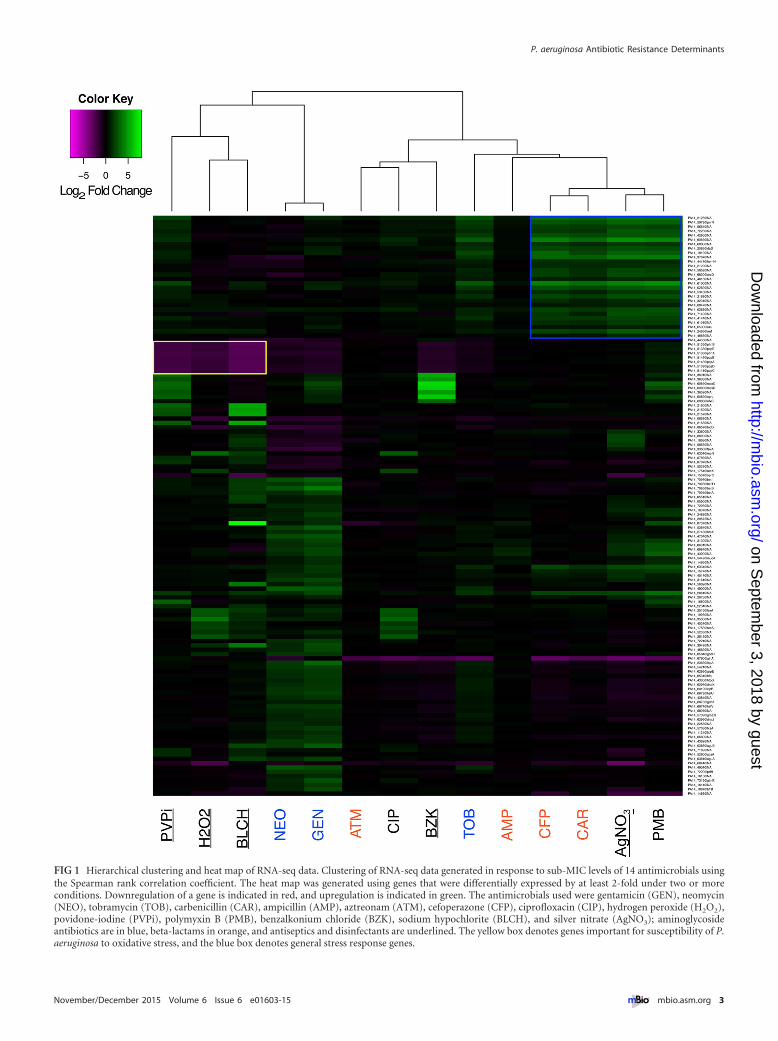
FIG 1 Hierarchical clustering and heat map of RNA-seq data. Clustering of RNA-seq data generated in response to sub-MIC levels of 14 antimicrobials usingthe Spearman rank correlation coefficient. The heat map was generated using genes that were differentially expressed by at least 2-fold under two or moreconditions. Downregulation of a gene is indicated in red, and upregulation is indicated in green. The antimicrobials used were gentamicin (GEN), neomycin(NEO), tobramycin (TOB), carbenicillin (CAR), ampicillin (AMP), aztreonam (ATM), cefoperazone (CFP), ciprofloxacin (CIP), hydrogen peroxide (H2O2),povidone-iodine (PVPi), polymyxin B (PMB), benzalkonium chloride (BZK), sodium hypochlorite (BLCH), and silver nitrate (AgNO3); aminoglycosideantibiotics are in blue, beta-lactams in orange, and antiseptics and disinfectants are underlined. The yellow box denotes genes important for susceptibility of P.aeruginosa to oxidative stress, and the blue box denotes general stress response genes.
P. aeruginosa Antibiotic Resistance Determinants
November/December 2015 Volume 6 Issue 6 e01603-15 ® mbio.asm.org 3
on Septem
ber 3, 2018 by guesthttp://m
bio.asm.org/
Dow
nloaded from
levels of the 14 antimicrobials. For these experiments, a trans-poson mutant library containing ~300,000 P. aeruginosa PA14mutants, each with a single transposon insertion, was constructed.This number of mutants represents an insertion approximatelyevery 20 bp along the ~6.5-Mb P. aeruginosa genome. The trans-poson mutant library was subjected to sub-MIC levels of each ofthe 14 antimicrobials and allowed to grow for approximately 12generations, and quantitative sequencing of genomic DNA adja-cent to the transposon allowed the abundance of each insertionmutant to be measured (19–21). By comparing mutant abun-dance in the presence and absence of an antimicrobial, transposoninsertion sites that affect fitness in the presence of an antimicrobialcan be identified.
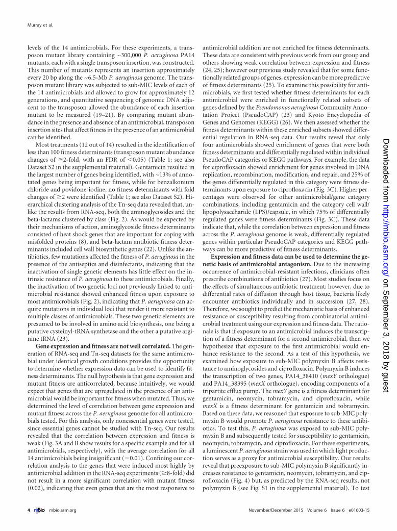
Most treatments (12 out of 14) resulted in the identification ofless than 100 fitness determinants (transposon mutant abundancechanges of �2-fold, with an FDR of �0.05) (Table 1; see alsoDataset S2 in the supplemental material). Gentamicin resulted inthe largest number of genes being identified, with ~13% of anno-tated genes being important for fitness, while for benzalkoniumchloride and povidone-iodine, no fitness determinants with foldchanges of �2 were identified (Table 1; see also Dataset S2). Hi-erarchical clustering analysis of the Tn-seq data revealed that, un-like the results from RNA-seq, both the aminoglycosides and thebeta-lactams clustered by class (Fig. 2). As would be expected bytheir mechanisms of action, aminoglycoside fitness determinantsconsisted of heat shock genes that are important for coping withmisfolded proteins (8), and beta-lactam antibiotic fitness deter-minants included cell wall biosynthetic genes (22). Unlike the an-tibiotics, few mutations affected the fitness of P. aeruginosa in thepresence of the antiseptics and disinfectants, indicating that theinactivation of single genetic elements has little effect on the in-trinsic resistance of P. aeruginosa to these antimicrobials. Finally,the inactivation of two genetic loci not previously linked to anti-microbial resistance showed enhanced fitness upon exposure tomost antimicrobials (Fig. 2), indicating that P. aeruginosa can ac-quire mutations in individual loci that render it more resistant tomultiple classes of antimicrobials. These two genetic elements arepresumed to be involved in amino acid biosynthesis, one being aputative cysteinyl-tRNA synthetase and the other a putative argi-nine tRNA (23).
Gene expression and fitness are not well correlated. The gen-eration of RNA-seq and Tn-seq datasets for the same antimicro-bial under identical growth conditions provides the opportunityto determine whether expression data can be used to identify fit-ness determinants. The null hypothesis is that gene expression andmutant fitness are anticorrelated, because intuitively, we wouldexpect that genes that are upregulated in the presence of an anti-microbial would be important for fitness when mutated. Thus, wedetermined the level of correlation between gene expression andmutant fitness across the P. aeruginosa genome for all antimicro-bials tested. For this analysis, only nonessential genes were tested,since essential genes cannot be studied with Tn-seq. Our resultsrevealed that the correlation between expression and fitness isweak (Fig. 3A and B show results for a specific example and for allantimicrobials, respectively), with the average correlation for all14 antimicrobials being insignificant (�0.01). Confining our cor-relation analysis to the genes that were induced most highly byantimicrobial addition in the RNA-seq experiments (�8-fold) didnot result in a more significant correlation with mutant fitness(0.02), indicating that even genes that are the most responsive to
antimicrobial addition are not enriched for fitness determinants.These data are consistent with previous work from our group andothers showing weak correlation between expression and fitness(24, 25); however our previous study revealed that for some func-tionally related groups of genes, expression can be more predictiveof fitness determinants (25). To examine this possibility for anti-microbials, we first tested whether fitness determinants for eachantimicrobial were enriched in functionally related subsets ofgenes defined by the Pseudomonas aeruginosa Community Anno-tation Project (PseudoCAP) (23) and Kyoto Encyclopedia ofGenes and Genomes (KEGG) (26). We then assessed whether thefitness determinants within these enriched subsets showed differ-ential regulation in RNA-seq data. Our results reveal that onlyfour antimicrobials showed enrichment of genes that were bothfitness determinants and differentially regulated within individualPseudoCAP categories or KEGG pathways. For example, the datafor ciprofloxacin showed enrichment for genes involved in DNAreplication, recombination, modification, and repair, and 25% ofthe genes differentially regulated in this category were fitness de-terminants upon exposure to ciprofloxacin (Fig. 3C). Higher per-centages were observed for other antimicrobial/gene categorycombinations, including gentamicin and the category cell wall/lipopolysaccharide (LPS)/capsule, in which 75% of differentiallyregulated genes were fitness determinants (Fig. 3C). These dataindicate that, while the correlation between expression and fitnessacross the P. aeruginosa genome is weak, differentially regulatedgenes within particular PseudoCAP categories and KEGG path-ways can be more predictive of fitness determinants.
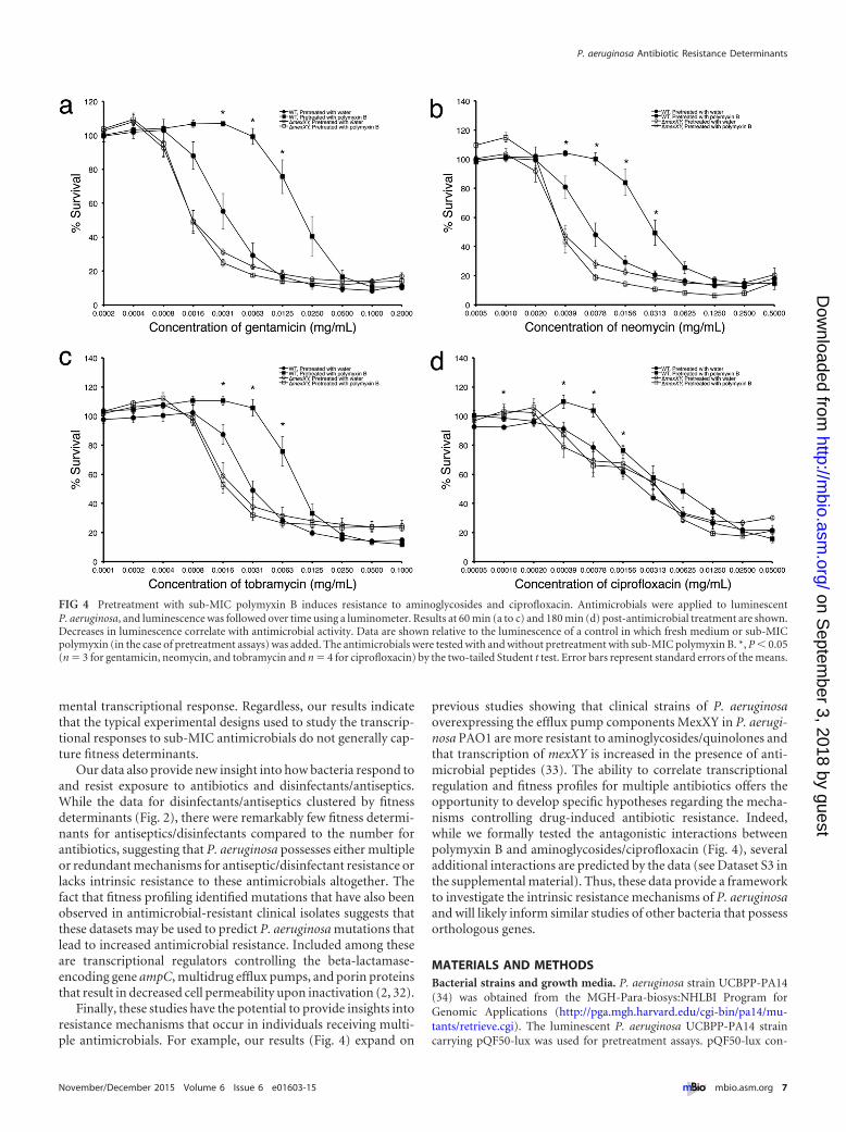
Expression and fitness data can be used to determine the ge-netic basis of antimicrobial antagonism. Due to the increasingoccurrence of antimicrobial-resistant infections, clinicians oftenprescribe combinations of antibiotics (27). Most studies focus onthe effects of simultaneous antibiotic treatment; however, due todifferential rates of diffusion through host tissue, bacteria likelyencounter antibiotics individually and in succession (27, 28).Therefore, we sought to predict the mechanistic basis of enhancedresistance or susceptibility resulting from combinatorial antimi-crobial treatment using our expression and fitness data. The ratio-nale is that if exposure to an antimicrobial induces the transcrip-tion of a fitness determinant for a second antimicrobial, then wehypothesize that exposure to the first antimicrobial would en-hance resistance to the second. As a test of this hypothesis, weexamined how exposure to sub-MIC polymyxin B affects resis-tance to aminoglycosides and ciprofloxacin. Polymyxin B inducesthe transcription of two genes, PA14_38410 (mexY orthologue)and PA14_38395 (mexX orthologue), encoding components of atripartite efflux pump. The mexY gene is a fitness determinant forgentamicin, neomycin, tobramycin, and ciprofloxacin, whilemexX is a fitness determinant for gentamicin and tobramycin.Based on these data, we reasoned that exposure to sub-MIC poly-myxin B would promote P. aeruginosa resistance to these antibi-otics. To test this, P. aeruginosa was exposed to sub-MIC poly-myxin B and subsequently tested for susceptibility to gentamicin,neomycin, tobramycin, and ciprofloxacin. For these experiments,a luminescent P. aeruginosa strain was used in which light produc-tion serves as a proxy for antimicrobial susceptibility. Our resultsreveal that preexposure to sub-MIC polymyxin B significantly in-creases resistance to gentamicin, neomycin, tobramycin, and cip-rofloxacin (Fig. 4) but, as predicted by the RNA-seq results, notpolymyxin B (see Fig. S1 in the supplemental material). To test
Murray et al.
4 ® mbio.asm.org November/December 2015 Volume 6 Issue 6 e01603-15
on Septem
ber 3, 2018 by guesthttp://m
bio.asm.org/
Dow
nloaded from
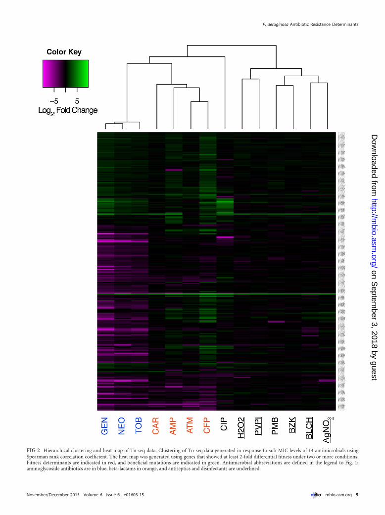
FIG 2 Hierarchical clustering and heat map of Tn-seq data. Clustering of Tn-seq data generated in response to sub-MIC levels of 14 antimicrobials usingSpearman rank correlation coefficient. The heat map was generated using genes that showed at least 2-fold differential fitness under two or more conditions.Fitness determinants are indicated in red, and beneficial mutations are indicated in green. Antimicrobial abbreviations are defined in the legend to Fig. 1;aminoglycoside antibiotics are in blue, beta-lactams in orange, and antiseptics and disinfectants are underlined.
P. aeruginosa Antibiotic Resistance Determinants
November/December 2015 Volume 6 Issue 6 e01603-15 ® mbio.asm.org 5
on Septem
ber 3, 2018 by guesthttp://m
bio.asm.org/
Dow
nloaded from
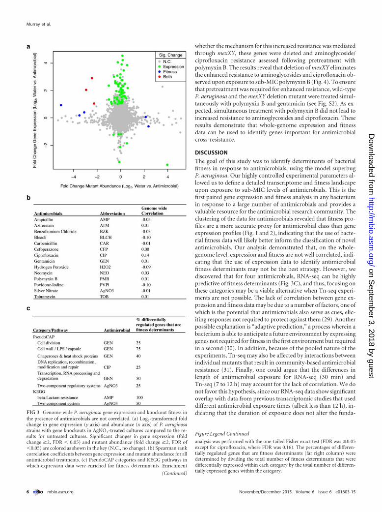
whether the mechanism for this increased resistance was mediatedthrough mexXY, these genes were deleted and aminoglycoside/ciprofloxacin resistance assessed following pretreatment withpolymyxin B. The results reveal that deletion of mexXY eliminatesthe enhanced resistance to aminoglycosides and ciprofloxacin ob-served upon exposure to sub-MIC polymyxin B (Fig. 4). To ensurethat pretreatment was required for enhanced resistance, wild-typeP. aeruginosa and the mexXY deletion mutant were treated simul-taneously with polymyxin B and gentamicin (see Fig. S2). As ex-pected, simultaneous treatment with polymyxin B did not lead toincreased resistance to aminoglycosides and ciprofloxacin. Theseresults demonstrate that whole-genome expression and fitnessdata can be used to identify genes important for antimicrobialcross-resistance.
DISCUSSION
The goal of this study was to identify determinants of bacterialfitness in response to antimicrobials, using the model superbugP. aeruginosa. Our highly controlled experimental parameters al-lowed us to define a detailed transcriptome and fitness landscapeupon exposure to sub-MIC levels of antimicrobials. This is thefirst paired gene expression and fitness analysis in any bacteriumin response to a large number of antimicrobials and provides avaluable resource for the antimicrobial research community. Theclustering of the data for antimicrobials revealed that fitness pro-files are a more accurate proxy for antimicrobial class than geneexpression profiles (Fig. 1 and 2), indicating that the use of bacte-rial fitness data will likely better inform the classification of novelantimicrobials. Our analysis demonstrated that, on the whole-genome level, expression and fitness are not well correlated, indi-cating that the use of expression data to identify antimicrobialfitness determinants may not be the best strategy. However, wediscovered that for four antimicrobials, RNA-seq can be highlypredictive of fitness determinants (Fig. 3C), and thus, focusing onthese categories may be a viable alternative when Tn-seq experi-ments are not possible. The lack of correlation between gene ex-pression and fitness data may be due to a number of factors, one ofwhich is the potential that antimicrobials also serve as cues, elic-iting responses not required to protect against them (29). Anotherpossible explanation is “adaptive prediction,” a process wherein abacterium is able to anticipate a future environment by expressinggenes not required for fitness in the first environment but requiredin a second (30). In addition, because of the pooled nature of theexperiments, Tn-seq may also be affected by interactions betweenindividual mutants that result in community-based antimicrobialresistance (31). Finally, one could argue that the differences inlength of antimicrobial exposure for RNA-seq (30 min) andTn-seq (7 to 12 h) may account for the lack of correlation. We donot favor this hypothesis, since our RNA-seq data show significantoverlap with data from previous transcriptomic studies that useddifferent antimicrobial exposure times (albeit less than 12 h), in-dicating that the duration of exposure does not alter the funda-FIG 3 Genome-wide P. aeruginosa gene expression and knockout fitness in
the presence of antimicrobials are not correlated. (a) Log2-transformed foldchange in gene expression (y axis) and abundance (x axis) of P. aeruginosastrains with gene knockouts in AgNO3-treated cultures compared to the re-sults for untreated cultures. Significant changes in gene expression (foldchange �2, FDR � 0.05) and mutant abundance (fold change �2, FDR of�0.05) are colored as shown in the key (N.C., no change). (b) Spearman rankcorrelation coefficients between gene expression and mutant abundance for allantimicrobial treatments. (c) PseudoCAP categories and KEGG pathways inwhich expression data were enriched for fitness determinants. Enrichment
(Continued)
Figure Legend Continued
analysis was performed with the one-tailed Fisher exact test (FDR was �0.05except for ciprofloxacin, where FDR was 0.16). The percentages of differen-tially regulated genes that are fitness determinants (far right column) weredetermined by dividing the total number of fitness determinants that weredifferentially expressed within each category by the total number of differen-tially expressed genes within the category.
Murray et al.
6 ® mbio.asm.org November/December 2015 Volume 6 Issue 6 e01603-15
on Septem
ber 3, 2018 by guesthttp://m
bio.asm.org/
Dow
nloaded from
mental transcriptional response. Regardless, our results indicatethat the typical experimental designs used to study the transcrip-tional responses to sub-MIC antimicrobials do not generally cap-ture fitness determinants.
Our data also provide new insight into how bacteria respond toand resist exposure to antibiotics and disinfectants/antiseptics.While the data for disinfectants/antiseptics clustered by fitnessdeterminants (Fig. 2), there were remarkably few fitness determi-nants for antiseptics/disinfectants compared to the number forantibiotics, suggesting that P. aeruginosa possesses either multipleor redundant mechanisms for antiseptic/disinfectant resistance orlacks intrinsic resistance to these antimicrobials altogether. Thefact that fitness profiling identified mutations that have also beenobserved in antimicrobial-resistant clinical isolates suggests thatthese datasets may be used to predict P. aeruginosa mutations thatlead to increased antimicrobial resistance. Included among theseare transcriptional regulators controlling the beta-lactamase-encoding gene ampC, multidrug efflux pumps, and porin proteinsthat result in decreased cell permeability upon inactivation (2, 32).
Finally, these studies have the potential to provide insights intoresistance mechanisms that occur in individuals receiving multi-ple antimicrobials. For example, our results (Fig. 4) expand on
previous studies showing that clinical strains of P. aeruginosaoverexpressing the efflux pump components MexXY in P. aerugi-nosa PAO1 are more resistant to aminoglycosides/quinolones andthat transcription of mexXY is increased in the presence of anti-microbial peptides (33). The ability to correlate transcriptionalregulation and fitness profiles for multiple antibiotics offers theopportunity to develop specific hypotheses regarding the mecha-nisms controlling drug-induced antibiotic resistance. Indeed,while we formally tested the antagonistic interactions betweenpolymyxin B and aminoglycosides/ciprofloxacin (Fig. 4), severaladditional interactions are predicted by the data (see Dataset S3 inthe supplemental material). Thus, these data provide a frameworkto investigate the intrinsic resistance mechanisms of P. aeruginosaand will likely inform similar studies of other bacteria that possessorthologous genes.
MATERIALS AND METHODSBacterial strains and growth media. P. aeruginosa strain UCBPP-PA14(34) was obtained from the MGH-Para-biosys:NHLBI Program forGenomic Applications (http://pga.mgh.harvard.edu/cgi-bin/pa14/mu-tants/retrieve.cgi). The luminescent P. aeruginosa UCBPP-PA14 straincarrying pQF50-lux was used for pretreatment assays. pQF50-lux con-
FIG 4 Pretreatment with sub-MIC polymyxin B induces resistance to aminoglycosides and ciprofloxacin. Antimicrobials were applied to luminescentP. aeruginosa, and luminescence was followed over time using a luminometer. Results at 60 min (a to c) and 180 min (d) post-antimicrobial treatment are shown.Decreases in luminescence correlate with antimicrobial activity. Data are shown relative to the luminescence of a control in which fresh medium or sub-MICpolymyxin (in the case of pretreatment assays) was added. The antimicrobials were tested with and without pretreatment with sub-MIC polymyxin B. *, P � 0.05(n � 3 for gentamicin, neomycin, and tobramycin and n � 4 for ciprofloxacin) by the two-tailed Student t test. Error bars represent standard errors of the means.
P. aeruginosa Antibiotic Resistance Determinants
November/December 2015 Volume 6 Issue 6 e01603-15 ® mbio.asm.org 7
on Septem
ber 3, 2018 by guesthttp://m
bio.asm.org/
Dow
nloaded from
tains a 1,489-bp fragment (genomic location, positions 6323103 to6324591 of the UCBPP-PA14 genome) from the P. aeruginosa genomethat provides a high level of expression of luxCDABE. Liquid cultures weregrown in chemically defined medium (CDM) supplemented with 20 mMsuccinate (35) at 37°C with shaking at 250 rpm.
Construction and confirmation of deletion mutants. The PA14�mexXY (PA14_38395-38410) deletion mutant was constructed byamplifying ~700-bp fragments flanking these two genes by PCR withPhusion hot start II DNA polymerase (Thermo Scientific, Waltham,MA) to replace the coding sequence of the genes with the sequence5=-GCGGCCGCC-3= flanked by the native start codon of PA14_38395and stop codon of PA14_38410. The primers used for PCR were 5=-TTCTGCAGGTCGACTCTAGACCAGGGTGCCGCAGATGC-3= and 5=-GCATCAGGCGGCCGCCATGGGTGTCCCTCGATTCGTG-3= for the up-stream region and 5=-CCATGGCGGCCGCCTGATGCCCCTAGCGAAACTCTCGC-3= and 5=-GAATTCGAGCTCGAGCCCGGGCCCGGAAGTTCTCCCTGGGC-3= for the downstream region. These two ampliconsand the suicide vector pEXG2 (36) were assembled using Gibson assemblyas described previously (37), transformed initially into Escherichia coliDH5� �pir, and then transformed into E. coli SM10 �pir for conjugationinto strain PA14. This construct was introduced into PA14 by conjugationwith selection for gentamicin-resistant transconjugates, followed by selec-tion on sucrose to obtain the chromosomal deletion. This mutation wasthen verified by PCR.
MIC determination. For MIC determination, logarithmic P. aerugi-nosa cells (optical density at 600 nm [OD600] of 0.5) were diluted to an ODof 0.001 and added to a 96-well microtiter plate. Antimicrobials wereadded, using serial 2-fold dilutions across the plate. The MIC was deter-mined as the lowest concentration with no visible growth.
RNA-seq growth conditions. Cultures for RNA-seq analysis weregrown overnight in CDM, diluted to an OD600 of 0.03 in 10 ml CDM,grown to an OD600 of 0.5, and then treated with 100-�l amounts of sub-MIC concentrations of antimicrobials or with 100 �l water as a control.Sub-MIC antibiotic levels were determined as the highest concentration atwhich P. aeruginosa growth was not inhibited (either 1/2 or 1/4 MIC)(Table 1). Cultures were grown for 30 min at 37°C with continuous shak-ing at 250 rpm. After 30 min, the OD600 was between 0.7 and 1.0, and theculture was immediately diluted with an equal volume of RNAlater.
Tn-seq growth conditions. Cultures for Tn-seq analysis were grownas follows: frozen aliquots of the P. aeruginosa PA14 transposon insertionlibrary (38) were washed three times with 1 ml 20 mM MOPS (morpho-linepropanesulfonic acid) buffered to pH 7.2, inoculated into 25 ml CDMat 2.5 � 105 CFU/ml, and grown for 30 min at 37°C with shaking at250 rpm. Then, 250-�l amounts of sub-MIC antimicrobials or water wereadded and the culture was grown for approximately 12 generations (to109 CFU/ml) and immediately placed on ice. Cells were pelleted, the su-pernatant was discarded, and the pellet was frozen.
RNA-seq Illumina library preparation. Cultures stored in RNAlaterwere pelleted, resuspended in RNA Bee, and transferred to 2-ml bead-beating tubes containing 0.1-mm beads (MP Biomedical). Cells werelysed by bead beating 3 times for 60 s, and the tubes placed on ice for 1 minbetween each homogenization. Amounts of 200 �l of chloroform wereadded, and the tubes were shaken vigorously for 30 s and incubated on icefor 5 min. Samples were centrifuged for 15 min at 4°C to separate theaqueous and organic phases. The top aqueous phase from each tube wastransferred to a new microcentrifuge tube to which 0.5 ml isopropanolwas added, and the tubes were incubated at room temperature for 10 min.Amounts of 20 �g of linear acrylamide were added to the tubes, and thesamples were centrifuged at 12,000 � g for 5 min at 4°C. The pellets werewashed with 1 ml 75% ethanol, air dried for 10 min, and resuspended in50 �l of RNase-free water. The RNA concentration for each sample wasdetermined with a NanoDrop spectrophotometer (Thermo Scientific).DNA contamination was assessed with PCR amplification of the clpXgene, and RNA integrity was verified with agarose gel electrophoresis ofglyoxylated samples (Ambion). Ribosomal RNA was depleted using the
RiboZero bacteria kit (Epicentre) and purified by ethanol precipitationusing 12.5 �g linear acrylamide to precipitate the RNA. The depleted RNAwas fragmented, and cDNA libraries were prepared as described previ-ously (39). Libraries were sequenced at the Genome Sequencing and Anal-ysis Facility at the University of Texas at Austin on an Illumina HiSeq 2000using a 1 � 100-bp single-end run.
Tn-seq Illumina library preparation. The frozen pellets were resus-pended in 1 ml 1� buffer A (40) with 0.1% SDS, homogenized in a bead-beating tube for 1 min, and then placed on ice. Proteinase K (1 mg/ml) wasadded, and the samples were incubated for 1 h. Samples were extractedwith equal volumes of 25:24:1 phenol-chloroform-isoamyl alcohol,pH 8.0. DNA was ethanol precipitated (0.1 volume 3 M sodium acetateand 3 volumes of 100% ethanol) from the aqueous phase, and the pelletwas washed with 75% ethanol 2 times, air dried for 10 min, and resus-pended in 100 �l water. DNA concentrations for each sample were deter-mined with a NanoDrop spectrophotometer (Thermo Scientific). DNAwas sheared to ~300 bp in a Q880R sonicator (Qsonica), and the size wasconfirmed on an agarose gel. The sheared DNA was treated with terminaldeoxynucleotidyltransferase (TdT), followed by two PCRs as describedpreviously (38, 41). The libraries were sequenced at the Genome Sequenc-ing and Analysis Facility at the University of Texas at Austin on an Illu-mina HiSeq 2500 using a 1 � 100-bp single-end run.
RNA-seq bioinformatic analyses. After discarding reads with no call(“N”) or low complexity (not containing all four nucleotides), we mappedreads to the P. aeruginosa UCBPP-PA14 genome (GenBank accessionnumber NC_008463.1; downloaded from http://www.pseudomonas.comon 31 July 2013) using the Burrows-Wheeler Aligner, Smith-Watermanalgorithm (BWA-SW) implemented on version 0.7.10 (42), and the bestnucleotide match for each read according to its mapping quality score wasselected. To remove ambiguous reads, we discarded the reads having mul-tiple best hits with the same mapping quality score. Because our librarypreparation method is designed to capture essentially all transcripts, in-cluding small RNAs (sRNAs), the aligned length of the reads was highlyvariable. Therefore, instead of read count, we counted the number ofnucleotides covering each gene (using the GFF3 annotation file obtainedfrom PseudoCAP [43]) and used the length of the gene and total numberof nucleotides mapped on genic regions of the genome as normalizationfactors, analogous to transcripts per millions of reads (TPM) (44). Weidentified genes showing significantly different RNA levels under eachcondition using edgeR (45), with false discovery rates (FDRs) of less than0.05 and differences of at least 2-fold. Detailed procedures and relatedscripts are available at https://github.com/marcottelab/HTseq-toolbox/wiki/ProkRNAseq.
Tn-seq bioinformatic analyses. As with the RNA-seq data, after dis-carding reads with no call or low complexity, we mapped the Tn-seq readsagainst the P. aeruginosa UCBPP-PA14 genome using BWA-SW and se-lected the best nucleotide match for each read according to its mappingquality score. We further filtered out mapped reads if their aligned lengthwas shorter than 10 bp (too short to infer the genomic DNA sequenceflanking the transposon). We then identified transposon insertion sites asa junction of the transposon flanking sequence (TAAGAGTCA) and themapped genomic DNA sequence. For fitness analysis, the occurrence of alltransposon insertion sites within a gene was summed based on Pseudo-CAP GFF3 annotation (43) and normalized by the total number of reads,and genes showing significant fitness changes under each condition, withFDRs of less than 0.05 and differences of at least 2-fold, were identified byusing edgeR. Detailed procedures and related scripts are available athttps://github.com/marcottelab/HTseq-toolbox/wiki/ProkTNseq.
Overlap between RNA-seq and existing gene expression data. Brazasand Hancock reported microarray data from sub-MIC ciprofloxacin-treated P. aeruginosa, and genes showing at least 2-fold differences inexpression and P values of less than 0.05 were compared to genes showingat least 2-fold differences in expression and FDRs of less than 0.05 in theRNA-seq data in this study (5). Chang et al. reported the 30 P. aeruginosagenes that were most highly upregulated in response to hydrogen perox-
Murray et al.
8 ® mbio.asm.org November/December 2015 Volume 6 Issue 6 e01603-15
on Septem
ber 3, 2018 by guesthttp://m
bio.asm.org/
Dow
nloaded from
ide, which were compared to genes showing at least 2-fold differences inexpression and FDRs of less than 0.05 in the RNA-seq data in this study(7). The significance of overlaps in these data was assessed using the Fisherexact test.
Heat map and cluster analyses. Heat maps were generated in R withthe function heatmap.2 of the gplots package. Clustering was performedusing genes exhibiting at least 2-fold differences in expression or fitnessand FDRs of less than 0.05 across at least 2 conditions using Spearmancorrelation coefficients.
Enrichment analyses. Enrichment of differentially regulated genesand differentially fit genes in a given PseudoCAP category or Kyoto En-cyclopedia of Genes and Genomes (KEGG) (46) pathway was determinedby comparing the prevalence of genes with differences of at least 2-foldand FDRs of less than 0.05 assigned to a specific PseudoCAP category orKEGG pathway to the prevalence of genes in the genome assigned to thatPseudoCAP category or KEGG pathway using the one-tailed Fisher exacttest. Only annotated genes were used in the analysis. P values for enrichedcategories were adjusted for multiple testing using a Benjamini-Hochbergcorrection, giving the resulting false discovery rate/q values (47).
Pretreatment assay. PA14 and PA14 �mexXY, constitutively express-ing luminescence from the plasmid pQF50-lux, were grown overnight inCDM or Lysogeny Broth with carbenicillin (300 �g/ml), washed 3� withfresh CDM containing no carbenicillin, diluted to an OD600 of 0.05 in10 ml CDM, and grown to mid-logarithmic phase (OD600 of 0.5). Thecultures were then treated with 100 �l of subinhibitory (1/2 MIC) poly-myxin B or with 100 �l water as a nontreatment control for 30 min.Treated cells were added to 96-well microtiter plates, and antibiotics (gen-tamicin, neomycin, tobramycin, or ciprofloxacin) were added, using se-rial dilutions. Luminescence was monitored at 0, 5, 15, 30, 60, and180 min using a Luminoskan Ascent microplate luminometer, and lumi-nescence at 60 min (gentamicin, neomycin, and tobramycin) and 180 min(ciprofloxacin) was reported. The internal temperature within the lumi-nometer was maintained at 37°C, and the plates were shaken at 240 rpm;prior to luminescence measurement, the plates were shaken at 1,200 rpmfor 10 s.
Microarray data accession numbers. RNA-seq and Tn-seq sequenc-ing data are available at the National Center for Biotechnology Informa-tion Sequence Read Archive (NCBI SRA) under accession numberSRP062243.
SUPPLEMENTAL MATERIALSupplemental material for this article may be found at http://mbio.asm.org/lookup/suppl/doi:10.1128/mBio.01603-15/-/DCSupplemental.
Figure S1, JPG file, 0.6 MB.Figure S2, JPG file, 0.6 MB.Dataset S1, XLSX file, 2.8 MB.Dataset S2, XLSX file, 2.9 MB.Dataset S3, XLSX file, 0.02 MB.
ACKNOWLEDGMENTS
This work was supported by a Burroughs Wellcome Investigator in thePathogenesis of Infectious Disease award to M.W. E.M.M. acknowledgesgrants from the NIH, NSF, Army Research Office (W911NF-12-1-0390),and Welch Foundation (F1515). T.K. was partially funded by the UNIST2015 Research Fund (1.150043.01).
REFERENCES1. D’Costa VM, McGrann KM, Hughes DW, Wright GD. 2006. Sampling
the antibiotic resistome. Science 311:374 –377. http://dx.doi.org/10.1126/science.1120800.
2. Breidenstein EBM, de la Fuente-Núñez C, Hancock REW. 2011. Pseu-domonas aeruginosa: all roads lead to resistance. Trends Microbiol 19:419 – 426. http://dx.doi.org/10.1016/j.tim.2011.04.005.
3. Gellatly SL, Hancock REW. 2013. Pseudomonas aeruginosa: new insightsinto pathogenesis and host defenses. Pathog Dis 67:159 –173. http://dx.doi.org/10.1111/2049-632X.12033.
4. Rice L. 2008. Federal funding for the study of antimicrobial resistance innosocomial pathogens: no ESKAPE. J Infect Dis 197:1079 –1081. http://dx.doi.org/10.1086/533452.
5. Brazas MD, Hancock REW. 2005. Ciprofloxacin induction of a suscep-tibility determinant in Pseudomonas aeruginosa. Antimicrob Agents Che-mother 49:3222–3227. http://dx.doi.org/10.1128/AAC.49.8.3222-3227.2005.
6. Brazas MD, Hancock REW. 2005. Using microarray gene signatures to elu-cidate mechanisms of antibiotic action and resistance. Drug Discov Today10:1245–1252. http://dx.doi.org/10.1016/S1359-6446(05)03566-X.
7. Chang W, Small DA, Toghrol F, Bentley WE. 2005. Microarray analysisof Pseudomonas aeruginosa reveals induction of pyocin genes in responseto hydrogen peroxide. BMC Genomics 6:115. http://dx.doi.org/10.1186/1471-2164-6-115.
8. Kindrachuk KN, Fernandez L, Bains M, Hancock REW. 2011. In-volvement of an ATP-dependent protease, PA0779/AsrA, in inducingheat shock in response to tobramycin in Pseudomonas aeruginosa. An-timicrob Agents Chemother 55:1874 –1882. http://dx.doi.org/10.1128/AAC.00935-10.
9. Cirz RT, O’Neill BM, Hammond JA, Head SR, Romesberg FE. 2006.Defining the Pseudomonas aeruginosa SOS response and its role in theglobal response to the antibiotic ciprofloxacin. J Bacteriol 188:7101–7110.http://dx.doi.org/10.1128/JB.00807-06.
10. Ezraty B, Henry C, Hérisse M, Denamur E, Barras F. 2014. Commerciallysogeny broth culture media and oxidative stress: a cautious tale. FreeR a d i c B i o l M e d 7 4 : 2 4 5 – 2 5 1 . h t t p : / / d x . d o i . o r g / 1 0 . 1 0 1 6 /j.freeradbiomed.2014.07.010.
11. Sezonov G, Joseleau-Petit D, D’Ari R. 2007. Escherichia coli physiology inLuria-Bertani broth. J Bacteriol 189:8746 – 8749. http://dx.doi.org/10.1128/JB.01368-07.
12. Fernandez L, Alvarez-Ortega C, Wiegand I, Olivares J, Kocincova D,Lam JS, Martinez JL, Hancock REW. 2013. Characterization of the poly-myxin B resistome of Pseudomonas aeruginosa. Antimicrob Agents Che-mother 57:110 –119. http://dx.doi.org/10.1128/AAC.01583-12.
13. Breidenstein EBM, Khaira BK, Wiegand I, Overhage J, HancockREW. 2008. Complex ciprofloxacin resistome revealed by screening aPseudomonas aeruginosa mutant library for altered susceptibility. An-timicrob Agents Chemother 52:4486 – 4491. http://dx.doi.org/10.1128/AAC.00222-08.
14. Alvarez-Ortega C, Wiegand I, Olivares J, Hancock REW, Martinez JL.2010. Genetic determinants involved in the susceptibility of Pseudomonasaeruginosa to beta-lactam antibiotics. Antimicrob Agents Chemother 54:4159 – 4167. http://dx.doi.org/10.1128/AAC.00257-10.
15. Schurek KN, Marr AK, Taylor PK, Wiegand I, Semenec L, Khaira BK,Hancock REW. 2008. Novel genetic determinants of low-level aminogly-coside resistance in Pseudomonas aeruginosa. Antimicrob Agents Che-mother 52:4213– 4219. http://dx.doi.org/10.1128/AAC.00507-08.
16. Brazas MD, Breidenstein EBM, Overhage J, Hancock REW. 2007. Roleof lon, an ATP-dependent protease homolog, in resistance of Pseudomo-nas aeruginosa to ciprofloxacin. Antimicrob Agents Chemother 51:4276 – 4283. http://dx.doi.org/10.1128/AAC.00830-07.
17. Marr AK, Overhage J, Bains M, Hancock REW. 2007. The Lon proteaseof Pseudomonas aeruginosa is induced by aminoglycosides and is involvedin biofilm formation and motility. Microbiology 153:474 – 482. http://dx.doi.org/10.1099/mic.0.2006/002519-0.
18. Häussler S, Becker T. 2008. The pseudomonas quinolone signal (PQS)balances life and death in Pseudomonas aeruginosa populations. PLoSPathog 4:e1000166. http://dx.doi.org/10.1371/journal.ppat.1000166.
19. Goodman AL, McNulty NP, Zhao Y, Leip D, Mitra RD, Lozupone CA,Knight R, Gordon JI. 2009. Identifying genetic determinants needed toestablish a human gut symbiont in its habitat. Cell Host Microbe6:279 –289. http://dx.doi.org/10.1016/j.chom.2009.08.003.
20. Van Opijnen T, Bodi KL, Camilli A. 2009. Tn-seq: high-throughputparallel sequencing for fitness and genetic interaction studies in microor-ganisms. Nat Methods 6:767–772. http://dx.doi.org/10.1038/nmeth.1377.
21. Langridge GC, Phan M, Turner DJ, Perkins TT, Parts L, Haase J,Charles I, Maskell DJ, Peters SE, Dougan G, Wain J, Parkhill J, TurnerAK. 2009. Simultaneous assay of every Salmonella Typhi gene using onemillion transposon mutants. Genome Res 19:2308 –2316. http://dx.doi.org/10.1101/gr.097097.109.
22. Lambert PA. 2002. Mechanisms of antibiotic resistance in Pseudomonasaeruginosa. J R Soc Med 95(Suppl 41):22–26.
23. Winsor GL, Lo R, Ho SSJ, Ung KS, Huang S, Cheng D, Ching WK,
P. aeruginosa Antibiotic Resistance Determinants
November/December 2015 Volume 6 Issue 6 e01603-15 ® mbio.asm.org 9
on Septem
ber 3, 2018 by guesthttp://m
bio.asm.org/
Dow
nloaded from
Hancock RE, Brinkman FS. 2004. Pseudomonas aeruginosa Genome Da-tabase and PseudoCAP: facilitating community-based, continually up-dated, genome annotation. Nucleic Acids Res 33:D338 –D343. http://dx.doi.org/10.1093/nar/gki047.
24. Giaever G, Chu AM, Ni L, Connelly C, Riles L, Véronneau S, Dow S,Lucau-Danila A, Anderson K, André B, Arkin AP, Astromoff A, ElBakkoury M, Bangham R, Benito R, Brachat S, Campanaro S, CurtissM, Davis K, Deutschbauer A, Entian KD, Flaherty P, Foury F, GarfinkelDJ, Gerstein M, Gotte D, Guldener U, Hegemann JH, Hempel S,Herman Z, Jaramillo DF, Kelly DE, Kelly SL, Kotter P, LaBonte D,Lamb DC, Lan N, Liang H, Liao H, Liu L, Luo CY, Lussier M, Mao R,Menard P, Ooi SL, Revuelta JL, Roberts CJ, Rose M, Ross-MacdonaldP, Scherens B, Schimmack G, et al. 2002. Functional profiling of theSaccharomyces cerevisiae genome. Nature 418:387–391. http://dx.doi.org/10.1038/nature00935.
25. Turner KH, Everett J, Trivedi U, Rumbaugh KP, Whiteley M. 2014.Requirements for Pseudomonas aeruginosa acute burn and chronic surgi-cal wound infection. PLoS Genet 10:e1004518. http://dx.doi.org/10.1371/journal.pgen.1004518.
26. Kanehisa M, Goto S. 2000. KEGG: Kyoto Encyclopedia of Genes andGenomes. Nucleic Acids Res 28:27–30. http://dx.doi.org/10.1093/nar/28.1.27.
27. Acar JF. 2000. Antibiotic synergy and antagonism. Med Clin North Am84:1391–1406. http://dx.doi.org/10.1016/S0025-7125(05)70294-7.
28. Haaber J, Friberg C, McCreary M, Lin R, Cohen SN, Ingmer H. 2015.Reversible antibiotic tolerance induced in Staphylococcus aureus by con-current drug exposure. mBio 6(1):e02268-14. http://dx.doi.org/10.1128/mBio.02268-14.
29. Yim G, Wang HH, Davies J. 2007. Antibiotics as signalling molecules.Philos Trans R Soc Lond B Biol Sci 362:1195–1200. http://dx.doi.org/10.1098/rstb.2007.2044.
30. Mitchell A, Romano GH, Groisman B, Yona A, Dekel E, Kupiec M,Dahan O, Pilpel Y. 2009. Adaptive prediction of environmental changesby microorganisms. Nature 460:220 –224. http://dx.doi.org/10.1038/nature08112.
31. West SA, Griffin AS, Gardner A, Diggle SP. 2006. Social evolution theoryfor microorganisms. Nat Rev Microbiol 4:597– 607. http://dx.doi.org/10.1038/nrmicro1461.
32. Livermore DM. 2002. Multiple mechanisms of antimicrobial resistance inPseudomonas aeruginosa: our worst nightmare? Clin Infect Dis 34:634 – 640. http://dx.doi.org/10.1086/338782.
33. Muller C, Plesiat P, Jeannot K. 2011. A two-component regulatory sys-tem interconnects resistance to polymyxins, aminoglycosides, fluoro-quinolones, and beta-lactams in Pseudomonas aeruginosa. AntimicrobAgents Chemother 55:1211–1221. http://dx.doi.org/10.1128/AAC.01252-10.
34. Rahme L, Stevens E, Wolfort S, Shao J, Tompkins R, Ausubel F. 1995.Common virulence factors for bacterial pathogenicity in plants and ani-mals. Science 268:1899 –1902. http://dx.doi.org/10.1126/science.7604262.
35. Brown SA, Whiteley M. 2007. A novel exclusion mechanism for carbonresource partitioning in Aggregatibacter actinomycetemcomitans. J Bacte-riol 189:6407– 6414. http://dx.doi.org/10.1128/JB.00554-07.
36. Rietsch A, Vallet-Gely I, Dove SL, Mekalanos JJ. 2005. ExsE, a secretedregulator of type III secretion genes in Pseudomonas aeruginosa. Proc NatlAcad Sci U S A 102:8006 – 8011. http://dx.doi.org/10.1073/pnas.0503005102.
37. Gibson DG, Young L, Chuang R, Venter JC, Hutchison CA III, SmithHO. 2009. Enzymatic assembly of DNA molecules up to several hundredkilobases. Nat Methods 6:343–345. http://dx.doi.org/10.1038/nmeth.1318.
38. Turner KH, Wessel AK, Palmer GC, Murray JL, Whiteley M. 2015.Essential genome of Pseudomonas aeruginosa in cystic fibrosis sputum.Proc Natl Acad Sci U S A 112:4110 – 4115. http://dx.doi.org/10.1073/pnas.1419677112.
39. Jorth P, Turner KH, Gumus P, Nizam N, Buduneli N, Whiteley M.2014. Metatranscriptomics of the human oral microbiome during healthand disease. mBio 5:e01012-14. http://dx.doi.org/10.1128/mBio.01012-14.
40. Goodman AL, Wu M, Gordon JI. 2011. Identifying microbial fitnessdeterminants by insertion sequencing using genome-wide transposonmutant libraries. Nat Protoc 6:1969 –1980. http://dx.doi.org/10.1038/nprot.2011.417.
41. Klein BA, Tenorio EL, Lazinski DW, Camilli A, Duncan MJ, Hu LT.2012. Identification of essential genes of the periodontal pathogen Por-phyromonas gingivalis. BMC Genomics 13:578. http://dx.doi.org/10.1186/1471-2164-13-578.
42. Li H, Durbin R. 2010. Fast and accurate long-read alignment withBurrows-Wheeler transform. Bioinformatics 26:589 –595. http://dx.doi.org/10.1093/bioinformatics/btp698.
43. Winsor GL, Lam DKW, Fleming L, Lo R, Whiteside MD, Yu NY,Hancock REW, Brinkman FSL. 2011. Pseudomonas Genome Database:improved comparative analysis and population genomics capability forPseudomonas genomes. Nucleic Acids Res 39:D596 –D600. http://dx.doi.org/10.1093/nar/gkq869.
44. Wagner VE, Bushnell D, Passador L, Brooks AI, Iglewski BH. 2003.Microarray analysis of Pseudomonas aeruginosa quorum-sensingregulons: effects of growth phase and environment. J Bacteriol 185:2080 –2095. http://dx.doi.org/10.1128/JB.185.7.2080-2095.2003.
45. Robinson MD, McCarthy DJ, Smyth GK. 2010. edgeR: a Bioconductorpackage for differential expression analysis of digital gene expression data.Bioinformatics 26:139 –140. http://dx.doi.org/10.1093/bioinformatics/btp616.
46. Kanehisa M, Goto S, Kawashima S, Nakaya A. 2002. The KEGG data-bases at GenomeNet. Nucleic Acids Res 30:42– 46. http://dx.doi.org/10.1093/nar/30.1.42.
47. Benjamini Y, Hochberg Y. 1995. Controlling the false discovery rate—apractical and powerful approach to multiple testing. J R Stat Soc B 57:289 –300.
Murray et al.
10 ® mbio.asm.org November/December 2015 Volume 6 Issue 6 e01603-15
on Septem
ber 3, 2018 by guesthttp://m
bio.asm.org/
Dow
nloaded from