Embed Size (px)
Citation preview
Intravenous Immunoglobulin with Enhanced Polyspecificity Improves Survival in
Experimental Sepsis and Aseptic Systemic Inflammatory Response Syndromes
Running Head: Modified IVIg improve survival in sepsis
Iglika Djoumerska-Alexieva1, Lubka Roumenina2, Anastas Pashov1, Jordan Dimitrov1,2, Maya
Hadzhieva1, Sandro Lindig3, Elisaveta Voynova4, Petya Dimitrova1, Nina Ivanovska1, Clemens
Bockmeyer7, Zvetanka Stefanova1, Catherine Fitting5, Markus Bläss3, Ralf Claus3, Stephan von
Gunten6, Srini Kaveri2, Jean-Marc Cavaillon5, Michael Bauer3 and Tchavdar Vassilev1,3*
Keywords: antibody polyspecificity, IVIg, passive immunotherapy, sepsis, systemic
inflammation
1 Department of Immunology, Stefan Angelov Institute of Microbiology, Bulgarian Academy of Sciences, 1113 Sofia, Bulgaria 2INSERM UMRS 1138, Centre de Recherche des Cordeliers, 75006 Paris, France 3Center for Sepsis Control and Care, University Hospital, Friedrich Schiller University, 07747 Jena, Germany 4Laboratory of Immunogenetics, National Institute of Allergy and Infectious Diseases, National Institutes of Health, Rockville MD, USA 5Cytokines & Inflammation Unit, Institut Pasteur, 75724 Paris, France 6Institute of Pharmacology, University of Bern, 3010 Bern, Switzerland 7Institute of Pathology, Hannover Medical School, Hannover, Germany
*Correspondence to: Tchavdar Vassilev, M.D., Ph.D. Department of Immunology, Steffan Angelov Institute of Microbiology Bulgarian Academy of Sciences 26, Acad. Georgi Bonchev St., 1113 Sofia, Bulgaria Tel.: +359-2-979 6348; Fax: +359-2-870 0109 e-mail: [email protected] and [email protected]
UN
CO
RR
EC
TED
PR
OO
FMolecular Medicine
www.molmed.org
Abstract
Sepsis is a major cause for death worldwide. Numerous interventional trials with agents
neutralizing single pro-inflammatory mediators have failed to improve survival in sepsis and
aseptic systemic inflammatory response syndromes. This failure could well be explained by the
widespread gene expression dysregulation known as “genomic storm” in these patients. A
multifunctional polyspecific therapeutic agent might be needed to thwart the effects of this
"storm". Licensed pooled intravenous immunoglobulin preparations seemed to be a promising
candidate but they have also failed in their present form to prevent sepsis-related death. We
report here the protective effect of a single dose of intravenous immunoglobulin preparations
with additionally enhanced polyspecificity in three models of sepsis and aseptic systemic
inflammation. The modification of the pooled immunoglobulin G molecules by exposure to
ferrous ions resulted in their newly acquired ability to bind some pro-inflammatory molecules,
complement components and endogenous “danger” signals. The improved survival in
endotoxemia was associated with serum levels of pro-inflammatory cytokines, diminished
complement consumption and normalization of the coagulation time. We suggest that
intravenous immunoglobulin preparations with additionally enhanced polyspecificity have a
clinical potential in sepsis and related systemic inflammatory syndromes.
UN
CO
RR
EC
TED
PR
OO
FMolecular Medicine
www.molmed.org
Introduction
Sepsis remains a leading cause of death in intensive care units. It is the result of a severe and
uncontrolled activation of inflammatory and coagulation pathways in response to infection,
accompanied by a variable degree of immune paralysis (1-3). Despite adequate antibiotic therapy
and the use of sophisticated life-supporting measures the prognosis of patients with this
syndrome has only marginally improved in recent years. This frustrating lack of progress,
especially when novel experimental treatments aimed to target individual mediators of
inflammation were used, has been hard to explain so far (1, 2, 4, 5).
A recent study has found significant changes of expression patterns of more than 80% of human
genes (referred to as “genomic storm”) in trauma patients and bacterial LPS-injected volunteers
with severe inflammatory response syndrome (SIRS) (6). This “genomic storm” could well
explain the discouraging results from the efforts to treat severe generalized inflammatory
syndromes by neutralizing a single pro-inflammatory mediator. Targeting only one or very few
components in a system-wide network disturbance, may not be successful in exerting control. A
multifunctional therapeutic agent may be needed instead. Passive immunotherapy with pooled
immunoglobulin preparations (administered as intravenous immunoglobulin, IVIg) is a logical
choice as they contain a vast array of antibody specificities, some of which could well affect key
products of the genomic storm. In addition to antibodies that neutralize pathogens and their
virulence factors, IVIg have diverse immunomodulatory and anti-inflammatory activities (7).
The latter are mediated through versatile interactions with receptors on immune cells,
components of the complement system, cytokines, etc. The outcome is down-regulation of T-, B-
lymphocyte activity and dendritic cell functions and modulation of the cytokine network (for UN
CO
RR
EC
TED
PR
OO
FMolecular Medicine
www.molmed.org
review see (7, 8)). The results from numerous clinical trials, using IVIg infusions as adjunctive
therapy in sepsis patients have been, however, inconclusive (3, 9-11).
All commercially available IVIg preparations are generally believed to have identical biological
and therapeutic properties. This may not be the case as the licensed therapeutic
immunoglobulins, produced using a fractionation step at pH 4.0 have been shown to possess an
increased potential to bind to self-antigens (12). Importantly, this increased reactivity to antigens
correlates with different functional activity of immunoglobulin preparation (13, 14). Thus, the
administration of the acid pH-treated, but not of the same unmodified preparation significantly
decreased mortality in animals with endotoxemia (12, 13). Previous studies by our and other
groups have proven that, in addition to low pH buffers, the exposure to a number of other
substances (e.g. ferrous ions, heme, reactive oxygen species, etc.) also increases the antigen-
binding polyspecificity of some IgG molecules (5, 15-17). IVIg modified by Fe(II) exposure
acquired the ability to bind to the human pro-inflammatory cytokine IFN-γ and to improve
survival in mice injected intravenously with 5.108 live E. coli or i.p with bacterial LPS (15, 18).
Infusions of Fe-ions modified IVIg were also shown to have an anti-inflammatory activity in an
experimental diabetes model (14). On the basis of these preliminary data we have hypothesized
that the passive immunotherapy with pooled immunoglobulin preparations with additionally
enhanced polyspecificity could neutralize some of the products of the genomic storm and thus
should be beneficial in systemic inflammatory syndromes regardless of their primary insult.
Three models of systemic inflammation in the presence or absence of infection were used to
check this hypothesis: induced by LPS, by zymosan and by cecal ligation and puncture (CLP).
While the infusion of native IVIg had no effect on survival, the administration of the same single
dose of the Fe(II)-exposed IVIg improved significantly the survival of mice in all three models.
UN
CO
RR
EC
TED
PR
OO
FMolecular Medicine
www.molmed.org
The studies of the mechanisms of beneficial action of the latter preparation revealed its ability to
bind to pro-inflammatory molecules, complement components and extracellular histones.
Materials and Methods
Mice
Outbred female ICR mice (8-12 weeks old, 18-22 g) were purchased from the Breeding Farm of
the Bulgarian Academy of Sciences and kept in a conventional animal facility. C57Bl/6 mice (8-
12 weeks old, 18-22 g) were from Charles River Laboratories, Sulzfeld, Germany. The animals
were housed under specific pathogen-free conditions, 6 per cage, with ad libitum access to food
and water and maintained in a temperature-controlled environment. The animals were allowed to
adapt to laboratory conditions for three days. The experimental protocols were approved by the
Animal Care Commission of the Institute of Microbiology and the Thuringian State Office for
Consumer Protection and Food Safety in accordance with National and European Regulations.
Blood (0.05 ml / mouse) was collected from the retro-orbital sinus by a Pasteur pipette after a
local anesthesia with 0.5% tetracaine hydrochloride and let to clot at +4 0C. Sera samples were
aliquoted and stored at -80 °C.
Experimental sepsis models
All survival experiments were performed using ICR mice in groups of twelve animals each.
Endotoxemia was induced by the i.p. injection of 10 mg/kg E. coli LPS (B 055:B5, #L2880,
Sigma-Aldrich St. Luis, MO). Cecal ligation and puncture (CLP) was performed as described in
the literature (19). Briefly, the mice were anesthetized i.p. with ketamine (80 mg/kg) and
xylazine (10 mg/kg), a 1.5 cm long laparotomic incision was made, the cecum was ligated at 2/3
UN
CO
RR
EC
TED
PR
OO
FMolecular Medicine
www.molmed.org
of its length and punctured once by a sterile 21G needle. The puncture was confirmed by a
delicate pressing and the abdomen was closed by suture. No antibiotics or fluids were
administered. Multiple organ dysfunction was induced by the intraperitoneal injection of 500
mg/kg zymosan (Sigma-Aldrich) (20). The dose of the LPS or zymosan was adjusted to cause 80
to 100% mortality in the control animals, treated with PBS only.
Modified pooled therapeutic human immunoglobulins
The intravenous immunoglobulin preparations Endobulin S/D (Baxter, Deerfield, IL) and a
special maltose- and albumin-free batch of Immunovenin-intact (BulBio Ltd, Sofia, Bulgaria,
kindly provided by Dr Julia Nacheva), were used in the experiments. Both preparations are
referred to as “native”, as they are produced without using a fractionation step at acid pH (12).
The pooled immunoglobulins were exposed to freshly prepared ferrous sulfate solution (1 mM
final concentration) for 2 h at 4°C and then dialyzed against phosphate-buffered saline (PBS; pH
7.4) for at least 48 h as previously described (15). In a separate experiment the ferrous ions-
modified IVIg was dialyzed against 4 mM EDTA in PBS and then against PBS.
Treatment schedules
Single dose of the native, the modified IVIg preparations or PBS alone was injected
intravenously (i.v.) 10 min before the administration of LPS, of zymosan or at the beginning of
CLP procedure. In all experiments survival was observed daily for seven days.
In a separate experiment groups of animals were treated i.v. with 50 mg/kg of the native or
Fe(II)-exposed IVIg at different time points – minutes before, one, or four hours post-LPS
injection.
UN
CO
RR
EC
TED
PR
OO
FMolecular Medicine
www.molmed.org
Determination of serum levels of cytokines
The levels of TNF-α, IFN-γ, IL-6, IL-10 and IL-22 in the sera obtained 4, 24 and 48 h after LPS
injection were measured using commercial ELISA kits (PeproTech EC Ltd., London, UK).
The Mouse inflammation antibody array kit (RayBiotech Inc., Norcross, GA). was used to
evaluate semi-quantitatively the levels of 40-inflammation-related molecules. Briefly,
membranes spotted with 40 capture antibodies were blocked with the manufacturer’s BSA buffer
and incubated with the studied serum samples for 2 h at room temperature. After washing, a
cocktail of biotinylated anti-cytokine antibodies was incubated with membranes for 2 h at room
temperature. Membranes were then washed, incubated with HRP-conjugated streptavidin for 2 h,
washed and exposed to the ECL peroxidase substrate for 2 minutes. Next, the membranes were
drained and exposed for 20 seconds to a X-ray film (Kodak X-Omat AR film). The gray color
intensity of the spots was quantified by densitometry and analyzed using the Image-Tool v2.0 for
Windows (UTHSCSA, San Antonio, TX, USA) software package.
Measurement of plasma C3 levels
The C3 levels in plasma were measured at 4, 24 and 48 hour after LPS administration using the
Mouse C3 ELISA kit (GenWay Biotech Inc., San Diego, CA).
Binding of modified IVIg to the C1q complement component and biological activity assays
Increasing concentrations of native and of ferrous ions exposure-modified IVIg were added to
the wells of a plate coated with human C1q (Calbiochem, Merck KGaA, Darmstadt, Germany). UN
CO
RR
EC
TED
PR
OO
FMolecular Medicine
www.molmed.org
Bound IgG was detected using anti-human IgG-HRP, (Fc-specific, from Southern Biotech) and
OPD substrate.
The ability of modified IVIg to interfere with the binding of human C1q to its ligands was
analyzed by ELISA. C-reactive protein (from Calbiochem) or human IgG1 were coated to the
plates at a concentration of 10 µg/ml and the wells were blocked with 2% BSA. IVIg (at 10
mg/ml) was pre-exposed to ferrous ions (0; 0,5 and 1mM) and was mixed at 1,5 mg/ml in the
wells of a second plate with increasing concentrations of C1q (7 serial dilutions starting from
10µg/ml). The mixtures were transferred to the CRP- and human IgG1-coated plates. After
incubation for 1 h at 37 oC and extensive washing, anti-C1q biotin-conjugated antibody (purified
from goat anti-C1q antiserum from Quidel, San Diego, CA and biotinilated in house) was added
in a 1:500 dilution. Finally streptavidin-HRP was added (from DAKO, diluted 1:1000) for 30
minutes. After washing TMB substrate was used, the reaction was stopped with 2M sulfuric acid
and the optical density was read at 450 nm.
Coagulation and organ dysfunction tests
Coagulation time was measured as previously described (21). Animals were bled 4 hours after
the injection of LPS and the plasma concentrations of bilirubin and creatinine were measured by
commercially available kits (from DIALAB GmbH, Wr. Neudorf, Austria).
Detection of antibodies to cytokines and endogenous «danger» molecules in the
immunoglobulin preparations
The Bio-Plex Cytokine Assay (BioRad, Richmond, CA) was used to detect antibodies to human
IL-1Ra, IL-6, IL-8, IL-10, IL-15, TNF, G-CSF, IFN-γ, IP-10, RANTES, MCP-1, MIP-1 alpha
UN
CO
RR
EC
TED
PR
OO
FMolecular Medicine
www.molmed.org
and MIP-1 beta in a) the native IVIg, b) Fe(II) –exposed IVIg and c) pooled human IgM (kindly
provided by Biotest AG, Dreieich, Germany). A human IgG1 monoclonal antibody was used as a
control (22). The immunoglobulin preparations were added (at a concentration 200 µg/ml) to
serial dilutions of the respective cytokines and incubated for 1 h at 37 °C following the
manufacturer’s protocol. The presence of antibodies to mouse IFN-γ, human MIG, HMGB1
(from HMGBiotech, Milan, Italy), recombinant human heat shock proteins (HSP 60 and HSP 70
obtained from Enzo Life Sciences, Lausen, Switzerland) and to human histones H3 and H4
(Roche Diagnostics, Mannheim, Germany) were tested for by ELISA. Plates (Nunc MaxiSorp)
were coated with 2 μg/ml of the respective antigens in 0.1 M carbonate buffer and incubated
overnight at 4 oC. After blocking and washing, the plates were incubated for 2 h at room
temperature with increasing concentrations of the immunoglobulin preparations under study. The
plates were then extensively washed and goat anti-human IgG antibody coupled to alkaline
phosphatase was added and incubated for 1 h at room temperature. Immunoreactivities were
revealed with p-nitrophenyl phosphate (Sigma-Aldrich) and the OD values were read at OD=405
nm.
Statistical analysis
All statistical analyses were performed using the GraphPad Prism 4 software (La Jolla, CA,
USA) and the R software (v.3.0.2). Data were expressed as mean ± SD. Differences in survival
curves were compared using the Mantel-Haenszel test. For other data, the differences in the
mean values between groups were analyzed with the two-tailed Student t test. Differences were
considered significant when p < 0.05. Pearson correlation coefficient was used to compare gene
expression profiles in different organs.
UN
CO
RR
EC
TED
PR
OO
FMolecular Medicine
www.molmed.org
Results
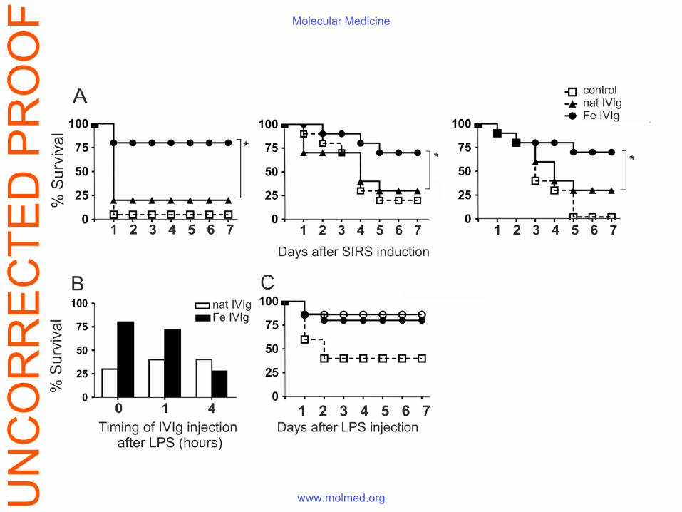
Protective effect of ferrous ions-exposed IVIg improved survival in sepsis and aseptic SIRS
The intravenous administration of a single dose of 50 mg/kg of the Fe(II)-ions-modified IVIg
significantly decreased the mortality of animals with endotoxemia (Fig. 1A - left panel). The
same treatment (at dose of 250 mg/kg) was protective in zymosan-induced SIRS and in
polymicrobial CLP sepsis (Fig. 1A, middle and right panels). The survival curves of all animals
in the experimental series are shown on Fig. S1. The native immunoglobulin preparation,
regardless of the dose used, had no effect thus confirming results from numerous clinical trials.
The mechanisms of protective activity of IVIg with enhanced polyspecificity in endotoxemia
were studied in more details. The infusion of the modified IVIg one hour post-LPS injection still
had a protective effect while the treatment delayed for 4 hours failed to improve the survival
(Fig. 1B). The iron content of IVIg increased after the Fe(II) treatment from 0.028 mg/g IgG to
7.2 mg/g IgG (Table S1). Nevertheless the beneficial effect of the preparation was still present
even upon dialysis against EDTA which decreased the iron concentration (Fig. 1C).
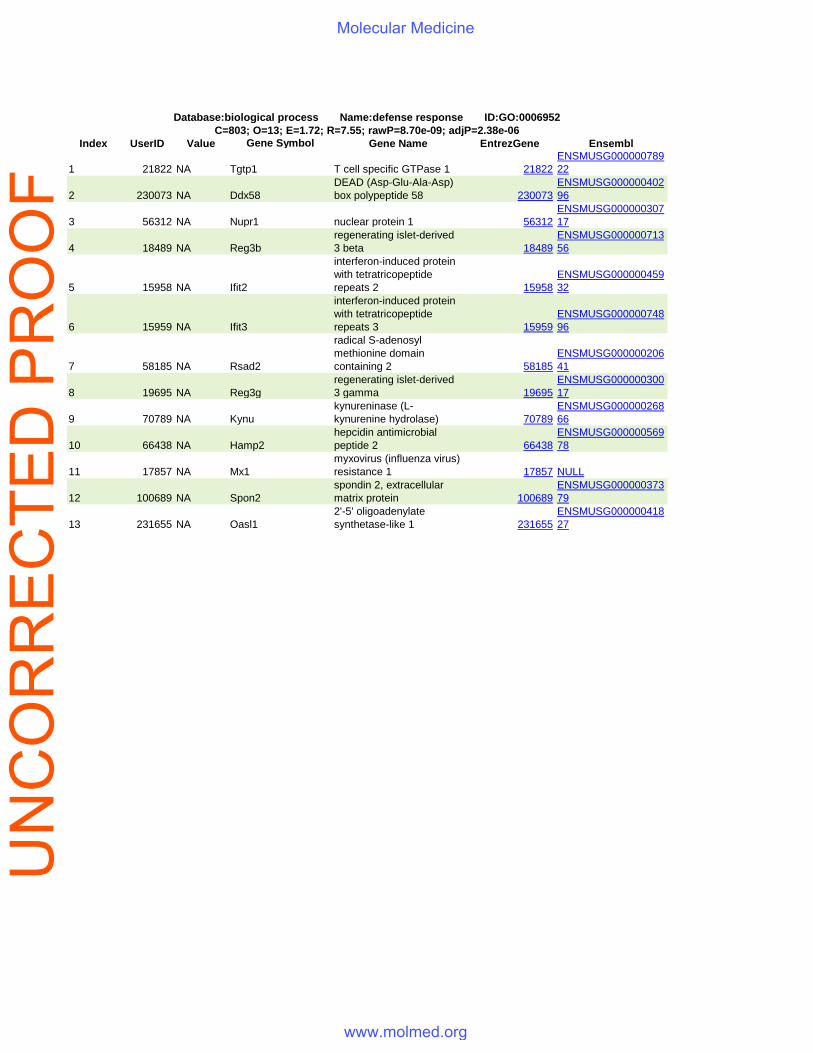
Gene expression analysis
Microarray data of organ samples from Fe(II)-exposed IVIg treated endotoxemic mice were
clustured by an unfiltered unsupervised approach. Unique responses to Fe(II)-exposed IVIg were
observed in all organs, but were most pronounced in the liver, supported by a weaker correlation
(Pearson correlation coefficient r (IVIg vs Fe(II)-exposed IVIg) = 0.83 in kidney and r = 0.45 in
liver). Although, several differentially expressed genes (DEG) could be identified, they did not
constitute an easily interpretable pattern of signaling pathways (Figure S2).
UN
CO
RR
EC
TED
PR
OO
FMolecular Medicine
www.molmed.org
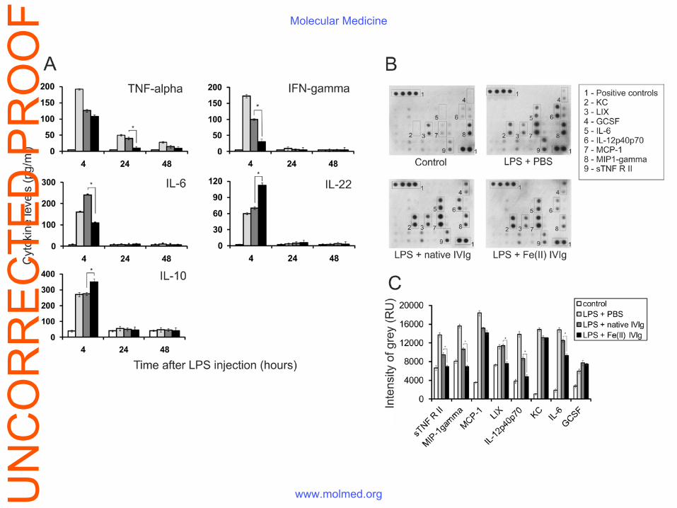
Treatment with IVIg with enhanced polyspecificity affects plasma levels of pro- and anti-
inflammatory cytokines
IVIg exposure to Fe(II) ions did not result in its enhanced binding to bacterial LPS (Fig. S3),
indicating that the beneficial effect of modified IVIg was due to its interactions with host defense
mechanisms rather than to a direct neutralization of LPS. The observed beneficial effect on
survival suggests that the levels of pro-inflammatory molecules might be influenced by the
treatment. Indeed, plasma levels of TNF-α, IL-6 and IFN-γ were significantly reduced in animals
injected with modified IVIg (Fig. 2A). In contrast, the levels of IL-10 and IL-22 were
significantly elevated. A semi-quantitative dot-blot protein array technique allowed the analysis
of a larger panel of inflammation-related molecules. Significantly lower levels of IL-6, LIX
(CXCL5), IL-12p70 and MIP-1gamma (CCL9) were detected after treatment with the IVIg with
increased polyspecificity (Fig. 2B, 2C).
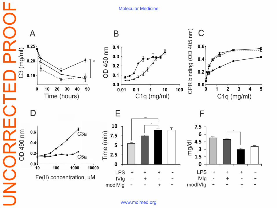
Effect of ferrous ions-exposed IVIg on the complement system, coagulation and organ
dysfunction of mice with LPS-induced sepsis
Complement activation and the release of anaphylatoxins play an important role in the
pathogenesis of sepsis (23, 24). The levels of C3 complement component in the circulation
mirror the severity of on-going inflammation. The LPS injection resulted in a sharp and
prolonged drop in C3 levels and the administration of the native IVIg did not significantly affect
this level. Interestingly, the infusion of the same single dose of the IVIg with enhanced
polyspecificity led to a significantly diminished consumption of C3 (Fig. 3A). We analyzed in
more detail the interaction of the immunoglobulin preparations with human complement
components. The binding of the pooled human IgG to the C1q component was enhanced after its
UN
CO
RR
EC
TED
PR
OO
FMolecular Medicine
www.molmed.org
exposure to ferrous ions (Fig. 3B). While the pre-incubation of C1q with Fe(II)-exposed IVIg did
not affect the interaction of the latter with immobilized IgG1 (not shown), it significantly
inhibited C1q binding to CRP (Fig. 3C). Interestingly, the modified immunoglobulin preparation
bound to C3a, but not to C5a anaphylatoxins (Fig. 3D).
Systemic inflammation in sepsis is accompanied by an over-activation of the coagulation cascade
and results in disseminated clots in small blood vessels, contributing to organ failure. A single
infusion of ferrous ions-exposed IVIg to endotoxemic mice normalized the blood clotting time
(Fig. 3E). This could well explain also the observation that liver injury was significantly
ameliorated in the group treated with the modified IVIg as assessed by the capacity of ferrous
ions-exposed IVIg to decrease the LPS-induced elevation of serum bilirubin levels (Fig. 3F).
Creatinine levels at the same early time point were not affected.
The Fe(II)-modified IVIg acquires additional polyspecificity that includes ability to bind
pro-inflammatory mediators and endogenous “danger” molecules
The exposure of therapeutic IVIg to increasing concentrations of ferrous ions results in its ability
to bind to a broader number of antigens in a HUVEC lysate (Fig. S4). In addition, anti-human IP-
10, IFN-gamma, anti-MIG and anti-HMGBox1 reactivities were observed in Fe(II)-exposed IVIg
using a BioPlex or ELISA assays, whereas native IVIg and pooled IgM preparations had no
detectable antibodies to the studied cytokines. Antibodies to endogenous "danger" molecules -
free Histone 3 and Histone 4, reported to possess a toxic effect on endothelial cells in sepsis (25),
as well as to heat shock proteins were also present in the modified preparation (Table 1 and Fig.
S5).
UN
CO
RR
EC
TED
PR
OO
FMolecular Medicine
www.molmed.org
Discussion
Passive immunotherapy with commercially available therapeutic IVIg exposed in vitro to a low
concentration of pro-oxidative ferrous ions, significantly improved survival in animals with
experimental sepsis and aseptic SIRS. The same immunoglobulin preparations were not
protective in their native form. The improved survival was accompanied by lower serum levels
of pro-inflammatory cytokines, normalization of the coagulation time, diminished consumption
of C3 complement component and attenuated organ injury or dysfunction. These multimodal
effects strongly suggest that the ferrous-exposed IVIg has acquired novel biological properties.
Pooled IVIg are highly polyspecific therapeutic preparations. Previous studies point that their
exposure to Fe(II)-ions or to ROS, released in inflammation sites, amplify further this
polyspecificity (15).
IVIg are known to suppress the harmful effects of activated complement cascade components by
preventing their binding to the respective receptors (26). In the fluid phase the IVIg with
enhanced polyspecificity did not perturb the capacity of C1q to recognize IgG immune
complexes. The latter is in agreement with previous studies, showing that even at high doses
IVIg do not alter C1q binding and complement activation on sensitized sheep erythrocytes used
as a model of cell-bound immune complexes (27). Therefore, the administration of Fe(II) ions-
modified IVIg will not affect C1q functions during the early phase of defense against pathogens.
Moreover, C1q binds better to the surface immobilized Fe(II) ions-modified IVIg (as a model of
immune complex) than to the native IVIg, suggesting that the modified preparation could be
more effective in opsonizing invading bacteria. When allowed to interact in a fluid phase with
C1q Fe(II) ions-exposed IVIg inhibited the binding of C1q to surface-bound CRP, suggesting
that attenuation of the complement activation on damaged host cells or on bacteria could result.
UN
CO
RR
EC
TED
PR
OO
FMolecular Medicine
www.molmed.org
Since CRP levels are increased up to 50 fold in sepsis (28) the inhibition of its effector functions
may be beneficial by reducing the ongoing inflammation. In our experiments the Fe(II) ions-
modified IVIg also showed enhanced binding to C3a anaphylatoxin expected to result in vivo in
its rapid neutralization and elimination.
The net beneficial effect of the blunted host response upon administration of IVIg with enhaced
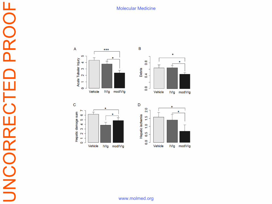
polyspecificity were reflected in less organ damage primarily in kidneys (Fig. S6). The
transcriptome analysis showed a high variability between the individual animals suggesting that
detection of the differential expression of particular genes might be a transitory phenomenon in a
highly disordered (chaotic) gene network dynamics, characteristic of the septic state (29). For
instance, some of the untreated septic animals clustered with the IVIG treated and some - with
the modified IVIg. Nevertheless, the experiment showed detectable changes in the gene
expression patterns, specific for the modified IVIg. A different set up with larger experimental
groups will be necessary to trace these changes of the cascade ultimately dampening the genomic
storm (Fig. S2).
The idea that polyspecific natural antibodies may act as a buffering system that prevents brisk
changes in the levels of circulating cytokines, hormones, etc., has been proposed twenty years
ago (30). The immune-suppressive properties of plasma during sepsis have been discussed
extensively before. Diverse factors are currently considered in this context: IL-10, extracellular
ubiquitin, circulating CD14, epinephrine and other inducible signals (31). Such a convergence of
different activities towards an anti-inflammatory plasma compartment indicates the importance
of this principle. It is thus not surprising that the quasi-complete repertoire of circulating natural
antibodies can be conditionally recruited to that function too. UN
CO
RR
EC
TED
PR
OO
FMolecular Medicine
www.molmed.org
It is counter-intuitive that naturally polyspecific IgG antibodies should generally limit or block
cytokine activities in the tissues. On the other hand, in vivo induced polyspecificity would be
limited in space (to the site of inflammation) and time (IgG polyspecificity would result in a
short half-life (32) and molecules gaining systemic access would be quickly diluted). Thus, its
effect would be tightly controlled. It is possible that the in vitro treatment of IVIg with protein-
destabilizing agents serendipitously activated this function on a large scale. Intravenous infusion
of immunoglobulin preparation with additionally enhanced polyspecificity would apply
systemically a “buffer” system that evolution have meant to have a local effect. During the event
of uncontrolled systemic inflammation (sepsis) this may be helpful as it would enhance the
inherent anti-inflammatory activity of plasma. Our data provide evidence to support this
hypothesis as only the modified, but not the native IVIg preparation binds to a) a panel of
antigens in a HUVEC lysate, b) mediators of inflammation – IFNγ, IP-10/CXCL10,
MIG/CXCL9 and HGMB1, c) the complement components C1q and C3a, and d) the
hydrophobic "danger signals" heat shock proteins and the free extracellular histones H3 and H4
(see Refs. (15, 25) and Figs. 3, S4 and S5). Importantly, our studies revealed that Fe(II) ions
exposure is associated with considerable increase in the hydrophobicity of antigen binding sites
of IgG (15). As endogenous danger signals have frequently hydrophobic nature, antibody-
mediated non-specific quenching of hydrophobicity may also well explain the systemic anti-
inflammatory effect of modified IVIg (33, 34).
Furthermore, the elimination of the formed immune complexes by Fc receptor-expressing
phagocytes may contribute to the down-regulation of the ongoing severe generalized
inflammatory reaction. UN
CO
RR
EC
TED
PR
OO
FMolecular Medicine
www.molmed.org
Although the dependence of the observed therapeutic effect of modified IVIg on lymphocytes
and macrophages is compatible with the “immunosomatics” mechanism (30), an effect, mediated
by a particular mononuclear cell population, cannot be ruled out. Innate response activator (IRA)
CD19lowCD5lowCD93+ B cells have been implicated recently in the control of experimental
sepsis in mice (35). Indeed, our recent studies show that, unlike native IVIg, ferrous ions
exposure-modified IVIg induced in the LPS-injected mice higher levels of IRA B cells and the
beneficial effect of the treatment on survival correlated with this increase (36).
Conclusion
This work contains evidence that pleiotropic therapeutic approaches offer a viable strategy for
treatment of sepsis and aseptic SIRS. Apart from the mechanistic explanation, the pleiotropy
principle also justifies the use of mouse models despite the controversy on differences in
transcriptome profiles in humans and mice with severe inflammatory syndromes (37, 38).
Acknowledgments
This work was supported by grants from the Bulgarian-Swiss Research Programme (BSRP
IZEBZO_142967), the Bulgarian National Science Fund (DFNI B02/29), the Pasteur Institute
(ACIP A07-2012), the NATO Science for Peace Program (SfP 982158), the German Federal
Ministry of Education and Research (BMBF; grant 01EO1002) and grant 03Z2J521 (Meta-ZIK)
- both to the Centre for Sepsis Control and Care. We thank D. Himsel for technical assistance.
UN
CO
RR
EC
TED
PR
OO
FMolecular Medicine
www.molmed.org
For More Information The microarray data generated in this publication have been deposited in
NCBI's Gene Expression Omnibus (GEO) and are accessible through the accession number
GSE55964.
Competing financial interests
The authors declare competing financial interests: TV is applicant and TV and JD are inventors
of a patent.
References:
1. Angus DC, van der Poll T. (2013) Severe sepsis and septic shock. N Engl J Med. 369:840-
851.
2. Jean-Marc Cavaillon CAE (2008) Sepsis and Non-infectious Systemic Inflammation: from
Biology to Critical Care, Weinheim: Wiley-VCH Verlag GmbH & Co.
3. The INIS Collaborative Group (2011) Treatment of Neonatal Sepsis with Intravenous Immune
Globulin. N Eng J Med. 365:1201-1211.
4. Rittirsch D, Flierl MA, Ward PA. (2008) Harmful molecular mechanisms in sepsis. Nat Rev
Immunol. 8:776-787.
5. Bouvet J, et al. (2001) Induction of natural autoantibody activity following treatment of
human immunoglobulin with dissociating agents. J Autoimmun. 16:163-172.
6. Xiao W, et al. (2011) A genomic storm in critically injured humans. J Exp Med. 208:2581-
2590. UN
CO
RR
EC
TED
PR
OO
FMolecular Medicine
www.molmed.org
7. Kazatchkine MD, Kaveri SV. (2001) Immunomodulation of Autoimmune and Inflammatory
Diseases with Intravenous Immune Globulin. N Engl J Med. 345: 747-755.
8. Lutz HU. (2012) Naturally Occurring Antibodies (NAbs). Landes Bioscience, Austin, TX,
USA.
9. Laupland KB, Kirkpatrick AW, Delaney A. (2007) Polyclonal intravenous immunoglobulin
for the treatment of severe sepsis and septic shock in critically ill adults: A systematic review and
meta-analysis*. Crit Care Med. 35:2686-2692.
10. Soares MO, et al. (2012) An evaluation of the feasibility, cost and value of information of a
multicentre randomised controlled trial of intravenous immunoglobulin for sepsis (severe sepsis
and septic shock): incorporating a systematic review, meta-analysis and value of information
analysis. Health Technology Assessment. 16:1-18611.
11. Werdan K, et al. (2007) Score-based immunoglobulin G therapy of patients with sepsis: The
SBITS study. Critical Care Medicine. 35:2693-2701.
12. Djoumerska I, Tchorbanov A, Pashov A, Vassilev T. (2005) The autoreactivity of
therapeutic intravenous immunoglobulin (IVIG) preparations depends on the fractionation
methods used. Scand J Immunol. 61:357-363.
13. Djoumerska-Alexieva IK, et al. (2010) Exposure of IgG to an acidic environment results in
molecular modifications and in enhanced protective activity in sepsis. FEBS J. 277:3039-3050.
14. Pavlovic S, et al. (2011) Intravenous immunoglobulins exposed to heme (heme IVIG) are
more efficient than IVIG in attenuating autoimmune diabetes. Clin Immunol. 138:162-171.
15. Dimitrov JD, et al. (2006) Ferrous ions and reactive oxygen species increase antigen-binding
and anti-inflammatory activities of immunoglobulin G. J Biol Chem. 281:439-446. UN
CO
RR
EC
TED
PR
OO
FMolecular Medicine
www.molmed.org
16. Dimitrov J, et al. (2007) Antibodies use heme as a cofactor to extend their pathogen-
eliminating activity and to acquire new effector functions. J Biol Chem. 282:26696-26706.
17. McIntyre J, Wagenknecht D, Faulk W. (2006) Redox-reactive autoantibodies: detection and
physiological relevance. Autoimmun Rev.5:76.
18. Djoumerska-Alexieva IK, Dimitrov JD, Nacheva J, Kaveri SV, Vassilev TL. (2009) Protein
destabilizing agents induce polyreactivity and enhanced immunomodulatory activity in IVIg
preparations. Autoimmunity. 42:365-367.
19. Hubbard WJ, et al. (2005) Cecal ligation and puncture. Shock. 24 Suppl 1:52-57
20. Volman TJ, Hendriks T, Goris RJ. (2005) Zymosan-induced generalized inflammation:
experimental studies into mechanisms leading to multiple organ dysfunction syndrome. Shock.
23:291-297.
21. Ivanovska ND, et al. (2008) Properdin deficiency in murine models of nonseptic shock. J
Immunol. 180:6962-6969.
22. Haeffner-Cavaillon N, Klein M, Dorrington KJ. (1979) Studies on the Fc gamma receptor of
the murine macrophage-like cell line P388D1. I. The binding of homologous and heterologous
immunoglobulin G1. J Immunol. 123:1905-1913.
23. Ward PA, Gao H. (2009) Sepsis, complement and the dysregulated inflammatory response. J
Cell Mol Med. 13:4154-4160.
24. Markiewski MM, DeAngelis RA, Lambris JD. (2008) Complexity of complement activation
in sepsis. J Cell Mol Med. 12:2245-2254.
25. Xu J, et al. (2009) Extracellular histones are major mediators of death in sepsis. Nat Med.
15:1318-1321. UN
CO
RR
EC
TED
PR
OO
FMolecular Medicine
www.molmed.org
26. Basta M. (2008) Ambivalent effect of immunoglobulins on the complement system:
activation versus inhibition. Mol Immunol. 45:4073-4079.
27. Basta M, Fries LF, Frank MM. (1991) High doses of intravenous Ig inhibit in vitro uptake of
C4 fragments onto sensitized erythrocytes. Blood. 77:376-380.
28. Devran O, et al. (2012) C-reactive protein as a predictor of mortality in patients affected with
severe sepsis in intensive care unit. Multidiscip Respir Med. 7:47-52.
29. Tang BM, Huang SJ, McLean AS. (2010) Genome-wide transcription profiling of human
sepsis: a systematic review. Crit Care 14: R237.
30. Coutinho A, Avrameas S. (1992) Speculation on immunosomatics : Potential diagnostic and
therapeutic value of immune homeostasis concepts. Scand J Immunol. 36:527-532.
31. Cavaillon JM, Adib-Conquy M. (2007) Determining the degree of immunodysregulation in
sepsis. Contrib Nephrol. 156:101-111.
32. Sigounas G, Harindranath N, Donadel G, Notkins AL. (1994) Half-life of polyreactive
antibodies. J Clin Immunol. 14:134-140.
33. Matzinger P. (2002) The danger model: a renewed sense of self. Science. 296:301-305.
34. Seong SY, Matzinger P. (2004) Hydrophobicity: an ancient damage-associated molecular
pattern that initiates innate immune responses. Nat Rev Immunol 4:469-478.
35. Rauch PJ, et al. (2012) Innate response activator B cells protect against microbial sepsis.
Science. 335:597-601.
36. Djoumerska-Alexieva I, Pashova S, Vassilev T, Pashov A. (2013) The protective effect of
modified intravenous immunoglobulin in LPS sepsis model is associated with an increased IRA
B cells response. Autoimmun Rev. 12:653-656. UN
CO
RR
EC
TED
PR
OO
FMolecular Medicine
www.molmed.org
37. Seok J, et al. (2013) Genomic responses in mouse models poorly mimic human inflammatory
diseases. PNAS 110:3507-3512.
38. Takao K, Miyakawa T. (2015) Genomic responses in mouse models greatly mimic human
inflammatory diseases. PNAS 112:1167-1172.
UN
CO
RR
EC
TED
PR
OO
FMolecular Medicine
www.molmed.org
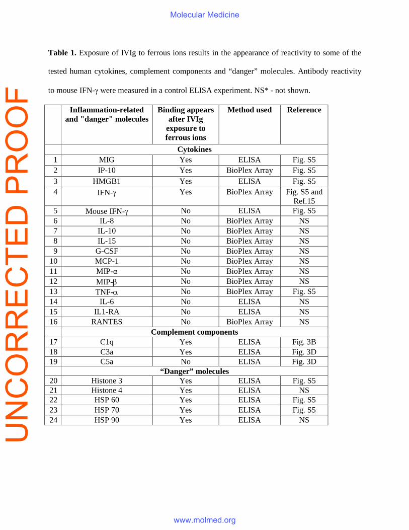
Table 1. Exposure of IVIg to ferrous ions results in the appearance of reactivity to some of the
tested human cytokines, complement components and “danger” molecules. Antibody reactivity
to mouse IFN-γ were measured in a control ELISA experiment. NS* - not shown.
Inflammation-related and "danger" molecules
Binding appears after IVIg
exposure to ferrous ions
Method used Reference
Cytokines 1 MIG Yes ELISA Fig. S5 2 IP-10 Yes BioPlex Array Fig. S5 3 HMGB1 Yes ELISA Fig. S5 4 IFN-γ Yes BioPlex Array Fig. S5 and
Ref.15 5 Mouse IFN-γ No ELISA Fig. S5 6 IL-8 No BioPlex Array NS 7 IL-10 No BioPlex Array NS 8 IL-15 No BioPlex Array NS 9 G-CSF No BioPlex Array NS
10 MCP-1 No BioPlex Array NS 11 MIP-α No BioPlex Array NS 12 MIP-β No BioPlex Array NS 13 TNF-α No BioPlex Array Fig. S5 14 IL-6 No ELISA NS 15 IL1-RA No ELISA NS 16 RANTES No BioPlex Array NS
Complement components 17 C1q Yes ELISA Fig. 3B 18 C3a Yes ELISA Fig. 3D 19 C5a No ELISA Fig. 3D
“Danger” molecules 20 Histone 3 Yes ELISA Fig. S5 21 Histone 4 Yes ELISA NS 22 HSP 60 Yes ELISA Fig. S5 23 HSP 70 Yes ELISA Fig. S5 24 HSP 90 Yes ELISA NS
UN
CO
RR
EC
TED
PR
OO
FMolecular Medicine
www.molmed.org
Figure legends:
Figure 1. Passive immunotherapy with IVIg modified by Fe(II) exposure improves animal
survival in two aseptic SIRS and one polymicrobial sepsis models. Outbred female ICR mice
were used in the experiments. (A) Endotoxemia was induced by the i.p. injection of 10 mg/kg E.
coli LPS (left panel). Zymosan-induced systemic inflammation (middle panel) was caused by i.p.
injection of 500 mg/kg zymosan. Polymicrobial CLP-induced sepsis (right panel) was performed
as described in the Materials and methods section. The survival was compared between the
control group, injected i.v. with PBS (open squares, n = 12), the group with endotoxemia treated
with 50 mg/kg or of 250 mg/kg (in zymosan-induced inflammation and CLP sepsis) native IVIg
(triangles, n = 12), and a group treated with 50 mg/kg (endotoxemia) or 250 mg/kg (zymosan-
induced inflammation and CLP sepsis) Fe(II)-exposed IVIg (circles, n = 12); *p<0.05, Mantel-
Haenszel logrank test). (B) The beneficial effect of the Fe(II)-exposed preparation on survival
was still observed if its administration was delayed. Endotoxemia was induced as described
above and the mice were treated i.v. with 50 mg/kg of the native (open bars) or Fe(II)-exposed
IVIg (black bars) at different time points – minutes before, one, or four hours after the i.p.
injection of LPS. (C) Residual iron ions in Fe(II)-exposed IVIg did not contribute to its
protective effect in LPS sepsis. ICR mice (n=12) were injected i.p with 10 mg/kg E. coli LPS
and treated i.v. with 50 mg/kg Fe(II)-exposed IVIg with high (7.2 mg/g IgG, black circles) or
low (0.66 mg/g IgG, white circles) iron concentration or with PBS only (open squares).
Figure 2. Serum levels of inflammation-related molecules in treated endotoxemic mice. (A)
Endotoxemia was induced by the i.p. injection of 10 mg/kg E. coli LPS. A control group was
UN
CO
RR
EC
TED
PR
OO
FMolecular Medicine
www.molmed.org
injected i.v. with PBS (n = 10, light grey bars), a second group was injected i.v. with 50 mg/kg of
native IVIg (n = 10, dark grey bars), and a third group - with 50 mg/kg of Fe(II)-exposed IVIg
(n=10, black bars). Cytokine levels in untreated mice are represented with white bars. Sera were
obtained 4, 24 and 48 h after LPS injection and cytokine levels were measured by ELISA. Data
represent mean pg/ml values±SD of quadruplicate wells, *p<0.05 (paired Student t-test). (B)
Scan of dot blots evaluating semi-quantitatively of serum levels of a larger panel of
inflammatory molecules two hours after LPS injection. Each blot is representative of three
individual experiments. (C) The individual dots (see panel B) were subjected to densitometry and
the data were analyzed using the Image-Tool v2.0 for Windows (UTHSCSA, San Antonio, TX,
USA) software package and were represented in relative units of grey intensity (n=3, mean±SD,
*p<0.05, paired Student t-test).
Figure 3. A single administration of Fe(II) exposure-modified IVIg overcomes the
coagulation and complement abnormalities in LPS-induced sepsis. (A) Endotoxemia was
induced to ICR mice by the i.p. injection of 10 mg/kg E. coli LPS. A control group (n=10, open
squares) was injected i.v. with PBS, a second group was treated with 50 mg/kg native IVIg (n =
10, black triangles), and a third was treated with 50 mg/kg Fe(II)-exposed IVIg (n=10, black
circles). Serum samples were collected at 4, 24 and 48 hour after LPS administration and the C3
levels in plasma were measured using a commercial Mouse C3 ELISA kit, *p<0.05, paired t-test.
(B) The exposure of IVIg to Fe(II)-ions results in its increased binding to C1q molecules (black
circles) compared to that of native IVIg (black triangles), mean±SD. (C) C1q pretreatment with
Fe(II)-exposure modified IVIg inhibits the binding of the former to CRP (black circles); in
comparison to C1q pretreated with native IVIg (black triangles). The binding of untreated C1q is
UN
CO
RR
EC
TED
PR
OO
FMolecular Medicine
www.molmed.org
presented with open squares. (D) The exposure of IVIg to increasing concentrations of ferrous
ions results in an enhanced binding to the C3a, but not the C5a component Anti-C3a and anti-
C5a reactivity was compared by ELISA in IVIg, exposed to increasing concentrations of ferrous
ions. (E) Endotoxemia was induced to ICR mice by the i.p. injection of 10 mg/kg E. coli LPS. A
control group was treated i.v. with PBS (n=10, light grey bars), a group was treated with 50
mg/kg native IVIg (n = 10, dark grey bars), and another was treated with 50 mg/kg Fe(II)-
exposed IVIg (n=10, black bars). Animals were bled 4 hours after the injection of LPS and
coagulation time was measured as previously described (21), *p<0.05; ***p<0.001, t-test. (F)
Bilirubin serum levels measured four hours after LPS administration in groups of mice treated
with native (n=10, dark grey bars), Fe(II) -exposed IVIg (n=10, black bars) or PBS alone (n=10,
light grey bars). *p<0.05, t-test.
UN
CO
RR
EC
TED
PR
OO
FMolecular Medicine
www.molmed.org
UN
CO
RR
EC
TED
PR
OO
F Molecular Medicine
www.molmed.org
UN
CO
RR
EC
TED
PR
OO
F Molecular Medicine
www.molmed.org
UN
CO
RR
EC
TED
PR
OO
F Molecular Medicine
www.molmed.org
Supporting information:
Materials and Methods Flame atomic absorption spectroscopy The concentration of iron ions in the IVIg preparations under study was measured by flame
atomic absorption spectrometry. The samples were diluted 1:2 with a 20% (W/V) trichloro
acetic acid solution, and heated in a heating block for 15 minutes at 90°C. They were next
cooled, centrifugated and the iron concentration was determined using a Perkin Elmer M 5000
atomic absorption spectrometer.
Gene expression analysis
C57Bl/6 mice were used in this analysis. Blood samples, liver and kidney homogenates were
obtained and processed by standard protocols. RNA was extracted using the RNeasy minikit
(Qiagen, Hilden, Germany) in a balanced design comprising three biological replicates from
blood and four from liver and kidney of each treatment group (vehicle, native IVIg, Fe(II)-
exposed IVIg) respectively. Biotinylated probes were prepared using the TargetAmp™-Nano
Labeling Kit for Illumina® Expression BeadChip® (Illumina, San Diego, CA). The arrays
were scanned using the Illumina BeadArray Reader 500 X.
Illumina mouseRef8 V2 chips were read by Genome Studio (v.1.9, and annotation MouseRef-
8_V2_0_R3_11278551_A) yielding 25,697 bead probes for each sample. Analysis was
performed consistently using R software version 3.02 (http://www.r-project.org/) and
Bioconductor (1) packages. Raw data from each organ was separately subjected to robust
spline normalization, quality control and background correction with the R package Lumi (2).
Two samples (one from blood cells, one from kidney) did not pass quality control and were
discarded from analysis. Bead types having detection values p < 0.05 of the RNA samples
UN
CO
RR
EC
TED
PR
OO
FMolecular Medicine
www.molmed.org
were called "present" and taken for further analysis. Differentially expressed genes (DEG)
were filtered according to microarray quality control criteria (MAQC, (3)) by: 1) at least two-
fold changes and 2) p-values from moderated t-statistics <0.05 using ”Limma” (24). Further,
these statistics were computed for all the contrasts (IVIg, Fe(II) exposure-modified IVIg
(modIVIg), vehicle) using linear model fits, moderated t-and F-statistics, and log-odds of
differential expression by empirical Bayes moderation. Gene set enrichment analysis was
performed by hypergeometric tests with the WebGestalt tool (4). Annotation, log2 fold
changes, top enriched categories and p-values are provided as Supplemental gene expression
data.
Binding of and modified IVIg to bacterial lipopolysaccharide ELISA plates were coated with E. coli LPS (B 055:B5, Sigma #L2880, at 5 ug/ml in PBS)
and blocked. Serial dilutions of the IVIg preparations were added in triplicate wells and the
binding of the pooled human IgG was measured by ELISA.
In a separate experiment the native- and the Fe(II)-exposed IVIg were mixed in the wells of
a blocked ELISA plate with increasing concentrations of E.coli LPS and incubated for two
hours at room temperature. These mixtures were transferred to a second LPS-coated plate
and the binding of human IgG was measured by ELISA as described in the Methods section.
Its inhibition in the presence of free LPS was determined and presented in arbitrary units.
Immunoblot analysis of binding to HUVEC antigens HUVEC cells lysates were subjected to 10% gel SDS-PAGE and transferred to nitrocellulose
membranes (Scheicher & Schuell, Dassel, Germany) by a Mini Transfer Blot system
(BioRad, Richmond, CA). Next, the membranes were incubated for 1 hour at room
temperature in TBS containing 0,4% Tween 20, cut into strips and incubated for 1 h at room
UN
CO
RR
EC
TED
PR
OO
FMolecular Medicine
www.molmed.org
temperature with native or Fe(II)-exposed IVIg preparations at concentrations of 0,2 mg/ml.
After extensive washing the strips were incubated with an anti-human IgG antibody (Fc-
specific), conjugated to alkaline phosphatase (Sigma Chemical Co, St. Louis, MO) and
revealed using the nitro blue tetrazolium/bromo-chloro-indolyl-phosphate substrates (Sigma).
Antibodies to pro-inflammatory and “danger” molecules in the modified IVIg
The presence of antibodies to cytokines, complement components and “danger” molecules
was studied using ELISA and BioPlex Array (see the Materials and Methods section of the
main text).
Histology ICR mice (8-12 weeks old, 18-22 g) were used in this experiment. Endotoxemia was induced
by the i.p. injection of 10 mg/kg E. coli LPS (B 055:B5, #L2880, Sigma-Aldrich St. Luis,
MO). Single dose of native, modified IVIg preparations or PBS alone was injected
intravenously (i.v.) 10 min before the administration of LPS. Kidneys and livers from each
experimental group were collected 24 h after the injection of LPS. The organs were fixed in
4% buffered formalin, embedded in paraffin, sectioned into 3.5 µm thick slices and stained
with Hematoxylin and Eosin (H&E). Pathologists blinded for the treatments estimated kidney
damage in mice challenged with LPS + vehicle (n=8), LPS + native IVIg (n=10), LPS +
Fe(II)-exposed IVIg (n=10) using the following criteria: the presence of brush-border defects,
flattening of epithelia, loss of nuclei and presence of debris all indicative of an acute tubular
injury (ATI) (5). Presence of liver damage was graded based on sinusoidal congestion,
nuclear and cytoplasmic irregularities, signs of cellular ischemia, sinusoidal infiltration and
single cell necrosis (6).
UN
CO
RR
EC
TED
PR
OO
FMolecular Medicine
www.molmed.org
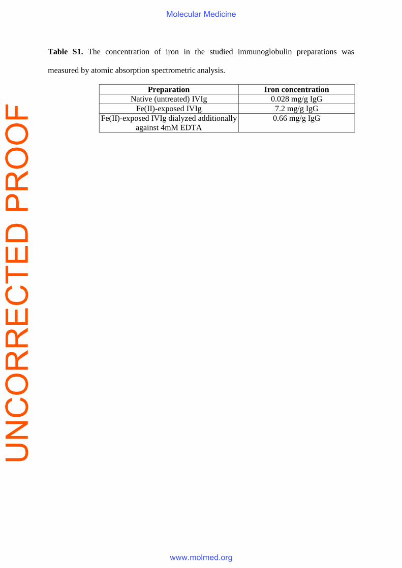
Table S1. The concentration of iron in the studied immunoglobulin preparations was
measured by atomic absorption spectrometric analysis.
Preparation Iron concentration Native (untreated) IVIg 0.028 mg/g IgG
Fe(II)-exposed IVIg 7.2 mg/g IgG Fe(II)-exposed IVIg dialyzed additionally
against 4mM EDTA 0.66 mg/g IgG
UN
CO
RR
EC
TED
PR
OO
FMolecular Medicine
www.molmed.org
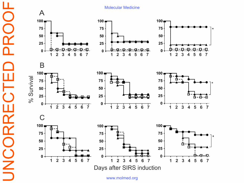
Figure legends: Figure S1. Reduced mortality in three different experimental models of sepsis after
passive immunotherapy with an IVIg preparation, modified by Fe(II) exposure. (A)
Survival curves of ICR mice with bacterial LPS-induced endotoxemia. Animals were injected
i.p. with 10 mg/kg E. coli LPS. A survival study was performed comparing a control group,
treated i.v. with PBS (open squares, n = 12), a group treated with 2 mg/kg (left panel), 10
mg/kg (middle panel) or 50 mg/kg (right panel) native IVIg (triangles, n = 12), and a group
treated with 2 mg/kg (left panel), 10 mg/kg (middle panel) or 50 mg/kg (right panel) Fe(II)-
exposed IVIg (circles, n = 12); *p<0.05, Mantel-Haenszel logrank test). (B) Survival curves
of ICR mice with zymosan-induced systemic inflammation. Animals were injected i.p. with
500 mg/kg zymosan. A survival study was performed comparing a control group, treated i.v.
with PBS (open squares, n = 12), a group treated with 10 mg/kg (left panel), 50 mg/kg
(middle panel) or 250 mg/kg (right panel) native IVIg (triangles, n = 12), and a group treated
with 10 mg/kg (left panel), 50 mg/kg (middle panel) or 250 mg/kg (right panel) Fe(II)-
exposed IVIg (circles, n = 12); *p<0.05, Mantel- Haenszel logrank test). (C) Survival curves
of ICR mice with polymicrobial CLP-induced sepsis. Survival was compared between a
control group, injected i.v. with PBS (open squares, n = 12), a group treated with 10 mg/kg
(left panel), 50 mg/kg (middle panel) or 250 mg/kg (right panel) of native IVIg (triangles, n =
12), and a group treated with 10 mg/kg (left panel), 50 mg/kg (middle panel) or 250 mg/kg
(right panel) of Fe(II)-exposed IVIg (circles, n = 12). *p<0.05, Mantel- Haenszel logrank
test).
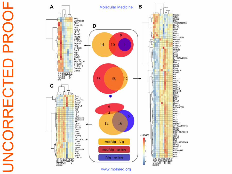
Figure S2. Blood cell, liver and kidney gene expression in LPS-injected mice treated
with native- or Fe(II)-exposed IVIg (modIVIg). Heatmaps depicting the differentially
expressed genes (DEG, average two-fold change and p<0.05) for Fe(II)-exposed IVIg versus
UN
CO
RR
EC
TED
PR
OO
FMolecular Medicine
www.molmed.org
native IVIg in blood (A), liver (B) and kidney (C), respectively. Resolved log2 signals of
biological replicates were row-wise scaled (z-score) accompanied by vertical and horizontal
hierarchical correlation clustering (technical probe replicates are indicated by ".1". Definition
of gene symbols is provided in the supplement files. The Venn diagrams depict the number of
DEG for all contrasts (D).
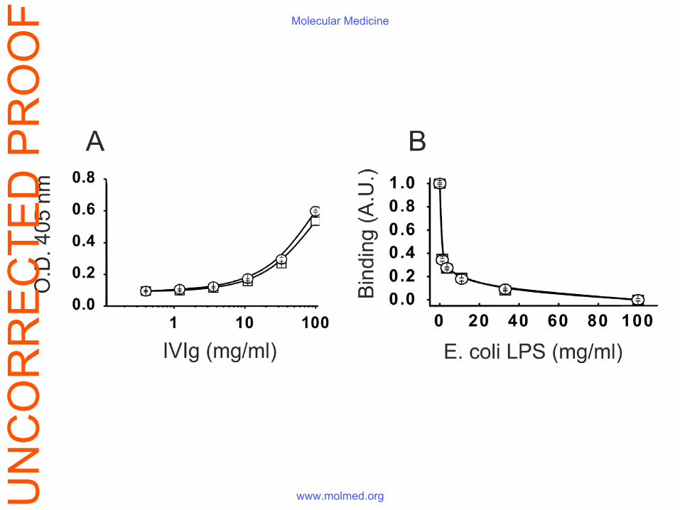
Figure S3. The exposure of IVIg to ferrous ions does not affect its binding to LPS. (A)
Increasing concentrations of native- and Fe(II)-exposed IVIg preparations (marked with
squares and circles respectively) were left to interact with immobilized LPS and human IgG
binding was evaluated by ELISA. (B) Both preparations were pre-incubated with increasing
concentrations of LPS and the mixtures were transferred to the LPS-coated wells. Next, the
binding of human IgG was measured by ELISA and its inhibition (presented in arbitrary units
- AU) was plotted against the concentration of free LPS used.
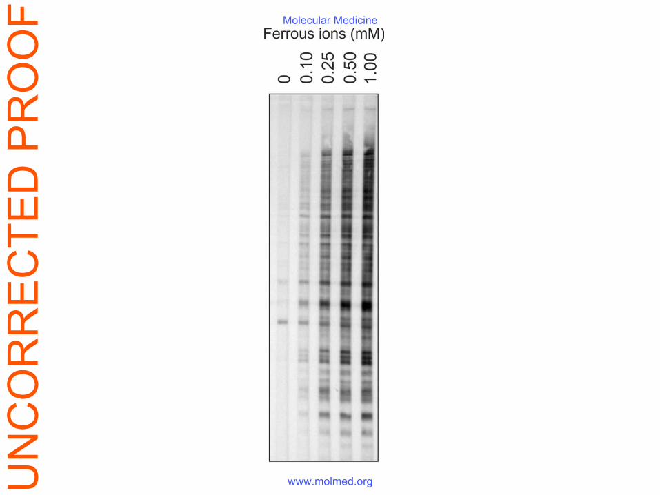
Figure S4. Exposure of IVIg to increasing concentrations of Fe(II) ions results in its
enhanced binding to antigens in a HUVEC lysate. The membranes were incubated with
200 µg/ml of the native IVIg or of the same preparation exposed to ferrous ions.
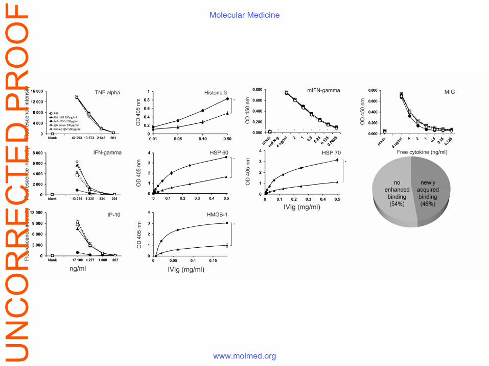
Figure S5. Ferrous ions-exposed IVIg acquires the ability to bind to various human
pro-inflammatory molecules. The figure presents several examples whereas all results are
showed in Table 1 of the main text. Reactivity to IFN-γ , ΙP−10 and ΤΝFα were measured
using BioPlex Array and in all other cases ELISA was used (see the Materials and Methods
section of the main text). In a control experiment the reactivities of both studied preparations
to mouse IFN-γ (mIFN-γ ) were compared. The pie-chart summarizes all data and points out
that modified IVIg acquires the ability bind to almost half of the unselected tested
inflammation-related molecules.
UN
CO
RR
EC
TED
PR
OO
FMolecular Medicine
www.molmed.org
Figure S6. Tissue damage score from histological specimen (kidney - A and B, liver - C
and D) from mice with endotoxemia treated with native or Fe(II)-exposed IVIg. Kidney
injury was evaluated by the presence of brush-border defects, flattening of epithelia, loss of
nuclei and presence of debris, summarized in the acute tubular injury (ATI) score (1). Liver
damage scoring was based on sinusoidal congestion, nuclear and cytoplasmic irregularities,
signs of cellular ischemia, sinusoidal infiltration and single cell necrosis. Summaries are
provided in A and C for kidney and liver, respectively. *p<0.05, t-test (Welch-modified,
accounting for differences in variances) Fe(II)-exposed IVIg vs the other groups given in
parametric plots.
References
1. Gentleman RC, et al. (2004) Bioconductor: open software development for computational
biology and bioinformatics. Genome Biol 5:R80.
2. Du P, Kibbe WA, Lin SM. (2008) lumi: a pipeline for processing Illumina microarray.
Bioinformatics 24:1547-1548.
3. Shi L, et al. (2006) The MicroArray Quality Control (MAQC) project shows inter- and
intraplatform reproducibility of gene expression measurements. Nat Biotechnol 24:1151-
1161.
4. Wang J, Duncan D, Shi Z, Zhang B. (2013) WEB-based GEne SeT AnaLysis Toolkit
(WebGestalt): update 2013. Nucleic Acids Res 41:W77-83.
5. Bockmeyer CL, et al. (2011) ADAMTS13 activity is decreased in a septic porcine model.
Significance for glomerular thrombus deposition. Thromb Haemost 105:145-153.
6. Recknagel P, et al. (2013) Mechanisms and functional consequences of liver failure
substantially differ between endotoxaemia and faecal peritonitis in rats. Liver Int 33:283-293.
7. Smyth GK. (2004) Linear models and empirical bayes methods for assessing differential
UN
CO
RR
EC
TED
PR
OO
FMolecular Medicine
www.molmed.org
expression in microarray experiments. Stat Appl Genet Mol Biol 3:Article3.
UN
CO
RR
EC
TED
PR
OO
FMolecular Medicine
www.molmed.org
UN
CO
RR
EC
TED
PR
OO
F Molecular Medicine
www.molmed.org
UN
CO
RR
EC
TED
PR
OO
F Molecular Medicine
www.molmed.org
UN
CO
RR
EC
TED
PR
OO
F Molecular Medicine
www.molmed.org
UN
CO
RR
EC
TED
PR
OO
F Molecular Medicine
www.molmed.org
UN
CO
RR
EC
TED
PR
OO
F Molecular Medicine
www.molmed.org
UN
CO
RR
EC
TED
PR
OO
F Molecular Medicine
www.molmed.org
ILMN_Gene average log2FC IVIg-Vehicle modIVIg-Vehicle modIVIg-IVIg P (moderated t-statistics) IVIg-Vehicle modIVIg-Vehicle modIVIg-IVIg Probe_Id Probe_Sequence Definition2900062L11RIK -1.416697695 -0.173654787 1.243042908 0.037431 0.775410146 0.0463929 ILMN_2609182 CCTTGTAGGTCCACATTGAATGCCGTTTTCTTATATCTTCTAGGGTGTGT Mus musculus RIKEN cDNA 2900062L11 gene (2900062L11Rik), non-coding RNA.ADFP -0.594814157 0.717284553 1.31209871 0.0968802 0.051508139 0.00135786 ILMN_1222246 AGAAAATTCAGGGTGCTCAGGATAAGCTCTATGTCTCGTGGGTGGAGTG Mus musculus adipose differentiation related protein (Adfp), mRNA.ADIPOQ -2.400097295 0.232938434 2.633035729 0.011571 0.771813749 0.00439888 ILMN_2738082 GACTGCAACTACCCATAGCCCATACACCAGGAGAATCATGGAACAGTCG Mus musculus adiponectin, C1Q and collagen domain containing (Adipoq), mRNA.AOC3 -1.531980725 -0.312202827 1.219777897 0.0366476 0.63477318 0.06509666 ILMN_2625920 GAGAGGGAATCAGGTTCTGCCTGCCCTTCTGAACATCATGGATTGGAAC Mus musculus amine oxidase, copper containing 3 (Aoc3), mRNA.APOC1 -1.299479683 -0.347932302 0.951547381 0.0479402 0.560706361 0.10537082 ILMN_2599794 AAAGTGAAGGAGAAGTTGAAGACCACGTTCTCCTGAGCACCTGGCGGGCMus musculus apolipoprotein C-I (Apoc1), mRNA.AQP4 -1.01024592 -1.330365875 -0.320119956 0.0777703 0.026900839 0.51716872 ILMN_2757232 GTGCAGAGCATATGGACACCTCTGTGAGGAAGCTGGCATTGTCCATCGT Mus musculus aquaporin 4 (Aqp4), mRNA.CAR3 -1.362158574 -0.203605447 1.158553127 0.0381835 0.729427688 0.05319186 ILMN_2630047 CCCTGGCTCTGCTAAGACCATCCTGAACAATGGGAAGACCTGCAGAGTT Mus musculus carbonic anhydrase 3 (Car3), mRNA.CCND1 0.381308916 -0.86206292 -1.243371836 0.3771588 0.062804125 0.00844535 ILMN_2601471 GCCATAGACTGGTGGTGGGTCCACGGAATCTGCCCTGTGACATGAAAGGCCCND1 0.398618927 -0.770912202 -1.169531129 0.3564935 0.090707034 0.01173754 ILMN_1221503 GGCCATAGACTGGTGGTGGGTCCACGGAATCTGCCCTGTGACATGAAAGGCD1D1 -1.46195839 -0.084498452 1.377459938 0.028787 0.886047215 0.02663035 ILMN_2734212 GAACTAGAAAGCATACTTCCTGCCCAAACAGACGCTCTGAGGTTAGTTG Mus musculus CD1d1 antigen (Cd1d1), mRNA.CDKN1A -0.598236148 0.649996258 1.248232406 0.2810181 0.243870608 0.02766547 ILMN_2634083 CATCCTGGTCTGGACTGTCTACCCTTAGCCCGCACCCCAAGAACATGTA Mus musculus cyclin-dependent kinase inhibitor 1A (P21) (Cdkn1a), mRNA.CFD -2.965653662 0.530765528 3.496419189 0.0049065 0.536798053 0.0010017 ILMN_2835423 CAGTCGAAGGTGTGGTTACGTGGGGCTCTCGCGTCTGTGGCAATGGCAAMus musculus complement factor D (adipsin) (Cfd), mRNA.CIDEA -1.801244902 -0.559559857 1.241685045 0.0150816 0.385987296 0.05452063 ILMN_1215446 GACGGGACAGTTCCTGGTCTATGCGGGCACATACATGCTCCGAGTACTGMus musculus cell death-inducing DNA fragmentation factor, alpha subunit-like effector A (Cidea), mRNA.CIDEC -1.456853318 -0.138919218 1.317934099 0.0081337 0.761195691 0.00924771 ILMN_1222679 GAGGTGTGCGAGAGAGTGTTAGAGGTGGAGATTAACTAAAGCCGGCATGMus musculus cell death-inducing DFFA-like effector c (Cidec), mRNA.CLDN11 -1.645730543 -1.42116018 0.224570363 0.0191203 0.036851878 0.69063747 ILMN_1246139 GTGTCTAAGACTCTGGATACTGCAAGCTCCGTCCGGTGCATTTGTTCAG Mus musculus claudin 11 (Cldn11), mRNA.COX8B -2.449745171 -0.250312233 2.199432937 0.0265011 0.796554407 0.03034856 ILMN_2727520 CGTTGGCTTCATGGTTCCAGCAGGATGGGTCTTAGCCCACTTGGAGAGC Mus musculus cytochrome c oxidase, subunit VIIIb (Cox8b), mRNA.GNA14 0.487424158 -0.52936533 -1.016789488 0.3000355 0.262600443 0.03319393 ILMN_2597778 GCGCTGTGCCTTTCTGGATACTAAGAATACACTGTTCTACCCTGAACACCMus musculus guanine nucleotide binding protein, alpha 14 (Gna14), mRNA.HAVCR1 -0.172270347 1.403494432 1.575764778 0.7857277 0.045698769 0.01981639 ILMN_2604307 AAGCTTTGCAGAACGCAGCGGTTGTGCATTCCCGAGCTGAAGACAACAT Mus musculus hepatitis A virus cellular receptor 1 (Havcr1), mRNA.HP -2.096151341 0.392517347 2.488668689 0.0132898 0.587818322 0.00317183 ILMN_2668509 CTGTTGTCACTCTCCTGCTCTGGGGTCAGCTTTTTGCTGTGGAGTTGGG Mus musculus haptoglobin (Hp), mRNA.HP -2.204083306 0.44858963 2.652672936 0.0217303 0.593855579 0.00538841 ILMN_2668510 CTCTCCTGCTCTGGGGTCAGCTTTTTGCTGTGGAGTTGGGCAATGATGC Mus musculus haptoglobin (Hp), mRNA.HP -2.332394507 0.611622015 2.944016523 0.0117124 0.44034818 0.00181362 ILMN_2944824 ACGAGTCCCGTTGGGGTCCAGCCTATCTTGAACGAGCACACCTTCTGTG Mus musculus haptoglobin (Hp), mRNA.IGFBP5 -0.499217044 -1.072298018 -0.573080974 0.272493 0.031436343 0.18029713 ILMN_2964324 GGATAGTACAGTTCAGACAAGACTCCTTCAGATTCCGAGACGCCTACCG Mus musculus insulin-like growth factor binding protein 5 (Igfbp5), mRNA.IL4I1 -0.391947595 -1.081478706 -0.689531111 0.3802022 0.029367298 0.11131137 ILMN_2733778 CCCAGCTCCTAAGCCAGCCCTCTTCAGGGCAGACAGACCACCTACACTA Mus musculus interleukin 4 induced 1 (Il4i1), mRNA.ITIH4 -0.746467905 0.417565978 1.164033883 0.1420903 0.394031883 0.02270197 ILMN_2718431 CACCTTATGCCAACAGGGACGCCTGTGAGGCCGAGACCTTGATGGGAAGMus musculus inter alpha-trypsin inhibitor, heavy chain 4 (Itih4), mRNA.KRT20 0.083357367 1.757084824 1.673727457 0.8930599 0.015346426 0.01327319 ILMN_2907285 GTGAACGGGTTCTCCAGGGAGGCAACTTCCCAGTTAATCTCATTGTCTCCMus musculus keratin 20 (Krt20), mRNA.LOC100043671 -1.456136503 -0.593052178 0.863084325 0.0369099 0.351242413 0.15502824 ILMN_1258600 TCTGTCCACTTGTTCACAAAGCAGCCTTCACACGTGTCAGTGAGAGTTCGPREDICTED: Mus musculus hypothetical protein LOC100043671 (LOC100043671), mRNA.MFSD2 -1.051508402 0.381609069 1.433117471 0.097658 0.522914016 0.02253908 ILMN_1225764 AGGGCTGCTACTGTGAATATGCCAAGGACTGACTGGGCCTAGCTCGGAAMus musculus major facilitator superfamily domain containing 2 (Mfsd2), mRNA.MYCL1 -0.022587085 1.064445119 1.087032205 0.9557901 0.022773331 0.01412011 ILMN_1221750 GGGACTCCTGGAACTATCTTGGGAGGACAAGTGGTGAACAGGCTAAAGTMus musculus v-myc myelocytomatosis viral oncogene homolog 1, lung carcinoma derived (avian) (Mycl1), mRNA.MYL1 -1.077625097 -0.343423488 0.734201609 0.0333534 0.451390027 0.10004509 ILMN_2878542 GATAATCTCAAGTCCACACACCACCGTTATCATCTACTTGGGTCCACAAC Mus musculus myosin, light polypeptide 1 (Myl1), mRNA.NNMT -1.199571192 0.25428225 1.453853442 0.0678093 0.674137856 0.0229501 ILMN_2885277 CTCCCTAGTGGGGCGAAAGCCAGGCAGATCTGAATGACATCCTGTGATC Mus musculus nicotinamide N-methyltransferase (Nnmt), mRNA.ORM1 -2.030115548 -0.101961618 1.92815393 0.0352627 0.905436355 0.03164228 ILMN_1226935 CCTTTGCCCACCTTATAGTGCTGAGGAAACATGGGGCCTTCATGCTTGC Mus musculus orosomucoid 1 (Orm1), mRNA.S3-12 -1.472158014 0.190004481 1.662162495 0.0228048 0.736759772 0.00826673 ILMN_2588249 CCTTAGTGTCTGCTTCACTCAAGCTGGTAGAACAGGCTACTCGCTGCAC Mus musculus plasma membrane associated protein, S3-12 (S3-12), mRNA.SAA3 -0.78514295 0.678302447 1.463445398 0.1322338 0.187289026 0.00786362 ILMN_2772632 GAGGACTCAAGAGCTGACCAGTTTGCCAATGAGTGGGGCCGGAGTGGC Mus musculus serum amyloid A 3 (Saa3), mRNA.SCD1 -1.647443514 -0.537645069 1.109798445 0.0044752 0.263470449 0.02437171 ILMN_1237375 CCAGTCAAAGTGCAAGACTACCTACCCACTGCCATGAAAACCATTGCAG Mus musculus stearoyl-Coenzyme A desaturase 1 (Scd1), mRNA.SCIN -0.002361753 -1.293221152 -1.290859399 0.9945273 0.003086022 0.00188657 ILMN_1259174 GCTTCTAAGCATTTCCCCGTCTGCTACTCTGTTTGCAGTGAGCTTTACTT Mus musculus scinderin (Scin), mRNA.SERPINA3C -1.001675207 0.217593835 1.219269043 0.0171768 0.550762992 0.00373395 ILMN_2730054 CAAGGGACTCATCTCAGACCTGGATACGGATACACTGATGGTGCTGGTG Mus musculus serine (or cysteine) peptidase inhibitor, clade A, member 3C (Serpina3c), mRNA.SERPINA3M -0.885792445 0.417494031 1.303286476 0.03249 0.270410176 0.00270448 ILMN_2728473 CCTCTTTATGGCCAAAGTCACTAACCCCAAGTGAACCTAAAGCTCCCCAAMus musculus serine (or cysteine) peptidase inhibitor, clade A, member 3M (Serpina3m), mRNA.SERPINA3N -1.378347276 0.440050877 1.818398152 0.0343426 0.453636705 0.00575328 ILMN_1246800 CCCCTGGCACTCCTACTTAGAACAAAGTAGCCTTTCTTTTAGTTCCCAGC Mus musculus serine (or cysteine) peptidase inhibitor, clade A, member 3N (Serpina3n), mRNA.SGK2 0.533528881 1.0746424 0.541113519 0.1507394 0.010434508 0.11899333 ILMN_2827080 GGCTGCCCAGAAACCGTCTGGTGGCTTCCTTTAGTGTGTCGTAAATCTC Mus musculus serum/glucocorticoid regulated kinase 2 (Sgk2), mRNA.SGK2 0.639027332 1.000316869 0.361289537 0.0772857 0.011458472 0.25746565 ILMN_2827081 GAGAGAAAGGGGAGGATGTAGGTGACCAGCACTCTGGGAGAGAGCCTC Mus musculus serum/glucocorticoid regulated kinase 2 (Sgk2), mRNA.SLC1A3 -1.259241472 0.321836021 1.581077493 0.040451 0.562001189 0.00954689 ILMN_2634317 CCCGGGCTCTGCCTATGTGTGTACCCACTGTGGACTGGGAAACTTCACT Mus musculus solute carrier family 1 (glial high affinity glutamate transporter), member 3 (Slc1a3), mRNA.SLC4A1 0.52824231 -0.744134243 -1.272376553 0.0751733 0.018739204 0.00039945 ILMN_1227675 CGCTCTTCCCCCACCGCCGTGCTTGGGAATAGGTTTTGCCAATAAACGT Mus musculus solute carrier family 4 (anion exchanger), member 1 (Slc4a1), mRNA.SLC5A3 0.750679233 1.037239005 0.286559772 0.0513856 0.012004863 0.38412317 ILMN_1233078 CCCAGTGGCTTCCATGGGTCATTCAGAGGCAGAAACACCAGTAGATGCT Mus musculus solute carrier family 5 (inositol transporters), member 3 (Slc5a3), mRNA.TMEM174 0.249557844 -0.848911432 -1.098469276 0.414559 0.015675558 0.00224195 ILMN_1216425 GGCTGCCCCCAAGAGAGATACAGGAATTGTCAGGTCTTGAAGGTTTTGG Mus musculus transmembrane protein 174 (Tmem174), mRNA.TRF -1.477109112 0.245021332 1.722130444 0.0175221 0.648774584 0.00498246 ILMN_2485323 CTGGGGTAGACAGAACCGCTGGTTGGAACATCCCTATGGGCATGCTGTAMus musculus transferrin (Trf), mRNA.
UN
CO
RR
EC
TED
PR
OO
FMolecular Medicine
www.molmed.org
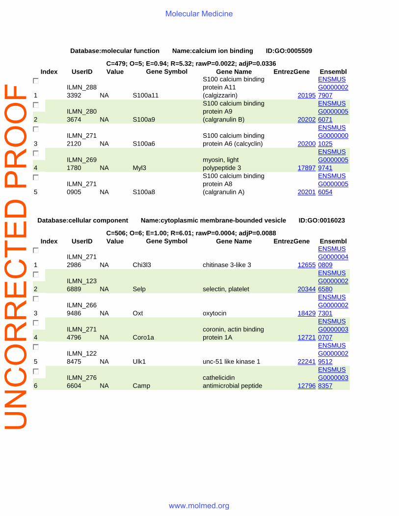
Index UserID Value Gene Symbol Gene Name EntrezGene Ensembl
1 ILMN_2883392 NA S100a11
S100 calcium binding protein A11 (calgizzarin) 20195
ENSMUSG00000027907
2 ILMN_2803674 NA S100a9
S100 calcium binding protein A9 (calgranulin B) 20202
ENSMUSG00000056071
3 ILMN_2712120 NA S100a6
S100 calcium binding protein A6 (calcyclin) 20200
ENSMUSG00000001025
4 ILMN_2691780 NA Myl3
myosin, light polypeptide 3 17897
ENSMUSG00000059741
5 ILMN_2710905 NA S100a8
S100 calcium binding protein A8 (calgranulin A) 20201
ENSMUSG00000056054
Index UserID Value Gene Symbol Gene Name EntrezGene Ensembl
1 ILMN_2712986 NA Chi3l3 chitinase 3-like 3 12655
ENSMUSG00000040809
2 ILMN_1236889 NA Selp selectin, platelet 20344
ENSMUSG00000026580
3 ILMN_2669486 NA Oxt oxytocin 18429
ENSMUSG00000027301
4 ILMN_2714796 NA Coro1a
coronin, actin binding protein 1A 12721
ENSMUSG00000030707
5 ILMN_1228475 NA Ulk1 unc-51 like kinase 1 22241
ENSMUSG00000029512
6 ILMN_2766604 NA Camp
cathelicidin antimicrobial peptide 12796
ENSMUSG00000038357
Database:molecular function Name:calcium ion binding ID:GO:0005509
C=479; O=5; E=0.94; R=5.32; rawP=0.0022; adjP=0.0336
Database:cellular component Name:cytoplasmic membrane-bounded vesicle ID:GO:0016023
C=506; O=6; E=1.00; R=6.01; rawP=0.0004; adjP=0.0088
UN
CO
RR
EC
TED
PR
OO
FMolecular Medicine
www.molmed.org
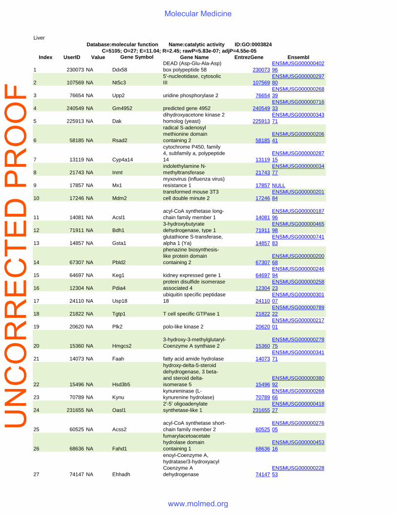
Liver
Index UserID Value Gene Symbol Gene Name EntrezGene Ensembl
1 230073 NA Ddx58DEAD (Asp-Glu-Ala-Asp) box polypeptide 58 230073
ENSMUSG00000040296
2 107569 NA Nt5c35'-nucleotidase, cytosolic III 107569
ENSMUSG00000029780
3 76654 NA Upp2 uridine phosphorylase 2 76654ENSMUSG00000026839
4 240549 NA Gm4952 predicted gene 4952 240549ENSMUSG00000071633
5 225913 NA Dakdihydroxyacetone kinase 2 homolog (yeast) 225913
ENSMUSG00000034371
6 58185 NA Rsad2
radical S-adenosyl methionine domain containing 2 58185
ENSMUSG00000020641
7 13119 NA Cyp4a14
cytochrome P450, family 4, subfamily a, polypeptide 14 13119
ENSMUSG00000028715
8 21743 NA Inmtindolethylamine N-methyltransferase 21743
ENSMUSG00000003477
9 17857 NA Mx1myxovirus (influenza virus) resistance 1 17857 NULL
10 17246 NA Mdm2transformed mouse 3T3 cell double minute 2 17246
ENSMUSG00000020184
11 14081 NA Acsl1acyl-CoA synthetase long-chain family member 1 14081
ENSMUSG00000018796
12 71911 NA Bdh13-hydroxybutyrate dehydrogenase, type 1 71911
ENSMUSG00000046598
13 14857 NA Gsta1glutathione S-transferase, alpha 1 (Ya) 14857
ENSMUSG00000074183
14 67307 NA Pbld2
phenazine biosynthesis-like protein domain containing 2 67307
ENSMUSG00000020068
15 64697 NA Keg1 kidney expressed gene 1 64697ENSMUSG00000024694
16 12304 NA Pdia4protein disulfide isomerase associated 4 12304
ENSMUSG00000025823
17 24110 NA Usp18ubiquitin specific peptidase 18 24110
ENSMUSG00000030107
18 21822 NA Tgtp1 T cell specific GTPase 1 21822ENSMUSG00000078922
19 20620 NA Plk2 polo-like kinase 2 20620ENSMUSG00000021701
20 15360 NA Hmgcs23-hydroxy-3-methylglutaryl-Coenzyme A synthase 2 15360
ENSMUSG00000027875
21 14073 NA Faah fatty acid amide hydrolase 14073ENSMUSG00000034171
22 15496 NA Hsd3b5
hydroxy-delta-5-steroid dehydrogenase, 3 beta- and steroid delta-isomerase 5 15496
ENSMUSG00000038092
23 70789 NA Kynukynureninase (L-kynurenine hydrolase) 70789
ENSMUSG00000026866
24 231655 NA Oasl12'-5' oligoadenylate synthetase-like 1 231655
ENSMUSG00000041827
25 60525 NA Acss2acyl-CoA synthetase short-chain family member 2 60525
ENSMUSG00000027605
26 68636 NA Fahd1
fumarylacetoacetate hydrolase domain containing 1 68636
ENSMUSG00000045316
27 74147 NA Ehhadh
enoyl-Coenzyme A, hydratase/3-hydroxyacyl Coenzyme A dehydrogenase 74147
ENSMUSG00000022853
Database:molecular function Name:catalytic activity ID:GO:0003824C=5105; O=27; E=11.04; R=2.45; rawP=5.83e-07; adjP=4.55e-05
UN
CO
RR
EC
TED
PR
OO
FMolecular Medicine
www.molmed.org
Index UserID Value Gene Symbol Gene Name EntrezGene Ensembl
1 21822 NA Tgtp1 T cell specific GTPase 1 21822ENSMUSG00000078922
2 230073 NA Ddx58DEAD (Asp-Glu-Ala-Asp) box polypeptide 58 230073
ENSMUSG00000040296
3 56312 NA Nupr1 nuclear protein 1 56312ENSMUSG00000030717
4 18489 NA Reg3bregenerating islet-derived 3 beta 18489
ENSMUSG00000071356
5 15958 NA Ifit2
interferon-induced protein with tetratricopeptide repeats 2 15958
ENSMUSG00000045932
6 15959 NA Ifit3
interferon-induced protein with tetratricopeptide repeats 3 15959
ENSMUSG00000074896
7 58185 NA Rsad2
radical S-adenosyl methionine domain containing 2 58185
ENSMUSG00000020641
8 19695 NA Reg3gregenerating islet-derived 3 gamma 19695
ENSMUSG00000030017
9 70789 NA Kynukynureninase (L-kynurenine hydrolase) 70789
ENSMUSG00000026866
10 66438 NA Hamp2hepcidin antimicrobial peptide 2 66438
ENSMUSG00000056978
11 17857 NA Mx1myxovirus (influenza virus) resistance 1 17857 NULL
12 100689 NA Spon2spondin 2, extracellular matrix protein 100689
ENSMUSG00000037379
13 231655 NA Oasl12'-5' oligoadenylate synthetase-like 1 231655
ENSMUSG00000041827
Database:biological process Name:defense response ID:GO:0006952C=803; O=13; E=1.72; R=7.55; rawP=8.70e-09; adjP=2.38e-06
UN
CO
RR
EC
TED
PR
OO
FMolecular Medicine
www.molmed.org
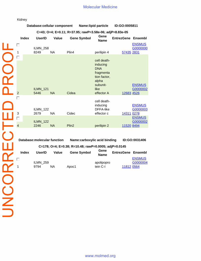
Kidney
Index UserID Value Gene Symbol Gene Name
EntrezGene Ensembl
1 ILMN_2588249 NA Plin4 perilipin 4 57435
ENSMUSG00000002831
2 ILMN_1215446 NA Cidea
cell death-inducing DNA fragmentation factor, alpha subunit-like effector A 12683
ENSMUSG00000024526
3 ILMN_1222679 NA Cidec
cell death-inducing DFFA-like effector c 14311
ENSMUSG00000030278
4 ILMN_1222246 NA Plin2 perilipin 2 11520
ENSMUSG00000028494
Index UserID Value Gene Symbol Gene Name
EntrezGene Ensembl
1 ILMN_2599794 NA Apoc1
apolipoprotein C-I 11812
ENSMUSG00000040564
Database:cellular component Name:lipid particle ID:GO:0005811
C=43; O=4; E=0.11; R=37.95; rawP=3.58e-06; adjP=8.83e-05
Database:molecular function Name:carboxylic acid binding ID:GO:0031406
C=178; O=4; E=0.38; R=10.48; rawP=0.0005; adjP=0.0145
UN
CO
RR
EC
TED
PR
OO
FMolecular Medicine
www.molmed.org
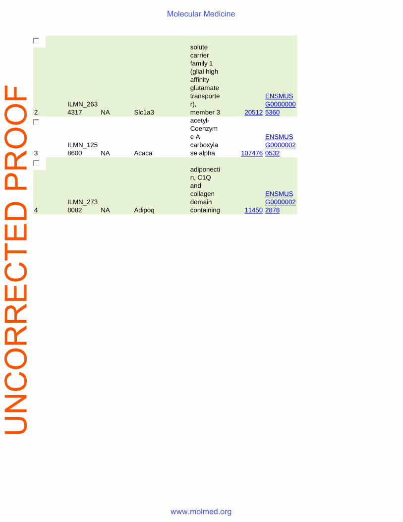
2 ILMN_2634317 NA Slc1a3
solute carrier family 1 (glial high affinity glutamate transporter), member 3 20512
ENSMUSG00000005360
3 ILMN_1258600 NA Acaca
acetyl-Coenzyme A carboxylase alpha 107476
ENSMUSG00000020532
4 ILMN_2738082 NA Adipoq
adiponectin, C1Q and collagen domain containing 11450
ENSMUSG00000022878
UN
CO
RR
EC
TED
PR
OO
FMolecular Medicine
www.molmed.org


![Intravenous Immunoglobulin for Preventing Infection in Preterm and-Or Low Birth Weight Infants[2]](https://img.pdfslide.us/doc/110x75/577ccfc81a28ab9e78909145/intravenous-immunoglobulin-for-preventing-infection-in-preterm-and-or-low-birth.jpg)