Embed Size (px)
Citation preview
Intravascular Ultrasound Guided IVC filter placement
Donald L. Jacobs, MD C Rollins Hanlon Professor and Chair
Department of Surgery Saint Louis University School of Medicine
Saint Louis, Missouri
Disclosure
I have the following potential conflicts of interest to report:
National PI for LUMIFI, an IDE study for the Lumifi with Crux® VCF System (Volcano)
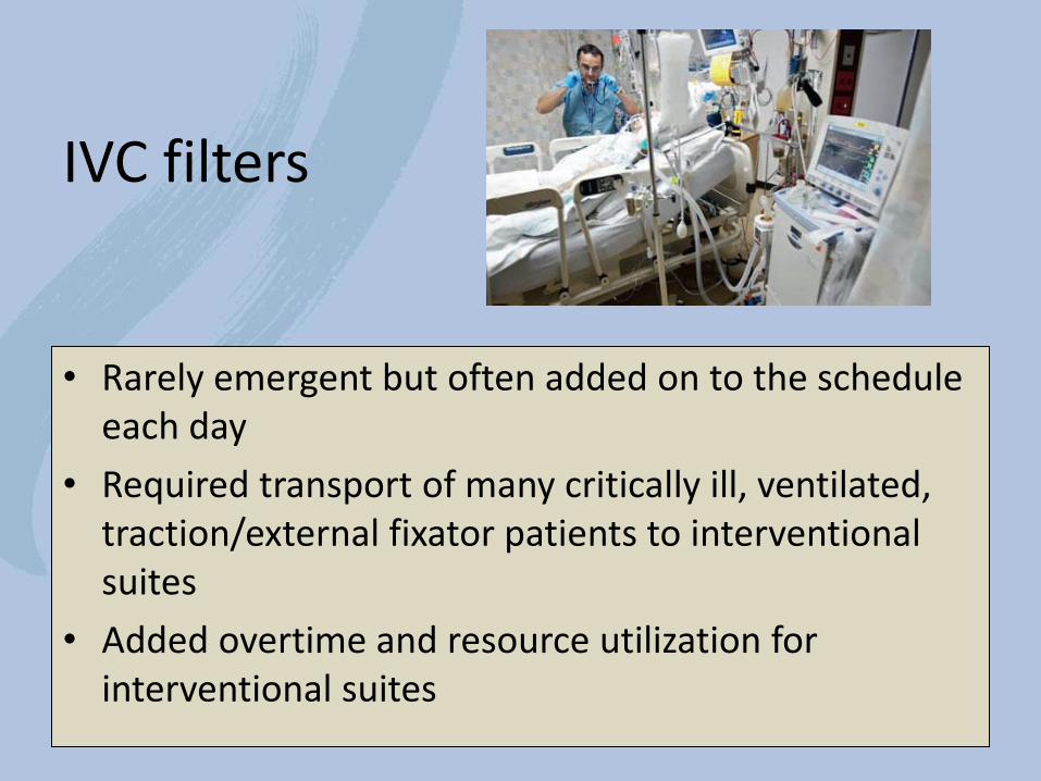
IVC filters
• Rarely emergent but often added on to the schedule each day
• Required transport of many critically ill, ventilated, traction/external fixator patients to interventional suites
• Added overtime and resource utilization for interventional suites
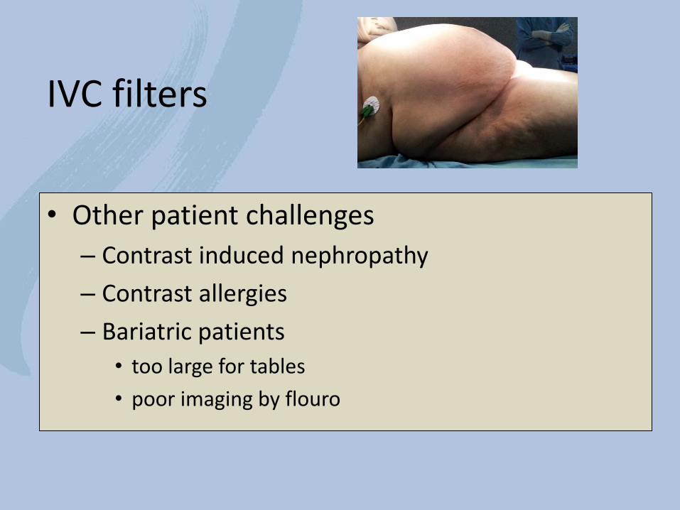
IVC filters
• Other patient challenges
– Contrast induced nephropathy
– Contrast allergies
– Bariatric patients
• too large for tables
• poor imaging by flouro
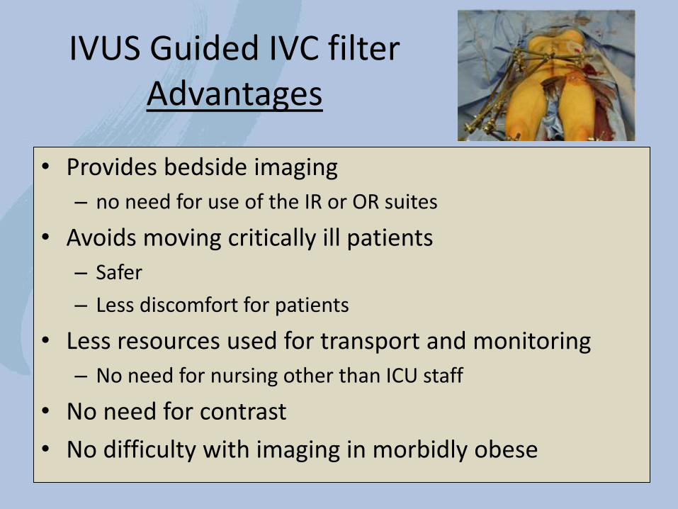
IVUS Guided IVC filter Advantages
• Provides bedside imaging
– no need for use of the IR or OR suites
• Avoids moving critically ill patients
– Safer
– Less discomfort for patients
• Less resources used for transport and monitoring
– No need for nursing other than ICU staff
• No need for contrast
• No difficulty with imaging in morbidly obese
Why has IVUS guided IVC filter placement not widely adopted?
–Concern of the cost of the IVUS catheter
– Lack of familiarity if IVUS imaging of the IVC
–Concern of need for two punctures • One to place the IVUS catheter
• One to place the IVC filter delivery catheter for real-time imaging of filter placement
Counter to these concerns:
• IVUS catheter costs less than the utilization costs of the angiography suite
• IVUS mapping of the IVC by protocol provides imaging superior to venographic imaging
• Single puncture technique can allow for real-time positioning of the IVC filter delivery sheath and there is no need for a second puncture
• New intergral IVUS catheter with the Filter Delivery catheter – The Lumifi with Crux® VCF System (Volcano)
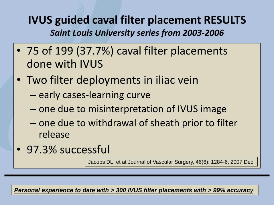
• 75 of 199 (37.7%) caval filter placements done with IVUS
• Two filter deployments in iliac vein – early cases-learning curve
– one due to misinterpretation of IVUS image
– one due to withdrawal of sheath prior to filter release
• 97.3% successful
IVUS guided caval filter placement RESULTS Saint Louis University series from 2003-2006
Jacobs DL, et at Journal of Vascular Surgery, 46(6): 1284-6, 2007 Dec
Personal experience to date with > 300 IVUS filter placements with > 99% accuracy
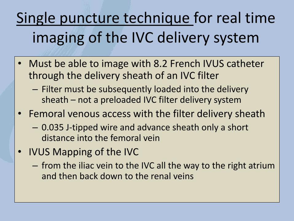
Single puncture technique for real time imaging of the IVC delivery system
• Must be able to image with 8.2 French IVUS catheter through the delivery sheath of an IVC filter – Filter must be subsequently loaded into the delivery
sheath – not a preloaded IVC filter delivery system
• Femoral venous access with the filter delivery sheath – 0.035 J-tipped wire and advance sheath only a short
distance into the femoral vein
• IVUS Mapping of the IVC – from the iliac vein to the IVC all the way to the right atrium
and then back down to the renal veins
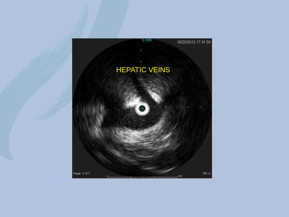
HEPATIC VEINS
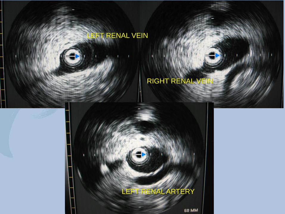
LEFT RENAL VEIN
RIGHT RENAL VEIN
LEFT RENAL ARTERY
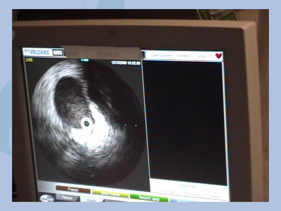
Positioning of the delivery sheath
• The radioopaque tip on the filter delivery sheath will obscure or “shadow” the IVUS image as it passes over the transducer
• With the IVUS transducer positioned at the level of the renal veins, you advance the filter delivery sheath to the point that the “shadowing” by the sheath tip occurs
• This confirms that the delivery sheath tip is then at the level of the renal veins where you want the filter tip to be when it is deployed
• Remove the IVUS catheter and keep the sheath stable in the position with it’s tip at the renal veins
Filter delivery
• Filter is then delivered into it’s delivery sheath up to the point that the tip of the filter is at the tip of the sheath based on the markings on the delivery shaft
• Having insured the tip of the sheath is at the renal veins, the filter inserted in the sheath is now positioned with the tip at the level of the renal veins – Then “pin and pull” delivery to release the filter
• Follow up imaging with the IVUS – use a 3cm lead of J-wire out the tip of the IVUS catheter
– confirms the accurate placement of the filter
Tips and Tricks
• Do not mistake the confluence of the iliacs for the renal veins
– Best avoided by mapping the complete vena cava
• Low intravascular volume will flatten the IVC and can make it difficult to see the renal veins
– imaging the right renal artery crossing behind the IVC is also a good landmark to aid in identification of the renal veins
Summary
• Well established technique – Over 30 peer reviewed articles on various aspects
of IVUS guided IVC filter placement
– Accurate placement of IVC at bedside
– Lower cost, better utilization of resources
– Increased patient safety

Intravascular Ultrasound Guided IVC filter placement
Donald L. Jacobs, MD C Rollins Hanlon Professor and Chair
Department of Surgery Saint Louis University School of Medicine
Saint Louis, Missouri