Embed Size (px)
Citation preview

Intraoperative Spinal Sonography in the Evaluation of Intramedullary Tumors
Joel F. Platt, MD•, Jonathan M. Rubin, MD, PhD•, William F. Chandler, MOt, Richard A. Bowerman, MD•, Michael A. DiPietro, MD•
Seventeen intraoperative spinal sonographic examinations were performed in 14 patients with intramedullary spinal cord neoplasms. Results of the ultrasound exams were correlated with preoperative imaging studies, surgical findings, and histopathologic analysis of the tumors.
Intraoperative spinal sonography accurately localized the intramedullary tumors, often revealing the need for extension of the initial laminectomy. The neoplasms appeared as expansile echogenic masses, with cystic com-
I ntraoperative spinal sonography (lOSS) is becoming widely accepted as a valuable aid in the intraoperative evaluation of a variety of intraspinal abnor
malities.1-10 Published data on lOSS evaluation of intramedullary spinal cord neoplasms is somewhat limited when compared to reports of other types of spinal cord pathology. 1- 6 Most previous reports on the evaluation of spinal cord neoplasms with intraoperative sonography are limited to smaller patient populations. We report our experience with 17 intraoperative sonographic examinations in 14 patients with intramedullary spinal cord tumors. We attempt to better define the role and value of lOSS by describing the benefits and potential pitfalls that may be encountered in these examinations.
Received August 10, 1987, from the Departments of •Radiology and fNeurosurgery. University of Michigan Medical Center, Ann Arbor, Michigan. Revised manuscript accepted for publication November 24, 1987.
Address correspondence and reprint requests to Dr. Platt: Depart• ment of Radiolog~ Box 0030, University of Michigan Hospital, 1500 East Medical Center Drive, Ann Arbor, Ml 48109-0030.
ponents in over half. Ultrasound was as accurate as preoperative imaging in the evaluation of solid neoplasms, and was superior to computed tomography (CT) and magnetic resonance imaging (MRI) for delineating the cystic components of neoplasms. Intraoperative sonog· raphy should be used routinely in all surgical cases of intramedullary spinal cord neoplasms. KEY woRos: neurosurgical ultrasound; intraoperative spinal sonography; spinal cord neoplasms. (/ Ultrasound Med 7:317, 1988)
MATERIALS AND METHODS
Fourteen patients (ten women, four men; age range, 7 to 70 years) underwent 17 lOSS examinations. Examinations were performed with an ATL NeurosectOR portable real-time unit (Advanced Technology Laboratories, Bellevue, WA) with an in-line scan head capable of imaging at 3, 5, or 7.5 MHz transducer frequency. All scans in our series were performed at 7.5 MHz. Before sonography, the scan head was draped as previously described?
The sonographic exams were performed via a poste-rior laminectomy with the patient lying prone. The laminectomy wound was filled with sterile saline to provide a fluid path for scanning. Initial sonographic evaluation was provided prior to opening the dura mater. Further use of sonography depended on the na• ture of the spinal surgery and on the neoplasm encountered. Scanning was performed in both longitudinal and transverse planes. Images were recorded on video tape and later photographed for the permanent patient record. Correlation of the lOSS exams with the operative
@ 1988 by the Amerif:an Institute of Ultrasound in Medicine • J Ultrasound Med 7:317-325, 1988 • 0278-4297/88/$3.50

318 SPINAL SONOGRAPHY
findings, pathologic findings, and preoperative imaging was performed retrospectively.
RESULTS
The spinal cord neoplasms imaged in our series are summarized in Table 1. The most common lesion encountered was an astrocytoma (five cases). Tumors occurred in all portions of the spinal canal (Table 2), but were most common in the thoracic or cervical thoracic region (nine cases).
Certain benefits of lOSS were evident in virtually all cases: localization, delineation of extent, and cystic vs. solid nature of the neoplasm.
Accurate localization of the intramedullary neoplasm before dural opening proved to be a useful feature of lOSS in all cases. In nine of our cases sonography demonstrated the need to extend the laminectomy.
Intraoperative spinal sonography delineated tumor extent by identifying zones of transition between normal and abnormal spinal cord based on definition of margins, and presence or absence of the central canal echo (Fig. 1). In 11 of our 14 patients good tumor border definition was present. In six of these 11 well-delineated neoplasms the surgeon could define margins as well, and a total resection was attempted. In the other five tumors with sonographically well-defined margins a total resection could not be performed, most commonly due to the long segment of tumor involvement.
In only three instances could lOSS not define tumor borders. One patient had a diffuse primitive neuroectodermal tumor involving the entire length of the spinal
Figure 1 Identification of tumor margin. Sagittal lOSS image (patient's head is toward viewer's left} shows transition from ex· panded spinal cord due to echogenic tumor mass (T). to normal appearing spinal cord with visible central canal (arrows).
J Ultrasound Med 7:317-325, 1988
Table 1: Types of Neoplasms Examined With Intraoperative Spinal Sonography
Spinal Cord Neoplasm
Astrocytoma Ependymoma Ependymoblastoma Dermoid Teratoma Low grade glioma Hemangioblastoma Primitive neuroectodermal tumor
No. of Patients
5 3 1 1 1 1 1 1
Table 2: Location of Neoplasms Examined With Intraoperative Spinal Sonography
Location of Neoplasm
Cervical Cervicothoracic Thoracic Conus Entire cord
No. of Patients
2 3 6 2 1
cord. The second patient had an infiltrative astrocytoma that at surgery was determined to be nonresectable. Intraoperative spinal sonography in this patient accurately reflected the infiltrating nature of this neoplasm by the lack of well-defined tumor margins and an absent central canal echo.
The third case demonstrated the one pitfall in evaluation of tumor extent. The patient was studied twice in a 10-day period. Preoperative magnetic resonance (MR)

J Ultrasound Med 7:317- 325, 1988 PLATI ET AL 319
A 8
c Figure 2 Ependymoma of spinal cord associated with cord edema, simulating a diffuse neoplasm. A, Sagittal Tl-weighted image (TR = .30 TE = 30) at 35T. Spinal cord is diffusely enlarged. B, Sagittal T2-weighted image (TR = 1.50 TE = 40). Diffuse intramedullary lesion of high signal intensity consistent with extensive neoplasm. C, Sagittal lOSS image (patient's head is toward viewer's left) from initial operation, shows diffuse widening of cervical spinal cord (arrows), absent central canal, and small amount of subarachnoid space anteriorly (S).

Figure 2 (continued) D, Sagittal lOSS image (pa· tient's head is toward viewer's left) from initial opera· tion, demonstrates focal echogenic region (arrows) in upper thoracic area. Biopsy of this region revealed ependymoma, E, Sagittal lOSS image (patient's head is tcward viewer's left) from reoperation 10 days later, shows local area of fluid within previous biopsy site (F), echogenic focal tumor (T) and normal appearing cervical spinal cord with central canal (arrows). F, Sagittal lOSS image (patient's head is toward viewer's left) from reoperation, demonstrates fluid within site of resected tumor (F), and normal spinal cord and cenlral canal (arrows).
E
F
D

J Ultrasound Med 7:317-325, 1988
Figure 3 Cystic lesion appearing solid. Transverse 1055 image (patient's head is toward viewer's left) of cystic teratoma (T) which sonographically appeared solid.
exam revealed a diffuse abnormality of the cervicothoracic cord. Initial lOSS exam revealed a diffusely abnormal spinal cord with no recognizable tumor margins (Fig. 2). Biopsy of a more focally echogenic expanded area in the upper thoracic cord was consistent with an ependymoma, and a second operation was performed 10 days later for total tumor resection. At reoperation lOSS demonstrated focal echogenic residual neoplasm with well-defined tumor margins, a local area of fluid within
PLATT ET AL 321
the previous biopsy site, and normal appearing spinal cord adjacent to the neoplasm, with a clearly recogniz· able central canal echo (Fig. 2). Presumably the changes on the initial lOSS exam were secondary to cord edema, simulating the appearance of a diffuse extensive neoplasm.
Intraoperative spinal sonography was very accurate in characterizing the nature (solid/cystic) of intramedullary tumors. In only one case did ultrasound predict a solid lesion, when pathologic analysis revealed a cystic abnormality. This lesion, a teratoma, was composed of two cysts filled with thick creamy debris (Fig. 3). The lesion also had a solid appearance on enhanced computed tomography (CT) scanning.
Certain characteristics of neoplasms' solid components were invariably present. The neoplasms resulted in expansion of the cord, loss of the central canal echo, and increased echogenicity when compared to normal spinal cord tissue.
In general, it was impossible to specifically distinguish among different tumor cell types based on lOSS alone.ln one case, a hemangioblastoma, distinctive fea· tures were noted that suggested the correct diagnosis. This partially cystic and solid tumor was associated with a large tortuous blood vessel with obvious flow during real-time examination (Fig. 4). These findings were not associated with any other neoplasm, and this appearance may be specific for hemangioblastoma. In this case, JOSS alerted the surgeon to the presence and location of the abnormal blood vessel, so that it could be avoided during biopsy and tumor resection.
Intraoperative spinal sonography was commonly used to guide the neurosurgeon in diagnostic tumor biopsy. This guidance was particularly useful in more difficult
Figure 4 Hemangioblastoma of thoracic cord. Sagittal lOSS image (pa· tient's head is toward viewer's left) of echogenic tumor (T) and cyst (C) asso· ciated with enlarged blood vessel (arrows) with obvious internal flow at real-time examination.
322 SPINAL SONOGRAPHY
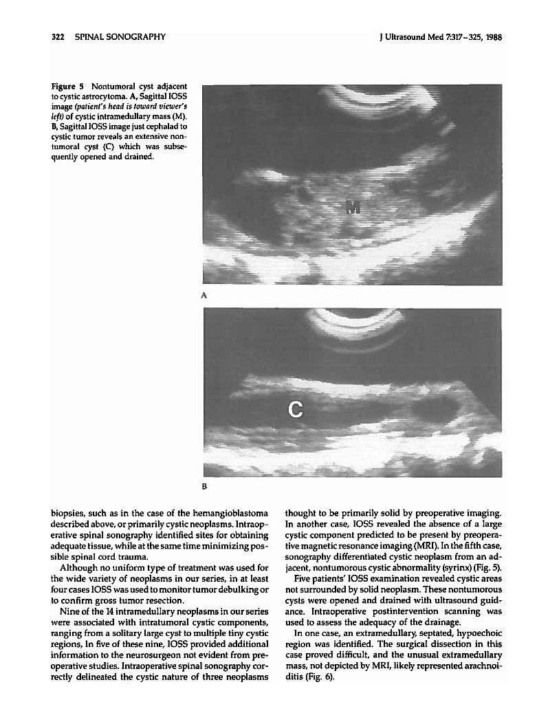
Figure 5 Nontumoral cyst adjacent to cystic astrocytoma. A, Sagittal lOSS image (patient's head is toward viewer's left) of cystic intramedullary mass (M). 8, Sagittal lOSS image just cephalad to cystic tumor reveals an extensive non· tumoral cyst (C) which was subse· quently opened and drained.
A
B
biopsies, such as in the case of the hemangioblastoma described above, or primarily cystic neoplasms. Intraoperative spinal sonography identified sites for obtaining adequate tissue, while at the same time minimizing possible spinal cord trauma.
Although no uniform type of treatment was used for the wide variety of neoplasms in our series, in at least four cases lOSS was used to monitor tumor debulking or to confirm gross tumor resection.
Nine of the 14 intramedullary neoplasms in our series were associated with intratumoral cystic components, ranging from a solitary large cyst to multiple tiny cystic regions, In five of these nine, lOSS provided additional information to the neurosurgeon not evident from preoperative studies. Intraoperative spinal sonography correctly delineated the cystic nature of three neoplasms
J Ultrasound Med 7:317- 325, 1988
thought to be primarily solid by preoperative imaging. In another case, lOSS revealed the absence of a large cystic component predicted to be present by preoperative magnetic resonance imaging (MRI). In the fifth case, sonography differentiated cystic neoplasm from an adjacent, nontumorous cystic abnormality (syrinx) (Fig. 5).
Five patients' lOSS examination revealed cystic areas not surrounded by solid neoplasm. These nontumorous cysts were opened and drained with ultrasound guidance. Intraoperative postintervention scanning was used to assess the adequacy of the drainage.
In one case, an extramedullary, septated, hypoechoic region was identified. The surgical dissection in this case proved difficult, and the unusual extramedullary mass, not depicted by MRI, likely represented arachnoiditis (Fig. 6).

J Ultrasound Med 7:317- 325, 1988
A
0
DISCUSSION
Intraoperative spinal sonography is gaining greater acceptance as a valuable adjunct in surgery for a variety of abnormalities.1- 10 Previous reports on the role of sonography in the evaluation of intramedullary tumors are limited by small patient groups. 3-' Our experience with a larger patient population with a wide variety of lesions reemphasizes many of the previously reported uses of JOSS, suggests additional benefits, and illustrates potential pitfalls.
PLATT ET AL 323
Figure 6 Arachonoiditis complicating surgery for intramedullary neoplasm. A, Sagittal lOSS image (patient's head is toward viewer's left) of hypoechoic septated mass-like area (M), with anterior displacement of spinal cord (C). B, Transverse lOSS image (patient's right is toward viewer's left) shows thickened septae (arrows) within the mass-like region. Displaced spinal cord (C) is noted anteriorly to the left. This area was very difficult to dissect and was thought to be arachnoiditis at surgery.
Clearly lOSS is useful in the localization of intrame+ dullary neoplasms before dural opening. In nine of our cases the initial sonographic examination revealed the need to extend the laminectomy either cranially or caudally. At the same time, it enabled the neurosurgeon to limit the laminectomy as much as possible, thereby decreasing the chance of progressive or long-term deformity of the spine.
A second definite benefit of lOSS was definition of tumor extent. This analysis was based on delineation of tumor margins and identification of the central canal

324 SPINAL SONOGRAPHY
echo. In 11 of our 14 patients good tumor border definition was present on the ultrasound exam. Subsequently, total tumor resection was attempted in six patients. However, sharp tumor margins do not necessarily indicate that a resection can be performed, because long segment tumors may be sharply delineated. Identification of tumor borders by lOSS, however, does indicate that complete resection can be considered unless contraindicated for other reasons. In two cases, no recognizable tumor border was evident by lOSS, which correlated well with the surgical·pathologic findings of extensive infiltrating neoplasm not amenable to surgical resection. We encountered only one case in which lOSS incorrectly depicted tumor extent. A focal ependymoma appeared as an extensive spinal cord tumor due to cord edema, which is echogenic similar to its appearance in the brain. 11 It is interesting to note that in a recent report edema was described as being hypoechoic. 12 This appearance is atypical in the neural axis and is different from our experience. In that report, the authors claimed that ultrasound could distinguish between edema and tumor, where MR could not.12 Clearly, this distinction was not possible in our case.
A third dear benefit of lOSS is to identify and localize cystic and solid components of neoplasms. Poser, in an autopsy series, reported 31% of spinal intramedullary neoplasms were associated with cord cavitation (cysts).13
Sixty-four percent (9 of 14) of spinal cord neoplasms in our series were associated with cysts, similar to a recent report that found 53% of intramedullary tumors had cystic regions.6 Five of our neoplasms were associated with cord cavitation not completely surrounded by tumor, apparently representing nontumorous cysts. It is interesting to note that all five patients with nontu· morous cysts had, in addition, a cystic component to their intramedullary neoplasms. No totally solid tumor was associated with a nontumorous type cyst. This observation suggests that a more thorough search for cystic regions, cephalad and caudal to the primary neoplasm, should be performed in patients with cystic neoplasms. A wide variety of cystic abnormalities, ranging from a solitary large cyst to innumerable tiny cystic regions, were associated with the tumors in our series. Fifty·six percent (5 of 9) of the cystic neoplasms in our series, were more accurately characterized by lOSS than preoperative imaging (CT or MRl). The most com· mon benefit was delineation of the primarily cystic nature of a tumor thought to be solid preoperatively. In no case was MRI or CT better than lOSS at demonstrating the cystic components of neoplasms. The one primarily cystic tumor incorrectly characterized by lOSS was a teratoma. The echogenic appearing cysts were filled with thick, creamy fluid and desquamated tissue. This case points out a potential pitfall in sonographic identi· fication of cystic structures. The cyst may be falsely
J Ultrasound Med 7:317- 325, 1988
deemed solid, if filled with material producing multiple reflective interfaces. This neoplasm was also incorrectly deemed solid by preoperative imaging studies.
A fourth benefit of lOSS is in the guidance and monitoring of interventional procedures performed during surgery. Intraoperative spinal sonography frequently played a prominent role in guiding diagnostic tumor biopsies. Ultrasound guidance was invaluable in tumors where a specific site was required for biopsy, such as a tumor nodule or a cyst wall. Accurate guidance reduced the number of biopsies required, thereby minimizing possible complications. Intraoperative spinal sonography also allowed the neurosurgeon to avoid specific areas of the cord, such as the enlarged blood vessel seen in the case of a spinal hemangioblastoma. The other major procedure in which lOSS proved beneficial was aspiration or drainage of cystic structures. Three intra· tumoral cysts were successfully aspirated under ultrasound guidance, and aspiration of one cyst relieved the degree of cord swelling. All five patients with nontumoral cysts underwent sonographically guided opening and drainage of these cysts. Intraoperative spinal sonography guided cyst puncture confirmed proper needle position and monitored adequacy of drainage.
Previously published data indicate that intramedullary tumors exhibit only a slight difference in echogenicity compared to normal spinal cord tissue. 3 The intra· medullary tumors in our series, however, were invariably more echogenic than normal cord.
Prediction of exact tumor cell type based solely on lOSS could not in general be performed. One neoplasm did exhibit findings allowing for exact tumor diagnosis. This spinal hemangioblastoma was noted to contain an enlarged tortuous blood vessel, with obvious flow at real-time examination. The appearance of spinal hemangioblastoma has been previously described.10
Intraoperative spinal sonography may also reveal unexpected findings not evident in preoperative studies. This benefit was seen in one case in which the intra· medullary neoplasm was associated with a septated ex· tramedullary mass. The spinal cord was displaced anteriorly as demonstrated sonographically. Surgical dissection in this case was difficuh, and the mass was thought to be secondary to arachnoiditis. It was not evident on preoperative MRI.
REFERENCES
1 Knake JE, Chandler WF, McGillicuddy JE, et aJ: Intraoperative sonography of intraspinal tumors: initial experience. AJNR 4:1199, 1983
2 Dohrmann GJ, Rubin JM: Intraoperative ultrasound imaging of the spinal cord syringomyelia, cysts, and tumors - a preliminary report. Surg Neurol 18;395, 1982

J Ultrasound Med 7:317-325, 1988
3. Quencer RM, Montalvo BM, Green BA, et al: Intraoperative spinal sonography of soft-tissue masses of the spinal cord and spinal canal. AJR 143:1307, 1984
4. Hutchins WM, Vogelzang RL, Neiman HL, et al: Differentiation of tumor from syringohydromyelia: intraoperative neurosonography of the spinal cord. Radiology 151:171, 1984
5. Enzmann DR, Irwin KM, Silverberg GD, eta!: Spinal cord tumor imaging with Cf and sonography. AJNR 6:95, 1985
6. Goy AM, Pinto RS, Raghavendra BN, et al: Intramedullary spinal cord tumors: MR imaging, with emphasis on associated cysts. Radiology 161:381, 1986
7. Rubin JM, Dohrmann GJ: Work in progress. Intraoperative ultrasonography of the spine. Radiology 146:173, 1983
8. Quencer RM, Morse BMM, Green BA, et al: Intraoperative spinal sonography: adjunct to metrizamide CT in the as-
PLATT ET AL 325
sessment and surgical decompression of post-traumatic spinal cord cysts. AJNR 5:71, 1984; AJR 142:593, 1984
9. I<nake JE, Gabrielsen TO, Chandler WF, et al: Real-time sonography during surgery. Radiology 151:461, 1984
10. Sanders WP, Ausman Jl, Dujouny M, et al: Ultrasonic features of two cases of spinal hemangioblastoma. Surg Neurol 26:453, 1986
11. Smith SJ, Vogelzang RL, Marzano MI. et al: Brain edema: ultrasound examination. Radiology 155:379, 1985
12. Post MJD, Quencer RM, Green BA, et al: Intramedullary spinal cord metastases, mainly of nonneurogenic origin. AJR 148:1015, 1987
13. Poser CM: The relationship between syringomyelia and neoplasm. In: American Lecture Series No. 262: Ameri· can Lectures in Neurology. Springfield, IL, Thomas, 1956