Embed Size (px)
Citation preview
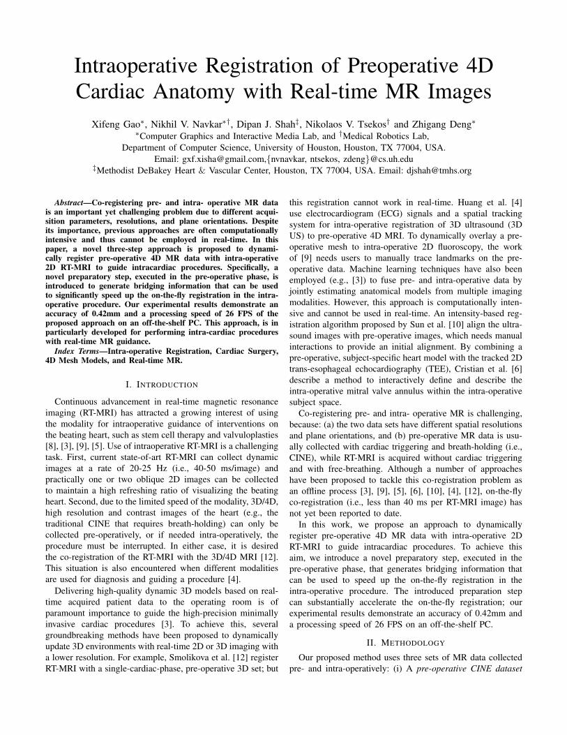
Intraoperative Registration of Preoperative 4DCardiac Anatomy with Real-time MR Images
Xifeng Gao⇤, Nikhil V. Navkar⇤†, Dipan J. Shah‡, Nikolaos V. Tsekos† and Zhigang Deng⇤⇤Computer Graphics and Interactive Media Lab, and †Medical Robotics Lab,
Department of Computer Science, University of Houston, Houston, TX 77004, USA.Email: [email protected],{nvnavkar, ntsekos, zdeng}@cs.uh.edu
‡Methodist DeBakey Heart & Vascular Center, Houston, TX 77004, USA. Email: [email protected]
Abstract—Co-registering pre- and intra- operative MR datais an important yet challenging problem due to different acqui-sition parameters, resolutions, and plane orientations. Despiteits importance, previous approaches are often computationallyintensive and thus cannot be employed in real-time. In thispaper, a novel three-step approach is proposed to dynami-cally register pre-operative 4D MR data with intra-operative2D RT-MRI to guide intracardiac procedures. Specifically, anovel preparatory step, executed in the pre-operative phase, isintroduced to generate bridging information that can be usedto significantly speed up the on-the-fly registration in the intra-operative procedure. Our experimental results demonstrate anaccuracy of 0.42mm and a processing speed of 26 FPS of theproposed approach on an off-the-shelf PC. This approach, is inparticularly developed for performing intra-cardiac procedureswith real-time MR guidance.
Index Terms—Intra-operative Registration, Cardiac Surgery,4D Mesh Models, and Real-time MR.
I. INTRODUCTION
Continuous advancement in real-time magnetic resonanceimaging (RT-MRI) has attracted a growing interest of usingthe modality for intraoperative guidance of interventions onthe beating heart, such as stem cell therapy and valvuloplasties[8], [3], [9], [5]. Use of intraoperative RT-MRI is a challengingtask. First, current state-of-art RT-MRI can collect dynamicimages at a rate of 20-25 Hz (i.e., 40-50 ms/image) andpractically one or two oblique 2D images can be collectedto maintain a high refreshing ratio of visualizing the beatingheart. Second, due to the limited speed of the modality, 3D/4D,high resolution and contrast images of the heart (e.g., thetraditional CINE that requires breath-holding) can only becollected pre-operatively, or if needed intra-operatively, theprocedure must be interrupted. In either case, it is desiredthe co-registration of the RT-MRI with the 3D/4D MRI [12].This situation is also encountered when different modalitiesare used for diagnosis and guiding a procedure [4].
Delivering high-quality dynamic 3D models based on real-time acquired patient data to the operating room is ofparamount importance to guide the high-precision minimallyinvasive cardiac procedures [3]. To achieve this, severalgroundbreaking methods have been proposed to dynamicallyupdate 3D environments with real-time 2D or 3D imaging witha lower resolution. For example, Smolikova et al. [12] registerRT-MRI with a single-cardiac-phase, pre-operative 3D set; but
this registration cannot work in real-time. Huang et al. [4]use electrocardiogram (ECG) signals and a spatial trackingsystem for intra-operative registration of 3D ultrasound (3DUS) to pre-operative 4D MRI. To dynamically overlay a pre-operative mesh to intra-operative 2D fluoroscopy, the workof [9] needs users to manually trace landmarks on the pre-operative data. Machine learning techniques have also beenemployed (e.g., [3]) to fuse pre- and intra-operative data byjointly estimating anatomical models from multiple imagingmodalities. However, this approach is computationally inten-sive and cannot be used in real-time. An intensity-based reg-istration algorithm proposed by Sun et al. [10] align the ultra-sound images with pre-operative images, which needs manualinteractions to provide an initial alignment. By combining apre-operative, subject-specific heart model with the tracked 2Dtrans-esophageal echocardiography (TEE), Cristian et al. [6]describe a method to interactively define and describe theintra-operative mitral valve annulus within the intra-operativesubject space.
Co-registering pre- and intra- operative MR is challenging,because: (a) the two data sets have different spatial resolutionsand plane orientations, and (b) pre-operative MR data is usu-ally collected with cardiac triggering and breath-holding (i.e.,CINE), while RT-MRI is acquired without cardiac triggeringand with free-breathing. Although a number of approacheshave been proposed to tackle this co-registration problem asan offline process [3], [9], [5], [6], [10], [4], [12], on-the-flyco-registration (i.e., less than 40 ms per RT-MRI image) hasnot yet been reported to date.
In this work, we propose an approach to dynamicallyregister pre-operative 4D MR data with intra-operative 2DRT-MRI to guide intracardiac procedures. To achieve thisaim, we introduce a novel preparatory step, executed in thepre-operative phase, that generates bridging information thatcan be used to speed up the on-the-fly registration in theintra-operative procedure. The introduced preparation stepcan substantially accelerate the on-the-fly registration; ourexperimental results demonstrate an accuracy of 0.42mm anda processing speed of 26 FPS on an off-the-shelf PC.
II. METHODOLOGY
Our proposed method uses three sets of MR data collectedpre- and intra-operatively: (i) A pre-operative CINE dataset
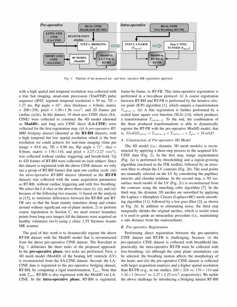
Fig 1
LONG AXIS
CINE
(LA-CINE)
SHORT AXIS
CINE
(SA-CINE)
REAL-TIME
Breath-hold (RT-BH)
Intra-operative Pre-operative
4D MODEL
(Mod4D)
STEP 1: BRIDGING
(Tpre)
STEP 2: COARSE
(Tintra-1)
REAL-TIME
Free Breathing (RT-FB)
Step 3: FINE-
TUNNED (Tintra-2)
Mod4D to
LA-CINE to
RT-BH
Mod4D to
RT-BH to
RT-FB
Fig. 1. Pipeline of the proposed pre- and intra- operative MR registration approach.
with a high spatial and temporal resolution was collected witha true fast imaging, stead-state precession (TrueFISP) pulsesequence (iPAT, segment temporal resolution = 39 ms, TE =1.27 ms, flip angle = 82�, slice thickness = 4.0mm, matrix= 200⇥256, pixel = 1.56⇥1.56 mm
2, and 20 frames percardiac cycle). In this dataset, 19 short axis CINE slices (SA-CINE) were collected to construct the 4D model (denotedas Mod4D), and long axis CINE slices (LA-CINE) werecollected for the first registration step. (ii) A pre-operative RT-MRI bridging dataset (denoted as the RT-BH dataset), witha high temporal but low spatial resolution which is the bestresolution we could achieve for real-time imaging (time perimage = 45.6 ms, TE = 0.98 ms, flip angle = 71�, slice =8.0mm, matrix = 176⇥110, and pixel = 2.27⇥2.27 mm
2),was collected without cardiac triggering and breath-hold. Upto 420 frames of RT-BH were collected on each subject. Sincethis dataset is registered with the above CINE dataset, we onlyuse a group of RT-BH frames that span one cardiac cycle. (iii)An intra-operative RT-MRI dataset (denoted as the RT-FBdataset) was collected with the same acquisition parametersas RT-BH, without cardiac triggering and with free breathing.We select the LA slice in the above three cases (i), (ii), and (iii)because of the following two reasons: 1) based on the analysisin [15], to minimize differences between the RT-BH and RT-FB sets so that the heart mainly translates along and rotatesaround without significant out-of-plane motion, 2) to performcoarse registration in Section C, we need extract boundarypoints from long-axis images.All the datasets were acquired onhealthy volunteers (n=3) using a clinic 1.5T Siemens AvantoMR scanner.
The goal of this work is to dynamically register the aboveRT-FB dataset with the Mod4D model that is reconstructedfrom the above pre-operative CINE dataset. The flowchart inFig. 1 delineates the three tasks of the proposed approach.In the pre-operative phase, two tasks are performed. First, a4D mesh model (Mod4D) of the beating left ventricle (LV)is reconstructed from the SA-CINE dataset. Second, the LA-CINE data is registered to the pre-operative bridging dataset,RT-BH, by computing a rigid transformation, Tpre. Note thatwith Tpre, RT-BH is also registered with the Mod4D via LA-CINE. In the intra-operative phase, RT-BH is registered,
frame-by-frame, to RT-FB. This intra-operative registration isperformed in a two-phase protocol: (i) A coarse registrationbetween RT-BH and RT-FB is performed by the iterative clos-est point (ICP) algorithm [1], which outputs a transformationTintra�1. (ii) A fine registration is further performed by ascaled least square cost function (SLS) [14], which producesa transformation Tintra�2. To the end, the combination ofthe three produced transformations is able to dynamicallyregister the RT-FB with the pre-operative Mod4D model, thatis, Mod4Dintra = Tintra�2 ⇥ Tintra�1 ⇥ Tpre ⇥Mod4D.
A. Construction of Pre-operative 4D Model
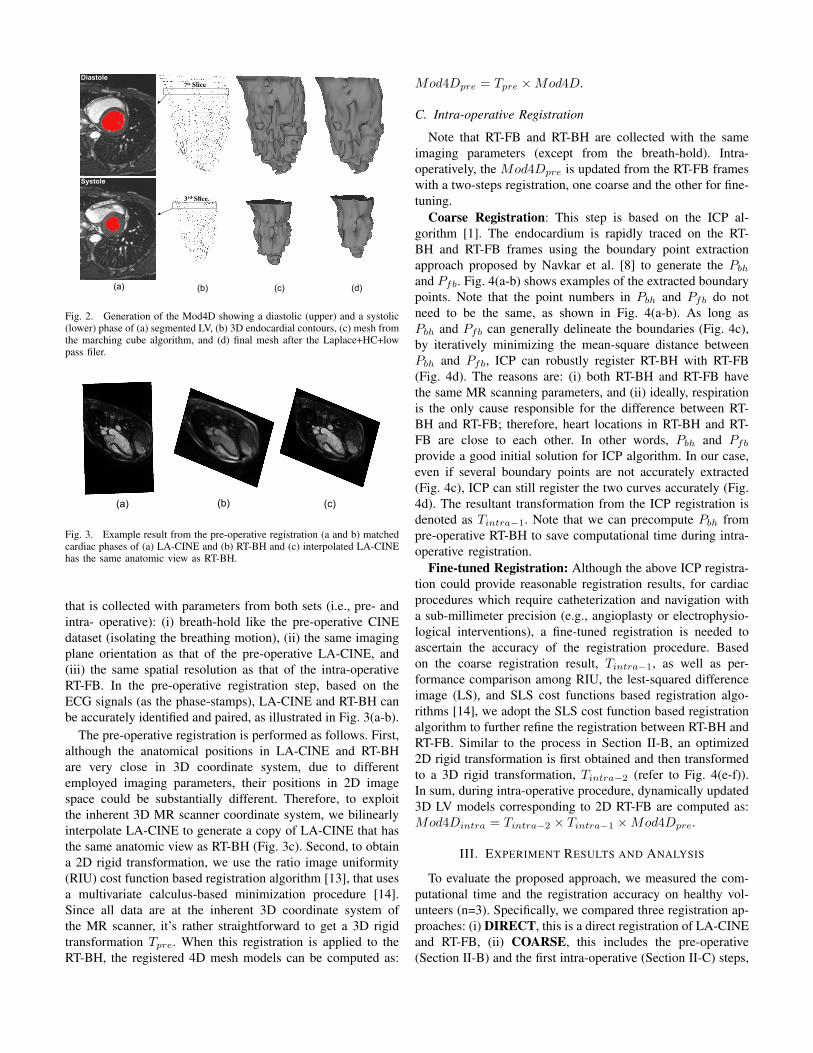
The 4D model (i.e., dynamic 3D mesh models) is recon-structed by applying a three-step process to the acquired SA-CINE data (Fig. 2). In the first step, image segmentation(Fig. 2a) is performed by thresholding and a region-growingalgorithm (enclosed in the ITK toolkit), followed by an inver-sion filter to obtain the LV contours (Fig. 2b). The seed pointsare manually selected on the LV by considering the papillarymuscles and chordae tendinae. In the second step, a 3D iso-surface mesh model of the LV (Fig. 2c) is reconstructed fromthe contours using the marching cube algorithm [7]. In thethird step, the dynamic 3D meshes are smoothed by applyingthe Laplace + Humphrey Classes (Laplace+HC) mesh smooth-ing algorithm [11], followed by a low pass filter [2], as shownin Fig. 2d. In addition to eliminating noise, the third stepmarginally shrinks the original meshes, which is useful whenit is used to guide an intracardiac procedure (i.e., maintaininga safe distance from the endocardium).
B. Pre-operative Registration
Performing direct registration between the pre-operativeCINE dataset and RT-FB is challenging, because: (i) thepre-operative CINE dataset is collected with breathhold but,practically, the intra-operative RT-FB must be collected withfree-breathing; (ii) although the same plane orientations canbe selected, the breathing motion affects the morphology ofthe heart; and (iii) the pre-operative CINE dataset is collectedwith larger acquisition matrices and a higher spatial resolutionthan RT-FB (e.g., in our studies, 200⇥ 256 vs. 176⇥ 110 and1.56⇥1.56mm
2 vs. 2.27⇥2.27mm
2, respectively). We tacklethe above challenge by introducing a bridging dataset RT-BH
(a) (b) (c) (d)
Diastole
Systole
3rd Slice
7th Slice
Fig. 2. Generation of the Mod4D showing a diastolic (upper) and a systolic(lower) phase of (a) segmented LV, (b) 3D endocardial contours, (c) mesh fromthe marching cube algorithm, and (d) final mesh after the Laplace+HC+lowpass filer.
(a) (b) (c)
Fig. 3. Example result from the pre-operative registration (a and b) matchedcardiac phases of (a) LA-CINE and (b) RT-BH and (c) interpolated LA-CINEhas the same anatomic view as RT-BH.
that is collected with parameters from both sets (i.e., pre- andintra- operative): (i) breath-hold like the pre-operative CINEdataset (isolating the breathing motion), (ii) the same imagingplane orientation as that of the pre-operative LA-CINE, and(iii) the same spatial resolution as that of the intra-operativeRT-FB. In the pre-operative registration step, based on theECG signals (as the phase-stamps), LA-CINE and RT-BH canbe accurately identified and paired, as illustrated in Fig. 3(a-b).
The pre-operative registration is performed as follows. First,although the anatomical positions in LA-CINE and RT-BHare very close in 3D coordinate system, due to differentemployed imaging parameters, their positions in 2D imagespace could be substantially different. Therefore, to exploitthe inherent 3D MR scanner coordinate system, we bilinearlyinterpolate LA-CINE to generate a copy of LA-CINE that hasthe same anatomic view as RT-BH (Fig. 3c). Second, to obtaina 2D rigid transformation, we use the ratio image uniformity(RIU) cost function based registration algorithm [13], that usesa multivariate calculus-based minimization procedure [14].Since all data are at the inherent 3D coordinate system ofthe MR scanner, it’s rather straightforward to get a 3D rigidtransformation Tpre. When this registration is applied to theRT-BH, the registered 4D mesh models can be computed as:
Mod4Dpre = Tpre ⇥Mod4D.
C. Intra-operative Registration
Note that RT-FB and RT-BH are collected with the sameimaging parameters (except from the breath-hold). Intra-operatively, the Mod4Dpre is updated from the RT-FB frameswith a two-steps registration, one coarse and the other for fine-tuning.
Coarse Registration: This step is based on the ICP al-gorithm [1]. The endocardium is rapidly traced on the RT-BH and RT-FB frames using the boundary point extractionapproach proposed by Navkar et al. [8] to generate the Pbh
and Pfb. Fig. 4(a-b) shows examples of the extracted boundarypoints. Note that the point numbers in Pbh and Pfb do notneed to be the same, as shown in Fig. 4(a-b). As long asPbh and Pfb can generally delineate the boundaries (Fig. 4c),by iteratively minimizing the mean-square distance betweenPbh and Pfb, ICP can robustly register RT-BH with RT-FB(Fig. 4d). The reasons are: (i) both RT-BH and RT-FB havethe same MR scanning parameters, and (ii) ideally, respirationis the only cause responsible for the difference between RT-BH and RT-FB; therefore, heart locations in RT-BH and RT-FB are close to each other. In other words, Pbh and Pfb
provide a good initial solution for ICP algorithm. In our case,even if several boundary points are not accurately extracted(Fig. 4c), ICP can still register the two curves accurately (Fig.4d). The resultant transformation from the ICP registration isdenoted as Tintra�1. Note that we can precompute Pbh frompre-operative RT-BH to save computational time during intra-operative registration.
Fine-tuned Registration: Although the above ICP registra-tion could provide reasonable registration results, for cardiacprocedures which require catheterization and navigation witha sub-millimeter precision (e.g., angioplasty or electrophysio-logical interventions), a fine-tuned registration is needed toascertain the accuracy of the registration procedure. Basedon the coarse registration result, Tintra�1, as well as per-formance comparison among RIU, the lest-squared differenceimage (LS), and SLS cost functions based registration algo-rithms [14], we adopt the SLS cost function based registrationalgorithm to further refine the registration between RT-BH andRT-FB. Similar to the process in Section II-B, an optimized2D rigid transformation is first obtained and then transformedto a 3D rigid transformation, Tintra�2 (refer to Fig. 4(e-f)).In sum, during intra-operative procedure, dynamically updated3D LV models corresponding to 2D RT-FB are computed as:Mod4Dintra = Tintra�2 ⇥ Tintra�1 ⇥Mod4Dpre.
III. EXPERIMENT RESULTS AND ANALYSIS
To evaluate the proposed approach, we measured the com-putational time and the registration accuracy on healthy vol-unteers (n=3). Specifically, we compared three registration ap-proaches: (i) DIRECT, this is a direct registration of LA-CINEand RT-FB, (ii) COARSE, this includes the pre-operative(Section II-B) and the first intra-operative (Section II-C) steps,
(b) RT-FB
Diastole
Systole
(a) RT-BH (c) RT-BH & RT-FB (d) RT-BH & RT-FB (e) RT-FB & Model (f) RT-FB & Model
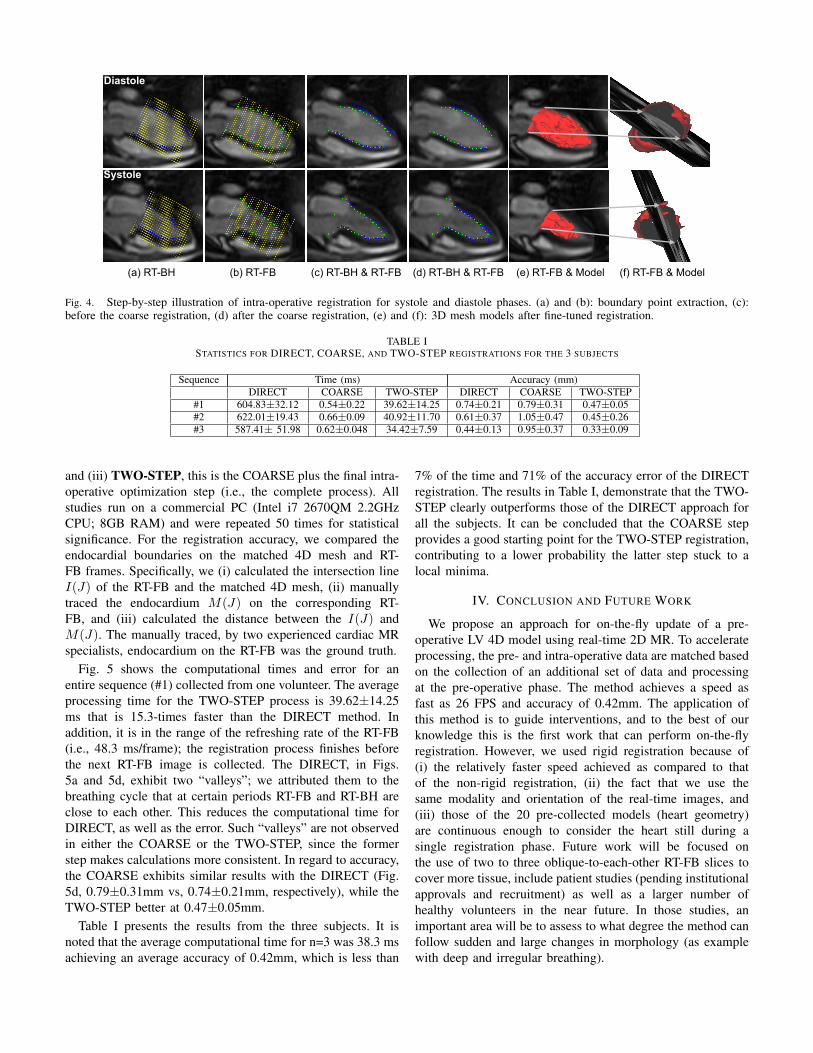
Fig. 4. Step-by-step illustration of intra-operative registration for systole and diastole phases. (a) and (b): boundary point extraction, (c):before the coarse registration, (d) after the coarse registration, (e) and (f): 3D mesh models after fine-tuned registration.
TABLE ISTATISTICS FOR DIRECT, COARSE, AND TWO-STEP REGISTRATIONS FOR THE 3 SUBJECTS
Sequence Time (ms) Accuracy (mm)DIRECT COARSE TWO-STEP DIRECT COARSE TWO-STEP
#1 604.83±32.12 0.54±0.22 39.62±14.25 0.74±0.21 0.79±0.31 0.47±0.05#2 622.01±19.43 0.66±0.09 40.92±11.70 0.61±0.37 1.05±0.47 0.45±0.26#3 587.41± 51.98 0.62±0.048 34.42±7.59 0.44±0.13 0.95±0.37 0.33±0.09
and (iii) TWO-STEP, this is the COARSE plus the final intra-operative optimization step (i.e., the complete process). Allstudies run on a commercial PC (Intel i7 2670QM 2.2GHzCPU; 8GB RAM) and were repeated 50 times for statisticalsignificance. For the registration accuracy, we compared theendocardial boundaries on the matched 4D mesh and RT-FB frames. Specifically, we (i) calculated the intersection lineI(J) of the RT-FB and the matched 4D mesh, (ii) manuallytraced the endocardium M(J) on the corresponding RT-FB, and (iii) calculated the distance between the I(J) andM(J). The manually traced, by two experienced cardiac MRspecialists, endocardium on the RT-FB was the ground truth.
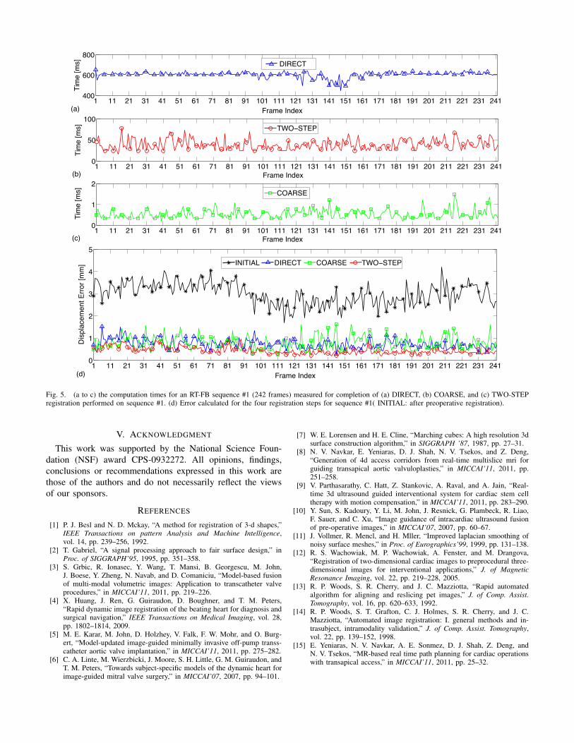
Fig. 5 shows the computational times and error for anentire sequence (#1) collected from one volunteer. The averageprocessing time for the TWO-STEP process is 39.62±14.25ms that is 15.3-times faster than the DIRECT method. Inaddition, it is in the range of the refreshing rate of the RT-FB(i.e., 48.3 ms/frame); the registration process finishes beforethe next RT-FB image is collected. The DIRECT, in Figs.5a and 5d, exhibit two “valleys”; we attributed them to thebreathing cycle that at certain periods RT-FB and RT-BH areclose to each other. This reduces the computational time forDIRECT, as well as the error. Such “valleys” are not observedin either the COARSE or the TWO-STEP, since the formerstep makes calculations more consistent. In regard to accuracy,the COARSE exhibits similar results with the DIRECT (Fig.5d, 0.79±0.31mm vs, 0.74±0.21mm, respectively), while theTWO-STEP better at 0.47±0.05mm.
Table I presents the results from the three subjects. It isnoted that the average computational time for n=3 was 38.3 msachieving an average accuracy of 0.42mm, which is less than
7% of the time and 71% of the accuracy error of the DIRECTregistration. The results in Table I, demonstrate that the TWO-STEP clearly outperforms those of the DIRECT approach forall the subjects. It can be concluded that the COARSE stepprovides a good starting point for the TWO-STEP registration,contributing to a lower probability the latter step stuck to alocal minima.
IV. CONCLUSION AND FUTURE WORK
We propose an approach for on-the-fly update of a pre-operative LV 4D model using real-time 2D MR. To accelerateprocessing, the pre- and intra-operative data are matched basedon the collection of an additional set of data and processingat the pre-operative phase. The method achieves a speed asfast as 26 FPS and accuracy of 0.42mm. The application ofthis method is to guide interventions, and to the best of ourknowledge this is the first work that can perform on-the-flyregistration. However, we used rigid registration because of(i) the relatively faster speed achieved as compared to thatof the non-rigid registration, (ii) the fact that we use thesame modality and orientation of the real-time images, and(iii) those of the 20 pre-collected models (heart geometry)are continuous enough to consider the heart still during asingle registration phase. Future work will be focused onthe use of two to three oblique-to-each-other RT-FB slices tocover more tissue, include patient studies (pending institutionalapprovals and recruitment) as well as a larger number ofhealthy volunteers in the near future. In those studies, animportant area will be to assess to what degree the method canfollow sudden and large changes in morphology (as examplewith deep and irregular breathing).
1 11 21 31 41 51 61 71 81 91 101 111 121 131 141 151 161 171 181 191 201 211 221 231 241400
600
800
Frame Index
Tim
e [m
s]
DIRECT
(a)
! !! "! #! $! %! &! '! (! )! !*! !!! !"! !#! !$! !%! !&! !'! !(! !)! "*! "!! ""! "#! "$!*
%*
!**
+,-./0-12
/
/
345-./678.9
+:;!<+=>
?@A
1 11 21 31 41 51 61 71 81 91 101 111 121 131 141 151 161 171 181 191 201 211 221 231 2410
1
2
Frame Index
Tim
e [
ms]
COARSE
(c)
! !! "! #! $! %! &! '! (! )! !*! !!! !"! !#! !$! !%! !&! !'! !(! !)! "*! "!! ""! "#! "$!*
!
"
#
$
%
+,-./0123/4
56789-:/./2;0<,,=,0>..?
0
0
1@1A1BC 51D<EA EFBDG< AHF!GA<I
J3K
Fig. 5. (a to c) the computation times for an RT-FB sequence #1 (242 frames) measured for completion of (a) DIRECT, (b) COARSE, and (c) TWO-STEPregistration performed on sequence #1. (d) Error calculated for the four registration steps for sequence #1( INITIAL: after preoperative registration).
V. ACKNOWLEDGMENT
This work was supported by the National Science Foun-dation (NSF) award CPS-0932272. All opinions, findings,conclusions or recommendations expressed in this work arethose of the authors and do not necessarily reflect the viewsof our sponsors.
REFERENCES
[1] P. J. Besl and N. D. Mckay, “A method for registration of 3-d shapes,”IEEE Transactions on pattern Analysis and Machine Intelligence,vol. 14, pp. 239–256, 1992.
[2] T. Gabriel, “A signal processing approach to fair surface design,” inProc. of SIGGRAPH’95, 1995, pp. 351–358.
[3] S. Grbic, R. Ionasec, Y. Wang, T. Mansi, B. Georgescu, M. John,J. Boese, Y. Zheng, N. Navab, and D. Comaniciu, “Model-based fusionof multi-modal volumetric images: Application to transcatheter valveprocedures,” in MICCAI’11, 2011, pp. 219–226.
[4] X. Huang, J. Ren, G. Guiraudon, D. Boughner, and T. M. Peters,“Rapid dynamic image registration of the beating heart for diagnosis andsurgical navigation,” IEEE Transactions on Medical Imaging, vol. 28,pp. 1802–1814, 2009.
[5] M. E. Karar, M. John, D. Holzhey, V. Falk, F. W. Mohr, and O. Burg-ert, “Model-updated image-guided minimally invasive off-pump transs-catheter aortic valve implantation,” in MICCAI’11, 2011, pp. 275–282.
[6] C. A. Linte, M. Wierzbicki, J. Moore, S. H. Little, G. M. Guiraudon, andT. M. Peters, “Towards subject-specific models of the dynamic heart forimage-guided mitral valve surgery,” in MICCAI’07, 2007, pp. 94–101.
[7] W. E. Lorensen and H. E. Cline, “Marching cubes: A high resolution 3dsurface construction algorithm,” in SIGGRAPH ’87, 1987, pp. 27–31.
[8] N. V. Navkar, E. Yeniaras, D. J. Shah, N. V. Tsekos, and Z. Deng,“Generation of 4d access corridors from real-time multislice mri forguiding transapical aortic valvuloplasties,” in MICCAI’11, 2011, pp.251–258.
[9] V. Parthasarathy, C. Hatt, Z. Stankovic, A. Raval, and A. Jain, “Real-time 3d ultrasound guided interventional system for cardiac stem celltherapy with motion compensation,” in MICCAI’11, 2011, pp. 283–290.
[10] Y. Sun, S. Kadoury, Y. Li, M. John, J. Resnick, G. Plambeck, R. Liao,F. Sauer, and C. Xu, “Image guidance of intracardiac ultrasound fusionof pre-operative images,” in MICCAI’07, 2007, pp. 60–67.
[11] J. Vollmer, R. Mencl, and H. Mller, “Improved laplacian smoothing ofnoisy surface meshes,” in Proc. of Eurographics’99, 1999, pp. 131–138.
[12] R. S. Wachowiak, M. P. Wachowiak, A. Fenster, and M. Drangova,“Registration of two-dimensional cardiac images to preprocedural three-dimensional images for interventional applications,” J. of MagneticResonance Imaging, vol. 22, pp. 219–228, 2005.
[13] R. P. Woods, S. R. Cherry, and J. C. Mazziotta, “Rapid automatedalgorithm for aligning and reslicing pet images,” J. of Comp. Assist.Tomography, vol. 16, pp. 620–633, 1992.
[14] R. P. Woods, S. T. Grafton, C. J. Holmes, S. R. Cherry, and J. C.Mazziotta, “Automated image registration: I. general methods and in-trasubject, intramodality validation,” J. of Comp. Assist. Tomography,vol. 22, pp. 139–152, 1998.
[15] E. Yeniaras, N. V. Navkar, A. E. Sonmez, D. J. Shah, Z. Deng, andN. V. Tsekos, “MR-based real time path planning for cardiac operationswith transapical access,” in MICCAI’11, 2011, pp. 25–32.