Embed Size (px)
Citation preview
Intramolecular Valence and Spin Interaction in meso and racDiastereomers of a p-Quinonoid-Bridged Diruthenium
Complex
Doyel Kumbhakar,† Biprajit Sarkar,‡ Somnath Maji,† Shaikh M. Mobin,† Jan Fiedler,§
Francisco A. Urbanos,⊥ Reyes Jimenez-Aparicio,*,⊥ Wolfgang Kaim,*,‡ andGoutam Kumar Lahiri*,†
Department of Chemistry, Indian Institute of Technology Bombay, Powai, Mumbai-400076,India, Institut fur Anorganische Chemie, UniVersitat Stuttgart, Pfaffenwaldring 55, D-70550
Stuttgart, Germany, J. HeyroVsky Institute of Physical Chemistry, V.V.i., Academy of Sciences ofthe Czech Republic, DolejskoVa 3, CZ-18223 Prague, Czech Republic, and Departamento de
Quımica Inorganica, Facultad de Ciencias Quımicas, UniVersidad Complutense, CiudadUniVersitaria, E-28040 Madrid, Spain
Received September 11, 2008; E-mail: [email protected]; [email protected]
Abstract: The complexes meso- and rac-[(acac)2Ru(µ-L)Ru(acac)2]n, 1 and 2, where L2- ) 1,4-dioxido-2,3-bis(3,5-dimethylpyrazol-1′-yl)benzene and acac- ) 2,4-pentanedionato, were characterized structurally,magnetically, electrochemically, and spectroscopically as well as spectroelectrochemically (UV-vis-NIR,EPR) in the accessible redox states (n ) 0, +, -, 2-). Due to steric interference, the neutral compoundscontain a severely twisted L2- bridging ligand with 43-48° dihedral angles between the planes of thehydroquinone dianion and those of the ortho positioned pyrazolyl substituents. The difference betweenmeso and rac isomers is rather pronounced in terms of the redox potentials (easier oxidation and reductionof the rac form 2) and with respect to the absorption spectra of the oxidized states. Susceptibility and EPRmeasurements confirm the {RuIII(µ-L2-)RuIII} configuration of the neutral species, showing J values of -37and -21 cm-1 for the spin-spin interaction between the ca. 7.75 Å separated metal centers in 1 and 2,respectively. Two-step reduction involves the metals and produces RuIIIRuII mixed-valent monoanions withcomproportionation constants of ca. 104, with RuIII-type EPR signals, and with broad intervalence chargetransfer bands at about 1200-1500 nm absorption maximum, suggesting localized valence (class II).Oxidation produces intense near-infrared absorption at 892 (1+) or 1027 nm (2+) and narrow isotropicEPR spectra at g ≈ 2.005, signifying unprecedented spin localization at the p-semiquinone bridge. Theseresults are not compatible with an (L2-)-bridged {RuIVRuIII} situation nor with an {RuIII(µ-L•-)RuIII} three-spinarrangement with up-down-up spin configuration in the ground state, which would result in metal-centeredspin through antiferromagnetic coupling between the adjacent individual spins. Only the {RuIII(µ-L•-)RuIII}situation, with up-up-down spin configuration, leads to ligand-centered resulting spin through the strongantiferromagnetic coupling between the remote metal spins, an unusual situation which is favored herebecause of weakened metal-radical coupling resulting from the pyrazolyl/p-semiquinone twist.
Introduction
As naturally occurring redox-active molecules, the quinonesare widely distributed, functioning in vital electron transportprocesses where they often interact with transition metal ions1a,b
or exhibit specific toxicity.1c ortho-Quinone-containing pros-thetic groups in metallo-quinoproteins (Chart 1) are well knownin the form of pyrrolo-quinoline-quinone (PQQ), tryptophan-tryptophyl-quinone (TTQ), topaquinone (TPQ), and lysine-tyrosyl-quinone (LTQ).2 Catechols as 2e-/2H+-reduced o-quino-
nes are being investigated as antioxidants (polyphenols), asneurotransmitters (catecholamines), and as precursors of melaninpigments.3
However, para-quinones (Chart 2) such as vitamin K deriva-tives, ubiquinones, or plastoquinones also play many importantroles in energy conversion (photosynthesis, respiration) andinformation transfer.3,4
In order to rationalize the intricate electronic interactionsbetween transition metal ions and quinone redox systems inbiochemical environments, there have been considerable inves-
† Indian Institute of Technology Bombay.‡ Universitat Stuttgart.§ Academy of Sciences of the Czech Republic.⊥ Universidad Complutense.
(1) (a) Nohl, H.; Jordan, W.; Youngman, R. I. AdV. Free Rad. Biol. Med.1986, 2, 211. (b) Thomson, S. D. Naturally Occurring Quinones IV;Springer: The Netherlands, 1996. (c) Bolton, J. L.; Trush, M. A.;Penning, T. M.; Dryhurst, G.; Monks, T. J. Chem. Res. Toxicol. 2000,13, 135.
(2) (a) Duine, J. A.; Jongejan, J. A. In Bioinorganic Catalysis; Reedijk,J., Ed.; Marcel Dekker: New York, 1993; p 447. (b) Klinman, J. P.Proc. Natl. Acad. Sci. U.S.A. 2001, 98, 705. (c) Mure, M.; Mills, S. A.;Klinman, J. P. Biochemistry 2002, 41, 9269. (d) Wang, S. X.;Nakamura, N.; Murell, M.; Klinman, J. P.; Sanders-Loehr, J. J. Biol.Chem. 1997, 272, 28841. (e) Mure, M.; Wang, S. X.; Klinman, J. P.J. Am. Chem. Soc. 2003, 125, 6113. (f) Anthony, C. Arch. Biochem.Biophys. 2004, 428, 2. (g) Anthony, C. Biochem. J. 1996, 320, 697.
Published on Web 11/20/2008
10.1021/ja807043s CCC: $40.75 2008 American Chemical Society J. AM. CHEM. SOC. 2008, 130, 17575–17583 9 17575
tigations at the molecular level of metal complexes with O,O′-chelating o-quinonoid ligands, particularly in assigning thevalence state distribution at the metal-quinone interface.5
However, despite the biochemical significance,1,4 far fewerresults have been reported for the coordination chemistry ofp-quinonoid ligands,6,7 including those with combined o,p-quinone functions.6r-w The present study is aimed at exploringthe electronic structural aspects of two diastereomeric diruthe-nium compounds, [(acac)2Ru(µ-L)Ru(acac)2], 1 and 2 (Scheme1), where L2- is the two-electron-reduced p-quinonoid ligand1,4-dioxido-2,3-bis(3,5-dimethylpyrazol-1′-yl)benzene and acac-
) acetylacetonate ) 2,4-pentanedionate. Although 1,4-dioxido-2,5-bis(pyrazol-1′-yl)benzene, (L′)2-, has been used previously
for the formation of polynuclear complexes,6a-d including[(bpy)2Ru(µ-L′)Ru(bpy)2]n,7e the new compounds 1 and 2represent the first set of polynuclear metal complexes bridgedby L2-.
The preferential stabilization of ruthenium ions in theparamagnetic 3+ oxidation state in 1 or 2 via the σ-donationeffect of anionic acac- and L2- introduces the possibility ofstudying L2--mediated magnetic exchange between RuIII centers.In addition, the initially ambivalent electronic situations in theone-electron-reduced form {Ru(µ-L)Ru}-, as well as in the one-electron-oxidized state {Ru(µ-L)Ru}+, were targets of ourinvestigation.Thepresentworkthusdescribesthemetal-ligand-metalvalence state distributions in both the isolated neutral systemsand the spectroelectrochemically accessible charged redox statesof diastereomeric 1n and 2n (n ) 0, +, -, 2-), using X-raystructure analysis, SQUID susceptometry, OTTLE spectroelec-trochemistry, and EPR.
Results and Discussion
Synthesis and Characterization. The diastereomeric com-plexes [meso (∆Λ), 1; rac (∆∆/ΛΛ), 2]8 were isolated fromthe reaction of RuII(acac)2(CH3CN)2 with 1,4-dihydroxy-2,3-bis(3,5-dimethylpyrazol-1′-yl)benzene (H2L) in the presence of
(3) Izumi, Y.; Sawada, H.; Sakka, N.; Yamamoto, N.; Kume, T.; Katsuki,H.; Shimohama, S.; Akaike, A. J. Neurosci. Res. 2005, 79, 849.
(4) (a) Furie, B.; Bouchard, B. A.; Furie, B. C. Blood 1999, 93, 1798. (b)Meganathan, R. Vitam. Horm. 2001, 61, 173. (c) He, M.; Sheldon,P. J.; Sherman, D. H. Proc. Natl. Acad. Sci. U.S.A. 2001, 98, 926.
(5) (a) Pierpont, C. G.; Lange, C. W. Prog. Inorg. Chem. 1994, 41, 331.(b) Pierpont, C. G. Coord. Chem. ReV. 2001, 219-221, 415. (c) Lever,A. B. P.; Gorelsky, S. I. Coord. Chem. ReV. 2000, 208, 153. (d)Gorelsky, S. I.; Lever, A. B. P.; Ebadi, M. Coord. Chem. ReV. 2002,230, 97. (e) Dei, A.; Gatteschi, D.; Sangregorio, C.; Sorace, L. Acc.Chem. Res. 2004, 37, 827. (f) Pierpont, C. G.; Attia, A. S. Collect.Czech. Chem. Commun. 2001, 66, 33. (g) DelMedico, A.; Dodsworth,E. S.; Lever, A. B. P.; Pietro, W. J. Inorg. Chem. 2004, 43, 2654. (h)da Cunha, C. J.; Dodsworth, E. S.; Monteiro, M. A.; Lever, A. B. P.Inorg. Chem. 1999, 38, 5399. (i) Salmonsen, R. B.; Abelleira, A.;Clarke, M. J. Inorg. Chem. 1984, 23, 385. (j) Patra, S.; Sarkar, B.;Mobin, S. M.; Kaim, W.; Lahiri, G. K. Inorg. Chem. 2003, 43, 6469.(k) Remenyi, C.; Kaupp, M. J. Am. Chem. Soc. 2005, 127, 11399. (l)Maji, S.; Patra, S.; Chakraborty, S.; Mobin, S. M.; Janardanan, D.;Sunoj, R. B.; Lahiri, G. K. Eur. J. Inorg. Chem. 2007, 314. (m) Dei,A.; Gatteschi, D.; Pardi, L. Inorg. Chim. Acta 1991, 189, 125. (n)Frantz, S.; Rall, J.; Hartenbach, I.; Schleid, T.; Zalis, S.; Kaim, W.Chem. Eur. J. 2004, 19, 149.
Chart 1
Chart 2
Scheme 1
17576 J. AM. CHEM. SOC. 9 VOL. 130, NO. 51, 2008
A R T I C L E S Kumbhakar et al.
NEt3 as a base under aerobic conditions, followed by chro-matographic separation using a neutral alumina column. Theligand L2- bridges two complex fragments {Ru(acac)2}+, eachthrough one anionic O- and one neutral pyrazolyl N donor,forming two six-membered chelate rings.
The combined electron donor effects from chelating terminalacac- ligands and from the doubly deprotonated anionichydroquinone moiety (L2-) facilitate the isolation of rutheniumin the paramagnetic 3+ oxidation state in 1 and 2 under aerobicreaction conditions, as observed similarly for many other{Ru(acac)2} derivatives.9,10
The neutral diastereomers 1 and 2 are identified by theirmicroanalytical data and by mass spectrometry (see Experi-mental Section). The paramagnetic 1 and 2 exhibit magneticmoments of about 2.4 µB at 298 K, which implies antiferro-magnetic coupling of RuIII (cf. below). Compounds 1 and 2display complex EPR spectra in acetonitrile at 110 K as wellas 1H NMR resonances (at 298 K) over a wide range, between+30 and -40 ppm in CDCl3 (Figures S1 and S2, SupportingInformation), resulting from paramagnetic contact shift.11 Therac isomer 2 shows signals due to four CH(acac), eightCH3(acac), two CH(quinone), four CH3(pyrazolyl), and twoCH(pyrazolyl) protons corresponding to the full molecule,whereas the meso isomer 1 displays signals equivalent to halfof the molecule.
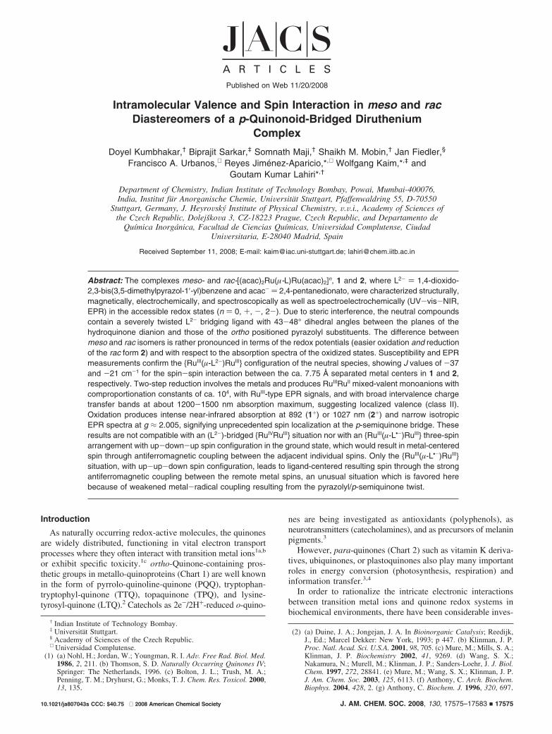
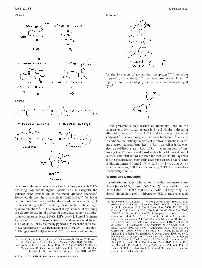
Crystal Structures. The isomeric identity of 1 and 2 corre-sponding to meso and rac, respectively, has been evidenced by
their crystal structures (Figures 1 and 2). Selected crystal-lographic parameters and comparative bond lengths and bondangles are given in Tables 1 and 2, respectively. Each ruthe-nium(III) ion is bonded to the bridging ligand (L2-) through apair of N,O- donors to form a six-membered chelate ring. TheRu(1)O5N and Ru(2)O5N arrangements in 1 and 2 are distortedoctahedral. The Ru-O distances with the bridging L2- in 1[1.974(6)/1.965(6) Å] and in 2 [1.976(2)/1.983(2) Å] lie withina close range and are slightly shorter than the Ru-O bondsinvolving the acac- groups (average 2.000 and 2.018 Å in 1and 2, respectively). This observation suggests a less balancedcharge from the chelate donor atoms in L2-, as evident fromthe Ru-N distances: the longest bonds for each ruthenium(III)ion are those to the pyrazolyl nitrogen atoms of the bridgingL2- ligand at 2.069(7)/2.070(8) Å for 1 and at 2.059(3)/2.072(3)Å for 2. In general, the bond lengths involving the metal ionsof 1 and 2 are in the expected range.12 The intramolecular
(6) (a) Kretz, T.; Bats, J. W.; Losi, S.; Wolf, B.; Lerner, H. -W.; Lang,M.; Zanello, P.; Wagner, M Dalton Trans. 2006, 4914. (b) Margraf,G.; Kretz, T.; de Biani, F. F.; Laschi, F.; Losi, S.; Zanello, P.; Bats,J. W.; Wolf, B.; Removic-Langer, K.; Lang, M.; Prokofiev, A.;Assmus, W.; Lerner, H.-W.; Wagner, M. Inorg. Chem. 2006, 45, 1277.(c) Wolf, B.; Bruehl, A.; Pashchenko, V.; Removic-Langer, K.; Kretz,T.; Bats, J. W.; Lerner, H.-W.; Wagner, M.; Salguero, A.; Saha-Dasgupta, T.; Rahaman, B.; Valenti, R.; Lang, M. C. R. Chim. 2007,10, 109. (d) Dinnebier, R.; Lerner, H.-W.; Ding, L.; Shankland, K.;David, W. I. F.; Stephens, P. W.; Wagner, M. Z. Anorg. Allg. Chem.2002, 628, 310. (e) Siri, O.; Braunstein, P. Chem. Commun. 2000,2223. (f) Elduque, A.; Garces, Y.; Oro, L. A.; Pinillos, M. T.;Tiripicchio, A.; Ugozzoli, F. J. Chem. Soc., Dalton Trans. 1996, 2155.(g) Dei, A.; Gatteschi, D.; Pardi, L.; Russo, U. Inorg. Chem. 1991,30, 2589. (h) Calvo, M. A.; Lanfredi, A. M. M.; Oro, L. A.; Pinillos,M. T.; Tejel, C.; Tiripicchio, A.; Ugozzoli, F. Inorg. Chem. 1993, 32,1147. (i) Johnston, R. F.; Holwerda, R. A. Inorg. Chem. 1985, 24,153. (j) Vlcek, A. A.; Danzlik, J. Inorg. Chem. 1967, 6, 2053. (k)Liu, S.; Shaikh, S. N.; Zubieta, J. Inorg. Chem. 1989, 28, 723. (l)Folgado, J. V.; Ibanez, R.; Coronado, E.; Beltran, D.; Savariault, J. M.;Galy, J. Inorg. Chem. 1988, 27, 19. (m) Lloret, F.; Julve, M.; Faus,J.; Solans, X.; Journaux, Y.; Morgenstern-Badarau, I. Inorg. Chem.1990, 29, 2232. (n) Pierpont, C. G.; Francescons, L. C.; Hendrickson,D. N. Inorg. Chem. 1978, 17, 3470. (o) Wrobleski, J. T.; Brown, D. B.Inorg. Chem. 1979, 18, 498. (p) Mathur, P.; Dismukes, G. C. J. Am.Chem. Soc. 1983, 105, 7093. (q) Cornago, P.; Escolastico, C.; SantaMaria, M. D.; Claramunt, R. M.; Carmona, D.; Esteban, M.; Oro, L. A.;Foces-Foces, C.; Llamas-Saiz, L.; Elguero, J. J. Organomet. Chem.1994, 467, 293. (r) Min, K. S.; Rheingold, A. L.; DiPasquale, A.;Miller, J. S. Inorg. Chem. 2006, 45, 6135. (s) Min, K. S.; DiPasquale,A.; Rheingold, A. L.; Miller, J. S. Inorg. Chem. 2007, 46, 1048. (t)Guo, D.; McCusker, J. K. Inorg. Chem. 2007, 46, 3257. (u) Tao, J.;Maruyama, H.; Sato, O. J. Am. Chem. Soc. 2006, 128, 1790. (v) Min,K. S.; DiPasquale, A. G.; Golen, J. A.; Rheingold, A. L.; Miller, J. S.J. Am. Chem. Soc. 2007, 129, 2360. (w) Cotton, F. A.; Jin, J.-Y.; Li,Z.; Murillo, C. A.; Reibenspies, J. H. Chem. Commun. 2008, 211.
(7) Ruthenium complexes: (a) Ernst, S.; Haenel, P.; Jordanov, J.; Kaim,W. V.; Kasack, V.; Roth, E. J. Am. Chem. Soc. 1989, 111, 1733. (b)Ghumaan, S.; Mukherjee, S.; Kar, S.; Roy, D.; Mobin, S. M.; Sunoj,R. B.; Lahiri, G. K. Eur. J. Inorg. Chem. 2006, 21, 4426. (c) Maji, S.;Sarkar, B.; Mobin, S. M; Fiedler, J.; Urbanos, F. A.; Jimenez-Aparicio,R.; Kaim, W.; Lahiri, G. K. Inorg. Chem. 2008, 47, 5204. (d) Kar, S.;Sarkar, B.; Ghumaan, S.; Janardanan, D.; van Slageren, J.; Fiedler,J.; Puranik, V. G.; Sunoj, R. B.; Kaim, W.; Lahiri, G. K. Chem. Eur.J. 2005, 11, 4901. (e) Keyes, T. E.; Forster, R. J.; Jayaweera, P. M.;Coates, C. G.; McGarvey, J. J.; Vos, J. G. Inorg. Chem. 1998, 37,5925. (f) Bond, A. M.; Marken, F.; Williams, C. T.; Beattie, D. A.;Keyes, T. E.; Forster, R. J.; Vos, J. G. J. Phys. Chem. B 2000, 104,1977. (g) Ward, M. D. Inorg. Chem. 1996, 35, 1712. (h) Dei, A.;Gatteschi, D.; Pardi, L. Inorg. Chem. 1990, 29, 1442. (i) Ura, Y.; Sato,Y.; Shiotsuki, M.; Suzuki, T.; Wada, K.; Kondo, T.; Mitsudo, T. -AOrganometallics 2003, 22, 77.
(8) (a) Keene, F. R. Chem. Soc. ReV. 1998, 27, 185. (b) D’Alessandro,D. M.; Keene, F. R. Chem. Phys. 2006, 324, 8.
(9) Kar, S.; Sarkar, B.; Ghumaan, S.; Roy, D.; Urbanos, F. A.; Fiedler,J.; Sunoj, R. B.; Jimenez-Aparicio, R.; Kaim, W.; Lahiri, G. K. Inorg.Chem. 2005, 44, 8715.
Figure 1. ORTEP diagram of 1. Ellipsoids are drawn at 50% probability.
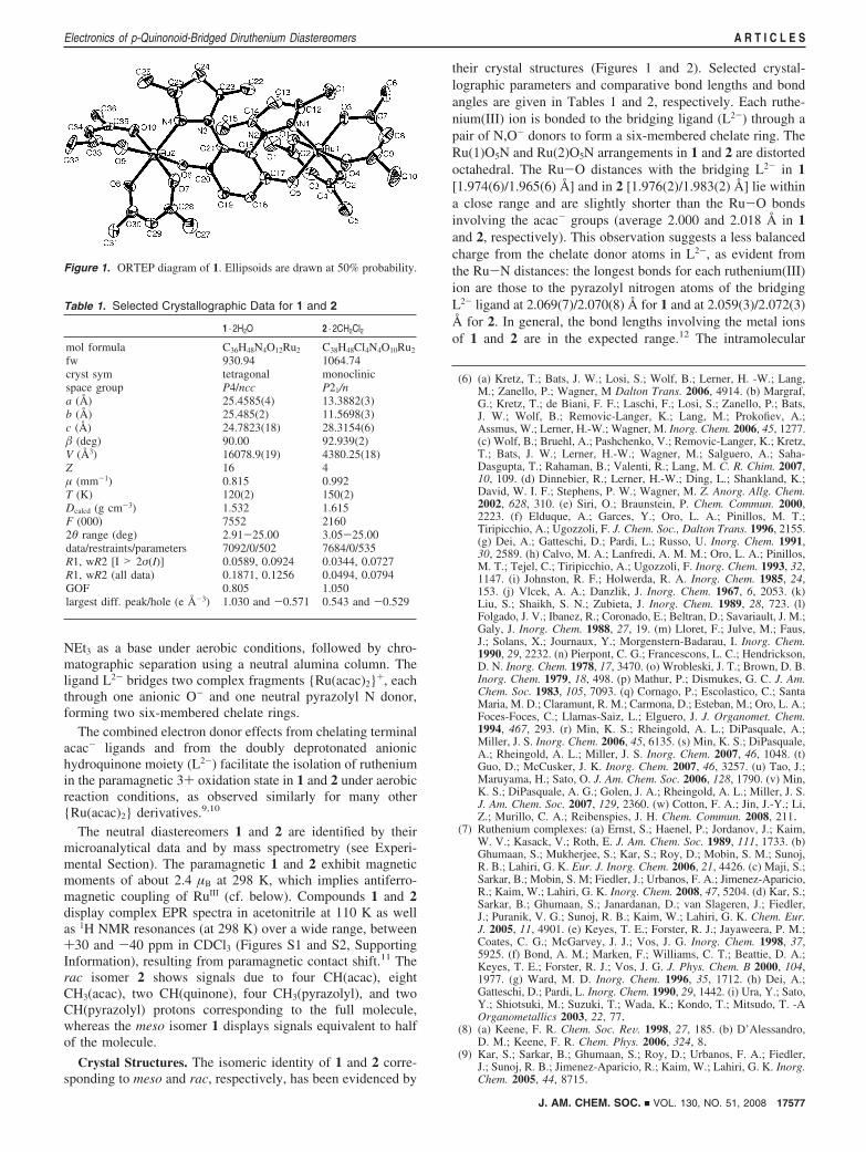
Table 1. Selected Crystallographic Data for 1 and 2
1 · 2H2O 2 · 2CH2Cl2
mol formula C36H48N4O12Ru2 C38H48Cl4N4O10Ru2
fw 930.94 1064.74cryst sym tetragonal monoclinicspace group P4/ncc P21/na (Å) 25.4585(4) 13.3882(3)b (Å) 25.485(2) 11.5698(3)c (Å) 24.7823(18) 28.3154(6)� (deg) 90.00 92.939(2)V (Å3) 16078.9(19) 4380.25(18)Z 16 4µ (mm-1) 0.815 0.992T (K) 120(2) 150(2)Dcalcd (g cm-3) 1.532 1.615F (000) 7552 21602θ range (deg) 2.91-25.00 3.05-25.00data/restraints/parameters 7092/0/502 7684/0/535R1, wR2 [I > 2σ(I)] 0.0589, 0.0924 0.0344, 0.0727R1, wR2 (all data) 0.1871, 0.1256 0.0494, 0.0794GOF 0.805 1.050largest diff. peak/hole (e Å-3) 1.030 and -0.571 0.543 and -0.529
J. AM. CHEM. SOC. 9 VOL. 130, NO. 51, 2008 17577
Electronics of p-Quinonoid-Bridged Diruthenium Diastereomers A R T I C L E S
distances between the two paramagnetic metal centers in 1 and2 are 7.709 and 7.771 Å, respectively.
The coordinated pyrazolyl rings are not coplanar with thep-quinone part of L2-. The angles between the planes comprisingthe benzo ring and the pyrazolyl ring N3,N4,C23,C24,C25 arearound 47.8° for both molecules, whereas the angles betweenthe former and the second pyrazolyl ring (N1,N2,C12,C13,C14)are 45.8° and 42.9° in 1 and 2, respectively. These valuesindicate a rather diminished π conjugation. The two pyrazolylrings of L2- are situated at angles of 46.3° and 54.7° in 1 and2, respectively.
Magnetic Properties. For both compounds, the change inmagnetization with increase in the magnetic field at 5 or 300 Kis linear, at least until 50 000 G. For both complexes, themagnetization values are lower than those predicted by theBrillouin function13 for two paramagnetic centers, S ) 1/2 (g )2.00) + 1/2 (g ) 2.00), suggesting the presence of antiferro-magnetic interactions, mainly at low temperatures (Figures S3and S4, Supporting Information).
The magnetic susceptibility curve of 1 versus temperatureshows a broad maximum at 69 K (Figure 3a), supporting thepresence of antiferromagnetic interactions. In addition, thesusceptibility curve shows a typical paramagnetic tail at verylow temperatures, ascribed to a small quantity of paramagnetic(S ) 1) impurity, which is tentatively attributed to a noncoupledphase of the same compound. The magnetic moment of 2.40µB at room temperature is close to that expected for the presenceof two isolated unpaired electrons associated with two RuIII ionsper molecule. The µeff decreases from 2.40 to 0.33 µB at 2 K,in accordance with an antiferromagnetic interaction.
The magnetic interaction between two RuIII (S ) 1/2) centersis given by the exchange spin Hamiltonian H ) -2JS1S2, whichleads to the analytical expression of eq 1 for the magneticsusceptibility.14
�) Ng2�2
kT2 exp(2J ⁄ kT)
1+ 3 exp(2J ⁄ kT)(1)
The presence of a temperature-independent paramagnetism(TIP) and that of a paramagnetic impurity (S ) 1) have beenconsidered, as shown in eq 2.
�′) (1-P)(�+TIP)+P2Ng2�2
3kT(2)
The terms N, g, �, k, J, and T in eqs 1 and 2 have the usualmeaning, and P is the mole fraction of the noncoupledparamagnetic impurity. The fit of the experimental data usingeq 2 gives good agreement between the experimental andcalculated curves (Figure 3a). The parameters obtained in the
(10) Sarkar, B.; Patra, S.; Fiedler, J.; Sunoj, R. B.; Janardanan, D.; Mobin,S. M.; Niemeyer, M.; Lahiri, G. K.; Kaim, W. Angew. Chem., Int.Ed. 2005, 44, 5655.
(11) (a) Patra, S.; Miller, T. A.; Sarkar, B.; Niemeyer, M.; Ward, M. D.;Lahiri, G. K. Inorg. Chem. 2003, 42, 4707. (b) Koiwa, T.; Masuda,Y.; Shono, J.; Kawamoto, Y.; Hoshino, Y.; Hashimoto, T.; Natarajan,K.; Shimizu, K. Inorg. Chem. 2004, 43, 6215. (c) Eaton, D. R. J. Am.Chem. Soc. 1965, 87, 3097. (d) Palmer, R. A.; Fay, R. C.; Piper, T. S.Inorg. Chem. 1964, 3, 875. (e) Holm, R. H.; Cotton, F. A. J. Am.Chem. Soc. 1958, 80, 5658. (f) Fay, R. C.; Piper, T. S. J. Am. Chem.Soc. 1963, 85, 500. (g) Chen, J.-L.; Zhang, X.-U.; Zhang, L.-Y.; Shi,L.X.; Chen, Z.-N. Inorg. Chem. 2005, 44, 1037.
(12) (a) Kar, S.; Chanda, N.; Mobin, S. M.; Datta, A.; Urbanos, F. A.;Puranik, V. G.; Jimenez-Aparicio, R.; Lahiri, G. K. Inorg. Chem. 2004,43, 4911. (b) Kar, S.; Chanda, N.; Mobin, S. M.; Urbanos, F. A.;Niemeyer, M.; Puranik, V. G.; Jimenez-Aparicio, R.; Lahiri, G. K.Inorg. Chem. 2005, 44, 1571. (c) Chao, G. K. J.; Sime, R. L.; Sime,R. J. Acta Crystallogr. 1973, B29, 2845. (d) Sarkar, B.; Laye, R. H.;Mondal, B.; Chakraborty, S.; Paul, R. L.; Jeffery, J. C.; Puranik, V. G.;Ward, M. D.; Lahiri, G. K. J. Chem. Soc., Dalton Trans. 2002, 2097.
(13) Kahn, O. Molecular Magnetism; VCH: Weinheim, 1993; p 107. (14) Carlin, R. L. Magnetochemistry, Springer-Verlag: Berlin, 1986; p 14.
Figure 2. ORTEP diagram of 2. Ellipsoids are drawn at 50% probability.
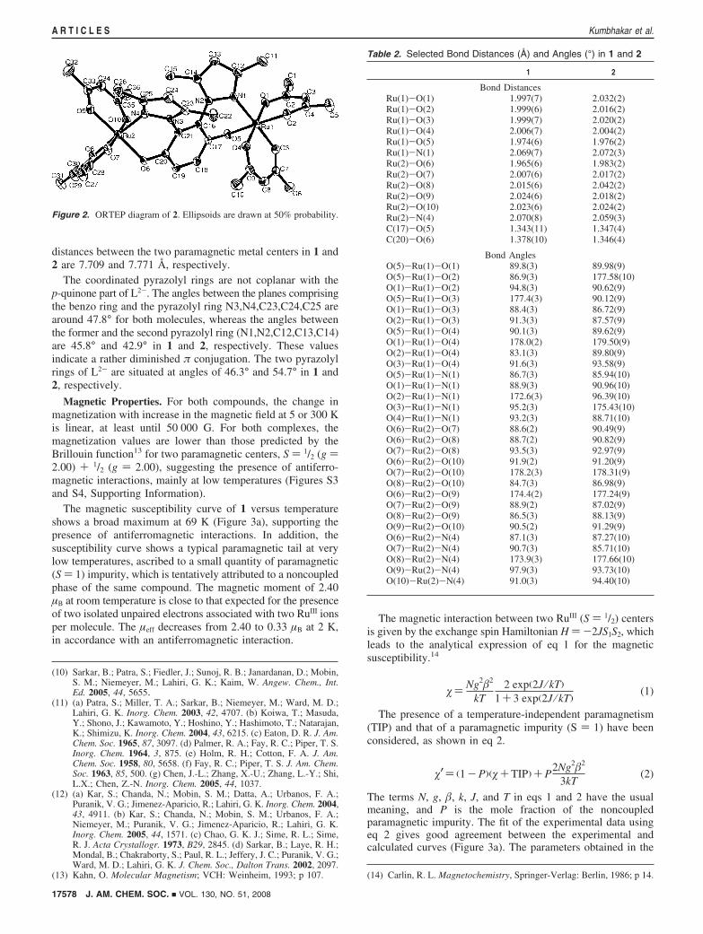
Table 2. Selected Bond Distances (Å) and Angles (°) in 1 and 2
1 2
Bond DistancesRu(1)-O(1) 1.997(7) 2.032(2)Ru(1)-O(2) 1.999(6) 2.016(2)Ru(1)-O(3) 1.999(7) 2.020(2)Ru(1)-O(4) 2.006(7) 2.004(2)Ru(1)-O(5) 1.974(6) 1.976(2)Ru(1)-N(1) 2.069(7) 2.072(3)Ru(2)-O(6) 1.965(6) 1.983(2)Ru(2)-O(7) 2.007(6) 2.017(2)Ru(2)-O(8) 2.015(6) 2.042(2)Ru(2)-O(9) 2.024(6) 2.018(2)Ru(2)-O(10) 2.023(6) 2.024(2)Ru(2)-N(4) 2.070(8) 2.059(3)C(17)-O(5) 1.343(11) 1.347(4)C(20)-O(6) 1.378(10) 1.346(4)
Bond AnglesO(5)-Ru(1)-O(1) 89.8(3) 89.98(9)O(5)-Ru(1)-O(2) 86.9(3) 177.58(10)O(1)-Ru(1)-O(2) 94.8(3) 90.62(9)O(5)-Ru(1)-O(3) 177.4(3) 90.12(9)O(1)-Ru(1)-O(3) 88.4(3) 86.72(9)O(2)-Ru(1)-O(3) 91.3(3) 87.57(9)O(5)-Ru(1)-O(4) 90.1(3) 89.62(9)O(1)-Ru(1)-O(4) 178.0(2) 179.50(9)O(2)-Ru(1)-O(4) 83.1(3) 89.80(9)O(3)-Ru(1)-O(4) 91.6(3) 93.58(9)O(5)-Ru(1)-N(1) 86.7(3) 85.94(10)O(1)-Ru(1)-N(1) 88.9(3) 90.96(10)O(2)-Ru(1)-N(1) 172.6(3) 96.39(10)O(3)-Ru(1)-N(1) 95.2(3) 175.43(10)O(4)-Ru(1)-N(1) 93.2(3) 88.71(10)O(6)-Ru(2)-O(7) 88.6(2) 90.49(9)O(6)-Ru(2)-O(8) 88.7(2) 90.82(9)O(7)-Ru(2)-O(8) 93.5(3) 92.97(9)O(6)-Ru(2)-O(10) 91.9(2) 91.20(9)O(7)-Ru(2)-O(10) 178.2(3) 178.31(9)O(8)-Ru(2)-O(10) 84.7(3) 86.98(9)O(6)-Ru(2)-O(9) 174.4(2) 177.24(9)O(7)-Ru(2)-O(9) 88.9(2) 87.02(9)O(8)-Ru(2)-O(9) 86.5(3) 88.13(9)O(9)-Ru(2)-O(10) 90.5(2) 91.29(9)O(6)-Ru(2)-N(4) 87.1(3) 87.27(10)O(7)-Ru(2)-N(4) 90.7(3) 85.71(10)O(8)-Ru(2)-N(4) 173.9(3) 177.66(10)O(9)-Ru(2)-N(4) 97.9(3) 93.73(10)O(10)-Ru(2)-N(4) 91.0(3) 94.40(10)
17578 J. AM. CHEM. SOC. 9 VOL. 130, NO. 51, 2008
A R T I C L E S Kumbhakar et al.
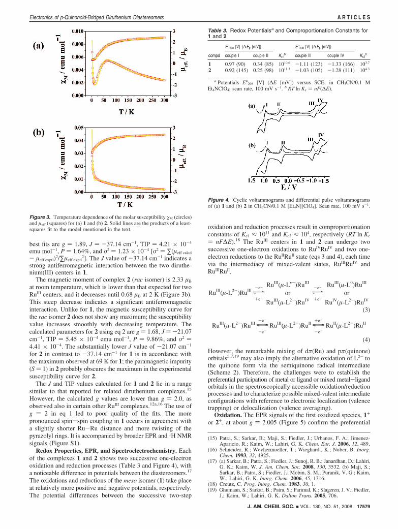
best fits are g ) 1.89, J ) -37.14 cm-1, TIP ) 4.21 × 10⁻⁴
emu mol-1, P ) 1.64%, and σ2 ) 1.23 × 10⁻⁴ [σ2 ) ∑(µeff calcd
- µeff exptl)2/∑µeff exptl2]. The J value of -37.14 cm-1 indicates a
strong antiferromagnetic interaction between the two diruthe-nium(III) centers in 1.
The magnetic moment of complex 2 (rac isomer) is 2.33 µB
at room temperature, which is lower than that expected for twoRuIII centers, and it decreases until 0.68 µB at 2 K (Figure 3b).This steep decrease indicates a significant antiferromagneticinteraction. Unlike for 1, the magnetic susceptibility curve forthe rac isomer 2 does not show any maximum; the susceptibilityvalue increases smoothly with decreasing temperature. Thecalculated parameters for 2 using eq 2 are g ) 1.68, J ) -21.07cm-1, TIP ) 5.45 × 10⁻⁴ emu mol-1, P ) 9.86%, and σ2 )4.41 × 10⁻⁴. The substantially lower J value of -21.07 cm-1
for 2 in contrast to -37.14 cm-1 for 1 is in accordance withthe maximum observed at 69 K for 1; the paramagnetic impurity(S ) 1) in 2 probably obscures the maximum in the experimentalsusceptibility curve for 2.
The J and TIP values calculated for 1 and 2 lie in a rangesimilar to that reported for related diruthenium complexes.15
However, the calculated g values are lower than g ) 2.0, asobserved also in certain other RuIII complexes.12a,16 The use ofg ) 2 in eq 1 led to poor quality of the fits. The morepronounced spin-spin coupling in 1 occurs in agreement witha slightly shorter Ru-Ru distance and more twisting of thepyrazolyl rings. It is accompanied by broader EPR and 1H NMRsignals (Figure S1).
Redox Properties, EPR, and Spectroelectrochemistry. Eachof the complexes 1 and 2 shows two successive one-electronoxidation and reduction processes (Table 3 and Figure 4), witha noticeable difference in potentials between the diastereomers.17
The oxidations and reductions of the meso isomer (1) take placeat relatively more positive and negative potentials, respectively.The potential differences between the successive two-step
oxidation and reduction processes result in comproportionationconstants of Kc1 ≈ 1011 and Kc2 ≈ 104, respectively (RT ln Kc
) nF∆E).18 The RuIII centers in 1 and 2 can undergo twosuccessive one-electron oxidations to RuIVRuIV and two one-electron reductions to the RuIIRuII state (eqs 3 and 4), each timevia the intermediacy of mixed-valent states, RuIIIRuIV andRuIIIRuII.
RuIII(µ-L2-)RuIII y\z-e-
+e-
RuIII(µ-L•-)RuIII
or
RuIII(µ-L2-)RuIV
y\z-e-
+e-
RuIII(µ-L0)RuIII
or
RuIV(µ-L2-)RuIV
(3)
RuIII(µ-L2-)RuIII y\z+e-
-e-RuIII(µ-L2-)RuII y\z
+e-
-e-RuII(µ-L2-)RuII
(4)
However, the remarkable mixing of dπ(Ru) and pπ(quinone)orbitals5,7,19 may also imply the alternative oxidation of L2- tothe quinone form via the semiquinone radical intermediate(Scheme 2). Therefore, the challenges were to establish thepreferential participation of metal or ligand or mixed metal-ligandorbitals in the spectroscopically accessible oxidation/reductionprocesses and to characterize possible mixed-valent intermediateconfigurations with reference to electronic localization (valencetrapping) or delocalization (valence averaging).
Oxidation. The EPR signals of the first oxidized species, 1+
or 2+, at about g ) 2.005 (Figure 5) confirm the preferential
(15) Patra, S.; Sarkar, B.; Maji, S.; Fiedler, J.; Urbanos, F. A.; Jimenez-Aparicio, R.; Kaim, W.; Lahiri, G. K. Chem. Eur. J. 2006, 12, 489.
(16) Schneider, R.; Weyhermueller, T.; Wieghardt, K.; Nuber, B. Inorg.Chem. 1993, 32, 4925.
(17) (a) Sarkar, B.; Patra, S.; Fiedler, J.; Sunoj, R. B.; Janardhan, D.; Lahiri,G. K.; Kaim, W. J. Am. Chem. Soc. 2008, 130, 3532. (b) Maji, S.;Sarkar, B.; Patra, S.; Fiedler, J.; Mobin, S. M.; Puranik, V. G.; Kaim,W.; Lahiri, G. K. Inorg. Chem. 2006, 45, 1316.
(18) Creutz, C. Prog. Inorg. Chem. 1983, 30, 1.(19) Ghumaan, S.; Sarkar, B.; Patra, S.; Parimal, K.; Slageren, J. V.; Fiedler,
J.; Kaim, W.; Lahiri, G. K. Dalton Trans. 2005, 706.
Figure 3. Temperature dependence of the molar susceptibility �M (circles)and µeff (squares) for (a) 1 and (b) 2. Solid lines are the products of a least-squares fit to the model mentioned in the text.
Table 3. Redox Potentialsa and Comproportionation Constants for1 and 2
E°298 [V] (∆Ep [mV]) E°298 [V] (∆Ep [mV])
compd couple I couple II Kc1b couple III couple IV Kc2
b
1 0.97 (90) 0.34 (85) 1010.6 -1.11 (123) -1.33 (166) 103.7
2 0.92 (145) 0.25 (98) 1011.3 -1.03 (105) -1.28 (111) 104.3
a Potentials E°298 [V] (∆E [mV]) versus SCE; in CH3CN/0.1 MEt4NClO4; scan rate, 100 mV s-1. b RT ln Kc ) nF(∆E).
Figure 4. Cyclic voltammograms and differential pulse voltammogramsof (a) 1 and (b) 2 in CH3CN/0.1 M [Et4N][ClO4]. Scan rate, 100 mV s-1.
J. AM. CHEM. SOC. 9 VOL. 130, NO. 51, 2008 17579
Electronics of p-Quinonoid-Bridged Diruthenium Diastereomers A R T I C L E S
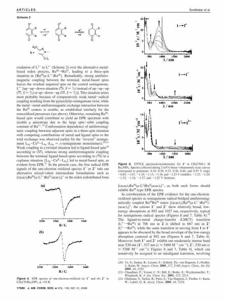
oxidation of L2- to L•- (Scheme 2) over the alternative metal-based redox process, RuIIIfRuIV, leading to a three-spinsituation in {RuIII(µ-L•-)RuIII}. Remarkably, strong antiferro-magnetic coupling between the terminal, metal-based spinsleaves the residual unpaired spin on the central semiquinone,L•- [up-up-down situation (vvV, S ) 1/2) instead of up-up-up(vvv, S ) 3/2) or up-down-up (vVv, S ) 1/2)]. This situation arisesmost probably because of comparatively weak metal-radicalcoupling resulting from the pyrazolyl/p-semiquinone twist, whilethe metal-metal antiferromagnetic exchange interaction betweenthe RuIII centers is sizable, as established similarly for thenonoxidized precursors (see above). Otherwise, remaining RuIII-based spin would contribute to yield an EPR spectrum withsizable g anisotropy due to the large spin-orbit couplingconstant of Ru3+.7a Conformation dependence of antiferromag-netic coupling between adjacent spins in a three-spin situationwith competing contributions of metal and ligand spins to thetotal exchange was observed earlier for the “reverse” arrange-ment LSq-CuII-LSq (LSq ) o-semiquinone monoimines).20,21
Weak coupling in a twisted situation led to ligand-based spin20
according to (vVv), whereas strong antiferromagnetic couplingbetween the terminal ligand-based spins according to (vvV) in acoplanar situation {LSq-CuII-LSq} led to metal-based spin, asevident from EPR.21 In the present case, the free radical EPRsignals of the one-electron oxidized species 1+ or 2+ rule outalternative mixed-valent intermediate formulations such as[(acac)2RuIII(µ-L2-)RuIV(acac)2]+ or the redox redistributed form
[(acac)2RuIII(µ-Lo)RuII(acac)2]+, as both such forms shouldexhibit RuIII-type EPR spectra.
In corroboration of the EPR evidence for the one-electron-oxidized species as semiquinone radical-bridged antiferromag-netically coupled RuIIIRuIII states [(acac)2{RuIII(µ-L•-)RuIII}-(acac)2]+, the cations 1+ and 2+ show relatively broad, low-energy absorptions at 892 and 1027 nm, respectively, typicalfor semiquinone radical species (Figures 6 and 7, Table 4).22
The ligand-to-metal charge-transfer (LMCT) transition(L2-fRuIII) at 706 nm in 2 is shifted to 687 nm in 2+
(L•-fRuIII), while the same transition in moving from 1 to 1+
appears to be obscured by the broad envelope of the low-energyabsorption centered at 892 nm (Figures 6 and 7, Table 4).Moreover, both 1+ and 2+ exhibit one moderately intense bandnear 530 nm [1+, 537 nm (ε ≈ 5400 M-1 cm-1); 2+, 530 nm (ε≈ 5300 M-1 cm-1); Figures 6 and 7, Table 4], which cantentatively be assigned to an intraligand transition, involving
(20) Ye, S.; Sarkar, B.; Lissner, F.; Schleid, Th.; van Slageren, J.; Fiedler,J.; Kaim, W. Angew. Chem. 2005, 117, 2140; Angew. Chem., Int. Ed.2005, 44, 2103.
(21) Chaudhuri, P.; Verani, C. N.; Bill, E.; Bothe, E.; Weyhermueller, T.;Wieghardt, K. J. Am. Chem. Soc. 2001, 123, 2213.
(22) Ghumaan, S.; Sarkar, B.; Patra, S.; Van Slageren, J.; Fiedler, J.; Kaim,W.; Lahiri, G. K. Inorg. Chem. 2005, 44, 3210.
Scheme 2
Figure 5. EPR spectra of one-electron-oxidized (a) 1+ and (b) 2+ inCH3CN/Bu4NPF6 at 110 K.
Figure 6. OTTLE spectroelectrochemistry for 1n in CH3CN/0.1 MBu4NPF6. Spectra collected during 2 mV/s linear voltammetric scan; curvescorrespond to potentials: 0.10, 0.30, 0.33, 0.36, 0.40, and 0.55 V (top);-0.85, -1.07, -1.10, -1.13, -1.16, and -1.23 V (middle); -1.23, -1.29,-1.32, -1.34, -1.37, and -1.52 V (bottom).
17580 J. AM. CHEM. SOC. 9 VOL. 130, NO. 51, 2008
A R T I C L E S Kumbhakar et al.
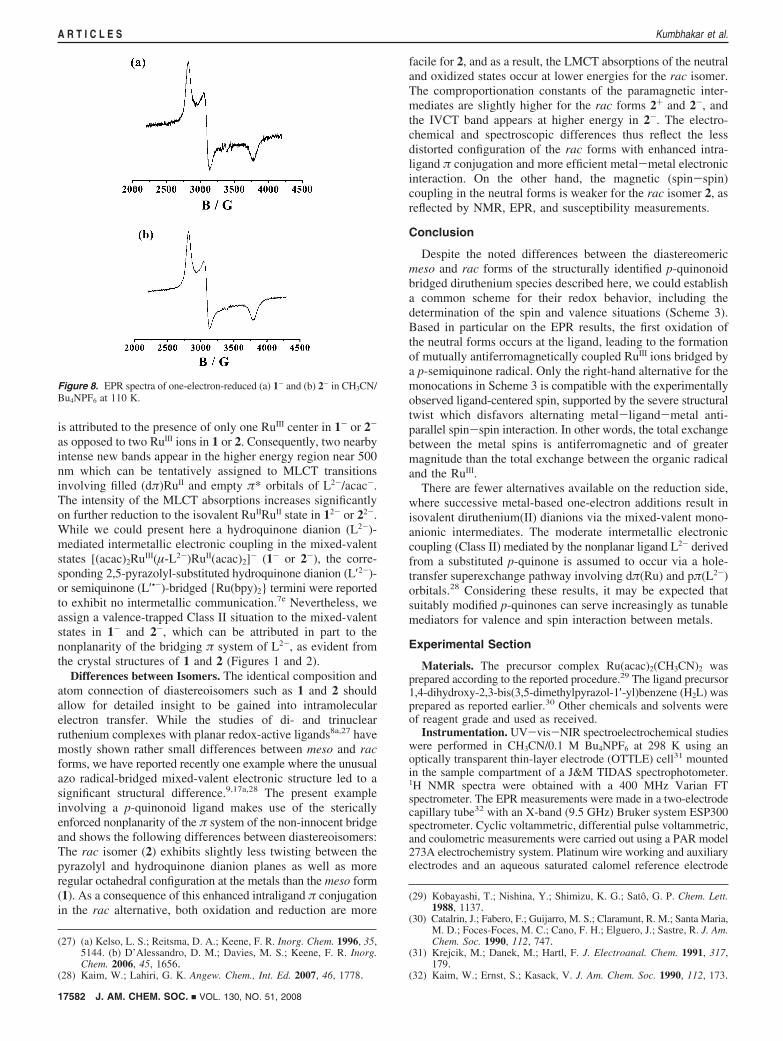
the SOMO of the semiquinone bridge. Although the two-electron-oxidized species (12+ and 22+) appear to be generatedreversibly on the cyclic voltammetry time scale (Figure 4), theywere not sufficiently stable on the electrolysis time scale. Inthe case of [(bpy)2RuII(µ-L′2-)RuII(bpy)2]2+, the first oxidationproduct was proposed to be [(bpy)2RuII(µ-L′•-)RuII(bpy)2]3+ viaspectroelectrochemistry and resonance Raman studies.7e Thisresult is probably favored through the RuII stabilization by thebpy ancillary ligands. In contrast, the one-electron oxidation of
[(acac)2RuIII(µ-L′′ 2-)RuIII(acac)2] (3, L′′ 2- ) 1,4-dioxido-9,10-anthraquinone) was found to take place at the metal, leading tothe formation of mixed-valent [(acac)2RuIII(µ-L′′ 2-)RuIV(acac)2]+
as suggested by EPR and by a long-wavelength RuIIIfRuIV
intervalence charge-transfer (IVCT) transition.7c
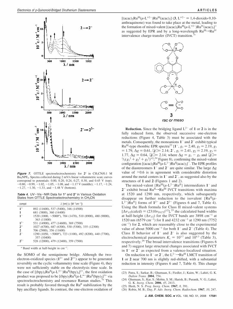
Reduction. Since the bridging ligand L2- of 1 or 2 is in thefully reduced form, the observed successive one-electronreductions (Figure 4, Table 3) must be associated with themetals. Consequently, the monoanions 1- and 2- exhibit typicalRuIII-type rhombic EPR spectra23 [1-, g1 ) 2.40, g2 ) 2.19, g3
) 1.79, ∆g ) 0.61, ⟨g⟩ ) 2.14; 2-, g1 ) 2.41, g2 ) 2.19, g3 )1.77, ∆g ) 0.64, ⟨g⟩ ) 2.14; where ∆g ) g1 - g3 and ⟨g⟩ )1/3(g1
2 + g22 + g3
2)1/2;24 Figure 8), confirming the mixed-valentconfiguration [(acac)2RuIII(µ-L2-)RuII(acac)2]-. The EPR profilesof the diastereomers 1- and 2- are quite similar. The large ∆gvalue of >0.6 is in agreement with considerable distortionaround the metal centers in 1- and 2-, as suggested also by thestructures of 1 and 2 (Figures 1 and 2).
The mixed-valent {RuIII(µ-L2-)RuII} intermediates 1- and2- exhibit broad RuIIfRuIII IVCT transitions with maximaat 1520 and 1290 nm, respectively, which subsequentlydisappear on further reduction to the isovalent {RuII(µ-L2-)RuII} forms of 12- and 22- (Figures 6 and 7, Table 4).Using the Hush formula for Class II mixed-valent systems[∆ν1/2(calcd) ) (2310νIVCT)1/2],25 the calculated band widthsat half-height (∆ν1/2) for the IVCT bands are 3898 cm-1 at1520 nm (6579 cm-1) for 1 and 4232 cm-1 at 1290 nm (7752cm-1) for 2, which are reasonably close to the experimentalvalue of about 5000 cm-1 for both 1- and 2- (Table 4). TheClass II behavior of 1- and 2- is also suggested by theelectrochemical parameters Kc ) 103.7 and 104.3 (Table 3),respectively.26 The broad intervalence transitions (Figures 6and 7) suggest large structural changes associated with IVCTin 1- or 2- as expected from a valence-localized situation.
On reduction to 1- or 2-, the L2-fRuIII LMCT transition of1 or 2 near 700 nm is slightly red-shifted, with a substantialreduction in intensity (Figures 6 and 7, Table 4). This change
(23) Patra, S.; Sarkar, B.; Ghumaan, S.; Fiedler, J.; Kaim, W.; Lahiri, G. K.Dalton Trans. 2004, 754.
(24) Ghumaan, S.; Kar, S.; Mobin, S. M.; Harish, B.; Puranik, V. G.; Lahiri,G. K. Inorg. Chem. 2006, 45, 2413.
(25) Hush, N. S. Prog. Inorg. Chem. 1967, 8, 391.(26) Robin, M. B.; Day, P. AdV. Inorg. Chem. Radiochem. 1967, 10, 247.
Figure 7. OTTLE spectroelectrochemistry for 2n in CH3CN/0.1 MBu4NPF6. Spectra collected during 2 mV/s linear voltammetric scan; curvescorrespond to potentials: 0.00, 0.20, 0.24, 0.27, 0.30, and 0.45 V (top);-0.80, -0.99, -1.02, -1.05, -1.08, and -1.17 V (middle); -1.17, -1.24,-1.27, -1.30, -1.33, and -1.48 V (bottom).
Table 4. UV-Vis-NIR Data for 1n and 2n in Various OxidationStates from OTTLE Spectroelectrochemistry in CH3CN
compd λ [nm] (ε [M-1cm-1])
1+ 892 (11600), 537 (5400), 346 (14500)1 681 (3800), 360 (14400)1- 1520 (1000, ∼5000a), 704 (1470), 510 (8900), 480 (9000),
363 (11800)12- 531 (14900), 477 (14600), 369 (7500)2+ 1027 (6700), 687 (6300), 530 (5300), 335 (12300)2 706 (2900), 356 (11600)2- 1290 (1050, ∼5000a), 723 (1100), 492 (8200), 440 (7700),
357 (10400)22- 528 (12000), 479 (12400), 359 (7500)
a Band width at half-height in cm-1.
J. AM. CHEM. SOC. 9 VOL. 130, NO. 51, 2008 17581
Electronics of p-Quinonoid-Bridged Diruthenium Diastereomers A R T I C L E S
is attributed to the presence of only one RuIII center in 1- or 2-
as opposed to two RuIII ions in 1 or 2. Consequently, two nearbyintense new bands appear in the higher energy region near 500nm which can be tentatively assigned to MLCT transitionsinvolving filled (dπ)RuII and empty π* orbitals of L2-/acac-.The intensity of the MLCT absorptions increases significantlyon further reduction to the isovalent RuIIRuII state in 12- or 22-.While we could present here a hydroquinone dianion (L2-)-mediated intermetallic electronic coupling in the mixed-valentstates [(acac)2RuIII(µ-L2-)RuII(acac)2]- (1- or 2-), the corre-sponding 2,5-pyrazolyl-substituted hydroquinone dianion (L′2-)-or semiquinone (L′•-)-bridged {Ru(bpy)2} termini were reportedto exhibit no intermetallic communication.7e Nevertheless, weassign a valence-trapped Class II situation to the mixed-valentstates in 1- and 2-, which can be attributed in part to thenonplanarity of the bridging π system of L2-, as evident fromthe crystal structures of 1 and 2 (Figures 1 and 2).
Differences between Isomers. The identical composition andatom connection of diastereoisomers such as 1 and 2 shouldallow for detailed insight to be gained into intramolecularelectron transfer. While the studies of di- and trinuclearruthenium complexes with planar redox-active ligands8a,27 havemostly shown rather small differences between meso and racforms, we have reported recently one example where the unusualazo radical-bridged mixed-valent electronic structure led to asignificant structural difference.9,17a,28 The present exampleinvolving a p-quinonoid ligand makes use of the stericallyenforced nonplanarity of the π system of the non-innocent bridgeand shows the following differences between diastereoisomers:The rac isomer (2) exhibits slightly less twisting between thepyrazolyl and hydroquinone dianion planes as well as moreregular octahedral configuration at the metals than the meso form(1). As a consequence of this enhanced intraligand π conjugationin the rac alternative, both oxidation and reduction are more
facile for 2, and as a result, the LMCT absorptions of the neutraland oxidized states occur at lower energies for the rac isomer.The comproportionation constants of the paramagnetic inter-mediates are slightly higher for the rac forms 2+ and 2-, andthe IVCT band appears at higher energy in 2-. The electro-chemical and spectroscopic differences thus reflect the lessdistorted configuration of the rac forms with enhanced intra-ligand π conjugation and more efficient metal-metal electronicinteraction. On the other hand, the magnetic (spin-spin)coupling in the neutral forms is weaker for the rac isomer 2, asreflected by NMR, EPR, and susceptibility measurements.
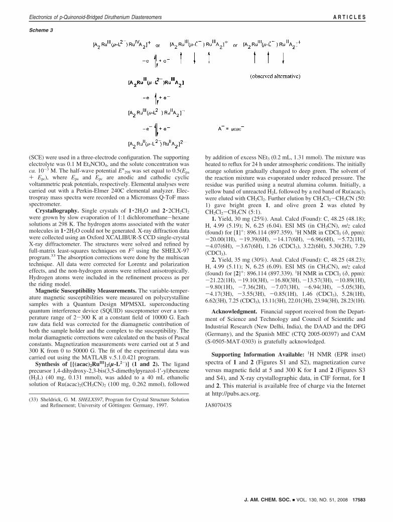
Conclusion
Despite the noted differences between the diastereomericmeso and rac forms of the structurally identified p-quinonoidbridged diruthenium species described here, we could establisha common scheme for their redox behavior, including thedetermination of the spin and valence situations (Scheme 3).Based in particular on the EPR results, the first oxidation ofthe neutral forms occurs at the ligand, leading to the formationof mutually antiferromagnetically coupled RuIII ions bridged bya p-semiquinone radical. Only the right-hand alternative for themonocations in Scheme 3 is compatible with the experimentallyobserved ligand-centered spin, supported by the severe structuraltwist which disfavors alternating metal-ligand-metal anti-parallel spin-spin interaction. In other words, the total exchangebetween the metal spins is antiferromagnetic and of greatermagnitude than the total exchange between the organic radicaland the RuIII.
There are fewer alternatives available on the reduction side,where successive metal-based one-electron additions result inisovalent diruthenium(II) dianions via the mixed-valent mono-anionic intermediates. The moderate intermetallic electroniccoupling (Class II) mediated by the nonplanar ligand L2- derivedfrom a substituted p-quinone is assumed to occur via a hole-transfer superexchange pathway involving dπ(Ru) and pπ(L2-)orbitals.28 Considering these results, it may be expected thatsuitably modified p-quinones can serve increasingly as tunablemediators for valence and spin interaction between metals.
Experimental Section
Materials. The precursor complex Ru(acac)2(CH3CN)2 wasprepared according to the reported procedure.29 The ligand precursor1,4-dihydroxy-2,3-bis(3,5-dimethylpyrazol-1′-yl)benzene (H2L) wasprepared as reported earlier.30 Other chemicals and solvents wereof reagent grade and used as received.
Instrumentation. UV-vis-NIR spectroelectrochemical studieswere performed in CH3CN/0.1 M Bu4NPF6 at 298 K using anoptically transparent thin-layer electrode (OTTLE) cell31 mountedin the sample compartment of a J&M TIDAS spectrophotometer.1H NMR spectra were obtained with a 400 MHz Varian FTspectrometer. The EPR measurements were made in a two-electrodecapillary tube32 with an X-band (9.5 GHz) Bruker system ESP300spectrometer. Cyclic voltammetric, differential pulse voltammetric,and coulometric measurements were carried out using a PAR model273A electrochemistry system. Platinum wire working and auxiliaryelectrodes and an aqueous saturated calomel reference electrode
(27) (a) Kelso, L. S.; Reitsma, D. A.; Keene, F. R. Inorg. Chem. 1996, 35,5144. (b) D’Alessandro, D. M.; Davies, M. S.; Keene, F. R. Inorg.Chem. 2006, 45, 1656.
(28) Kaim, W.; Lahiri, G. K. Angew. Chem., Int. Ed. 2007, 46, 1778.
(29) Kobayashi, T.; Nishina, Y.; Shimizu, K. G.; Sato, G. P. Chem. Lett.1988, 1137.
(30) Catalrin, J.; Fabero, F.; Guijarro, M. S.; Claramunt, R. M.; Santa Maria,M. D.; Foces-Foces, M. C.; Cano, F. H.; Elguero, J.; Sastre, R. J. Am.Chem. Soc. 1990, 112, 747.
(31) Krejcik, M.; Danek, M.; Hartl, F. J. Electroanal. Chem. 1991, 317,179.
(32) Kaim, W.; Ernst, S.; Kasack, V. J. Am. Chem. Soc. 1990, 112, 173.
Figure 8. EPR spectra of one-electron-reduced (a) 1- and (b) 2- in CH3CN/Bu4NPF6 at 110 K.
17582 J. AM. CHEM. SOC. 9 VOL. 130, NO. 51, 2008
A R T I C L E S Kumbhakar et al.
(SCE) were used in a three-electrode configuration. The supportingelectrolyte was 0.1 M Et4NClO4, and the solute concentration wasca. 10-3 M. The half-wave potential E°298 was set equal to 0.5(Epa
+ Epc), where Epa and Epc are anodic and cathodic cyclicvoltammetric peak potentials, respectively. Elemental analyses werecarried out with a Perkin-Elmer 240C elemental analyzer. Elec-trospray mass spectra were recorded on a Micromass Q-ToF massspectrometer.
Crystallography. Single crystals of 1 ·2H2O and 2 ·2CH2Cl2
were grown by slow evaporation of 1:1 dichloromethane-hexanesolutions at 298 K. The hydrogen atoms associated with the watermolecules in 1 ·2H2O could not be generated. X-ray diffraction datawere collected using an Oxford XCALIBUR-S CCD single-crystalX-ray diffractometer. The structures were solved and refined byfull-matrix least-squares techniques on F2 using the SHELX-97program.33 The absorption corrections were done by the multiscantechnique. All data were corrected for Lorentz and polarizationeffects, and the non-hydrogen atoms were refined anisotropically.Hydrogen atoms were included in the refinement process as perthe riding model.
Magnetic Susceptibility Measurements. The variable-temper-ature magnetic susceptibilities were measured on polycrystallinesamples with a Quantum Design MPMSXL superconductingquantum interference device (SQUID) susceptometer over a tem-perature range of 2-300 K at a constant field of 10000 G. Eachraw data field was corrected for the diamagnetic contribution ofboth the sample holder and the complex to the susceptibility. Themolar diamagnetic corrections were calculated on the basis of Pascalconstants. Magnetization measurements were carried out at 5 and300 K from 0 to 50000 G. The fit of the experimental data wascarried out using the MATLAB v.5.1.0.421 program.
Synthesis of [{(acac)2RuIII}2(µ-L2-)] (1 and 2). The ligandprecursor 1,4-dihydroxy-2,3-bis(3,5-dimethylpyrazol-1′-yl)benzene(H2L) (40 mg, 0.131 mmol), was added to a 40 mL ethanolicsolution of Ru(acac)2(CH3CN)2 (100 mg, 0.262 mmol), followed
by addition of excess NEt3 (0.2 mL, 1.31 mmol). The mixture washeated to reflux for 24 h under atmospheric conditions. The initiallyorange solution gradually changed to deep green. The solvent ofthe reaction mixture was evaporated under reduced pressure. Theresidue was purified using a neutral alumina column. Initially, ayellow band of unreacted H2L followed by a red band of Ru(acac)3
were eluted with CH2Cl2. Further elution by CH2Cl2-CH3CN (50:1) gave bright green 1, and olive green 2 was eluted byCH2Cl2-CH3CN (5:1).
1. Yield, 30 mg (25%). Anal. Calcd (Found): C, 48.25 (48.18);H, 4.99 (5.19); N, 6.25 (6.04). ESI MS (in CH3CN), m/z calcd(found) for [1]+: 896.114 (897.359). 1H NMR in CDCl3 (δ, ppm):-20.00(1H), -19.39(6H), -14.17(6H), -6.96(6H), -5.72(1H),-4.07(6H), -3.67(6H), 1.26 (CDCl3), 3.22(6H), 5.30(2H), 7.29(CDCl3).
2. Yield, 35 mg (30%). Anal. Calcd (Found): C, 48.25 (48.23);H, 4.99 (5.11); N, 6.25 (6.09). ESI MS (in CH3CN), m/z calcd(found) for [2]+: 896.114 (897.339). 1H NMR in CDCl3 (δ, ppm):-21.22(1H), -19.10(3H), -16.80(3H), -13.57(3H), -10.89(1H),-9.80(1H), -7.36(2H), -7.07(3H), -6.94(3H), -5.05(3H),-4.17(3H), -3.55(3H), -0.85(1H), 1.46 (CDCl3), 5.28(1H),6.62(3H), 7.25 (CDCl3), 13.11(3H), 22.01(3H), 23.94(3H), 28.23(1H).
Acknowledgment. Financial support received from the Depart-ment of Science and Technology and Council of Scientific andIndustrial Research (New Delhi, India), the DAAD and the DFG(Germany), and the Spanish MEC (CTQ 2005-00397) and CAM(S-0505-MAT-0303) is gratefully acknowledged.
Supporting Information Available: 1H NMR (EPR inset)spectra of 1 and 2 (Figures S1 and S2), magnetization curveversus magnetic field at 5 and 300 K for 1 and 2 (Figures S3and S4), and X-ray crystallographic data, in CIF format, for 1and 2. This material is available free of charge via the Internetat http://pubs.acs.org.
JA807043S(33) Sheldrick, G. M. SHELXS97, Program for Crystal Structure Solution
and Refinement; University of Gottingen: Germany, 1997.
Scheme 3
J. AM. CHEM. SOC. 9 VOL. 130, NO. 51, 2008 17583
Electronics of p-Quinonoid-Bridged Diruthenium Diastereomers A R T I C L E S