Embed Size (px)
Citation preview
Central Annals of Clinical Pathology
Cite this article: Velez1 M, Ganguly E (2014) Intraductal Papillary Mucinous Neoplasms of Pancreas- A Review. Ann Clin Pathol 2(2): 1021.
*Corresponding authorMario Velez, The University of Vermont/ Fletcher Allen Health Care, Address: 111 Colchester Avenue, Burlington, VT. Tel: 802-847-4892; Email:
Submitted: 19 July 2014
Accepted: 26 July 2014
Published: 28 July 2014
Copyright© 2014 Velez et al.
OPEN ACCESS
Keywords•Intraductal papillary mucinous neoplasms•Classifications•Main duct IPMN•Branch duct IPNM•Malignancy•Clinical management
Review Article
Intraductal Papillary Mucinous Neoplasms of Pancreas- A ReviewMario Velez1* and Eric Ganguly2
1Department of Gastroenterology, University of Vermont/ Fletcher Allen Health, USA2Department of Gastroenterology, University of Vermont/ Fletcher Allen Health USA
Abstract
Intraductal papillary mucinous neoplasms of pancreas are relatively common, often arise from the pancreatic head, and comprise more than one third of resected pancreatic cysts. In these lesions, the risk of dysplasia or carcinoma varies according to the site of ductal involvement or histologic subtypes. For instance, there is a higher risk of malignancy associated with main rather than branch duct IPMNs and/or with pancreaticobiliary rather than gastric subtype. Other factors altering this risk include cyst size and associated solid mural component. Although in general surgical excision is advocated for all MD-IPMNs when possible, a specific protocol is to follow for management of BD-IPMNs. Additional to specifics on the aforementioned, the present study reviews clinic-pathologic classification schema and provides recently updated guidelines on surveillance and clinical management of IPMNs. Certain molecular features and mutations such as P53 aberration have been linked to the presence of malignancy in IPMNs. Cellular/molecular techniques, newly proposed to increase the yield of diagnosis and/or potential measures of prognosis are also reported in the following review.
AbbreviAtionsIPMN (Intraductal papillary mucinous neoplasm); MD-
IPMN (Main duct IPMN); BD-IPMN (Branch duct IPMN); SCAs (Serous cystadenomas); MRI (Magnetic resonance imaging); CT (Computed tomography); EUS (Endoscopic ultrasound); CEA (Carcinoembryonic antigen)
introduction Since initial reports in the 1980s, the classification and
management of intraductal papillary mucinous neoplasm (IPMN) has evolved dramatically. Historically, these cystic neoplasms were grouped together with other mucinous cysts (cystadenomas/cystadenocarcinomas) until they were recognized as a distinct entity and a major precursor to pancreatic ductal adenocarcinoma. IPMNs are tumors composed of papillary proliferations of mucin-producing epithelium that cause excessive mucus production and cystic dilatation of the pancreatic ducts. Recent epidemiologic studies have suggested a growing incidence in IPMN. Increased recognition and attention to these lesions has led to development and revision of international consensus guidelines on diagnostic approach and management. This article will review the definition, classification and management of IPMNs, with a focus on preoperative diagnosis and management, particularly involving diagnostic imaging and cyst fluid analysis.
clAssificAtion Pancreatic cysts are broadly divided into inflammatory type
and neoplastic type. Inflammatory cysts, or pseudocysts, are not true cysts and lack an epithelial lining. They are one of the potential sequela of pancreatitis and do not harbor malignant potential. Neoplastic pancreatic cysts are further classified by their ability to produce mucinous fluid. Non-mucinous pancreatic cysts primarily include serous cyst adenomas (SCAs) as well as cystic variants of solid lesions such as solid pseudopapillary tumors and pancreatic neuroendocrine tumors.
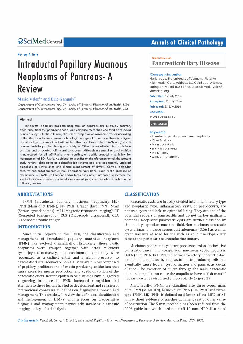
Mucinous pancreatic cysts are precursor lesions to invasive pancreatic cancer and comprise of mucinous cystic neoplasm (MCN) and IPMN. In IPMN, the normal excretory pancreatic duct epithelium is replaced by neoplastic, mucin-producing cells that eventually cause locular cyst formation and pancreatic ductal dilation. The excretion of mucin through the main pancreatic duct and ampulla can cause the ampulla to have a “fish-mouth” appearance when visualized endoscopically (Figure 1).
Anatomically, IPMNs are classified into three types: main duct IPMN (MD-IPMN), branch duct IPMN (BD-IPMN) and mixed type IPMN. MD-IPMN is defined as dilation of the MPD of >5 mm without evidence of another dominant cyst or other cause of obstruction. The 5 mm threshold has been reduced from the 2006 guidelines which used a cut-off 10 mm. MPD dilation of
Special Issue on
Pancreaticobiliary disease
Central
Velez et al. (2014)Email:
Ann Clin Pathol 2(2): 1021 (2014) 2/8
5-9 mm is now considered a “worrisome feature”, while an MPD diameter of over10 mm is considered “high-risk stigmata”. In contrast, branch-duct IPMN (BD-IPMN) are cysts >5mm that arise from the side branches of the pancreatic ductal system and communicate with a minimally dilated/normal pancreatic duct. A mixed type IPMN has features of both main and branch duct types. The impact of anatomic subtype on natural history is described below.
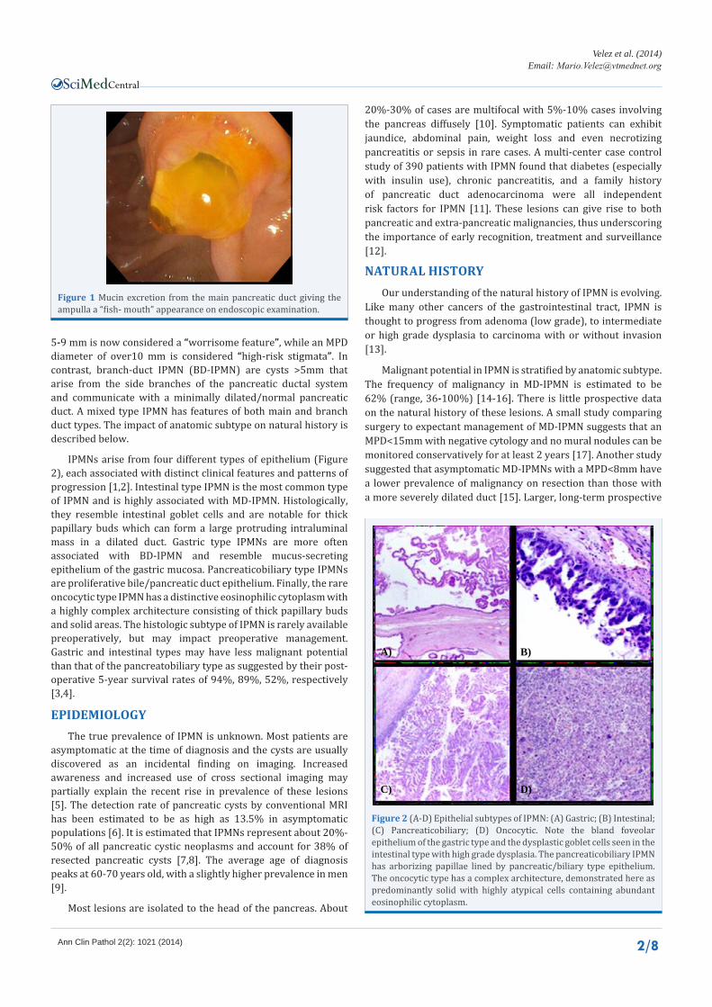
IPMNs arise from four different types of epithelium (Figure 2), each associated with distinct clinical features and patterns of progression [1,2]. Intestinal type IPMN is the most common type of IPMN and is highly associated with MD-IPMN. Histologically, they resemble intestinal goblet cells and are notable for thick papillary buds which can form a large protruding intraluminal mass in a dilated duct. Gastric type IPMNs are more often associated with BD-IPMN and resemble mucus-secreting epithelium of the gastric mucosa. Pancreaticobiliary type IPMNs are proliferative bile/pancreatic duct epithelium. Finally, the rare oncocytic type IPMN has a distinctive eosinophilic cytoplasm with a highly complex architecture consisting of thick papillary buds and solid areas. The histologic subtype of IPMN is rarely available preoperatively, but may impact preoperative management. Gastric and intestinal types may have less malignant potential than that of the pancreatobiliary type as suggested by their post-operative 5-year survival rates of 94%, 89%, 52%, respectively [3,4].
ePidemiology The true prevalence of IPMN is unknown. Most patients are
asymptomatic at the time of diagnosis and the cysts are usually discovered as an incidental finding on imaging. Increased awareness and increased use of cross sectional imaging may partially explain the recent rise in prevalence of these lesions [5]. The detection rate of pancreatic cysts by conventional MRI has been estimated to be as high as 13.5% in asymptomatic populations [6]. It is estimated that IPMNs represent about 20%-50% of all pancreatic cystic neoplasms and account for 38% of resected pancreatic cysts [7,8]. The average age of diagnosis peaks at 60-70 years old, with a slightly higher prevalence in men [9].
Most lesions are isolated to the head of the pancreas. About
20%-30% of cases are multifocal with 5%-10% cases involving the pancreas diffusely [10]. Symptomatic patients can exhibit jaundice, abdominal pain, weight loss and even necrotizing pancreatitis or sepsis in rare cases. A multi-center case control study of 390 patients with IPMN found that diabetes (especially with insulin use), chronic pancreatitis, and a family history of pancreatic duct adenocarcinoma were all independent risk factors for IPMN [11]. These lesions can give rise to both pancreatic and extra-pancreatic malignancies, thus underscoring the importance of early recognition, treatment and surveillance [12].
nAturAl History Our understanding of the natural history of IPMN is evolving.
Like many other cancers of the gastrointestinal tract, IPMN is thought to progress from adenoma (low grade), to intermediate or high grade dysplasia to carcinoma with or without invasion [13].
Malignant potential in IPMN is stratified by anatomic subtype. The frequency of malignancy in MD-IPMN is estimated to be 62% (range, 36-100%) [14-16]. There is little prospective data on the natural history of these lesions. A small study comparing surgery to expectant management of MD-IPMN suggests that an MPD<15mm with negative cytology and no mural nodules can be monitored conservatively for at least 2 years [17]. Another study suggested that asymptomatic MD-IPMNs with a MPD<8mm have a lower prevalence of malignancy on resection than those with a more severely dilated duct [15]. Larger, long-term prospective
figure 1 Mucin excretion from the main pancreatic duct giving the ampulla a “fish- mouth” appearance on endoscopic examination.
A) B)
C) D)
figure 2 (A-D) Epithelial subtypes of IPMN: (A) Gastric; (B) Intestinal; (C) Pancreaticobiliary; (D) Oncocytic. Note the bland foveolar epithelium of the gastric type and the dysplastic goblet cells seen in the intestinal type with high grade dysplasia. The pancreaticobiliary IPMN has arborizing papillae lined by pancreatic/biliary type epithelium. The oncocytic type has a complex architecture, demonstrated here as predominantly solid with highly atypical cells containing abundant eosinophilic cytoplasm.

Central
Velez et al. (2014)Email:
Ann Clin Pathol 2(2): 1021 (2014) 3/8
trials are still needed to estimate the risk of malignancy and help guide decisions for surgical intervention.
BD-IPMNs have a much lower incidence of malignancy (15-25%), and have an estimated 2-3% yearly risk of malignant transformation [18,19]. Cyst size greater than 3cm, presence of mural nodules or main pancreatic duct dilation>6mm have all been associated with greater risks for malignancy [16]. A recent analysis has suggested high malignant potential in cysts growing more than 2mm/year, and those with atypical epithelial cells on cytological analysis [20-23]. Obesity has also been associated with an increased frequency of malignant transformation in BD-IPMN [24].
PreoPerAtive diAgnosis Pancreatic cysts are often discovered incidentally on CT
or MRI. As mucinous cysts (including IPMN) have malignant potential and non-mucinous cysts do not, the initial challenge involves making this distinction. While serous cystadenomas are often microcystic and with central scar, mucinous cysts have thick epithelial walls that show isodense or hypodense enhancement on venous phase. Mucinous cystic neoplasms are most often solitary cysts located in the pancreatic body and tail and show no connection to the pancreatic duct and can have minor calcifications. On the other hand, IPMNs are found throughout the pancreas and by definition communicate with the pancreatic duct. Although MRI has a small benefit in describing these morphological features in smaller cysts, both CT and MRI have been shown to be similarly accurate in differentiating pancreatic masses as benign or malignant [25, 26]. MD-IPMNs are more accurately diagnosed preoperatively than BD-IPMNs, however CT imaging can often identify side-branching pancreatic duct cysts seen in BD-IPMN and mixed type (Figure 3) [27]. In practice, many small pancreatic cysts are assumed to be BD-IPMNs based clinical factors.
Endoscopic ultrasound (EUS) provides imaging that is complementary to CT and MRI. It is particularly useful in accurately assessing pancreatic ductal dilation (Figure 4) and in identifying high-risk stigmata for malignancy in IPMN such as internal septations, mural nodules, solid masses, vascular invasion and lymph node metastasis [28]. EUS can be also be used to sample cyst fluid or high-risk stigmata via fine-needle aspiration (FNA). Cyst fluid contains cells shed from the epithelium and protein/DNA/RNA biomarkers that may potentially help classify cysts as mucinous. Although imperfect, EUS with or without FNA has been found superior to CT or MRI in accurately describing neoplastic cysts [29]. Head to head trials have shown a trend towards increased sensitivity for EUS-FNA compared to CT-guided biopsy, with a much lower risk of peritoneal seeding as compared with percutaneous methodologies [30,31]. The current international guidelines recommend EUS in the evaluation of suspected IPMN with any worrisome clinical or radiographic features.
Hybrid PET/CT with F-FDG has shown promise in diagnosing malignancy and tumor invasion in IPMN [32-33]. However, studies on the sensitivity and specificity of PET-CT remain controversial and vary widely.[34] Given the costs and uncertain benefit over CT/MRI/EUS, there is currently not enough data to support using PET/CT in the evaluation of IPMN.
cyst flud AnAlysis Imaging alone is often inadequate to accurately diagnose
pancreatic cystic lesions. Fine needle aspiration with analysis of cyst fluid is becoming an increasingly used method of characterizing cystic lesions. While the current international guidelines still consider cyst fluid analysis investigational, they recommend it for small BD-IPMN at centers with expertise in EUS-FNA and cytological evaluation. Many centers now have such expertise.
Protein tumor markers
Pancreatic cyst fluid contains glycoproteins including cancer antigen (CA) 19-9 and carcinoembryonic antigen (CEA). CEA is the most often used marker. High CEA levels (>192ng/ml) are associated with mucinous cysts (including IPMN), with lower levels (<5 ng/ml) found in non-mucinous cysts (sensitivity 50%, specificity 95%) [35, 36]. CEA cannot differentiate IPMN from MCN, or accurately identify malignancy [37].
cytology
Cyst fluid cytology has a limited role in the diagnosis of IPMN. It can differentiate between mucinous and non-mucinous cysts by identifying columnar epithelium or extracelluar mucin. The fluid
figure 3 CT image of a mixed type IPMN characterized by main pancreatic ductal dilatation and several side-branching pancreatic cysts.
figure 4 Endoscopic ultrasound image of the common bile duct (CBD) and dilated main pancreatic duct (PD), representative of MD-IPMN.
Central
Velez et al. (2014)Email:
Ann Clin Pathol 2(2): 1021 (2014) 4/8
often is viscous. Although the specificity of identifying lesions as mucinous is high, the sensitivity of cytology is only 50%. The low sensitivity is often attributed to insufficient sample volume, scant cellularity in cyst fluid, and contamination from gastrointestinal wall cells. Accurate grading of these lesions on cytology can be difficult and the rate of under-diagnosis is high [38-40]. The detection of high-grade atypical epithelial cells (AEC) may prove more accurate than standard cytology in diagnosing pancreatic cysts. AECs have been found to correspond with histologically confirmed high-grade dysplasia and invasive carcinoma in mucinous pancreatic cysts [22,23]. In IPMN, cells showing an increased nuclear to cytoplasm ratio, abnormal chromatin pattern and background necrosis correspond with the risk of malignancy [21]. Identifying and using AECs as a predictor of malignancy in mucinous cysts is estimated to improve the sensitivity and specificity of diagnosis to 72% and 85%, respectively [22].
dnA biomarkers
Point mutations in the K-ras oncogene are found in over 70% of pancreatic adenocarcinomas and roughly 50% of mucin-producing tumors [41, 42]. This has led to its study in the evaluation of pancreatic cysts. In 2009, Khalid et. al showed that K-ras point mutations have a high specificity (96%), but low sensitivity(45%) for mucinous differentiation [43]. A long-term follow-up study showed similar results [44]. Combining K-ras and CEA testing increased the sensitivity for mucinous cysts without compromising specificity. Early K-ras mutation followed by allelic loss of heterozygosity has also been shown to predict malignancy (sensitivity, 91%; specificity, 93%). Other studies, however, do not support an additive role for DNA testing beyond cytology and imaging, in diagnosing malignant pancreatic cysts [45]. Specific to IPMN, K-ras mutations from fluid samples obtained via EUS-FNA have been correlated with advanced cystic pancreatic neoplasia [46,47]. As these mutations are less often seen in low-grade dysplasia, there is a possible role in K-ras analysis in the surveillance of un-resected IPMN.
Larger genomic sequencing of IPMNs has identified recurrent mutations at codon 201 of GNAS in nearly 65% of IPMNs, particularly the intestinal type [48,49]. This specific mutations leads to upregulation of intracellular cyclic adenine monophosphate (cAMP), which results in upregulated expression of mucin producing genes MUC2 and MUC5AC in pancreatic ductal epithelium [50,51]. In one study, K-ras and/or GNAS mutations were identified in 96% of IPMNs and advanced carcinomas arising from IPMNs. GNAS mutations have not been seen in other types of cystic neoplasms of the pancreas or invasive adenocarcinomas not associated with IPMNs [48,51]. Other DNA markers such at TP53 and BRAF show promise in identifying IPMNs with high-grade dysplasia or malignancy [52, 53].
micro-rnA biomarkers
MicroRNAs (miRNA) are small noncoding RNA segments that can regulate gene expression via promotion or inhibition. They are involved with many normal cellular functions that range from proliferation to differentiation to apoptosis, and thus have been implicated in the development of malignancy, including pancreatic cancer [54-56]. miRNA profiling of surgical specimens has been able to differentiate benign serous cyst adenomas from
pancreatic cystic neoplasms and pancreatic duct cancer [57]. Aberrant expression of miRNA segments have already been observed and correlated in pancreatic cancer arising from IPMN [58].
Recent literature has correlated miRNA sequences with varying degrees of dysplasia and invasive adenocarcinoma in IPMN [54-60]. Interestingly, these studies have few up-regulated miRNAs sequences in common, emphasizing that the role of miRNA in all malignancy is still evolving.
mAnAgement
Given the high risk of malignancy, current guidelines recommend resection for all surgically fit patients with MD-IPMN. Main pancreatic duct dilation of 5-9 mm is considered a “worrisome feature” and while there is no recommendation for immediate resection, no established surveillance protocols have been established for this sub-group of IPMN [61]. Surgical resection of BD-IPMN is indicated in those with high-risk stigmata (ie obstructive jaundice, enhanced solid component, main pancreatic duct >10mm, rapidly growing cyst size and the presence of high grade atypia). Patients with worrisome features such as acute pancreatitis, cyst size>3cm, thickened enhanced cyst walls, MPD 5-9mm, mural nodules, or an abrupt change in the MPD caliber with distal pancreatic atrophy and lymphadenopathy should be referred for EUS with FNA to confirm the presence of mural nodules, main duct involvement, or suspicious cytology. If any of these are present, resection is warranted.
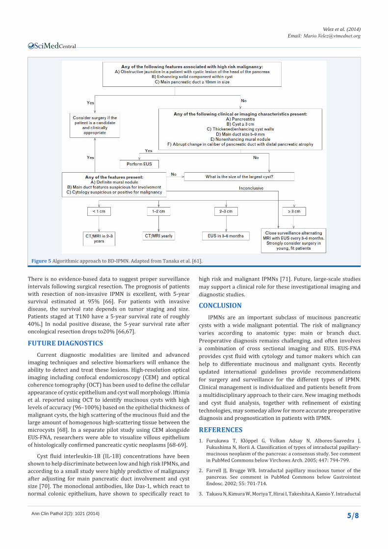
Because most BD-IPMNs are diagnosed in the elderly and the annual malignancy rate is estimated at 2-3%, new consensus guidelines have advised that lesions without worrisome features can undergo routine surveillance based on cyst size. Patients with cysts <1cm should receive CT/MRI surveillance imaging every 2-3years. Those with cysts measuring 1-2cm should have a CT/MRI every year. If no change is found after 2 years, patients can extend their surveillance intervals to every 2 years. Finally, cysts between 2-3cm should have a EUS every 3-6 months, and cysts>3cm should be surveyed by alternating MRI and EUS every 3-6months (see Figure 5) [61]. A scoring system has been proposed based on a multivariate analysis of 5 identified predictors of malignancy (age>60, history of pancreatitis, presence of mural nodules and main pancreatic duct diameter >6mm, elevated CA 19-9); however this system is estimated to have a specificity of 90% and a sensitivity rate of only 50% [62].
Some investigators have called for a more aggressive surgical approach in patients under 65 years old due to a cumulative risk of malignancy, especially with recent literature suggesting invasive carcinoma derived from BD-IPMN is more aggressive than that derived from MD-IPMN [63, 64]. Patients with IPMN of all types often require a multi-disciplinary approach, with decisions for resection individualized based each patient’s specific circumstances.
Following complete resection with negative margins, patients should continue surveillance imaging for recurrent IPMN or development of pancreatic ductal adenocarcinoma. Although the recurrence of IPMN varies across studies, one recent analysis observed new IPMN in 62% of patients within 10 years, with 18% of those lesions meeting criteria for surgical resection.
Central
Velez et al. (2014)Email:
Ann Clin Pathol 2(2): 1021 (2014) 5/8
There is no evidence-based data to suggest proper surveillance intervals following surgical resection. The prognosis of patients with resection of non-invasive IPMN is excellent, with 5-year survival estimated at 95% [66]. For patients with invasive disease, the survival rate depends on tumor staging and size. Patients staged at T1N0 have a 5-year survival rate of roughly 40%.] In nodal positive disease, the 5-year survival rate after oncological resection drops to20% [66,67].
future diAgnostics Current diagnostic modalities are limited and advanced
imaging techniques and selective biomarkers will enhance the ability to detect and treat these lesions. High-resolution optical imaging including confocal endomicroscopy (CEM) and optical coherence tomography (OCT) has been used to define the cellular appearance of cystic epithelium and cyst wall morphology. Iftimia et al. reported using OCT to identify mucinous cysts with high levels of accuracy (96–100%) based on the epithelial thickness of malignant cysts, the high scattering of the mucinous fluid and the large amount of homogenous high-scattering tissue between the microcysts [68]. In a separate pilot study using CEM alongside EUS-FNA, researchers were able to visualize villous epithelium of histologically confirmed pancreatic cystic neoplasms [68-69].
Cyst fluid interleukin-1B (IL-1B) concentrations have been shown to help discriminate between low and high risk IPMNs, and according to a small study were highly predictive of malignancy after adjusting for main pancreatic duct involvement and cyst size [70]. The monoclonal antibodies, like Das-1, which react to normal colonic epithelium, have shown to specifically react to
high risk and malignant IPMNs [71]. Future, large-scale studies may support a clinical role for these investigational imaging and diagnostic studies.
conclusion IPMNs are an important subclass of mucinous pancreatic
cysts with a wide malignant potential. The risk of malignancy varies according to anatomic type: main or branch duct. Preoperative diagnosis remains challenging, and often involves a combination of cross sectional imaging and EUS. EUS-FNA provides cyst fluid with cytology and tumor makers which can help to differentiate mucinous and malignant cysts. Recently updated international guidelines provide recommendations for surgery and surveillance for the different types of IPMN. Clinical management is individualized and patients benefit from a multidisciplinary approach to their care. New imaging methods and cyst fluid analysis, together with refinement of existing technologies, may someday allow for more accurate preoperative diagnosis and prognostication in patients with IPMN.
references1. Furukawa T, Klöppel G, Volkan Adsay N, Albores-Saavedra J,
Fukushima N, Horii A. Classification of types of intraductal papillary-mucinous neoplasm of the pancreas: a consensus study. See comment in PubMed Commons below Virchows Arch. 2005; 447: 794-799.
2. Farrell JJ, Brugge WR. Intraductal papillary mucinous tumor of the pancreas. See comment in PubMed Commons below Gastrointest Endosc. 2002; 55: 701-714.
3. Takasu N, Kimura W, Moriya T, Hirai I, Takeshita A, Kamio Y. Intraductal
figure 5 Algorithmic approach to BD-IPMN. Adapted from Tanaka et al. [61].
Central
Velez et al. (2014)Email:
Ann Clin Pathol 2(2): 1021 (2014) 6/8
papillary-mucinous neoplasms of the gastric and intestinal types may have less malignant potential than the pancreatobiliary type. See comment in PubMed Commons below Pancreas. 2010; 39: 604-610.
4. Furukawa T, Hatori T, Fujita I, Yamamoto M, Kobayashi M, Ohike N. Prognostic relevance of morphological types of intraductal papillary mucinous neoplasms of the pancreas. See comment in PubMed Commons below Gut. 2011; 60: 509-516.
5. Klibansky DA, Reid-Lombardo KM, Gordon SR, Gardner TB. The clinical relevance of the increasing incidence of intraductal papillary mucinous neoplasm. See comment in PubMed Commons below Clin Gastroenterol Hepatol. 2012; 10: 555-558.
6. Lee KS, Sekhar A, Rofsky NM, Pedrosa I. Prevalence of incidental pancreatic cysts in the adult population on MR imaging. See comment in PubMed Commons below Am J Gastroenterol. 2010; 105: 2079-2084.
7. Grützmann R, Niedergethmann M, Pilarsky C, Klöppel G, Saeger HD. Intraductal papillary mucinous tumors of the pancreas: biology, diagnosis, and treatment. See comment in PubMed Commons below Oncologist. 2010; 15: 1294-1309.
8. Valsangkar NP, Morales-Oyarvide V, Thayer SP, Ferrone CR, Wargo JA, Warshaw AL. 851 resected cystic tumors of the pancreas: a 33-year experience at the Massachusetts General Hospital. See comment in PubMed Commons below Surgery. 2012; 152: S4-12.
9. Sohn TA, Yeo CJ, Cameron JL, Hruban RH, Fukushima N, Campbell KA. Intraductal papillary mucinous neoplasms of the pancreas: an updated experience. See comment in PubMed Commons below Ann Surg. 2004; 239: 788-797.
10. Campbell F, Azadeh B. Cystic neoplasms of the exocrine pancreas. See comment in PubMed Commons below Histopathology. 2008; 52: 539-551.
11. Capurso G, Boccia S, Salvia R, Del Chiaro M, Frulloni L, Arcidiacono PG. Risk factors for intraductal papillary mucinous neoplasm (IPMN) of the pancreas: a multicentre case-control study. See comment in PubMed Commons below Am J Gastroenterol. 2013; 108: 1003-1009.
12. Kamisawa T, Tu Y, Egawa N, Nakajima H, Tsuruta K, Okamoto A. Malignancies associated with intraductal papillary mucinous neoplasm of the pancreas. See comment in PubMed Commons below World J Gastroenterol. 2005; 11: 5688-5690.
13. Adsay NV, F.N, Furukawa T, Hruban RH, Klimstra DS, Kloppel G, et al. Intraductal neoplasm of the pancreas. In: Bosman FT, Carneiro F,Hruban RH, Theise ND, editors. WHO classi!cation of tumors of digestive system. Lyon: WHO Press. 2010; 304-13.
14. Schmidt CM, White PB, Waters JA, Yiannoutsos CT, Cummings OW, Baker M. Intraductal papillary mucinous neoplasms: predictors of malignant and invasive pathology. See comment in PubMed Commons below Ann Surg. 2007; 246: 644-651.
15. Abdeljawad K, Vemulapalli KC2, Schmidt CM3, Dewitt J2, Sherman S2, Imperiale TF2. Prevalence of malignancy in patients with pure main duct intraductal papillary mucinous neoplasms. See comment in PubMed Commons below Gastrointest Endosc. 2014; 79: 623-629.
16. Anand N, Sampath K, Wu BU. Cyst features and risk of malignancy in intraductal papillary mucinous neoplasms of the pancreas: a meta-analysis. See comment in PubMed Commons below Clin Gastroenterol Hepatol. 2013; 11: 913-921.
17. Ogura T, Masuda D, Kurisu Y, Edogawa S, Imoto A, Hayashi M. Potential predictors of disease progression for main-duct intraductal papillary mucinous neoplasms of the pancreas. See comment in PubMed Commons below J Gastroenterol Hepatol. 2013; 28: 1782-1786.
18. Khannoussi W, Vullierme MP, Rebours V, Maire F, Hentic O, Aubert
A. The long term risk of malignancy in patients with branch duct intraductal papillary mucinous neoplasms of the pancreas. See comment in PubMed Commons below Pancreatology. 2012; 12: 198-202.
19. Lévy P, Jouannaud V, O’Toole D, Couvelard A, Vullierme MP, Palazzo L. Natural history of intraductal papillary mucinous tumors of the pancreas: actuarial risk of malignancy. See comment in PubMed Commons below Clin Gastroenterol Hepatol. 2006; 4: 460-468.
20. Kang MJ, Jang JY, Kim SJ, Lee KB, Ryu JK, Kim YT. Cyst growth rate predicts malignancy in patients with branch duct intraductal papillary mucinous neoplasms. See comment in PubMed Commons below Clin Gastroenterol Hepatol. 2011; 9: 87-93.
21. Pitman MB, Centeno BA, Daglilar ES, Brugge WR, Mino-Kenudson M. Cytological criteria of high-grade epithelial atypia in the cyst fluid of pancreatic intraductal papillary mucinous neoplasms. See comment in PubMed Commons below Cancer Cytopathol. 2014; 122: 40-47.
22. Pitman MB, Genevay M, Yaeger K, Chebib I, Turner BG, Mino-Kenudson M. High-grade atypical epithelial cells in pancreatic mucinous cysts are a more accurate predictor of malignancy than “positive” cytology. See comment in PubMed Commons below Cancer Cytopathol. 2010; 118: 434-440.
23. Pitman MB, Yaeger KA, Brugge WR, Mino-Kenudson M. Prospective analysis of atypical epithelial cells as a high-risk cytologic feature for malignancy in pancreatic cysts. See comment in PubMed Commons below Cancer Cytopathol. 2013; 121: 29-36.
24. Sturm EC, Roch AM, Shaffer KM, Schmidt CM 2nd, Lee SJ, Zyromski NJ. Obesity increases malignant risk in patients with branch-duct intraductal papillary mucinous neoplasm. See comment in PubMed Commons below Surgery. 2013; 154: 803-808.
25. Visser BC, Yeh BM, Qayyum A, Way LW, McCulloch CE, Coakley FV. Characterization of cystic pancreatic masses: relative accuracy of CT and MRI. See comment in PubMed Commons below AJR Am J Roentgenol. 2007; 189: 648-656.
26. Sainani NI, Saokar A, Deshpande V, Fernández-del Castillo C, Hahn P, Sahani DV. Comparative performance of MDCT and MRI with MR cholangiopancreatography in characterizing small pancreatic cysts. See comment in PubMed Commons below AJR Am J Roentgenol. 2009; 193: 722-731.
27. Correa-Gallego C, Ferrone CR, Thayer SP, Wargo JA, Warshaw AL, Fernández-Del Castillo C. Incidental pancreatic cysts: do we really know what we are watching? See comment in PubMed Commons below Pancreatology. 2010; 10: 144-150.
28. Pais SA, Attasaranya S, Leblanc JK, Sherman S, Schmidt CM, DeWitt J. Role of endoscopic ultrasound in the diagnosis of intraductal papillary mucinous neoplasms: correlation with surgical histopathology. See comment in PubMed Commons below Clin Gastroenterol Hepatol. 2007; 5: 489-495.
29. Khashab MA, Kim K, Lennon AM, Shin EJ, Tignor AS, Amateau SK. Should we do EUS/FNA on patients with pancreatic cysts? The incremental diagnostic yield of EUS over CT/MRI for prediction of cystic neoplasms. See comment in PubMed Commons below Pancreas. 2013; 42: 717-721.
30. Horwhat JD, Paulson EK, McGrath K, Branch MS, Baillie J, Tyler D. A randomized comparison of EUS-guided FNA versus CT or US-guided FNA for the evaluation of pancreatic mass lesions. See comment in PubMed Commons below Gastrointest Endosc. 2006; 63: 966-975.
31. Micames C, Jowell PS, White R, Paulson E, Nelson R, Morse M. Lower frequency of peritoneal carcinomatosis in patients with pancreatic cancer diagnosed by EUS-guided FNA vs. percutaneous FNA. See
Central
Velez et al. (2014)Email:
Ann Clin Pathol 2(2): 1021 (2014) 7/8
comment in PubMed Commons below Gastrointest Endosc. 2003; 58: 690-695.
32. Sperti C, Bissoli S, Pasquali C, Frison L, Liessi G, Chierichetti F, et al. 18-fluorodeoxyglucose positron emission tomography enhances computed tomography diagnosis of malignant intraductal papillary mucinous neoplasms of the pancreas. Ann Surg. 2007; 246: 932-937.
33. Tomimaru Y, Takeda Y, Tatsumi M, Kim T, Kobayashi S, Marubashi S, et al. Utility of 2-[18F] fluoro-2-deoxy-D-glucose positron emission tomography in differential diagnosis of benign and malignant intraductal papillary-mucinous neoplasm of the pancreas. Oncol Rep. 2010; 24: 613-620.
34. Mansour JC, Schwartz L, Pandit-Taskar N, D’Angelica M, Fong Y, Larson SM. The utility of F-18 fluorodeoxyglucose whole body PET imaging for determining malignancy in cystic lesions of the pancreas. See comment in PubMed Commons below J Gastrointest Surg. 2006; 10: 1354-1360.
35. Brugge WR, Lewandrowski K, Lee-Lewandrowski E, Centeno BA, Szydlo T, Regan S. Diagnosis of pancreatic cystic neoplasms: a report of the cooperative pancreatic cyst study. See comment in PubMed Commons below Gastroenterology. 2004; 126: 1330-1336.
36. van der Waaij LA, van Dullemen HM, Porte RJ. Cyst fluid analysis in the differential diagnosis of pancreatic cystic lesions: a pooled analysis. See comment in PubMed Commons below Gastrointest Endosc. 2005; 62: 383-389.
37. Ngamruengphong S, Bartel MJ, Raimondo M. Cyst carcinoembryonic antigen in differentiating pancreatic cysts: a meta-analysis. See comment in PubMed Commons below Dig Liver Dis. 2013; 45: 920-926.
38. Thornton GD, McPhail MJ, Nayagam S, Hewitt MJ, Vlavianos P, Monahan KJ. Endoscopic ultrasound guided fine needle aspiration for the diagnosis of pancreatic cystic neoplasms: a meta-analysis. See comment in PubMed Commons below Pancreatology. 2013; 13: 48-57.
39. Stelow EB, Stanley MW, Bardales RH, Mallery S, Lai R, Linzie BM. Intraductal papillary-mucinous neoplasm of the pancreas. The findings and limitations of cytologic samples obtained by endoscopic ultrasound-guided fine-needle aspiration. See comment in PubMed Commons below Am J Clin Pathol. 2003; 120: 398-404.
40. Layfield LJ, Cramer H. Fine-needle aspiration cytology of intraductal papillary-mucinous tumors: a retrospective analysis. See comment in PubMed Commons below Diagn Cytopathol. 2005; 32: 16-20.
41. Kimura W, Zhao B, Futakawa N, Muto T, Makuuchi M. Significance of K-ras codon 12 point mutation in pancreatic juice in the diagnosis of carcinoma of the pancreas. See comment in PubMed Commons below Hepatogastroenterology. 1999; 46: 532-539.
42. Matsubayashi H, Watanabe H, Yamaguchi T, Ajioka Y, Nishikura K, Iwafuchi M. Multiple K-ras mutations in hyperplasia and carcinoma in cases of human pancreatic carcinoma. See comment in PubMed Commons below Jpn J Cancer Res. 1999; 90: 841-848.
43. Khalid A, Zahid M, Finkelstein SD, LeBlanc JK, Kaushik N, Ahmad N. Pancreatic cyst fluid DNA analysis in evaluating pancreatic cysts: a report of the PANDA study. See comment in PubMed Commons below Gastrointest Endosc. 2009; 69: 1095-1102.
44. Nikiforova MN, Khalid A, Fasanella KE, McGrath KM, Brand RE, Chennat JS. Integration of KRAS testing in the diagnosis of pancreatic cystic lesions: a clinical experience of 618 pancreatic cysts. See comment in PubMed Commons below Mod Pathol. 2013; 26: 1478-1487.
45. Lee LS, Wu BU, Banks PA, Kadiyala V, Mehta S, Saltzman JR. Utility of commercial DNA analysis in detecting malignancy within pancreatic cysts. See comment in PubMed Commons below JOP. 2013; 15: 182-188.
46. Schoedel KE, Finkelstein SD, Ohori NP. K-Ras and microsatellite marker analysis of fine-needle aspirates from intraductal papillary mucinous neoplasms of the pancreas. See comment in PubMed Commons below Diagn Cytopathol. 2006; 34: 605-608.
47. Lubezky N, Ben-Haim M, Marmor S, Brazowsky E, Rechavi G, Klausner JM. High-throughput mutation profiling in intraductal papillary mucinous neoplasm (IPMN). See comment in PubMed Commons below J Gastrointest Surg. 2011; 15: 503-511.
48. Wu J, Matthaei H, Maitra A, Dal Molin M, Wood LD, Eshleman JR. Recurrent GNAS mutations define an unexpected pathway for pancreatic cyst development. See comment in PubMed Commons below Sci Transl Med. 2011; 3: 92ra66.
49. Dal Molin M, Matthaei H, Wu J, Blackford A, Debeljak M, Rezaee N. Clinicopathological correlates of activating GNAS mutations in intraductal papillary mucinous neoplasm (IPMN) of the pancreas. See comment in PubMed Commons below Ann Surg Oncol. 2013; 20: 3802-3808.
50. Komatsu H, Tanji E2, Sakata N3, Aoki T3, Motoi F3, Naitoh T3. A GNAS mutation found in pancreatic intraductal papillary mucinous neoplasms induces drastic alterations of gene expression profiles with upregulation of mucin genes. See comment in PubMed Commons below PLoS One. 2014; 9: e87875.
51. Furukawa T, Kuboki Y, Tanji E, Yoshida S, Hatori T, Yamamoto M. Whole-exome sequencing uncovers frequent GNAS mutations in intraductal papillary mucinous neoplasms of the pancreas. See comment in PubMed Commons below Sci Rep. 2011; 1: 161.
52. Kanda M, Sadakari Y, Borges M, Topazian M, Farrell J, Syngal S. Mutant TP53 in duodenal samples of pancreatic juice from patients with pancreatic cancer or high-grade dysplasia. See comment in PubMed Commons below Clin Gastroenterol Hepatol. 2013; 11: 719-730.
53. Amato E, Molin MD, Mafficini A, Yu J, Malleo G, Rusev B. Targeted next-generation sequencing of cancer genes dissects the molecular profiles of intraductal papillary neoplasms of the pancreas. See comment in PubMed Commons below J Pathol. 2014; 233: 217-227.
54. Habbe N, Koorstra JB, Mendell JT, Offerhaus GJ, Ryu JK, Feldmann G. MicroRNA miR-155 is a biomarker of early pancreatic neoplasia. See comment in PubMed Commons below Cancer Biol Ther. 2009; 8: 340-346.
55. Ryu JK, Hong SM, Karikari CA, Hruban RH, Goggins MG, Maitra A. Aberrant MicroRNA-155 expression is an early event in the multistep progression of pancreatic adenocarcinoma. See comment in PubMed Commons below Pancreatology. 2010; 10: 66-73.
56. Li A, Yu J, Kim H, Wolfgang CL, Canto MI, Hruban RH, et al. MicroRNA array analysis finds elevated serum miR-1290 accurately distinguishes patients with low-stage pancreatic cancer from healthy and disease controls. Clin Cancer Res. 2013; 19: 3600-3610.
57. Lee LS, Szafranska-Schwarzbach AE2, Wylie D2, Doyle LA3, Bellizzi AM4, Kadiyala V5. Investigating MicroRNA Expression Profiles in Pancreatic Cystic Neoplasms. See comment in PubMed Commons below Clin Transl Gastroenterol. 2014; 5: e47.
58. Park YG, Lee KH, Lee JK, Lee KT, Choi DW, Choi SH. [MicroRNA expression pattern in intraductal papillary mucinous neoplasm]. See comment in PubMed Commons below Korean J Gastroenterol. 2011; 58: 190-200.
59. Lubezky N, Loewenstein S, Ben-Haim M, Brazowski E, Marmor S, Pasmanik-Chor M. MicroRNA expression signatures in intraductal papillary mucinous neoplasm of the pancreas. See comment in PubMed Commons below Surgery. 2013; 153: 663-672.
60. Caponi S, Funel N, Frampton AE, Mosca F, Santarpia L, Van der Velde
Central
Velez et al. (2014)Email:
Ann Clin Pathol 2(2): 1021 (2014) 8/8
Velez1 M, Ganguly E (2014) Intraductal Papillary Mucinous Neoplasms of Pancreas- A Review. Ann Clin Pathol 2(2): 1021.
Cite this article
AG. The good, the bad and the ugly: a tale of miR-10, miR-21 and miR-155 in pancreatic intraductal papillary mucinous neoplasms. See comment in PubMed Commons below Ann Oncol. 2013; 24: 734-741.
61. Tanaka M, Fernández-del Castillo C, Adsay V, Chari S, Falconi M, Jang JY. International consensus guidelines 2012 for the management of IPMN and MCN of the pancreas. See comment in PubMed Commons below Pancreatology. 2012; 12: 183-197.
62. Shin SH, Han DJ, Park KT, Kim YH, Park JB, Kim SC. Validating a simple scoring system to predict malignancy and invasiveness of intraductal papillary mucinous neoplasms of the pancreas. See comment in PubMed Commons below World J Surg. 2010; 34: 776-783.
63. Okabayashi T, Shima Y, Kosaki T, Sumiyoshi T, Kozuki A, Iiyama T. Invasive carcinoma derived from branch duct-type IPMN may be a more aggressive neoplasm than that derived from main duct-type IPMN. See comment in PubMed Commons below Oncol Lett. 2013; 5: 1819-1825.
64. Fritz S, Klauss M, Bergmann F, Hackert T, Hartwig W, Strobel O. Small (Sendai negative) branch-duct IPMNs: not harmless. See comment in PubMed Commons below Ann Surg. 2012; 256: 313-320.
65. He J, Cameron JL, Ahuja N, Makary MA, Hirose K, Choti MA. Is it necessary to follow patients after resection of a benign pancreatic intraductal papillary mucinous neoplasm? See comment in PubMed Commons below J Am Coll Surg. 2013; 216: 657-665.
66. Crippa S, Partelli S, Falconi M. Extent of surgical resections for
intraductal papillary mucinous neoplasms. See comment in PubMed Commons below World J Gastrointest Surg. 2010; 2: 347-351.
67. Niedergethmann M, Grützmann R, Hildenbrand R, Dittert D, Aramin N, Franz M. Outcome of invasive and noninvasive intraductal papillary-mucinous neoplasms of the pancreas (IPMN): a 10-year experience. See comment in PubMed Commons below World J Surg. 2008; 32: 2253-2260.
68. Iftimia N, Cizginer S, Deshpande V, Pitman M, Tatli S, Iftimia NA. Differentiation of pancreatic cysts with optical coherence tomography (OCT) imaging: an ex vivo pilot study. See comment in PubMed Commons below Biomed Opt Express. 2011; 2: 2372-2382.
69. Konda VJ, Meining A, Jamil LH, Giovannini M, Hwang JH, Wallace MB, et al. A pilot study of in vivo identification of pancreatic cystic neoplasms with needle-based confocal laser endomicroscopy under endosonographic guidance. Endoscopy. 2013; 45: 1006-1013.
70. Maker AV, Katabi N, Qin LX, Klimstra DS, Schattner M, Brennan MF. Cyst fluid interleukin-1beta (IL1beta) levels predict the risk of carcinoma in intraductal papillary mucinous neoplasms of the pancreas. See comment in PubMed Commons below Clin Cancer Res. 2011; 17: 1502-1508.
71. Das KK, Xiao H, Geng X, Fernandez-Del-Castillo C, Morales-Oyarvide V, Daglilar E. mAb Das-1 is specific for high-risk and malignant intraductal papillary mucinous neoplasm (IPMN). See comment in PubMed Commons below Gut. 2013;.


![Mucinous Neoplasm: A Case Report A Rare Case of Low-grade ... · cell adenocarcinoma, or neuroendocrine carcinoma [3]. Mucinous adenocarcinoma accounts for Mucinous adenocarcinoma](https://img.pdfslide.us/doc/110x75/5d66f73588c993283a8b59a1/mucinous-neoplasm-a-case-report-a-rare-case-of-low-grade-cell-adenocarcinoma.jpg)