Embed Size (px)
Citation preview
1
IJP-D-14-00737 REVISED VERSION 1
2
Intracellular targeting of CD44+ cells with 3
self-assembling, protein only nanoparticles 4
Mireia Pesarrodona 1,2,3, Neus Ferrer-Miralles 1,2,3, Ugutz Unzueta 1,2,3, Petra Gener 3, 4, 5
Witold Tatkiewicz 3, 5, Ibane Abasolo 3, 5, Imma Ratera 3, 4, Jaume Veciana 3, 4, Simó 6
Schwartz Jr 3, 5, Antonio Villaverde 1,2,3 *, Esther Vazquez 1,2,3 7
8
1Departament de Genètica i de Microbiologia, Universitat Autònoma de Barcelona, 9
Bellaterra, 08193 Barcelona, Spain 10
2Institut de Biotecnologia i de Biomedicina, Universitat Autònoma de Barcelona, 11
Bellaterra, 08193 Barcelona, Spain 12
3CIBER en Bioingeniería, Biomateriales y Nanomedicina (CIBER-BBN), Zaragoza, 13
Spain 14
4 CIBBIM-Nanomedicine, Hospital Universitari Vall d'Hebron and Vall d'Hebron Institut 15
de Recerca, Universitat Autònoma de Barcelona, 08035 Barcelona, Spain. 16
5 Department of Molecular Nanoscience and Organic Materials, Institut de Ciència de 17
Materials de Barcelona (CSIC), Bellaterra, 08193 Barcelona, Spain 18
* Corresponding author: A. Villaverde. Institut de Biotecnologia i de Biomedicina, 19
Universitat Autònoma de Barcelona, Bellaterra, 08193 Barcelona, Spain. Phone: (+34) 20
935813086; Fax (+34) 935812011; Email [email protected] 21
22
This is the author’s version of a work that was accepted for publication in International journal of pharmaceutics
(Elsevier). Changes resulting from the publishing process, such as peer review, editing, corrections, structural
formatting, and other quality control mechanisms may not be reflected in this document. Changes may have be-
en made to this work since it was submitted for publication. A definitive version was subsequently published in
International journal of pharmaceutics, Vol. 473, Issue 1-2 (Oct. 2014) , p. 286-295.
DOI: 10.1016/j.ijpharm.2014.07.016
2
Abstract 23
CD44 is a multifunctional cell surface protein involved in proliferation and 24
differentiation, angiogenesis and signaling. The expression of CD44 is up-regulated in 25
several types of human tumors and particularly in cancer stem cells, representing an 26
appealing target for drug delivery in the treatment of cancer. We have explored here 27
several protein ligands of CD44 for the construction of self-assembling modular 28
proteins designed to bind and internalize target cells. Among five tested ligands, two of 29
them (A5G27 and FNI/II/V) drive the formation of protein-only, ring-shaped 30
nanoparticles of about 14 nm that efficiently bind and penetrate CD44+ cells by an 31
endosomal route. The potential of these newly designed nanoparticles is evaluated 32
regarding the need of biocompatible nanostructured materials for drug delivery in 33
CD44-linked conditions. 34
35
Keywords: CD44; Multifunctional protein; Nanoparticle; Self-assembling; Biomaterials; 36
Drug delivery 37
38

3
1. Introduction 39
CD44 is a transmembrane adhesion glycoprotein which participates in cell-cell and cell-40
extracellular matrix interactions, being hyaluronic acid (HA), fibronectin and laminin its 41
natural ligands. CD44 is expressed in many cell lines including leukocytes and 42
fibroblasts and it participates in a wide range of physiological processes such as cell 43
migration, lymphocyte homing, cell activation and hematopoiesis (Gee et al. 2004). 44
Among several surface receptors overexpressed in cancer stem cells (CSCs), including 45
CD133, CD44, CD49 and ITGA6, CD44 is the most frequent molecular marker, being 46
present in a large variety of tumor types. It is prevalent in highly recurrent colon, liver, 47
prostate or breast cancers (Zoller, 2011).The tumorigenic and metastatic potential of 48
CSCs have been associated to CD44 expression. Many evidences strongly support 49
that an alteration of CD44 expression levels promotes tumor cell survival and 50
aggressiveness and it also induces tumorogenesis and metastasis. In this regard, cell 51
lines which highly express CD44 are capable of forming more aggressive tumors in the 52
invading tissue (Goodison, Urquidi et al., 1999). In breast cancer, CD44 is not only a 53
useful stem cell marker but also a promising therapeutic target (Marangoni, Lecomte et 54
al., 2009; Sauter, Kloft et al., 2007; Tijink, Buter et al., 2006), and targeting to CD44 55
reduces tumor growth and prevents post-chemotherapy relapse of human breast 56
cancer xenografts (Marangoni, Lecomte et al., 2009). In humans, anti-CD44 antibodies 57
used as targeting agents for either radiolabels or anticancer chemotherapeutics have 58
shown promise in clinical trials, and disease stabilization was observed in patients with 59
breast or head and neck tumors treated with anti-CD44-based conjugates (Sauter, Kloft 60
et al., 2007; Tijink, Buter et al., 2006). 61
In the drug delivery scenario, a diversity of materials including natural polymers, carbon 62
nanotubes and lipid-based and inorganic nanoparticles have been proposed for the 63
specific targeting CD44-expressing cells, upon convenient functionalization. Most of 64
them have been formulated by the conjugation of a given nano-vehicle with HA acid 65
4
(Peer, Karp et al., 2007), but in general, preliminary results have been not dramatically 66
promising. Apart from toxicity issues (Goodison, Urquidi et al., 1999), not all CD44 + 67
cells constitutively bind HA, as the binding capacity appears to be influenced by 68
structural variation and/or CD44 glycosilation patterns (Gee, Kryworuchko et al., 2004; 69
Zoller, 2011). In this regard, CD44-binding proteins are promising alternatives to HA as 70
targeting agents, and the use of these proteins as functionalizing agents would offer 71
the flexibility of protein engineering in nanoparticle design. However, while CD44 has 72
been described to undergo receptor-mediated endocytosis when bound to HA and 73
collagen (Koo, Huh et al., 2011; Rezler, Khan et al., 2007), evidences supporting 74
internalisation of laminin and fibronectin are still missing. 75
In the present study and by applying a nano-architectonic principle based on the 76
combined use of end terminal cationic peptides and polyhistidines (Ferrer-Miralles, 77
Corchero et al., 2011; Unzueta, Cespedes et al., 2012; Unzueta, Ferrer-Miralles et al., 78
2012), we have explored five CD44-specific protein ligands as components of 79
multifunctional chimerical proteins, with potential for oligomerization. Among them, two 80
peptides from laminin and fibronectin, respectively, efficiently target CD44+ cells and in 81
addition, they promote the self-assembling of the carrier protein as functional 82
nanoparticles of 14 nm. Such particulate organization confers added value properties 83
to protein constructs, favouring cellular penetrability and opening a plethora of 84
possibilities for the rational design of protein-based, fully biocompatible nano-85
medicines, that in form of viral biomimetics, target CD44-overexpressing cells. 86
87
88
5
2. Materials and methods 89
2.1. Cell lines and media. MDA-MB-231 cell line was maintained in RPMI 1640 90
supplemented with 10 % foetal calf serum (FBS) and 6 mM GlutaMAX (Invitrogen), and 91
MCF-7 in Dulbecco's Modified Eagle Medium (DMEM) F12 supplemented with 10 % 92
foetal bovine serum (FBS) and 2 mM GlutaMAX (Invitrogen). HepG2 (ATCC HB-8065) 93
cell line was maintained in MEM-α (Invitrogen) supplemented with 10 % FBS and 2 94
mMGlutaMAX. All cell types were incubated at 37°C and 5 % CO2 except HEK-293-T, 95
which was maintained at 10% CO2 in DMEM supplemented with 10% FBS and 2 96
mMGlutaMAX. 97
2.2. Protein design, production, purification, and characterization. Five chimeric 98
genes were designed in-house and provided by GenScript (Hong Kong, China). The 99
resulting fusion proteins (Figure 1 A) were named according to their modular 100
organization as Nterm-Peptide-GFP- H6-Cterm, being all N terminal peptides known CD44 101
ligands (Table 1). Using NdeI/HindIII restriction sites, segments of these genes were 102
inserted into pET22b expression plasmid (Novagen 69744-3). Fibroblast growth factor 103
2 (FGF2-H6), as CD44 overexpression activator, was produced as described for the 104
proteins above. All the encoded proteins were produced in Escherichia coli BL21 (F–105
ompThsdSB(rB– mB
–) gal dcmDE3, Novagen) overnight at 16°C for A5G27-GFP-106
H6,FNI/II/V-GFP-H6 and FGF2-H6, overnight at 20ºC for FNI-GFP-H6 and P7-GFP-H6 107
and during 3 h at 37ºC for FNV-GFP-H6. Gene expression was induced upon the 108
addition of 1 mM IPTG. Bacterial cells were then centrifuged for 45 min (5000 g at 4°C) 109
and resuspended in Tris buffer (20 mM Tris, pH 8.0, 500 mM NaCl, 10 mM imidazole) 110
in the presence of protease inhibitor (Complete EDTA-Free, Roche, Basel, 111
Switzerland). The cells were disrupted at 1200 psi in a French press (Thermo 112
Scioentific). Lysates were centrifuged at 15,000g for 45 min and soluble fractions were 113
filtrated before His-tag affinity chromatography using HiTrap Chelating HP 1 mL 114
6
columns (GE Healthcare, Piscataway, NJ) with an AKTA purifier FPLC (GE 115
Healthcare). Elution was achieved by a linear gradient of 20 mM Tris, pH 8.0, 500 mM 116
NaCl, and 500 mM imidazole, and the eluted material was analyzed by Western 117
Blotting with an anti-His monoclonal antibody (Santa Cruz Biotechnology Inc., 118
Heidelberg, Germany) to observe the presence of the protein of interest. An anion-119
exchange chromatography was additionally performed for A5G27-GFP-H6 and 120
FNI/II/V-GFP-H6 proteins purification using DEAE HP and QFF HP 1 ml columns (GE 121
Healthcare) respectively and a linear gradient of 10 mM Tris-HCl pH 8.0, 1 M NaCl for 122
A5G27-GFP-H6 and 166 mMNaHCO3, 1 M NaCl for FNI/II/V-GFP-H6. Proteins were 123
finally dialyzed overnight at 4ºC against sodium bicarbonate buffer (166 mM NaHCO3 at 124
pH 7.4) except for FGF2-H6 that was dialysed with phosphate buffer (0.1 M NaPO3, pH 125
6.3). Protein integrity and purity were checked by Coomassie Brilliant Blue staining, by 126
mass spectrometry (MALDI-TOF) and N-terminal sequencing using the Edman 127
degradation method. Protein concentration was determined by Bradford assay as 128
described elsewhere (Bradford, 1976). A5G27-GFP-H6, FNV-GFP-H6 and FNI/II/V-129
GFP-H6 had been preliminarily screened for self-assembling in a previous study 130
(Unzueta, Ferrer-Miralles et al., 2012). Protein production was supported by Protein 131
Production Platform (CIBER-BBN – UAB, http://www.ciber-bbn.es/en/programas/89-132
plataforma-de-produccion-de-proteinas-ppp). 133
2.3. Atomic force microscopy, fluorescence determination, and dynamic light 134
scattering. The fluorescence of chimerical proteins was determined in a Cary Eclipse 135
fluorescence spectrophotometer (Varian Inc, Palo Alto, CA) at 510 nm using an 136
excitation wavelength of 450 nm. Volume size distribution of nanoparticles was 137
determined by dynamic light scattering at 633 nm (Zetasizer Nano ZS, Malvern 138
Instruments Limited, Malvern, UK). Atomic force microscopy (AFM) analyses were 139
performed with a commercial atomic force microscope (PicoSPM 5100 from Molecular 140
Imaging Agilent Technologies, Inc., Santa Clara, CA, USA) operating in acoustic mode. 141
7
Proteins suspended in 166 mM NaCO3H at pH 7.4 were dropped onto a freshly cleaved 142
mica surface. The substrate was rinsed gently with miliQ water to eliminate the salts of 143
the buffer solution and let to air dry before imaged. For the acoustic mode 144
measurements, a monolithic supersharp silicon SSS-NCH-50 (Nanosensors, Inc.) tip, 145
with a radius of 2 nm, a nominal spring constant of 10–130 N/m and a resonance 146
frequency of 204–497 kHz were used. 147
2.4. Flow cytometry. MDA-MB-231, MCF-7, HEK-293-T and HepG2 cells were 148
cultured on a 24-well plate at 8·104, 1·105, 8·104 and 1.2·105 cells/well respectively with 149
their correspondent medium for 24 h until reaching 70 % confluence. Medium was 150
removed and cells were washed twice with PBS (Sigma-Aldrich Chemie GmbH, 151
Steinheim, Germany), and then OptiPro medium supplemented with L-glutamine and 152
peptide at the desired concentration was added and incubated for 24 h at 37ºC and 5% 153
CO2 in a humidified atmosphere. Cells were detached using 1 mg/mL trypsin for 15 min 154
followed by the addition of complete medium and centrifuged at 1200 rpm for 15 min. 155
After supernatant was removed, the cell pellet was resuspended in 300 µl DPBS Ca2+, 156
Mg2+-free (Invitrogen). Protein internalization was analyzed using a FACS-Canto 157
system (Becton Dickinson, Franklin Lakes, NJ) using a 15 mW air-cooled argon ion 158
laser at 488 nm excitation. Experiments were performed in duplicate. 159
2.5. Protein internalisation monitored by confocal laser scanning microscopy. 160
MDA-MB-231 cells were plated on a MatTek culture dish (MatTek Corporation, 161
Ashland, MA) at 200.000 cells/plate for 24 h. Medium was removed and cells were 162
washed with DPBS, OptiPro medium supplemented with L-glutamine and peptide at 163
1.5 µM was added and incubated for 24 h at 37ºC and 5 % CO2 in a humidified 164
atmosphere before confocal analysis. Plasma membranes were labelled with 2.5 165
μg/mL CellMaskTM Deep Red (Molecular Probes, Eugene, OR) and cell nuclei with 0.2 166
μg/mL Hoechst 33342 (Molecular Probes) for 10 min. in the dark before confocal 167
analysis. Cells were washed in PBS and complete medium was added. Analysis was 168
8
performed using a TCS-SP5 confocal laser scanning microscope (Leica Microsystems, 169
Heidelberg, Germany) using a Plan Apo 63 ×/1.4 (oil HC × PL APO lambda blue) 170
objective as described elsewhere (Vazquez, Roldan et al., 2010). Images were 171
processed using Imaris version 6.1.0 software (Bitplane, Zürich, Switzerland). 172
2.6. Analysis of protein stability and cytoxicity. Stability of A5G27-GFP-H6 and 173
FNI/II/V-GFP-H6 was analysed in duplicate in human serum (S2257-5ML, Sigma, St 174
Louis, MO) at 37°C, with agitation and at a final concentration of 0.115 μg/μL and 0.055 175
μg/μL respectively. Fluorescence was determined as previously described. Cell viability 176
was determined by 3-(4,5-dimethylthiazol-2-yl)-2,5-diphenyltetrazolium bromide (MTT) 177
assay using VICTOR3V 1420 (Waltham, USA). 178
2.7. Determination of CD44 expression. 1.5·106 of each MDA-MB-231, MCF-7, HEK 179
293 T and HepG2 cells were tripsinised and centrifuged (1,200 rpm, 5 min, 4ºC) and 180
fixed in 100 µl of 2 % formaldehyde for 10 min at room temperature. The pellet was 181
washed with PBS-BSA. Cells were then resuspended in 75 µl of blocking solution 182
(Human Ig 200 µg/mL PBS) for 15 min at room temperature, and aliquoted into 3x25 µl 183
fractions. 5 µl of APC Mouse IgG2b κ Isotype control (BD Pharmingen, 555745) and 5 184
µl of APC Mouse Anti-Human CD44 (BD Pharmingen, 559942) were added to two of 185
these samples respectively. 45 min after incubation at 4ºC out from light, samples were 186
centrifuged (8,000 rpm, 15 sec, 4ºC) and washed with 1 ml PBS-BSA (0.5 %). Pellet 187
was resuspended in 500 µl PBS. Samples were analysed in duplicate with a 188
FacsCalibur cytometer (Becton Dickinson, Franklin Lakes, NJ). 189
2.9. Peptide internalisation under CD44 deregulation and competition assays. 190
MDA-MB-231 cells were cultured on a 24 well-plate at 8·104 c/w for 24 h until 70 % 191
confluence. Medium was removed and cells washed twice with PBS. 250 µl of OptiPro 192
medium supplemented with L-glutamine with the CD44 expression regulator/competitor 193
was added. Competitor anti-CD44 and downregulator anti-IL-10 polyclonal antibodies 194
9
(HCAM sc-7946 and IL-10 sc-7888 respectively, Santa Cruz Biotechnology Inc., 195
Heidelberg, Germanyand) were used at 0.3 µM whereas positive regulatorFGF2 was 196
added at different ratios (1:1, 1:10, 1:30). After 1 h incubation of regulator/competitor at 197
37ºC, proteins were added at 0.3 µM. Cells were detached and prepared for cytometry 198
analysis as described above. 199
2.10. Statistical analysis. Mean data and other statistics were calculated with 200
Sigmaplot 10.0.201
10
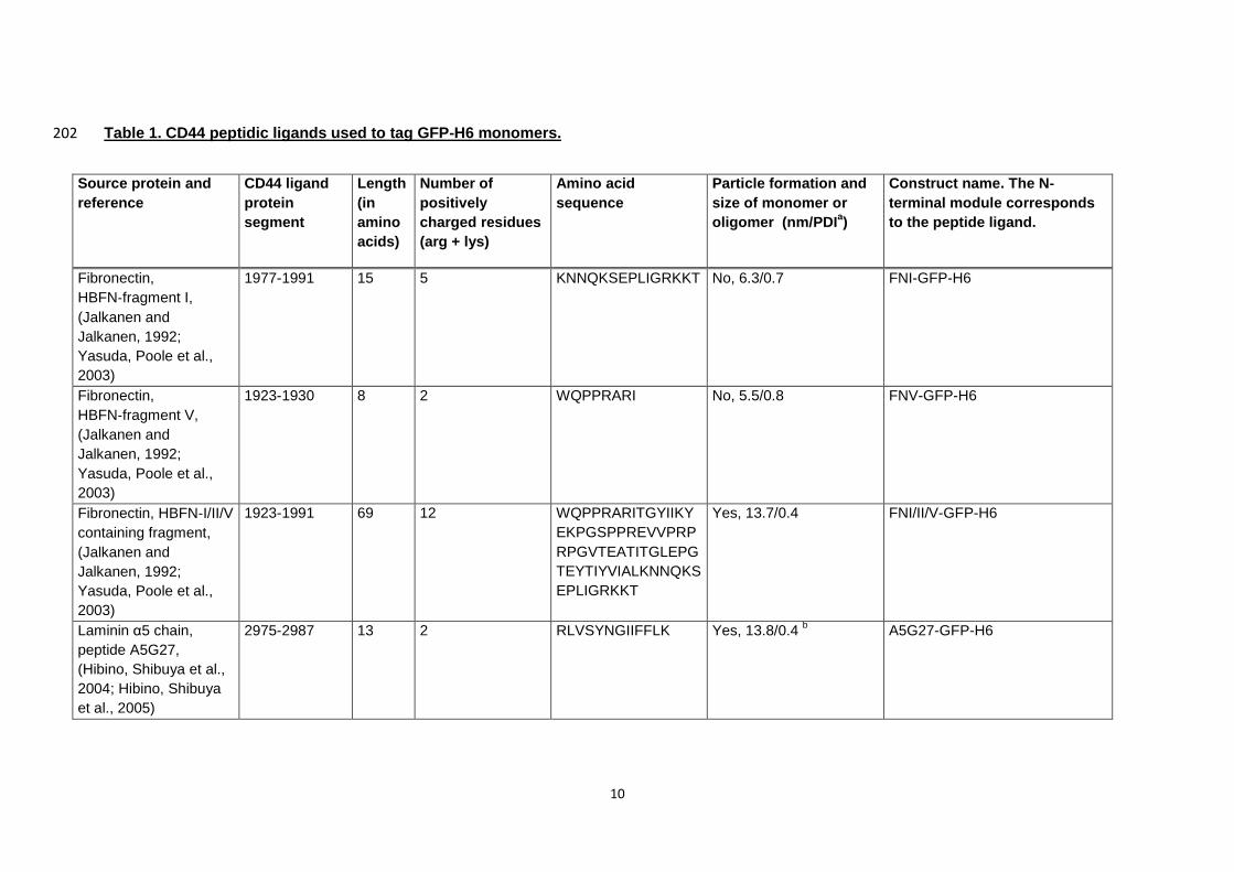
Table 1. CD44 peptidic ligands used to tag GFP-H6 monomers. 202
Source protein and
reference
CD44 ligand
protein
segment
Length
(in
amino
acids)
Number of
positively
charged residues
(arg + lys)
Amino acid
sequence
Particle formation and
size of monomer or
oligomer (nm/PDIa)
Construct name. The N-
terminal module corresponds
to the peptide ligand.
Fibronectin,
HBFN‐fragment I,
(Jalkanen and
Jalkanen, 1992;
Yasuda, Poole et al.,
2003)
1977-1991 15 5 KNNQKSEPLIGRKKT No, 6.3/0.7 FNI-GFP-H6
Fibronectin,
HBFN‐fragment V,
(Jalkanen and
Jalkanen, 1992;
Yasuda, Poole et al.,
2003)
1923-1930 8 2 WQPPRARI No, 5.5/0.8 FNV-GFP-H6
Fibronectin, HBFN‐I/II/V
containing fragment,
(Jalkanen and
Jalkanen, 1992;
Yasuda, Poole et al.,
2003)
1923-1991 69 12 WQPPRARITGYIIKY
EKPGSPPREVVPRP
RPGVTEATITGLEPG
TEYTIYVIALKNNQKS
EPLIGRKKT
Yes, 13.7/0.4 FNI/II/V-GFP-H6
Laminin α5 chain,
peptide A5G27,
(Hibino, Shibuya et al.,
2004; Hibino, Shibuya
et al., 2005)
2975-2987 13 2 RLVSYNGIIFFLK Yes, 13.8/0.4 b A5G27-GFP-H6
11
P7, phage display
derived peptide, (Park,
Lee et al., 2012)
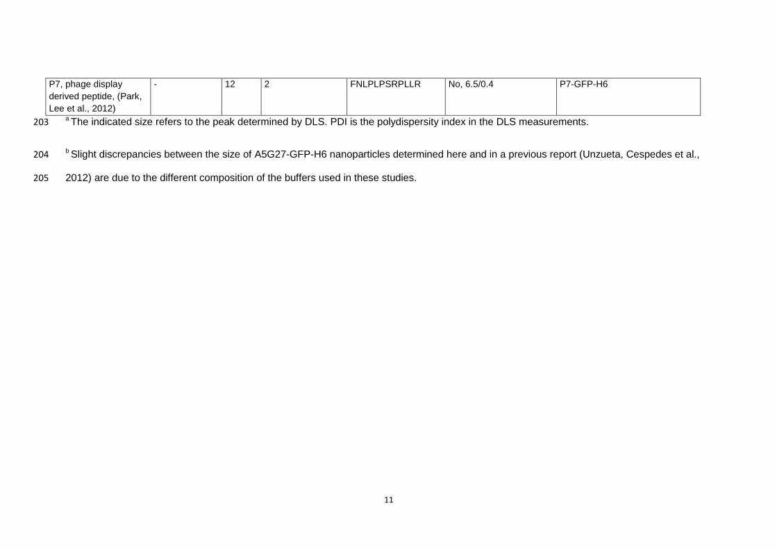
- 12 2 FNLPLPSRPLLR No, 6.5/0.4 P7-GFP-H6
a The indicated size refers to the peak determined by DLS. PDI is the polydispersity index in the DLS measurements. 203
b Slight discrepancies between the size of A5G27-GFP-H6 nanoparticles determined here and in a previous report (Unzueta, Cespedes et al., 204
2012) are due to the different composition of the buffers used in these studies.205
12
3. Results 206
Five known peptidic ligands of CD44 (FNI, FNV, FNI/II/V, A5G27 and P7, Table 1) 207
were fused to C-terminal H6-tagged GFP following a previously described approach 208
(Figure 1 A, (Unzueta, Ferrer-Miralles et al., 2012)), to construct multidomain GFP 209
protein versions with affinity for CD44+ cells. All these constructs were efficiently 210
produced in bacteria, resulting in full length polypeptides with expected mass and 211
predicted N-terminal amino acid sequence (see Supplementary Figure 1). Before 212
testing for biological properties, the potential self-assembling of protein monomers into 213
higher order entities was explored. According to a previously proposed model 214
(Unzueta, Ferrer-Miralles et al., 2012), the highly cationic peptide FNI/II/V was 215
expected to promote the formation of ordered oligomers within the nanoscale. In this 216
regard, FNI/II/V-GFP-H6 in solution peaked at around 14 nm by DLS (Figure 1 B, Table 217
1), indicative of nanoparticle formation. The unassembled GFP-H6 protein showed a 218
size of 6 nm (Vazquez, Roldan et al., 2010), and FNI, FNV and P7-empowered GFP 219
proteins peaked at the same size, indicating that they remained unassembled like the 220
parental GFP-H6 (Figure 1 B, Table 1). Unexpectedly, A5G27, even being poorly 221
cationic (Table 1), also promoted the formation of supramolecular entities of 14 nm 222
(Figure 1 B, Table 1). 223
When the nanoparticulate architecture of FNI/II/V-GFP-H6 and A5G27-GFP-H6 was 224
assessed by AFM, these proteins clearly organized as regular nanoparticles with a size 225
fully compatible with DLS determinations (Figure 1C-G). Some A5G27-GFP-H6 226
particles appeared as rings or pseudorings with a centred cavity (Figure 1E), and such 227
a ring-based architecture was generic in FNI/II/V-GFP-H6 samples, in which some of 228
the nanoparticles showed a pentameric organization (Figure 1F). In this regard, 229
molecular modelling of R9-GFP-H6 nanoparticles indicated a pentameric organization 230
of the constructs (Unzueta, Ferrer-Miralles et al., 2012; Vazquez, Cubarsi et al., 2010; 231
Vazquez, Roldan et al., 2010) and size-exclusion chromatography of several GFP and 232
13
iRFP-based nanoparticles also showed pentamers as a basic module resulting from 233
self-assembling in protein particles empowered by cationic stretches plus H6 234
(Cespedes, Unzueta et al., 2014). 235
To evaluate the biological properties of the fusion proteins, namely cell penetration and 236
eventual intracellular trafficking, we first checked the specific fluorescence of all fusion 237
proteins, as fluorescence emission is a convenient reporter to monitor cell binding and 238
internalization. As observed (Figure 2 A), fluorescence emission of the enhanced GFP 239
was not straightforward affected by protein fusion but it was reduced up to around 30-240
40 % of the parental protein in A5G27-GFP-H6 and FNI/II/V-GFP-H6. The coincidence 241
between fluorescence dropping and nanoparticle formation might be indicative of a 242
slight quenching effect associated to oligomer formation. Irrespectively of the precise 243
cause, fluorescence levels were well acceptable and sufficient for further analyses. An 244
important fraction of CD44+ MDA-MB-231 cells were fluorescent when exposed to 245
A5G27-GFP-H6 and FNI/II/V-GFP-H6 (around 80 % vs 40 % or less in the case of 246
other fusions, Figure 2B), and a higher global fluorescence emission was observed in 247
cell cultures exposed to these proteins when compared with alternative GFP versions 248
(between 1000 and 2000 vs up to 600, Figure 2C). In both experimental approaches 249
and as it was expected, GFP-H6 only rendered background values. In agreement with 250
quantitative data, confocal analyses of protein-exposed MDA-MB-231 cell cultures 251
confirmed the high penetrability of A5G27-GFP-H6 and FNI/II/V-GFP-H6 vs alternative 252
GFP versions, mostly showing a background uptake (Figure 2D). Note that as indicated 253
above, the lower specific fluorescence of these two constructs resulted in an 254
underestimation of the internalized material when compared to alternative ligands. 255
Considering the values presented in the Figure 2B, A5G27-GFP-H6 and FNI/II/V-GFP-256
H6 internalized 20 times more efficiently than the alternative fusion proteins. Again, the 257
parental, H6-tagged GFP was seen as totally excluded from cultured cells. The 258

14
incorporation of the internalized recombinant proteins into membranous vesicles was 259
clearly observed and it was indicative of endosomal uptake (Figure 2D). 260
Internalization of A5G27-GFP-H6 and FNI/II/V-GFP-H6 occurred very fast, and the 261
fraction of target cells and the amount of intracellular protein reached a plateau at 262
about 10 h (Figure 3A). Interestingly, the penetration of these nanoparticles did take 263
place without compromising cell viability (Figure 3B), again in agreement with the 264
occurrence of an endosomal route and supporting the full biocompatibility nature of 265
protein-only nanoparticles. In this context, both A5G27-GFP-H6 and FNI/II/V-GFP-H6 266
were fully stable when incubated in human serum (Figure 3C), a fact that confirmed the 267
structural robustness of the particles and prompted us to envisage a potential for 268
proper biodistribution of these materials in targeted drug delivery or diagnostic 269
applications. 270
To assess the receptor-driven specificity of the cell penetration, the uptake of protein 271
nanoparticles was explored in several cell lines, expressing and not expressing CD44, 272
namely MDA-MB-231, MCF-7, Hep G2 and HEK-293T. A differential cell penetrability 273
of A5G27-GFP-H6 and FNI/II/V-GFP-H6 was observed (Figure 4A), coincident with the 274
amount of cellular CD44 in the target cells (Figure 4B). This fact strongly supported the 275
CD44-dependence of cell binding. In this context, a commercial polyclonal anti-CD44 276
serum inhibited the entrance of both type of nanoparticles (Figure 5). Finally, we 277
wanted to determine if the externally mediated up- or down-regulation of CD44 278
expression could have enhancing or inhibiting effects on the penetration of 279
nanoparticles, as expected. The fibroblast growth factor 2 (FGF-2), a positive regulator 280
of CD44 (Grimme, Termeer et al., 1999; Jones, Tussey et al., 2000), stimulated the cell 281
penetrability of A5G27-GFP-H6 (Figure 6A), while the blocking of interleukin-10 (IL-10; 282
also a positive regulator of CD44, (Gee, Kryworuchko et al., 2004)) by a specific 283
antibody reduced the uptake of the nanoparticle (Figure 6B). By the combination of 284
15
these data, the CD44-targeting of the protein constructs developed here was fully 285
demonstrated. 286
287
16
4. Discussion 288
Developing tools for targeted drug delivery is a priority in targeted medicines of cancer 289
and other prevalent diseases (Ruoslahti, Bhatia et al., 2010; Vicent and Duncan, 2006). 290
While of the number of cell surface proteins identified as valuable markers are rapidly 291
expanding (Gonzalez-Angulo, Hennessy et al., 2010; Klonisch, Wiechec et al., 2008; 292
Mocellin, Lise et al., 2005; Nguyen and Massague, 2007; Ruoslahti, Bhatia et al., 2010; 293
Sawyers, 2008; Tjalsma, 2010), efficient and secure vehicles are still missing. While 294
consensus exists in that nanoscale containers are ideal for competent systemic 295
transport, diffusion in the tissue and cell penetrability (Mastrobattista, van der Aa et al., 296
2006), a fully biocompatible material remains to be developed. In this regard, the 297
number of nanomedicines so far approved by the medicament agencies is still limited 298
(Duncan and Gaspar, 2011). 299
A promising route to the generation of efficient vehicles for drug delivery is 300
conventional genetic engineering, since proteins are fully biocompatible and functional 301
macromolecules. The ability to recruit diverse peptides from distinct origins and with 302
different complementing activities in a single chain protein offers promise to generate 303
constructs showing the biological properties exhibited by viruses during infection. 304
These include stable systemic circulation, receptor targeting, internalization, 305
intracellular trafficking and accumulation into the appropriate compartment (Aris and 306
Villaverde, 2004; Ferrer-Miralles, Vazquez et al., 2008; Vazquez, Ferrer-Miralles et al., 307
2009; Vazquez, Ferrer-Miralles et al., 2008). Despite this potential, the exploration of 308
how protein-protein contacts could be engineered to construct protein-only 309
nanoparticles has been in general neglected. The adaptation of virus-like particles 310
(VLP) (Ma, Nolte et al., 2012), bacterial micro compartments (BMC) (Corchero and 311
Cedano, 2011), eukaryotic vaults (Rome and Kickhoefer, 2012) or other natural protein 312
constructs (Rodriguez-Carmona and Villaverde, 2010) poses severe limitations, as 313
conferring new tropisms might in general alters the stability of the particle. However, 314
17
recent lessons about how protein-protein interactions can be engineered for self-315
assembling in fully de novo designed protein constructs {Neus Ferrer-Miralles, 2013 316
1106 /id;Unzueta, 2014 1308 /id;Villaverde, 2012 7494 /id} should permit to approach 317
an ‘artificial virus’ strategy for the design of novel nanomedicines (Mastrobattista, van 318
der Aa et al., 2006). Importantly, the recent advances in systems and synthetic 319
biotechnology and industrial microbiology (Lee, Mattanovich et al., 2012) allow the 320
large scale biosynthesis of natural proteins and protein constructs, being biological 321
biofabrication highly versatile (Vazquez and Villaverde, 2013) and progressively more 322
competitive with regard to conventional chemical synthesis (Chen, 2012). 323
We have here constructed a set of five modular polypeptides intended to target CD44+ 324
cells (Table 1). Among them, two constructs (empowered by peptides A5G27 and 325
FNI/II/V respectively), self-organize as stable nanoparticles (Figure 1 and 3C) that 326
efficiently bind and internalize CD44+ target cells (Figure 4), accumulating in the 327
perinuclear and nuclear regions (Figure 2). Since the five peptides used here are very 328
well known ligands of CD44 (Table 1), the coincidence between nanoparticle formation 329
and internalization strongly suggests that the presentation of a given protein in form of 330
nanoparticle (versus the plain monomeric form) stimulates cell penetration. This is 331
probably because of the multiple ligand display and multivalent cross-linking at the cell 332
surface, favoring membrane wrapping (Jiang, Kim et al., 2008), and in the line of 333
nanoparticle size being a main determinant of interactions with cells (Jiang, Kim et al., 334
2008). Also, this is in agreement with the high penetrability found in natural oligomers 335
when displaying cell-binding peptides (Aris and Villaverde, 2003; Villaverde, Feliu et al., 336
1998). 337
While other engineering principles have been recently proposed to control protein self-338
assembling (Bai, Luo et al., 2013) (King, Sheffler et al., 2012), the approach based on 339
the end terminal fusion of a cationic peptide and a polyhistidine is not restricted to a 340
unique core protein. This versatility would be convenient to avoid immunogenicity of the 341
18
constructs by selecting homologous proteins in next generation-constructions. By using 342
the end-terminal peptide-pair strategy, the formation of nanoparticles with pentameric, 343
toroid-like organization had been previously predicted (Unzueta, Ferrer-Miralles et al., 344
2012; Vazquez, Roldan et al., 2010) and demonstrated by FESEM for a certain 345
category of proteins (those empowered by the cationic peptide T22) (Cespedes, 346
Unzueta et al., 2014). The highly resolutive AFM has confirmed this particular 347
architecture also for A5G27-GFP-H6 and FNI/II/V- GFP-H6 (Figure 1E,F), indicative 348
that the ring shaped distribution of the building blocks is not restricted to an unique type 349
of end-terminal tags. Being highly cationic (Table 1), the oligomerization of FNI/II/V-350
GFP-H6 was fully anticipated at the upstream stage, while the formation of A5G27-351
GFP-H6-based nanoparticles (being A5G27 poorly cationic) was unexpected. Acting 352
the amino terminal stretch both as architectonic tag and cell ligand, the promotion of 353
protein self-assembling by a non-cationic peptide expands, in any case, the spectrum 354
of potential ligands usable for the formation of cell-targeted nanoparticles, so far 355
restricted to cationic protein regions. 356
Laminin and fibronectin ligands bind CD44 through chondroitin and heparin-like GAG 357
side chains, especially by the heparin-sulphate found in CD44v3 and CD44v6 isoforms. 358
On the other hand, the alternative CD44 ligand HA binds to a binding site termed the 359
“link module” which is a domain expressed in all CD44 isoforms and it is located on 360
CD44 most exposed region (far from V3 and v6 variant regions) (Peach, Hollenbaugh 361
et al., 1993). Therefore, although HA could not be used as a competitor to probe the 362
CD44 targeting of the protein constructs developed here, the specificity in binding and 363
internalization was successfully demonstrated by the coincidence between CD44 levels 364
and penetrability (Figure 4), by inhibition mediated by a polyclonal anti-CD44 serum 365
(Figure 5), and though the external alternate regulation of CD44 levels and the 366
consequent variation in the efficiency of nanoparticle uptake (Figure 6). 367
19
In summary, we have developed smart and stable protein-only nanoparticles (A5G27-368
GFP-H6 and FNI/II/V-GFP-H6) as plastic agents that bind specifically CD44+ cells and 369
that are efficiently internalized by receptor-mediated endocytosis in absence of cell 370
toxicity. Interestingly, the vehicle itself is composed by fully functional (fluorescent, in 371
our model system) proteins, what opens a plethora of possibilities regarding the 372
targeted delivery of therapeutic polypeptides in form of nanoparticles. The 373
nanostructure gained by these proteins would desirably allow them escaping from renal 374
clearance, as the final size of the constructs is largely over the 6-7 nm cut-off. In 375
addition, protein nanoparticles could be loaded with chemically coupled conventional 376
drugs, as the principle of protein-drug coupling has been largely proved in already 377
licensed drugs (Elzoghby, Samy et al., 2012). Altogether, and according to recent data 378
proving the high stability in vivo (Cespedes, Unzueta et al., 2014) and efficient 379
biodistribution of similar protein-only modular constructs (Unzueta, Cespedes et al., 380
2012), the tools generated here are promising platforms as vehicles for drugs and 381
imaging agents, in the context of emerging nanomedines for breast cancer and other 382
metastatic CD44-linked tumours based on biocompatible and versatile protein 383
materials. 384
385
Acknowledgments 386
We appreciate the technical support of Fran Cortés from the Cell Culture Unit of Servei 387
de Cultius Cel.lulars Producció d’Anticossos i Citometria (SCAC, UAB), of the Servei 388
de Microscòpia and of Amable Bernabé from Soft Materials Service (ICMAB-389
CSIC/CIBER-BBN) and from Proteomics facility from UAB ( a member of ProteoRed-390
ISCIII network) . We are also indebted to the Protein Production Platform (CIBER-BBN 391
- UAB) for helpful technical assistance in protein production and purification 392
(http://www.ciber-bbn.es/en/programas/89-plataforma-de-produccion-de-proteinas-393
20
ppp). The authors also acknowledge the financial support granted to E.V. from FIS 394
(PI12/00327), to S. S. from FIS (PI11/01079), to E.V. and S.S. from The Marató de TV3 395
(TV32013-133930), to J.V from DGI (Grant POMAs CTQ2010-19501) and to A.V. from 396
MINECO (BIO2013-41019-P), from Generalitat de Catalunya (2014SGR-132) and from 397
the Centro de Investigación Biomédica en Red (CIBER) de Bioingeniería, 398
Biomateriales y Nanomedicina (NANOPROTHER and PENTI projects), financed by the 399
Instituto de Salud Carlos III with assistance from the European Regional Development 400
Fund. M.P. and U.U. received PhD fellowships from ISCIII and from UAB respectively. 401
W.T.is grateful to the Consejo Superior de Investigaciones Científicas (CSIC) for a 402
“JAE-pre”fellowship. A.V. has been distinguished with an ICREA ACADEMIA Award. 403
404
405
21
Reference List 406
407
Allen,M.J., Hud,N.V., Balooch,M., Tench,R.J., Siekhaus,W.J., Balhorn,R., 1992. Tip-radius-408
induced artifacts in AFM images of protamine-complexed DNA fibers. Ultramicroscopy, 42-44 ( 409
Pt B), 1095-1100. 410
Aris,A., Villaverde,A., 2003. Engineering nuclear localization signals in modular protein vehicles 411
for gene therapy. Biochem. Biophys. Res. Commun., 304, 625-631. 412
Aris,A., Villaverde,A., 2004. Modular protein engineering for non-viral gene therapy. Trends 413
Biotechnol., 22, 371-377. 414
Bai,Y., Luo,Q., Zhang,W., Miao,L., Xu,J., Li,H., Liu,J., 2013. Highly ordered protein nanorings 415
designed by accurate control of glutathione s-transferase self-assembly. J. Am. Chem Soc., 135, 416
10966-10969. 417
Bradford,M.M., 1976. A rapid and sensitive method for the quantitation of microgram 418
quantities of protein utilizing the principle of protein-dye binding. Anal. Biochem., 72, 248-254. 419
Cespedes,M.V., Unzueta,U., Tatkiewicz,W., Sanchez-Chardi,A., Conchillo-Sole,O., Alamo,P., 420
Xu,Z., Casanova,I., Corchero,J.L., Pesarrodona,M., Cedano,J., Daura,X., Ratera,I., Veciana,J., 421
Ferrer-Miralles,N., Vazquez,E., Villaverde,A., Mangues,R., 2014. In Vivo Architectonic Stability 422
of Fully de Novo Designed Protein-Only Nanoparticles. ACS Nano., 8, 4166-4176. 423
Chen,G.Q., 2012. New challenges and opportunities for industrial biotechnology. Microb. Cell 424
Fact., 11, 111. 425
22
Corchero,J.L., Cedano,J., 2011. Self-assembling, protein-based intracellular bacterial 426
organelles: emerging vehicles for encapsulating, targeting and delivering therapeutical 427
cargoes. Microb Cell Fact., 10, 92. 428
Duncan,R., Gaspar,R., 2011. Nanomedicine(s) under the microscope. Mol. Pharm., 8, 2101-429
2141. 430
Elzoghby,A.O., Samy,W.M., Elgindy,N.A., 2012. Albumin-based nanoparticles as potential 431
controlled release drug delivery systems. J. Control Release, 157, 168-182. 432
Ferrer-Miralles,N., Corchero,J.L., Kumar,P., Cedano,J.A., Gupta,K.C., Villaverde,A., Vazquez,E., 433
2011. Biological activities of histidine-rich peptides; merging Biotechnology and Nanomedicine. 434
Microb Cell Fact., 10, 101. 435
Ferrer-Miralles,N., Vazquez,E., Villaverde,A., 2008. Membrane-active peptides for non-viral 436
gene therapy: making the safest easier. Trends Biotechnol., 26, 267-275. 437
Gee,K., Kryworuchko,M., Kumar,A., 2004. Recent advances in the regulation of CD44 438
expression and its role in inflammation and autoimmune diseases. Arch. Immunol. Ther. Exp. 439
(Warsz. ), 52, 13-26. 440
Gonzalez-Angulo,A.M., Hennessy,B.T., Mills,G.B., 2010. Future of personalized medicine in 441
oncology: a systems biology approach. J. Clin. Oncol., 28, 2777-2783. 442
Goodison,S., Urquidi,V., Tarin,D., 1999. CD44 cell adhesion molecules. Mol. Pathol., 52, 189-443
196. 444
23
Grimme,H.U., Termeer,C.C., Bennett,K.L., Weiss,J.M., Schopf,E., Aruffo,A., Simon,J.C., 1999. 445
Colocalization of basic fibroblast growth factor and CD44 isoforms containing the variably 446
spliced exon v3 (CD44v3) in normal skin and in epidermal skin cancers. Br. J. Dermatol., 141, 447
824-832. 448
Hibino,S., Shibuya,M., Engbring,J.A., Mochizuki,M., Nomizu,M., Kleinman,H.K., 2004. 449
Identification of an active site on the laminin alpha 5 chain globular domain that binds to CD44 450
and inhibits malignancy. Cancer Res, 64, 4810-4816. 451
Hibino,S., Shibuya,M., Hoffman,M.P., Engbring,J.A., Hossain,R., Mochizuki,M., Kudoh,S., 452
Nomizu,M., Kleinman,H.K., 2005. Laminin alpha5 chain metastasis- and angiogenesis-inhibiting 453
peptide blocks fibroblast growth factor 2 activity by binding to the heparan sulfate chains of 454
CD44. Cancer Res., 65, 10494-10501. 455
Jalkanen,S., Jalkanen,M., 1992. Lymphocyte CD44 binds the COOH-terminal heparin-binding 456
domain of fibronectin. J. Cell Biol., 116, 817-825. 457
Jiang,W., Kim,B.Y., Rutka,J.T., Chan,W.C., 2008. Nanoparticle-mediated cellular response is 458
size-dependent. Nat. Nanotechnol., 3, 145-150. 459
Jones,M., Tussey,L., Athanasou,N., Jackson,D.G., 2000. Heparan sulfate proteoglycan isoforms 460
of the CD44 hyaluronan receptor induced in human inflammatory macrophages can function 461
as paracrine regulators of fibroblast growth factor action. J. Biol. Chem, 275, 7964-7974. 462
King,N.P., Sheffler,W., Sawaya,M.R., Vollmar,B.S., Sumida,J.P., Andre,I., Gonen,T., Yeates,T.O., 463
Baker,D., 2012. Computational design of self-assembling protein nanomaterials with atomic 464
level accuracy. Science, 336, 1171-1174. 465
24
Klonisch,T., Wiechec,E., Hombach-Klonisch,S., Ande,S.R., Wesselborg,S., Schulze-Osthoff,K., 466
Los,M., 2008. Cancer stem cell markers in common cancers - therapeutic implications. Trends 467
Mol. Med., 14, 450-460. 468
Koo,H., Huh,M.S., Sun,I.C., Yuk,S.H., Choi,K., Kim,K., Kwon,I.C., 2011. In vivo targeted delivery 469
of nanoparticles for theranosis. Acc. Chem. Res., 44, 1018-1028. 470
Lee,S.Y., Mattanovich,D., Villaverde,A., 2012. Systems metabolic engineering, industrial 471
biotechnology and microbial cell factories. Microb. Cell Fact., 11, 156. 472
Ma,Y., Nolte,R.J., Cornelissen,J.J., 2012. Virus-based nanocarriers for drug delivery. Adv. Drug 473
Deliv. Rev., 64, 811-825. 474
Marangoni,E., Lecomte,N., Durand,L., de,P.G., Decaudin,D., Chomienne,C., Smadja-Joffe,F., 475
Poupon,M.F., 2009. CD44 targeting reduces tumour growth and prevents post-chemotherapy 476
relapse of human breast cancers xenografts. Br. J. Cancer, 100, 918-922. 477
Mastrobattista,E., van der Aa,M.A., Hennink,W.E., Crommelin,D.J., 2006. Artificial viruses: a 478
nanotechnological approach to gene delivery. Nat. Rev. Drug Discov., 5, 115-121. 479
Mocellin,S., Lise,M., Nitti,D., 2005. Targeted therapy for colorectal cancer: mapping the way. 480
Trends Mol. Med., 11, 327-335. 481
Neus Ferrer-Miralles, Escarlata Rodriguez-Carmona, Jose Luis Corchero, Elena Garcia-Fruitos, 482
Esther Vazquez, Antonio Villaverde, 2013. Engineering protein self-assembling in protein-based 483
nanomedicines for drug delivery and gene therapy. Crit Rev. Biotechnol, in press. 484
25
Nguyen,D.X., Massague,J., 2007. Genetic determinants of cancer metastasis. Nat. Rev. Genet., 485
8, 341-352. 486
Park,H.Y., Lee,K.J., Lee,S.J., Yoon,M.Y., 2012. Screening of peptides bound to breast cancer 487
stem cell specific surface marker CD44 by phage display. Mol. Biotechnol, 51, 212-220. 488
Peach,R.J., Hollenbaugh,D., Stamenkovic,I., Aruffo,A., 1993. Identification of hyaluronic acid 489
binding sites in the extracellular domain of CD44. J. Cell Biol., 122, 257-264. 490
Peer,D., Karp,J.M., Hong,S., Farokhzad,O.C., Margalit,R., Langer,R., 2007. Nanocarriers as an 491
emerging platform for cancer therapy. Nat. Nanotechnol., 2, 751-760. 492
Rezler,E.M., Khan,D.R., Lauer-Fields,J., Cudic,M., Baronas-Lowell,D., Fields,G.B., 2007. Targeted 493
drug delivery utilizing protein-like molecular architecture. J. Am. Chem. Soc., 129, 4961-4972. 494
Rodriguez-Carmona,E., Villaverde,A., 2010. Nanostructured bacterial materials for innovative 495
medicines. Trends Microbiol., 18, 423-430. 496
Rome,L.H., Kickhoefer,V.A., 2012. Development of the Vault Particle as a Platform Technology. 497
ACS Nano.. 498
Ruoslahti,E., Bhatia,S.N., Sailor,M.J., 2010. Targeting of drugs and nanoparticles to tumors. J. 499
Cell Biol., 188, 759-768. 500
Sauter,A., Kloft,C., Gronau,S., Bogeschdorfer,F., Erhardt,T., Golze,W., Schroen,C., Staab,A., 501
Riechelmann,H., Hoermann,K., 2007. Pharmacokinetics, immunogenicity and safety of 502
26
bivatuzumab mertansine, a novel CD44v6-targeting immunoconjugate, in patients with 503
squamous cell carcinoma of the head and neck. Int. J. Oncol., 30, 927-935. 504
Sawyers,C.L., 2008. The cancer biomarker problem. Nature, 452, 548-552. 505
Tijink,B.M., Buter,J., de,B.R., Giaccone,G., Lang,M.S., Staab,A., Leemans,C.R., van Dongen,G.A., 506
2006. A phase I dose escalation study with anti-CD44v6 bivatuzumab mertansine in patients 507
with incurable squamous cell carcinoma of the head and neck or esophagus. Clin. Cancer Res., 508
12, 6064-6072. 509
Tjalsma,H., 2010. Identification of biomarkers for colorectal cancer through proteomics-based 510
approaches. Expert. Rev. Proteomics., 7, 879-895. 511
Unzueta,U., Cespedes,M.V., Ferrer-Miralles,N., Casanova,I., Cedano JA, Corchero JL, Domingo-512
Espin,J., Villaverde A, Mangues,R., Vazquez E, 2012. Intracellular CXCR4+ cell targeting with 513
T22-empowered protein-only nanoparticles. Int. J. Nanomedicine, 7, 4533-4544. 514
Unzueta,U., Ferrer-Miralles,N., Cedano,J., Zikung,X., Pesarrodona,M., Saccardo,P., Garcia-515
Fruitos,E., Domingo-Espin,J., Kumar,P., Gupta,K.C., Mangues,R., Villaverde,A., Vazquez,E., 516
2012. Non-amyloidogenic peptide tags for the regulatable self-assembling of protein-only 517
nanoparticles. Biomaterials, 33, 8714-8722. 518
Unzueta,U., Saccardo,P., Domingo-Espin,J., Cedano,J., Conchillo-Sole,O., Garcia-Fruitos,E., 519
Cespedes,M.V., Corchero,J.L., Daura,X., Mangues,R., Ferrer-Miralles,N., Villaverde,A., 520
Vazquez,E., 2014. Sheltering DNA in self-organizing, protein-only nano-shells as artificial 521
viruses for gene delivery. Nanomedicine.10:535-541. 522
27
Vazquez,E., Cubarsi,R., Unzueta,U., Roldan,M., Domingo-Espin,J., Ferrer-Miralles,N., 523
Villaverde,A., 2010. Internalization and kinetics of nuclear migration of protein-only, arginine-524
rich nanoparticles. Biomaterials, 31, 9333-9339. 525
Vazquez,E., Ferrer-Miralles,N., Mangues,R., Corchero,J.L., Schwartz S Jr, Villaverde,A., 2009. 526
Modular protein engineering in emerging cancer therapies. Curr. Pharm. Des, 15, 893-916. 527
Vazquez,E., Ferrer-Miralles,N., Villaverde,A., 2008. Peptide-assisted traffic engineering for 528
nonviral gene therapy. Drug Discov. Today, 13, 1067-1074. 529
Vazquez,E., Roldan,M., Diez-Gil,C., Unzueta,U., Domingo-Espin,J., Cedano,J., Conchillo,O., 530
Ratera,I., Veciana,J., Daura,X., Ferrer-Miralles,N., Villaverde,A., 2010. Protein nanodisk 531
assembling and intracellular trafficking powered by an arginine-rich (R9) peptide. 532
Nanomedicine. (Lond), 5, 259-268. 533
Vazquez,E., Villaverde,A., 2013. Microbial biofabrication for nanomedicine: biomaterials, 534
nanoparticles and beyond. Nanomedicine (Lond), 8, 1895-1898. 535
Vicent,M.J., Duncan,R., 2006. Polymer conjugates: nanosized medicines for treating cancer. 536
Trends Biotechnol., 24, 39-47. 537
Villaverde,A., Feliu,J.X., Aris,A., Harbottle,R.P., Benito,A., Coutelle,C., 1998. A cell adhesion 538
peptide from foot-and-mouth disease virus can direct cell targeted delivery of a functional 539
enzyme. Biotechnol Bioeng, 59, 294-301. 540

28
Villaverde,A., Garcia-Fruitos,E., Rinas,U., Seras-Franzoso,J., Kosoy,A., Corchero,J.L., Vazquez,E., 541
2012. Packaging protein drugs as bacterial inclusion bodies for therapeutic applications. 542
Microb Cell Fact., 11, 76. 543
Yasuda,T., Poole,A.R., Shimizu,M., Nakagawa,T., Julovi,S.M., Tamamura,H., Fujii,N., 544
Nakamura,T., 2003. Involvement of CD44 in induction of matrix metalloproteinases by a 545
COOH-terminal heparin-binding fragment of fibronectin in human articular cartilage, in 546
culture. Arthritis and Rheumatism, 48, 1271-1280. 547
Zoller,M., 2011. CD44: can a cancer-initiating cell profit from an abundantly expressed 548
molecule? Nat. Rev. Cancer, 11, 254-267. 549
550
551
552

29
Figure legends: 553
554
Figure 1. Construction and nanoscale characterization of CD44-targeted protein 555
nanoparticles. A) Schematic representation of the gene fusion scheme used in this 556
study. L represents a CD44 ligand that in some cases also has an architectonic role. B) 557
Size distribution of the protein constructs determined by DLS. Numerical values are 558
given in Table 1. C) AFM images of randomly selected A5G27-GFP-H6 nanoparticles. 559
D) Topography cross- section of one randomly selected isolated A5G27-GFP-H6 560
nanoparticle. E) Topography cross- section of two ring shaped A5G27-GFP-H6 nano 561
particles. F) AFM images of randomly selected FNI/II/V-GFP-H6 nanoparticles, 562
showing the topography cross- section of one isolated particle. G) Images of a 563
pentameric particle are shown. Measurements have been done with a tip radius of 2 564
nm and thus the width (but not the high) of the particles is inherently overestimated. An 565
AFM image is a convolution of the imaging tip shape/size with the actual shape of the 566
imaged object (Allen, Hud et al., 1992). Thus, one will observe broadening of the 567
sample features. 568
569
Figure 2. Internalization of CD44-targeted protein nanoparticles. A) Specific 570
fluorescence of the different protein constructs in comparison to that of the parental 571
GFP-H6 (in green). The specific green fluorescence of the parental protein is 1,021 572
fluorescence units (FU)/ug. B) Percentage of MDA-MB-231 cells that are fluorescent 573
after 24 h exposure to the multidomain proteins. C) Green fluorescence emitted by 574
MDA-MB-231 cells after 24 h exposure to multidomain proteins. D) Confocal sections 575
or projections of MDA-MB-231 cells upon 24 h of exposure to multidomain proteins. 576
Bars indicate 20 µm. E) Details of target cells during the uptake of fusion proteins, 577
indicating the exogenous material (in green) included in endosomes (red signal). 578

30
Merging into yellow is evident in some cases. A 3D projection is included in the case of 579
FNI/II/V-GFP-H6. 580
581
Figure 3. Kinetics of cellular internalization of CD44-targeted protein nanoparticles, 582
cytotoxicity and protein serum stability. A) Time course cell penetration of protein-only 583
nanoparticles at 1.5 µM. Percentatge of protein-internalised cells (left) and cell EGFP 584
fluorescence intensity (right). B) MTT viability analysis of MDA-MB-231cells upon 585
exposure to different doses of protein nanoparticles for 24,48 and 72h.. C) Stability of 586
protein nanoparticles in human. Fluorescence emission of serum samples with 587
nanoparticle incubation at different time point up to 24 hours. Soluble 24h corresponds 588
to fluorescence of soluble fraction from 24 hours sample after centrifugation to discard 589
nanoparticle aggregation/precipitation 590
Figure 4: Specific internalization of CD44-targeted protein nanoparticles in CD44-591
expressing and not expressing cells. A) Percentage of cells uptaking A5-GFP-H6 (left) 592
and FNI/II/V-GFP-H6 (right) in CD44-overexpressing MDA-MB-231 and MCF-7 lines 593
and in Hep G2 and HEK 293 T lines. B) Histograms of CD44-expressing cell population 594
from CD44+ cell lines MDA-MB-231 and MCF-7 and CD44- cell lines HepG2 and HEK-595
293-T. APC-anti-CD44 marked cells (red) are compared with isotopic control cells 596
(black). FL4-H axis corresponds to APC intensity. The percentage of CD44+ cells is 597
indicated. 598
Figure 5: Anti-CD44 mediated inhibition of nanoparticle internalization. Decrease on 599
the intensity of intracellular fluorescence mediated by 0.3 µM of either A5G27-GFP-H6 600
or FNI/II/V-GFP-H6 (control: back bar) when adding 0.3 µM polyclonal anti-CD44 (ratio 601
1:1). A control of GFP-H6 incubation with anti-CD44 is presented. 602
603
31
Figure 6: Modulation of nanoparticle internalization through CD44 regulation. A) 604
Enhanced cell entry (left) and fluorescence intensity (right) of A5G27-GFP-H6 605
incubation at 0.3 µM after 1 h of cell exposure to increasing amounts of FGF2 (ratios 606
1:1, 1:10 and 1:30) due to CD44 receptor overexpression mediated by FGF2-H6. B) 607
Reduced cell entry (left) and fluorescence intensity (right) of A5G27-GFP-H6 incubation 608
at 0.3 µM after 1 h of cell exposure to anti-IL10 antibody due to an inhibition of IL-10-609
induced CD44 expression mediated by polyclonal anti-IL10. 610