Embed Size (px)
Citation preview
The Journal of Cell Biology, Volume 146, Number 1, July 12, 1999 113–124http://www.jcb.org 113
Intracellular Localization of Proteasomal Degradation of aViral Antigen
Luis C. Antón,* Ulrich Schubert,*
‡
Igor Bacík,* Michael F. Princiotta,* Pamela A. Wearsch,*
James Gibbs,* Patricia M. Day,
§
Claudio Realini,
i
Martin C. Rechsteiner,
i
Jack R. Bennink,*and Jonathan W. Yewdell*
*Laboratory of Viral Diseases, National Institute of Allergy and Infectious Diseases, Bethesda, Maryland 20892;
‡
Heinrich-Pette Institute, University of Hamburg, Hamburg, Germany;
§
Laboratory of Cellular Oncology, National Cancer Institute, Bethesda,
Maryland 20892; and
i
Department of Biochemistry, University of Utah, Salt Lake City, Utah 84112
Abstract.
To better understand proteasomal degrada-tion of nuclear proteins and viral antigens we studied mutated forms of influenza virus nucleoprotein (NP) that misfold and are rapidly degraded by proteasomes. In the presence of proteasome inhibitors, mutated NP (dNP) accumulates in highly insoluble ubiquitinated and nonubiquitinated species in nuclear substructures known as promyelocytic leukemia oncogenic domains (PODs) and the microtubule organizing center (MTOC). Immunofluorescence revealed that dNP re-cruits proteasomes and a selective assortment of molec-ular chaperones to both locales, and that a similar (though less dramatic) effect is induced by proteasome inhibitors in the absence of dNP expression. Biochemi-cal evidence is consistent with the idea that dNP is de-
livered to PODs/MTOC in the absence of proteasome inhibitors. Restoring proteasome activity while block-ing protein synthesis results in disappearance of dNP from PODs and the MTOC and the generation of a ma-jor histocompatibility complex class I–bound peptide derived from dNP but not NP. These findings demon-strate that PODs and the MTOC serve as sites of pro-teasomal degradation of misfolded dNP and probably cellular proteins as well, and imply that antigenic pep-tides are generated at one or both of these sites.
Key words: antigen presentation • molecularchaperone • nuclear proteins proteolysis • ubiquitin/immunology • proteasome
P
ROTEASOMES
are complex multisubunit proteasesabundant in the nucleus and cytosol that function inall eukaryotic cells to degrade damaged or un-
wanted proteins. The major means of tagging proteins forproteasomal destruction is covalent modification by multi-ple chains of ubiquitin (Ub)
1
(Ciechanover, 1998). Incuba-tion of cells with proteasome inhibitors results in the ac-cumulation of polyubiquitinated proteins, and in mostcircumstances, cell death (Bogyo et al., 1997; Lee andGoldberg, 1998).
Proteasomes participate in the generation of many ofthe peptides that are displayed on the cell surface in acomplex with class I molecules of the major histocompati-bility complex for surveillance by the immune system(York and Rock, 1996). Peptides are generated indiscrimi-nately from host gene products and gene products of vi-ruses and other intracellular pathogens (Pamer and Cress-well, 1998). Discrimination between self and nonselfpeptides is performed at the level of the antigen receptorexpressed by CD8
1
T lymphocytes. There is sufficientplasticity in the system to enable T cell recognition ofsome self antigens, which accounts for the generation ofmost defined T cell responses to tumors.
Although there has been rapid progress in understand-ing the function of the proteasome-Ub system in cellularmetabolism, and more specifically, its role in generatingantigenic peptides, many important questions remain, in-cluding the involvement of molecular chaperones in theprocess, the intracellular sites of ubiquitination and pro-teasomal degradation, and the nature of substrates thatprovide antigenic peptides. In the present communicationwe describe findings with a metabolically unstable form of
Address all correspondence to J.W. Yewdell or J.R. Bennink, Room 213,Building 4, 4 Center Drive, NIH, Bethesda, MD 20892-0440. Tel.: 301-402-4602. Fax: 301-402-7362. E-mail: [email protected] or [email protected]
1.
Abbreviations used in this paper:
APL, acute PML; dNP, misfolded nu-cleoprotein; GFP, green fluorescent protein; LC, lactacystin; LCSM, laserconfocal scanning microscope; MTOC, microtubule organizing center;NP, influenza virus nucleoprotein; NP
pep
, NP with SIINFEKL appendedto the COOH terminus; OVA, ovalbumin; PBC, primary biliary cirrhosis;PML, promyelocytic leukemia; POD, PML oncogenic domain; RAR, ret-inoic acid receptor; rVV, recombinant vaccinia virus; Ub, ubiquitin; zLLL,cbz-LeuLeuLeucinal.
CORE Metadata, citation and similar papers at core.ac.uk
Provided by PubMed Central
The Journal of Cell Biology, Volume 146, 1999 114
influenza virus nucleoprotein (NP) that provide some ini-tial answers to these questions.
Materials and Methods
Cells
143B human osteosarcoma cells lacking thymidine kinase and HeLa cellswere obtained from the American Type Culture Collection and main-tained in DMEM supplemented with 7.5% (vol/vol) fetal bovine serum inan air/CO
2
(91%/9%) atmosphere at 37
8
C. L929 cells expressing K
b
froma transfected gene were maintained similarly.
Viruses
Recombinant vaccinia viruses (rVVs) were constructed by standard meth-odology (Chakrabarti et al., 1985) using a modified form of pSC11 withmultiple cloning sites to express inserted genes under the control of thep7.5 promoter active early and late in VV replication. dNPpep was con-structed by inserting an oligonucleotide encoding KEKEKNKLKR-KKLENKDKKDEERNKIREE at position 333 in NP
pep
. Green fluores-cent protein (GFP) constructs were created by fusing NP
pep
or dNP
pep
encoding constructs with a cDNA encoding EGFP (Clontech), a red-shifted version of GFP modified by two substitutions (Phe
64
→
Leu,Ser
65
→
Thr), and 190 synonymous coding alterations to match humancodon usage. For cloning convenience, sequences encoding the spacerpeptide ARDPPVAT were inserted between the COOH terminus of theNP
pep
constructs and initiating Met of GFP. Most VV infections were per-formed with media containing cytosine arabinoside at 40
m
g/ml to limit ex-pression to viral gene products expressed under the control of early pro-moters. This reduces the morphological alterations in cells induced by VVand also controls for the blockade in late VV gene expression induced byproteasome inhibitors (Antón et al., 1998).
Antibodies
Antipeptide antisera were prepared by immunizing rabbits with syntheticpeptides conjugated to KLH. Peptides corresponded to PR8 NP se-quences 2-12
1
Cys and Cys
1
488-498; the extraneous Cys being used forconjugation and coupling to beads. The NH
2
-terminal–specific serum wasaffinity purified against the synthetic peptide disulfide coupled to Sul-foLink
®
beads (Pierce Chemical Co.) and absorbed multiple times againstuninfected fixed and permeabilized cells to remove antibodies specific forcellular proteins. The COOH-terminal antiserum could be used withoutfurther purification. Monoclonal antibodies specific for the proteasome(clones MCP20 and MCP21) were generously provided by K.B. Hendil(University of Copenhagen, Copenhagen, Denmark). Human sera fromprimary biliary cirrhosis (PBC) patients containing anti–promyelocyticleukemia (PML) oncogenic domain (POD) antibodies were generouslyprovided by D.B. Bloch (Harvard Medical School, Charlestown, MA) andJ. Liang (NIDDK, Bethesda, MD). The colocalization of human antibodystaining of PODs in TK
2
cells with the anti-PML mAb was confirmed.The commercial sources of the following antibodies are as listed: poly Ubmouse mAb, clone FK2, Nippon Bio-Test Laboratories; PML mouse mAb,clone PG-M3, Santa Cruz Biotechnologies;
g
-tubulin mouse mAb, clonegtu-88, Sigma Chemical Co.; HSP27 mouse mAb, clone G3.1, HSP40 rab-bit Ab (SPA-400), HSP47 mouse mAb, clone M16.10A1, HSP56 mousemAb, clone KN382/EC1, HSP60 mouse mAb, clone LK-1, HSC70 ratmAb, clone 1B5, HSP70 mouse mAb, clone C92F3A-5, HSP90, rat mAb,clone 16F1, and HSP110 rabbit Ab (SPA-1101), all from StressGen Bio-technologies.
Immunofluorescence
Cells were grown overnight on acid-cleaned, 0.17-mm-thick, 12-mm-diam-eter glass coverslips placed in 24-well plates, infected with virus, and incu-bated as indicated in the text and figure legends. At the appropriate time,cells were fixed by incubation with 3% (wt/vol) paraformaldehyde in PBSfor 20 min and permeabilized by 2 min of treatment with 1% (vol/vol) NP-40 in PBS. After quenching of formaldehyde with 200 mM glycine/PBS,cells were incubated with a mixture of primary antibodies diluted in PBSsupplemented with 5% (vol/vol) donkey serum, usually overnight, at 4
8
C,washed, and incubated for 2–8 h in the same diluent containing secondarydonkey antibodies conjugated to DTAF, Texas red, or Cy5, specific for
mouse, human, rabbit, or rat Ig (Jackson ImmunoResearch). Coverslipswere mounted on glass slides with Fluoromount-G (Southern Biotechnol-ogy) containing 15-
m
m-diameter beads to prevent cell compression, andimages collected with a Bio-Rad MRC1024 laser scanning confocal ZeissAxioplan microscope, using a 63
3
planapochromat oil immersion objec-tive. Controls established the specificity of fluorochrome-conjugated anti-bodies for their respective Igs, and that signals in green, red, and far redchannels were derived from the respective fluor. Digital images were as-sembled using Adobe Photoshop software and printed with a Fujix Pic-trography digital printer (Fuji). For cytofluorography, cells were incu-bated with mAbs for 30 min on ice, washed, and incubated with rabbitanti–mouse Ig conjugated to fluorescein (Dako). Cells were suspended inPBS containing ethidium homodimer (Molecular Probes), and analyzedusing a FACScalibur
®
cytofluorograph (Becton Dickinson). Live cellswere gated based on scattering properties and low ethidium homodimerstaining.
Biochemical Procedures
Western Blotting.
Confluent 143B cells grown in 6-well plates were in-fected with rVVs and incubated for 150 min and then for 360 min in thepresence or absence of 20
m
M cbz-LeuLeuLeucinal (zLLL). Plates weretransferred to an ice bath, and subjected to a sequential extraction to pre-pare the nuclear matrix, according to the protocol of Staufenbiel and Dep-pert (1984), with the exception that all buffers contained Complete
®
pro-tease inhibitor cocktail (EDTA-free; Boehringer Mannheim) and 10
m
MzLLL. Equivalent amounts of samples from each step of the procedurewere acetone precipitated, and precipitates resuspended in boiling SDS-PAGE sample buffer (Laemmli, 1970). Samples were also preparedfrom rVV-infected HeLa cells by suspending cells in ice-cold buffer con-taining 50 mM Tris-hydrochloride (pH 8.0), 5 mM EDTA, 100 mM NaCl,0.5% (wt/vol) CHAPS ([3-[(3-cholamidopropyl)-dimethyl-ammonio]-1-pro-panesulfonate]), 0.2% (wt/vol) deoxycholate, and then mixed with anequal volume of boiling SDS-PAGE sample buffer. After electrophoresis,proteins were transferred to Immobilon
P membranes (Millipore) intransfer buffer (Towbin et al., 1979) lacking SDS. Membranes were incu-bated overnight with TBS-Casein (Bio-Rad), and then with rabbit anti-NPantibodies, followed by peroxidase-labeled anti–rabbit IgG (BoehringerMannheim). Blots were developed using the ECL system (Pierce Chemi-cal Co.), and luminescence recorded by Biomax MR film (Kodak). Imageswere digitized by a flat bed scanner, assembled using Adobe Photoshopsoftware, and printed with a Fujix Pictrography digital printer.
[
35
S]Met Labeling.
143 cells infected 3 h previously were incubated inMet-free DMEM with 100
m
M lactacystin (LC) for 40 min, and then radi-olabeled by 5 min of incubation with [
35
S]Met. After washing, cells werechased at 37
8
C for up to 120 min in Met containing DMEM. At appropri-ate times, 2
3
10
6
cells were removed to ice. Cells were incubated with 1%(vol/vol) Triton X-100 (TX100) containing buffer and centrifuged at15,000
g
for 10 min. Supernatants and pellets were suspended in boilingSDS-PAGE sample buffer boiled for 5 min, and analyzed by SDS-PAGE.Images of autoradiographs of the dried gels were digitized by a flat bedscanner, assembled using Adobe Photoshop software and printed with aFujix Pictrography digital printer. The radioactivity present in dried gelswas quantitated using a PhosphorImager (Molecular Devices) and thescreens supplied by the manufacturer. The VV protein shown in Fig. 2 awas used as an internal standard for normalization of the amount of pro-tein recovered from each sample.
Results
Modification of NP Results in Enhanced Generation of Antigenic Peptides
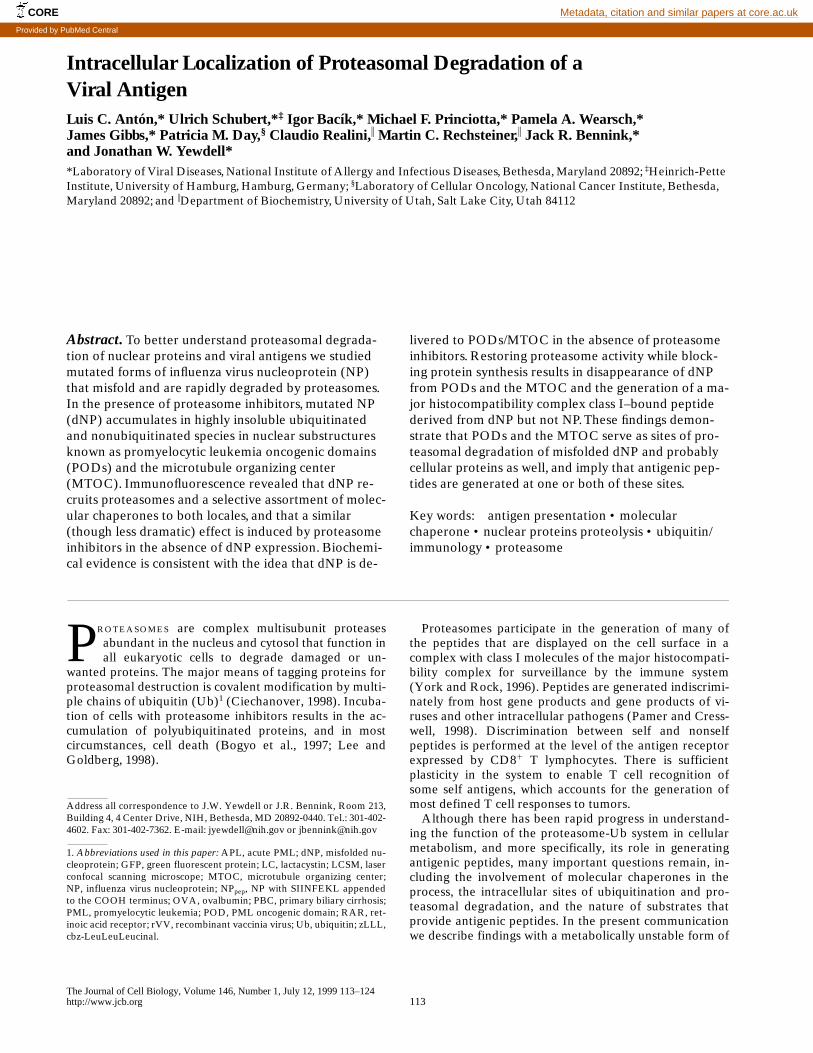
The NP from the PR8 influenza virus is a 498-residue pro-tein that is transported to the nucleus via multiple nuclearlocalization sequences (Wang et al., 1997). We geneticallyengineered NP to contain a 29-residue sequence nearlyidentical to that from JAK1 kinase proposed to enhancethe generation of antigenic peptides by targeting the pro-tein to proteasomes (Realini et al., 1994). In addition, weappended to the COOH terminus a peptide correspondingto residues 257–264 from chicken ovalbumin (OVA). This
Antón et al.
Viral Antigen Degradation
115
peptide binds tightly to the H-2 K
b
MHC class I molecule,and K
b–
Ova
257-264
complexes can be easily quantitatedcytofluorographically using a mAb (25-D1.16) specific forthis complex (Porgador et al., 1997). As a control, thepeptide was also expressed at the COOH terminus ofwild-type NP (this is termed NP
pep
and the other constructdNP
pep
).
After 6 h of infection of L-K
b
cells with rVVs express-ing NP
pep
or dNP
pep
, approximately threefold more K
b–
Ova
257-264
complexes were present on the surface of VV-dNP
pep
–infected cells as determined cytofluorographicallyafter indirect immunofluorescence (Fig. 1, top histogram).Incubation of cells with the highly specific irreversible pro-teasome inhibitor LC resulted in the nearly complete inhi-bition of complex expression from the chimeric proteinsand from OVA, the parent protein (Fig. 1, bottom histo-gram). There was only a slight effect on cells infected witha rVV expressing Ova
257-264
as a cytosolic minigene prod-uct (a single Met is appended to the NH
2
terminus to en-able efficient translation), consistent with the interpreta-tion that LC acts by preventing proteasome liberation ofOva
257-264
(or a proteolytic intermediate) from NP
pep
,dNP
pep
, and OVA, and not by interfering with VV geneexpression or delivery and loading of peptides onto K
b
molecules.
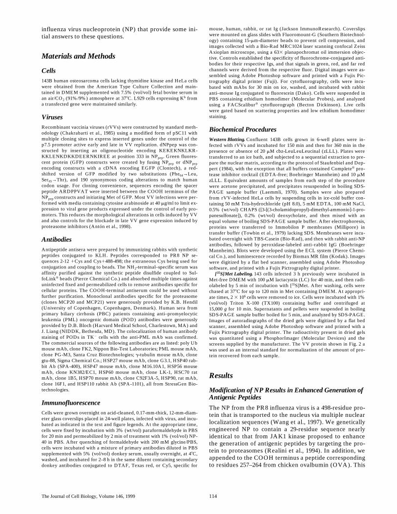
Metabolic Stability of dNP
pep
and NP
pep
Increased protein degradation is associated with enhancedgeneration of antigenic peptides (Tevethia et al., 1983;Townsend et al., 1988). To investigate the more efficientproduction of Ova
257-264
from dNP
pep
, we examined themetabolic stability of dNP
pep
and NP
pep
in the presenceand absence of LC. rVV-infected cells were labeled for 5min with [
35
S]Met and chased for up to 2 h at 37
8
C. Pro-teins present in TX100-soluble and insoluble materialwere separated by SDS-PAGE and the amounts of NPpresent in gel migrating with the expected mobility weredetermined by PhosphorImager analysis (Fig. 2). The totalamount of NP
pep
recovered remained nearly constantthroughout the chase period, with the solubility decreasing
Figure 1. Proteasome-dependent production of Ova257-264 fromVV-encoded proteins. L-Kb cells incubated for 90 min in the ab-sence (top) or presence (bottom) of 50 mM LC were infected for8 h with the indicated rVV in the presence or absence of LC, re-spectively. Cells were stained with 25-D1.16 mAb and analyzedby cytofluorography.
Figure 2. Proteasome-dependent degradation ofdNPpep. 143B cells were pulse radiolabeled with[35S]Met and chased for up to 120 min at 378C inthe presence or absence of LC. Radioactive pro-teins soluble in 1% TX100 (sol) or insoluble(ins) were separated by SDS-PAGE and thebands corresponding to dNPpep or NPpep locatedin the dried gel (a) and quantitated (b) after nor-malization using a VV-encoded protein as an in-ternal standard (indicated as VV).
The Journal of Cell Biology, Volume 146, 1999 116
in a time-dependent manner to a plateau value. This corre-sponds with the transport of NP
pep
into the nucleus whereit is partially TX100 insoluble. As expected, the processwas unaffected by LC. By contrast, in the absence of LC,recovery of both soluble and insoluble dNP
pep
decreasedwith time. Importantly, LC selectively increased the recov-ery of insoluble dNP
pep
, without affecting soluble dNP
pep
.We interpret this data to indicate that, first, insertion ofthe JAK1 sequence into dNP
pep
greatly enhances its degra-dation by proteasomes, and, second, that the form di-gested by proteasomes is insoluble in TX100. These find-ings predict that incubation of cells with proteasomeinhibitors should result in the accumulation of dNP
pep
incells.
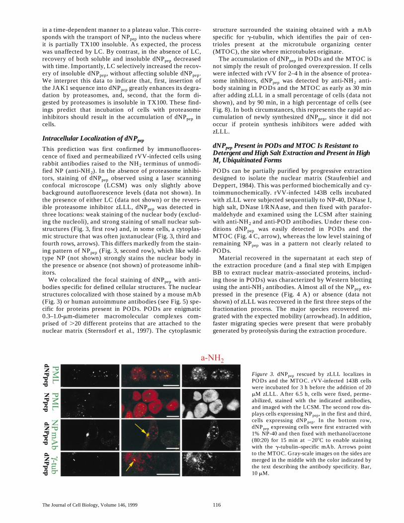
Intracellular Localization of dNP
pep
This prediction was first confirmed by immunofluores-cence of fixed and permeabilized rVV-infected cells usingrabbit antibodies raised to the NH
2
terminus of unmodi-fied NP (anti-NH
2
). In the absence of proteasome inhibi-tors, staining of dNP
pep
observed using a laser scanningconfocal microscope (LCSM) was only slightly abovebackground autofluorescence levels (data not shown). Inthe presence of either LC (data not shown) or the revers-ible proteasome inhibitor zLLL, dNP
pep
was detected inthree locations: weak staining of the nuclear body (exclud-ing the nucleoli), and strong staining of small nuclear sub-structures (Fig. 3, first row) and, in some cells, a cytoplas-mic structure that was often juxtanuclear (Fig. 3, third andfourth rows, arrows). This differs markedly from the stain-ing pattern of NP
pep
(Fig. 3, second row), which like wild-type NP (not shown) strongly stains the nuclear body inthe presence or absence (not shown) of proteasome inhib-itors.
We colocalized the focal staining of dNP
pep
with anti-bodies specific for defined cellular structures. The nuclearstructures colocalized with those stained by a mouse mAb(Fig. 3) or human autoimmune antibodies (see Fig. 5) spe-cific for proteins present in PODs. PODs are enigmatic0.3–1.0-
m
m-diameter macromolecular complexes com-prised of
.
20 different proteins that are attached to thenuclear matrix (Sternsdorf et al., 1997). The cytoplasmic
structure surrounded the staining obtained with a mAbspecific for
g
-tubulin, which identifies the pair of cen-trioles present at the microtubule organizing center(MTOC), the site where microtubules originate.
The accumulation of dNP
pep
in PODs and the MTOC isnot simply the result of prolonged overexpression. If cellswere infected with rVV for 2–4 h in the absence of protea-some inhibitors, dNP
pep
was detected by anti-NH
2
anti-body staining in PODs and the MTOC as early as 30 minafter adding zLLL in a small percentage of cells (data notshown), and by 90 min, in a high percentage of cells (seeFig. 8). In both circumstances, this represents the rapid ac-cumulation of newly synthesized dNP
pep
, since it did notoccur if protein synthesis inhibitors were added withzLLL.
dNP
pep
Present in PODs and MTOC Is Resistant to Detergent and High Salt Extraction and Present in High M
r
Ubiquitinated Forms
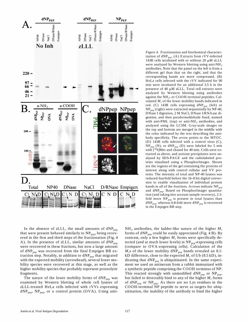
PODs can be partially purified by progressive extractiondesigned to isolate the nuclear matrix (Staufenbiel andDeppert, 1984). This was performed biochemically and cy-toimmunochemically. rVV-infected 143B cells incubatedwith zLLL were subjected sequentially to NP-40, DNase I,high salt, DNase I/RNAase, and then fixed with parafor-maldehyde and examined using the LCSM after stainingwith anti-NH
2
and anti-POD antibodies. Under these con-ditions dNP
pep
was easily detected in PODs and theMTOC (Fig. 4 C, arrow), whereas the low level staining ofremaining NP
pep
was in a pattern not clearly related toPODs.
Material recovered in the supernatant at each step ofthe extraction procedure (and a final step with EmpigenBB to extract nuclear matrix–associated proteins, includ-ing those in PODs) was characterized by Western blottingusing the anti-NH
2
antibodies. Almost all of the NP
pep
ex-pressed in the presence (Fig. 4 A) or absence (data notshown) of zLLL was recovered in the first three steps of thefractionation process. The major species recovered mi-grated with the expected mobility (arrowhead). In addition,faster migrating species were present that were probablygenerated by proteolysis during the extraction procedure.
Figure 3. dNPpep rescued by zLLL localizes inPODs and the MTOC. rVV-infected 143B cellswere incubated for 3 h before the addition of 20mM zLLL. After 6.5 h, cells were fixed, perme-abilized, stained with the indicated antibodies,and imaged with the LCSM. The second row dis-plays cells expressing NPpep, in the first and third,cells expressing dNPpep. In the bottom row,dNPpep expressing cells were first extracted with1% NP-40 and then fixed with methanol/acetone(80:20) for 15 min at 2208C to enable stainingwith the g-tubulin–specific mAb. Arrows pointto the MTOC. Gray-scale images on the sides aremerged in the middle with the color indicated bythe text describing the antibody specificity. Bar,10 mM.
Antón et al.
Viral Antigen Degradation
117
In the absence of zLLL, the small amounts of dNP
pep
that were present behaved similarly to NP
pep
, being recov-ered in the first and third steps of the fractionation (Fig. 4A). In the presence of zLLL, similar amounts of dNP
pep
were recovered in these fractions, but now a large amountof dNP
pep
was recovered from the final Empigen BB ex-traction step. Notably, in addition to dNP
pep
that migratedwith the expected mobility (arrowhead), several lower mo-bility species were recovered at this stage, as well as thehigher mobility species that probably represent proteolyticfragments.
The nature of the lower mobility forms of dNP
pep
wasexamined by Western blotting of whole cell lysates ofzLLL-treated HeLa cells infected with rVVs expressingdNP
pep
, NPpep, or a control protein (OVA). Using anti-
NH2 antibodies, the ladder-like nature of the higher Mrforms of dNPpep could be easily appreciated (Fig. 4 B). Bycontrast, only a few higher Mr forms were specifically de-tected (and at much lower levels) in NPpep-expressing cells(compare to OVA-expressing cells). Calculation of theMrs of the lower mobility dNPpep bands revealed an 8.1-kD difference, close to the expected Mr of Ub (8.5 kD), in-dicating that dNPpep is ubiquitinated. In the same experi-ment we used an antiserum from a rabbit immunized witha synthetic peptide comprising the COOH terminus of NP.This reacted strongly with unmodified dNPpep or NPpep,but failed to detectably bind to any of the higher Mr formsof dNPpep or NPpep. As there are no Lys residues in theCOOH-terminal NP peptide to serve as targets for ubiq-uitination, the inability of the antibody to bind the higher
Figure 4. Fractionation and biochemical character-ization of dNPpep. (A) Extracts from rVV-infected143B cells incubated with or without 20 mM zLLLwere analyzed by Western blotting using anti-NH2antibodies. Note that the panel on the left is from adifferent gel than that on the right, and that thecorresponding bands are more compressed. (B)HeLa cells infected with the rVV indicated for 90min were incubated for an additional 3.5 h in thepresence of 40 mM zLLL. Total cell extracts wereanalyzed by Western blotting using antibodiesagainst the NH2- or COOH-terminal peptides. Cal-culated Mr of the lower mobility bands indicated inred. (C) 143B cells expressing dNPpep (left) orNPpep (right) were extracted sequentially by NP-40,DNase I digestion, 2 M NaCl, DNase I/RNAase di-gestion, and then paraformaldehyde fixed, stainedwith anti-PML (top) or anti-NH2 antibodies, andanalyzed using the LCSM. Gray-scale images onthe top and bottom are merged in the middle withthe color indicated by the text describing the anti-body specificity. The arrow points to the MTOC.(D) 143B cells infected with a control virus (C),NPpep (N), or dNPpep (D) were labeled for 5 minwith [35S]Met and chased for 40 min. Cells were ex-tracted as above, and acetone precipitates were an-alyzed by SDS-PAGE and the radiolabeled pro-teins visualized using a PhosphorImager. Shownare the regions of the gel containing the proteins ofinterest along with control cellular and VV pro-teins. The intensity of total and NP-40 lysates wasreduced fourfold before the 16–8 bit digital conver-sion to enable visualization of individual proteinbands in all of the fractions. Arrows indicate NPpepand dNPpep. Based on PhosphorImager quantita-tion (and taking into account sample recovery), 2.3-fold more NPpep is present in total lysates thandNPpep, whereas 6.8-fold more dNPpep is recoveredin the Empigen BB step.
The Journal of Cell Biology, Volume 146, 1999 118
Mr forms may be due to either cleavage of a short segmentof the COOH terminus, or to steric effects of ubiquitina-tion on antibody access to the COOH terminus.
Based on these findings we conclude first that the bulkof dNPpep that is normally degraded by proteasomes accu-mulates in TX100/NP-40–insoluble forms concentrated inPODs and the MTOC when proteasomes are inhibited,and second, that a fraction of this material is present inmodified high Mr forms resulting at least in part fromubiquitination. Due to uncertainties associated with effi-ciencies of recovering and detecting antigens in Westernblots, the ratio of ubiquitinated to nonubiquitinateddNPpep cannot be determined by this method. It is worthnoting, however, that at least some of the loss in the totalamount of [35S]Met labeled dNPpep in LC-treated cells re-covered over the 2-h chase period (Fig. 2 b) is due to ubiq-uitination with its attendant alteration in electrophoreticmobility.
Transport of dNPpep to PODs/MTOC in the Absence of Proteasome Inhibitors
The failure to detect dNPpep in PODs/MTOC in the ab-sence of proteasome inhibitors raises two possibilities: thedelivery of dNPpep to PODs/MTOC occurs only when pro-teasomes are blocked; and dNPpep is delivered to PODs/MTOC in the absence of proteasome inhibitors but is de-graded too rapidly for cytochemical detection.
To address this issue, we labeled rVV-infected cells ex-pressing NPpep or dNPpep with [35S]Met for 5 min, chasedfor 40 min, sequentially fractionated cells as above, andanalyzed the fractions by SDS-PAGE, again taking advan-tage of the shut down of host proteins to visualize NP anddNPpep in an antibody-independent manner (Fig. 4 D).Consistent with the prior results, more NPpep (z2.3-fold) ispresent in total cell lysates than dNPpep, and the bulk of
NPpep is recovered in the first three fractionation steps. Bycontrast, less dNPpep is recovered from the DNase andhigh salt extracts, whereas its recovery in the Empigen BBextraction step is enhanced approximately sevenfold rela-tive to NPpep. This finding is consistent with the idea thatdNPpep is delivered to PODs in the absence of proteasomeinhibitors.
Effects of zLLL and dNPpep Expression on the Distribution of Cellular Constituents of the Degradation Machinery
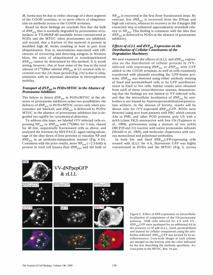
We next examined the effects of zLLL and dNPpep expres-sion on the distribution of cellular proteins in rVV-infected cells expressing dNPpep or dNPpep with GFPadded to the COOH terminus, as well as cells transientlytransfected with plasmids encoding the GFP-fusion pro-teins. dNPpep was detected using either antibody stainingof fixed and permeabilized cells or by GFP autofluores-cence in fixed or live cells. Similar results were obtainedfrom each of these vector/detection systems, demonstrat-ing that the findings are not limited to VV-infected cells,and that the intracellular localization of dNPpep by anti-bodies is not biased by fixation/permeabilization/penetra-tion artifacts. In the interest of brevity, results will beshown only for rVV-expressed dNPpepGFP. PODs weredetected using sera from patients with PBC which containAbs to PML and other POD proteins, poly Ub with amAb (clone FK2) nonreactive with free Ub (Fujimuro etal., 1994), proteasomes using a mixture of two mAbs(MCP20 and 21) reactive with native proteasome subunits(Hendil et al., 1995), and molecular chaperones with vari-ous monoclonal and polyclonal antibodies.
In both live and fixed dNPpepGFP-expressing cellstreated with zLLL for 4 h, fluorescent GFP was highlyconcentrated in PODs and the MTOC (Fig. 5, arrows
Figure 5. Effect of dNP expression on intracellularlocalization of components of the Ub-proteasomepathway. 143B cells infected for 4 h with VV-dNPpepGFP were incubated for an additional 4 h inthe presence of 10 mM zLLL, fixed, permeabilizedand stained for cellular components using the anti-bodies indicated. dNPpepGFP was located by its au-tofluorescence. Gray-scale images in each columnare merged on the bottom with the color indicatedby the text describing the antibody specificity. Ar-rows point to the MTOC. Bar, 10 mm.
Antón et al. Viral Antigen Degradation 119
point to the MTOC). The autofluorescence of dNPpepGFPindicates that the GFP domain is properly conformed,demonstrating that dNPpepGFP need not be completelydenatured to localize to these structures. The accumula-tion of dNPpepGFP in these sites was accompanied by re-cruitment of poly Ub, proteasomes, and HSC70 from theirnormal diffuse distribution in the nucleus and cytoplasm,often to the extent that staining was reduced elsewhere inthe cell (compare to Fig. 6; the distribution of protea-somes, not shown, is similar to poly Ub). While dNPpepand poly Ub filled the MTOC, in many cells proteasomesand HSC70 formed a ring around MTOC. The redistribu-tion of cellular proteins is a specific effect of inhibitingproteasomes, as similar results were obtained with LC(data not shown). A survey of mAbs specific for other mo-lecular chaperones (data not shown) revealed HSP27 re-cruited PODs similarly to HSC70 and somewhat lessstrongly to the MTOC, and HSP70 was recruited weaklyto both sites. The distribution of a number of other cytoso-lic chaperones (HSP110, HSP90, HSP60, HSP56, HSP47,and HSP40) was not noticeably affected by dNPpep expres-sion, and none were concentrated in either PODs or theMTOC.
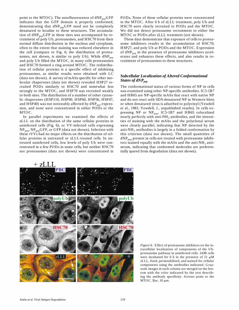
In parallel experiments we examined the effects ofzLLL on the distribution of the same cellular proteins inuninfected cells (Fig. 6), or VV-infected cells expressingNPpep, NPpepGFP, or GFP (data not shown). Infection withthese rVVs had no major effects on the distribution of cel-lular proteins in untreated or zLLL-treated cells. In un-treated uninfected cells, low levels of poly Ub were con-centrated in a few PODs in some cells, but neither HSC70nor proteasomes (data not shown) were concentrated in
PODs. None of these cellular proteins were concentratedin the MTOC. After 6 h of zLLL treatment, poly Ub andHSC70 were clearly recruited to PODs and the MTOC.We did not detect proteasome recruitment to either theMTOC or PODs after zLLL treatment (not shown).
These data demonstrate that exposure of cells to protea-some inhibitors results in the accumulation of HSC70,HSP27, and poly Ub at PODs and the MTOC. Expressionof dNPpep in the presence of proteasome inhibitors accel-erates and enhances these effects, and also results in re-cruitment of proteasomes to these structures.
Subcellular Localization of Altered Conformational States of dNPpep
The conformational status of various forms of NP in cellswas examined using other NP-specific antibodies. IC5-1B7and HB65 are NP-specific mAbs that react with native NPand do not react with SDS-denatured NP in Western blotsor when denatured virus is adsorbed to polyvinyl (Yewdellet al., 1981; Yewdell, J., unpublished results). In cells ex-pressing NP or NPpep, IC5-1B7 and HB65 colocalizednearly perfectly with anti-NH2 antibodies, and the intensi-ties of staining with the mAbs and the polyclonal serumwere closely parallel, indicating that NP detected by theanti-NH2 antibodies is largely in a folded conformation bythis criterion (data not shown). The small quantities ofdNPpep present in cells not treated with proteasome inhibi-tors stained equally with the mAbs and the anti-NH2 anti-serum, indicating that conformed molecules are preferen-tially spared from degradation (data not shown).
Figure 6. Effect of proteasome inhibitors on the in-tracellular localization of components of the Ub-proteasome pathway in uninfected cells. 143B cellswere incubated for 6 h in the presence of 21 mMzLLL, fixed, permeabilized, and stained for cellularcomponents using the antibodies indicated. Gray-scale images in each column are merged on the bot-tom with the color indicated by the text describ-ing the antibody specificity. Arrows point to theMTOC. Bar, 10 mm.
The Journal of Cell Biology, Volume 146, 1999 120
In dNPpep-expressing cells incubated with proteasomeinhibitors, the mAbs failed to stain PODs, while intenselystaining the MTOC (Fig. 3). This indicates that dNPpep res-cued by proteasome inhibitors exists in multiple confor-mations, and that most or all dNPpep in PODs is at leastpartially unfolded.
In additional experiments (data not shown), we studiedthe intracellular distribution of NP constructs using theanti-COOH antiserum for immunofluorescence. Whentested against VV-NP– or VV-NPpep–infected cells, stain-ing with this serum closely paralleled staining with HB65or IC5-1B7 as detected by double immunofluorescence.When used to stain dNPpep rescued by proteasome inhibi-tors, it strongly stained both the MTOC and PODs. Sincethis antiserum does not bind to ubiquitinated forms ofdNPpep (Fig. 4 B), this extends the biochemical data todemonstrate that nonubiquitinated dNPpep is present inPODs and the MTOC.
Extension of Findings to Other Forms of NP
We examined the behavior of two other forms of rapidlydegraded PR8 NP, one consisting of the first 168 residuesof the protein (NP1-168), the other full length NP withamino acid substitutions at residues 148 (Y→H) and 282(G→R) (NPDM). Both colocalized to PODs and theMTOC in a proteasome inhibitor–dependent manner asdemonstrated using anti-NH2 Abs, and recruited the samearray of cellular proteins as dNPpep (data not shown). Incontrast to dNPpep, NPDM was detected in PODs by theHB65 mAb, demonstrating that a more conformed formof NP can localize to PODs.
It was even possible to induce wild-type NP to localize
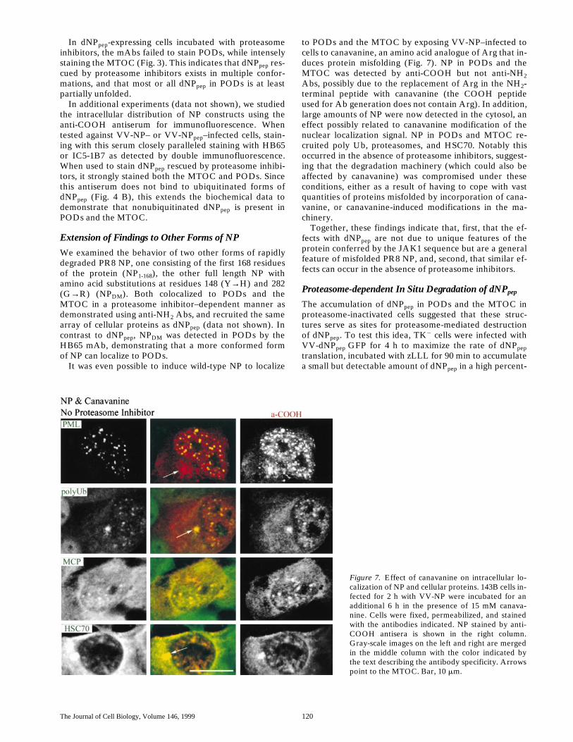
to PODs and the MTOC by exposing VV-NP–infected tocells to canavanine, an amino acid analogue of Arg that in-duces protein misfolding (Fig. 7). NP in PODs and theMTOC was detected by anti-COOH but not anti-NH2Abs, possibly due to the replacement of Arg in the NH2-terminal peptide with canavanine (the COOH peptideused for Ab generation does not contain Arg). In addition,large amounts of NP were now detected in the cytosol, aneffect possibly related to canavanine modification of thenuclear localization signal. NP in PODs and MTOC re-cruited poly Ub, proteasomes, and HSC70. Notably thisoccurred in the absence of proteasome inhibitors, suggest-ing that the degradation machinery (which could also beaffected by canavanine) was compromised under theseconditions, either as a result of having to cope with vastquantities of proteins misfolded by incorporation of cana-vanine, or canavanine-induced modifications in the ma-chinery.
Together, these findings indicate that, first, that the ef-fects with dNPpep are not due to unique features of theprotein conferred by the JAK1 sequence but are a generalfeature of misfolded PR8 NP, and, second, that similar ef-fects can occur in the absence of proteasome inhibitors.
Proteasome-dependent In Situ Degradation of dNPpep
The accumulation of dNPpep in PODs and the MTOC inproteasome-inactivated cells suggested that these struc-tures serve as sites for proteasome-mediated destructionof dNPpep. To test this idea, TK2 cells were infected withVV-dNPpep GFP for 4 h to maximize the rate of dNPpeptranslation, incubated with zLLL for 90 min to accumulatea small but detectable amount of dNPpep in a high percent-
Figure 7. Effect of canavanine on intracellular lo-calization of NP and cellular proteins. 143B cells in-fected for 2 h with VV-NP were incubated for anadditional 6 h in the presence of 15 mM canava-nine. Cells were fixed, permeabilized, and stainedwith the antibodies indicated. NP stained by anti-COOH antisera is shown in the right column.Gray-scale images on the left and right are mergedin the middle column with the color indicated bythe text describing the antibody specificity. Arrowspoint to the MTOC. Bar, 10 mm.
Antón et al. Viral Antigen Degradation 121
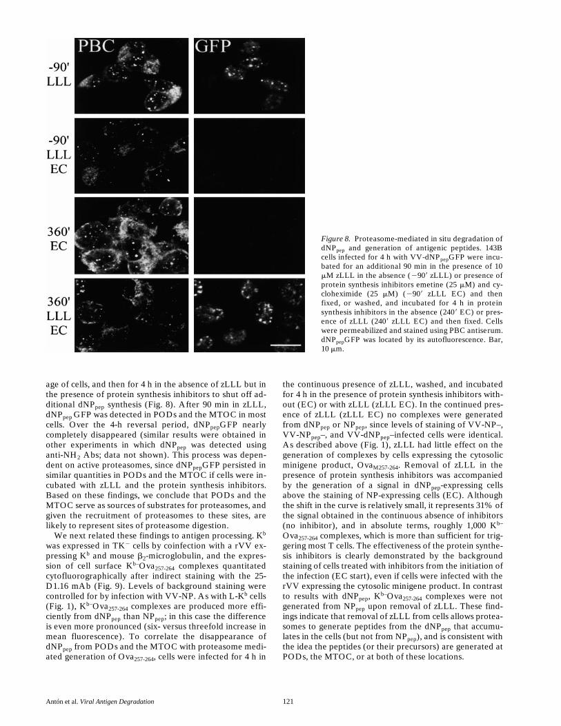
age of cells, and then for 4 h in the absence of zLLL but inthe presence of protein synthesis inhibitors to shut off ad-ditional dNPpep synthesis (Fig. 8). After 90 min in zLLL,dNPpep GFP was detected in PODs and the MTOC in mostcells. Over the 4-h reversal period, dNPpepGFP nearlycompletely disappeared (similar results were obtained inother experiments in which dNPpep was detected usinganti-NH2 Abs; data not shown). This process was depen-dent on active proteasomes, since dNPpepGFP persisted insimilar quantities in PODs and the MTOC if cells were in-cubated with zLLL and the protein synthesis inhibitors.Based on these findings, we conclude that PODs and theMTOC serve as sources of substrates for proteasomes, andgiven the recruitment of proteasomes to these sites, arelikely to represent sites of proteasome digestion.
We next related these findings to antigen processing. Kb
was expressed in TK2 cells by coinfection with a rVV ex-pressing Kb and mouse b2-microglobulin, and the expres-sion of cell surface Kb–Ova257-264 complexes quantitatedcytofluorographically after indirect staining with the 25-D1.16 mAb (Fig. 9). Levels of background staining werecontrolled for by infection with VV-NP. As with L-Kb cells(Fig. 1), Kb–Ova257-264 complexes are produced more effi-ciently from dNPpep than NPpep; in this case the differenceis even more pronounced (six- versus threefold increase inmean fluorescence). To correlate the disappearance ofdNPpep from PODs and the MTOC with proteasome medi-ated generation of Ova257-264, cells were infected for 4 h in
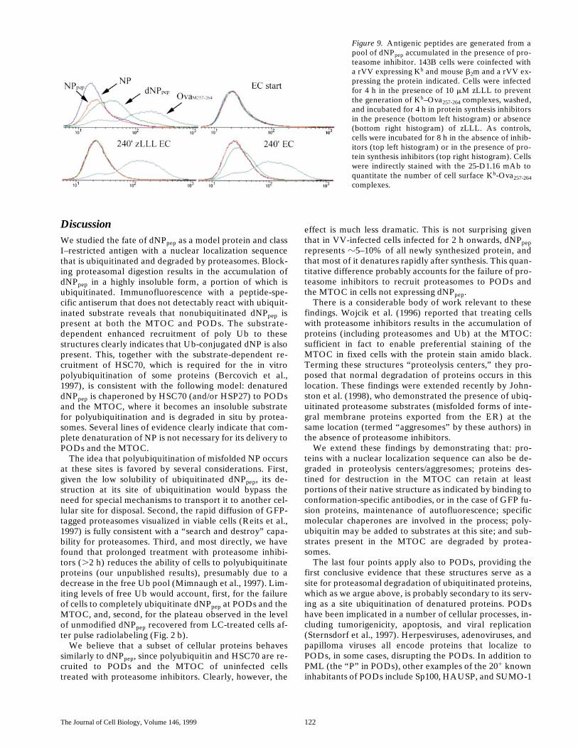
the continuous presence of zLLL, washed, and incubatedfor 4 h in the presence of protein synthesis inhibitors with-out (EC) or with zLLL (zLLL EC). In the continued pres-ence of zLLL (zLLL EC) no complexes were generatedfrom dNPpep or NPpep, since levels of staining of VV-NP–,VV-NPpep–, and VV-dNPpep–infected cells were identical.As described above (Fig. 1), zLLL had little effect on thegeneration of complexes by cells expressing the cytosolicminigene product, OvaM257-264. Removal of zLLL in thepresence of protein synthesis inhibitors was accompaniedby the generation of a signal in dNPpep-expressing cellsabove the staining of NP-expressing cells (EC). Althoughthe shift in the curve is relatively small, it represents 31% ofthe signal obtained in the continuous absence of inhibitors(no inhibitor), and in absolute terms, roughly 1,000 Kb–
Ova257-264 complexes, which is more than sufficient for trig-gering most T cells. The effectiveness of the protein synthe-sis inhibitors is clearly demonstrated by the backgroundstaining of cells treated with inhibitors from the initiation ofthe infection (EC start), even if cells were infected with therVV expressing the cytosolic minigene product. In contrastto results with dNPpep, Kb–Ova257-264 complexes were notgenerated from NPpep upon removal of zLLL. These find-ings indicate that removal of zLLL from cells allows protea-somes to generate peptides from the dNPpep that accumu-lates in the cells (but not from NPpep), and is consistent withthe idea the peptides (or their precursors) are generated atPODs, the MTOC, or at both of these locations.
Figure 8. Proteasome-mediated in situ degradation ofdNPpep and generation of antigenic peptides. 143Bcells infected for 4 h with VV-dNPpepGFP were incu-bated for an additional 90 min in the presence of 10mM zLLL in the absence (2909 zLLL) or presence ofprotein synthesis inhibitors emetine (25 mM) and cy-cloheximide (25 mM) (2909 zLLL EC) and thenfixed, or washed, and incubated for 4 h in proteinsynthesis inhibitors in the absence (2409 EC) or pres-ence of zLLL (2409 zLLL EC) and then fixed. Cellswere permeabilized and stained using PBC antiserum.dNPpepGFP was located by its autofluorescence. Bar,10 mm.
The Journal of Cell Biology, Volume 146, 1999 122
DiscussionWe studied the fate of dNPpep as a model protein and classI–restricted antigen with a nuclear localization sequencethat is ubiquitinated and degraded by proteasomes. Block-ing proteasomal digestion results in the accumulation ofdNPpep in a highly insoluble form, a portion of which isubiquitinated. Immunofluorescence with a peptide-spe-cific antiserum that does not detectably react with ubiquit-inated substrate reveals that nonubiquitinated dNPpep ispresent at both the MTOC and PODs. The substrate-dependent enhanced recruitment of poly Ub to thesestructures clearly indicates that Ub-conjugated dNP is alsopresent. This, together with the substrate-dependent re-cruitment of HSC70, which is required for the in vitropolyubiquitination of some proteins (Bercovich et al.,1997), is consistent with the following model: denatureddNPpep is chaperoned by HSC70 (and/or HSP27) to PODsand the MTOC, where it becomes an insoluble substratefor polyubiquitination and is degraded in situ by protea-somes. Several lines of evidence clearly indicate that com-plete denaturation of NP is not necessary for its delivery toPODs and the MTOC.
The idea that polyubiquitination of misfolded NP occursat these sites is favored by several considerations. First,given the low solubility of ubiquitinated dNPpep, its de-struction at its site of ubiquitination would bypass theneed for special mechanisms to transport it to another cel-lular site for disposal. Second, the rapid diffusion of GFP-tagged proteasomes visualized in viable cells (Reits et al.,1997) is fully consistent with a “search and destroy” capa-bility for proteasomes. Third, and most directly, we havefound that prolonged treatment with proteasome inhibi-tors (.2 h) reduces the ability of cells to polyubiquitinateproteins (our unpublished results), presumably due to adecrease in the free Ub pool (Mimnaugh et al., 1997). Lim-iting levels of free Ub would account, first, for the failureof cells to completely ubiquitinate dNPpep at PODs and theMTOC, and, second, for the plateau observed in the levelof unmodified dNPpep recovered from LC-treated cells af-ter pulse radiolabeling (Fig. 2 b).
We believe that a subset of cellular proteins behavessimilarly to dNPpep, since polyubiquitin and HSC70 are re-cruited to PODs and the MTOC of uninfected cellstreated with proteasome inhibitors. Clearly, however, the
effect is much less dramatic. This is not surprising giventhat in VV-infected cells infected for 2 h onwards, dNPpeprepresents z5–10% of all newly synthesized protein, andthat most of it denatures rapidly after synthesis. This quan-titative difference probably accounts for the failure of pro-teasome inhibitors to recruit proteasomes to PODs andthe MTOC in cells not expressing dNPpep.
There is a considerable body of work relevant to thesefindings. Wojcik et al. (1996) reported that treating cellswith proteasome inhibitors results in the accumulation ofproteins (including proteasomes and Ub) at the MTOC:sufficient in fact to enable preferential staining of theMTOC in fixed cells with the protein stain amido black.Terming these structures “proteolysis centers,” they pro-posed that normal degradation of proteins occurs in thislocation. These findings were extended recently by John-ston et al. (1998), who demonstrated the presence of ubiq-uitinated proteasome substrates (misfolded forms of inte-gral membrane proteins exported from the ER) at thesame location (termed “aggresomes” by these authors) inthe absence of proteasome inhibitors.
We extend these findings by demonstrating that: pro-teins with a nuclear localization sequence can also be de-graded in proteolysis centers/aggresomes; proteins des-tined for destruction in the MTOC can retain at leastportions of their native structure as indicated by binding toconformation-specific antibodies, or in the case of GFP fu-sion proteins, maintenance of autofluorescence; specificmolecular chaperones are involved in the process; poly-ubiquitin may be added to substrates at this site; and sub-strates present in the MTOC are degraded by protea-somes.
The last four points apply also to PODs, providing thefirst conclusive evidence that these structures serve as asite for proteasomal degradation of ubiquitinated proteins,which as we argue above, is probably secondary to its serv-ing as a site ubiquitination of denatured proteins. PODshave been implicated in a number of cellular processes, in-cluding tumorigenicity, apoptosis, and viral replication(Sternsdorf et al., 1997). Herpesviruses, adenoviruses, andpapilloma viruses all encode proteins that localize toPODs, in some cases, disrupting the PODs. In addition toPML (the “P” in PODs), other examples of the 201 knowninhabitants of PODs include Sp100, HAUSP, and SUMO-1
Figure 9. Antigenic peptides are generated from apool of dNPpep accumulated in the presence of pro-teasome inhibitor. 143B cells were coinfected witha rVV expressing Kb and mouse b2m and a rVV ex-pressing the protein indicated. Cells were infectedfor 4 h in the presence of 10 mM zLLL to preventthe generation of Kb–Ova257-264 complexes, washed,and incubated for 4 h in protein synthesis inhibitorsin the presence (bottom left histogram) or absence(bottom right histogram) of zLLL. As controls,cells were incubated for 8 h in the absence of inhib-itors (top left histogram) or in the presence of pro-tein synthesis inhibitors (top right histogram). Cellswere indirectly stained with the 25-D1.16 mAb toquantitate the number of cell surface Kb-Ova257-264complexes.
Antón et al. Viral Antigen Degradation 123
(also known as PIC1). Significantly, both SUMO-1 andparticularly HAUSP are related to the Ub-proteasomepathway. SUMO-1 is a Ub homologue that covalentlymodifies both PML and Sp100. Unlike Ub however,SUMO-1 modification appears to mainly affect the local-ization of its substrates and not their degradation, as bothPML and Sp100 are localized to PODs only in their modi-fied forms which are metabolically stable (as are otherSUMO-1–modified proteins) (Saitoh et al., 1997). Whenused as an alternative for Ub, SUMO-1 is even known toprevent proteasomal degradation of proteins (Desterro etal., 1998). HAUSP is a Ub-dependent hydrolase, that re-moves Ub, but not SUMO-1 from substrates (Everett etal., 1998), and its presence in PODs is consistent with theidea that PODs serve as a center for protein ubiquitinationand deubiquitination.
A chromosomal translocation characteristic for acutePML (APL) results in the creation of a fusion proteincomprised of PML and the retinoic acid receptor a(RARa). The PML-RARa fusion protein acts as a domi-nant negative mutant, disrupting the integrity of PODs.Exposure of APL cells to retinoic acid returns the cells toa nontransformed phenotype concomitantly with the ref-ormation of PODs and the degradation of the fusion pro-tein (Daniel et al., 1993; Dyck et al., 1994; Weis et al.,1994). Alternatively, As2O3 treatment of normal or APLcells results in the recruitment of both PML and PML-RARa to PODs as well as their degradation (Zhu et al.,1997). The retinoic acid–induced degradation of PML-RARa is blocked by LC, implicating proteasomes in theprocess (Yoshida et al., 1996). Our findings are consistentwith the idea that these proteins are degraded in PODs.
Everett et al. (1998) have shown that herpes simplex vi-rus–induced destruction of PODs is mediated by the viralprotein Vmw110 and is blocked by proteasome inhibitors.Vmw110 induces the proteasome-mediated destruction ofPML and nuclear protein kinase. Vmw110 binds toHAUSP, but this is not required for its localization toPODs, or POD disruption of destruction of the kinase(Parkinson et al., 1999). These findings again support ourconclusion that PODs serve as a general site of protea-some degradation, but Vmw110 probably induces protea-some degradation in multiple cellular sites, since Vmw110mutants that do not localize to PODs can induce kinasedegradation (Everett et al., 1999).
The involvement of PODs in the degradation of mis-folded proteins also helps explain findings regarding mu-tant alleles of ataxin 1 that encode multiple copies of apolyglutamine domain present in the normal protein.These alleles are associated with a variety of inherited dis-eases of the nervous system. Ataxin 1 is normally presentin small nuclear dots distinct from PODs. Mutant forms ofataxin 1 expressed in the absence of proteasome inhibitorsare present in POD-like structures that contain PML andrecruit Ub, proteasomes, HSP70, and HSP40 (Skinner etal., 1997; Cummings et al., 1998). It is uncertain to what ex-tent ataxin versus other polyglutamine-containing proteinsrecruited into ataxin-initiated structures accounts for therecruitment of Ub, proteasomes, and chaperones (Perezet al., 1998). Our findings suggest that one (or more) ofthese misfolded proteins is recruited to PODs in associa-tion with HSP40 and HSP70, where it is polyubiquitinated
but for some reason cannot be degraded by proteasomes,similar to what we describe for NP synthesized in the pres-ence of canavanine.
Finally, we have linked the destruction of dNPpep inPODs and the MTOC to the generation of an antigenicpeptide present in the protein. Given that all protein syn-thesis occurs in the cytosol, and that nuclear and ER pro-teins are commonly transported to the cytosol for degra-dation (Ciechanover, 1998), the MTOC is probably a moregeneral site of proteasome-mediated peptide generation,whereas peptide generation at PODs is expected to be lim-ited largely to the subset of proteins located in the nucleus.It should be noted that the inner portion of the nuclearmembrane forms part of the ER, and that peptides gener-ated in the nucleus would not necessarily need to be deliv-ered to the cytosol to access TAP (the MHC-encodedtransporter that delivers class I ligands to the ER).
Due to the low efficiency of antigen processing, we can-not be certain that peptides are generated from dNPpep ac-cumulated at PODs/MTOC and not from lesser amountsof antigen present elsewhere in the cell. Given the func-tion of the MTOC as proteolytic centers/aggresomes, how-ever, it would be surprising if this were not a common siteof peptide generation. Regarding PODs, there are severalpublished findings that would support a role in antigen pro-cessing, perhaps even a specialized role in regulating theprocess. First, the expression of PML and other POD con-stituent proteins is enhanced by exposure of cells to inter-ferons, which increase the expression of genes encodingclass I molecules and the other dedicated components ofthe class I–processing pathway (Sternsdorf et al., 1997).Second, PML itself has been directly implicated in the reg-ulation of antigen processing, as modifications in PML thatdisrupt PODs result in decreased transcription of antigen-processing genes (Zheng et al., 1998). Together with ourfindings, these observations suggest the following hypothe-sis: a signal emanating from ubiquitination/proteolysis oc-curring at PODs is involved in a positive feedback loopthat regulates antigen processing gene transcription.
We are grateful to Drs. Bloch, Hendil, and Liang for their generous giftsof antibodies. Bethany Buschling provided outstanding technical assis-tance.
Submitted: 3 February 1999Revised: 26 April 1999Accepted: 4 June 1999
References
Antón, L.C., H.L. Snyder, J.R. Bennink, A. Vinitsky, M. Orlowski, A. Porga-dor, and J.W. Yewdell. 1998. Dissociation of proteasomal degradation ofbiosynthesized viral proteins from generation of MHC class I-associated an-tigenic peptides. J. Immunol. 160:4859–4868.
Bercovich, B., I. Stancovski, A. Mayer, N. Blumenfeld, A. Laszlo, A.L.Schwartz, and A. Ciechanover. 1997. Ubiquitin-dependent degradation ofcertain protein substrates in vitro requires the molecular chaperone Hsc70.J. Biol. Chem. 272:9002–9010.
Bogyo, M., M. Gaczynska, and H.L. Ploegh. 1997. Proteasome inhibitors andantigen presentation. Biopolymers. 43:269–280.
Chakrabarti, S., K. Brechling, and B. Moss. 1985. Vaccinia virus expression vec-tor: coexpression of b-galactosidase provides visual screening of recombi-nant virus plaques. Mol. Cell. Biol. 5:3403–3409.
Ciechanover, A. 1998. The ubiquitin-proteasome pathway: on protein deathand cell life. EMBO (Eur. Mol. Biol. Organ.) J. 17:7151–7160.
Cummings, C.J., M.A. Mancini, B. Antalffy, D.B. DeFranco, H.T. Orr, andH.Y. Zoghbi. 1998. Chaperone suppression of aggregation and altered sub-cellular proteasome localization imply protein misfolding in SCA1. Nat.Genet. 19:148–154.
The Journal of Cell Biology, Volume 146, 1999 124
Daniel, M.T., M. Koken, O. Romagne, S. Barbey, A. Bazarbachi, M. Stadler,M.C. Guillemin, L. Degos, C. Chomienne, and H. de The. 1993. PML pro-tein expression in hematopoietic and acute promyelocytic leukemia cells.Blood. 82:1858–1867.
Desterro, J.M., M.S. Rodriguez, and R.T. Hay. 1998. SUMO-1 modification ofIkappaBalpha inhibits NF-kappaB activation. Mol. Cell. 2:233–239.
Dyck, J.A., G.G. Maul, W.H.J. Miller, J.D. Chen, A. Kakizuka, and R.M.Evans. 1994. A novel macromolecular structure is a target of the promyelo-cyte-retinoic acid receptor oncoprotein. Cell. 76:333–343.
Everett, R.D., P. Freemont, H. Saitoh, M. Dasso, A. Orr, M. Kathoria, and J.Parkinson. 1998. The disruption of ND10 during herpes simplex virus infec-tion correlates with the Vmw110- and proteasome-dependent loss of severalPML isoforms. J. Virol. 72:6581–6591.
Everett, R.D., M. Meredith, and A. Orr. 1999. The ability of herpes simplex vi-rus type 1 immediate-early protein Vmw110 to bind to a ubiquitin-specificprotease contributes to its roles in the activation of gene expression andstimulation of virus replication. J. Virol. 73:417–426.
Fujimuro, M., H. Sadada, and H. Yokosawa. 1994. Production and character-ization of monoclonal antibodies specific to multi-ubiquitin chains of poly-ubiquitinated proteins. FEBS Lett. 349:173–180.
Hendil, K.B., P. Kristensen, and W. Uerkvitz. 1995. Human proteasome analy-sed with monoclonal antibodies. Biochem. J. 305:245–252.
Johnston, J.A., C.L. Ward, and R.R. Kopito. 1998. Aggresomes: a cellular re-sponse to misfolded proteins. J. Cell Biol. 143:1883–1898.
Laemmli, U.K. 1970. Cleavage of structural proteins during the assembly of thehead of bacteriophage T4. Nature. 227:680–685.
Lee, D.H., and A.L. Goldberg. 1998. Proteasome inhibitors: valuable new toolsfor cell biologists. Trends Cell Biol. 8:397–403.
Mimnaugh, E.G., H.Y. Chen, J.R. Davie, J.E. Celis, and L. Neckers. 1997.Rapid deubiquitination of nucleosomal histones in human tumor cellscaused by proteasome inhibitors and stress response inducers: effects onreplication, transcription, translation, and the cellular stress response. Bio-chemistry. 36:14418–14429.
Pamer, E., and P. Cresswell. 1998. Mechanisms of MHC class I–restricted anti-gen processing. Annu. Rev. Immunol. 16:323–358.
Parkinson, J., S.P. Lees-Miller, and R.D. Everett. 1999. Herpes simplex virustype 1 immediate-early protein Vmw110 induces the proteasome-dependentdegradation of the catalytic subunit of DNA-dependent protein kinase. J.Virol. 73:650–657.
Perez, M.K., H.L. Paulson, S.J. Pendse, S.J. Saionz, N.M. Bonini, and R.N. Pitt-man. 1998. Recruitment and the role of nuclear localization in poly-glutamine-mediated aggregation. J. Cell Biol. 143:1457–1470.
Porgador, A., J.W. Yewdell, Y. Deng, J.R. Bennink, and R.N. Germain. 1997.Localization, quantitation, and in situ detection of specific peptide-MHCclass I complexes using a monoclonal antibody. Immunity. 6:715–726.
Realini, C., S.W. Rogers, and M. Rechsteiner. 1994. Proposed roles in protein-protein association and presentation of peptides by MHC class I receptors.FEBS Lett. 348:109–113.
Reits, E.A.J., A.M. Benham, B. Plougastel, J. Neefjes, and J. Trowsdale. 1997.Dynamics of proteasome distribution in living cells. EMBO (Eur. Mol. Biol.
Organ.) J. 16:6087–6094.Saitoh, H., R.T. Pu, and M. Dasso. 1997. SUMO-1: wrestling with a new ubiq-
uitin-related modifier. Trends Biochem. Sci. 22:374–376.Skinner, P.J., B.T. Koshy, C.J. Cummings, I.A. Klement, K. Helin, A. Servadio,
H.Y. Zoghbi, and H.T. Orr. 1997. Ataxin-1 with an expanded glutaminetract alters nuclear matrix-associated structures. Nature. 389:971–974.
Staufenbiel, M., and W. Deppert. 1984. Preparation of nuclear matrices fromcultured cells: subfractionation of nuclei in situ. J. Cell Biol. 98:1886–1894.
Sternsdorf, T., T. Grotzinger, K. Jensen, and H. Will. 1997. Nuclear dots: actorson many stages. Immunobiology. 198:307–331.
Tevethia, S., M. Tevethia, A. Lewis, V. Reddy, and S. Weissman. 1983. Biologyof simian virus 40 (SV40) transplantation antigen (TrAg). IX. Analysis ofTrAg in mouse cells synthesizing truncated SV40 large T antigen. Virology.128:319–330.
Towbin, H., T. Staehelin, and J. Gordon. 1979. Electrophoretic transfer of pro-teins from polyacrylamide gels to nitrocellulose sheets: procedure and someapplications. Proc. Natl. Acad. Sci. USA. 76:4350–4354.
Townsend, A., J. Bastin, K. Gould, G. Brownlee, M. Andrew, B. Coupar,D. Boyle, S. Chan, and G. Smith. 1988. Defective presentation to classI–restricted cytotoxic T lymphocytes in vaccinia-infected cells is overcomeby enhanced degradation of antigen. J. Exp. Med. 168:1211–1224.
Wang, P., P. Palese, and R.E. O’Neill. 1997. The NPI-1/NPI-3 (karyopherin a)binding site on the influenza A virus nucleoprotein NP is a nonconventionalnuclear localization signal. J. Virol. 71:1850–1856.
Weis, K., S. Rambaud, C. Lavau, J. Jansen, T. Carvalho, M. Carmo-Fonseca, A.Lamond, and A. Dejean. 1994. Retinoic acid regulates aberrant nuclear lo-calization of PML-RAR alpha in acute promyelocytic leukemia cells. Cell.76:345–356.
Wojcik, C., D. Schroeter, S. Wilk, J. Lamprecht, and N. Paweletz. 1996. Ubiq-uitin-mediated proteolysis centers in HeLa cells: indication from studies ofan inhibitor of the chymotrypsin-like activity of the proteasome. Eur. J. CellBiol. 71:311–318.
Yewdell, J.W., E. Frank, and W. Gerhard. 1981. Expression of influenza A vi-rus internal antigens on the surface of infected P815 cells. J. Immunol. 126:1814–1819.
York, I., and K.L. Rock. 1996. Antigen processing and presentation by the classI major histocompatibility complex. Annu. Rev. Immunol. 14:369–396.
Yoshida, H., K. Kitamura, K. Tanaka, S. Omura, T. Miyazaki, T. Hachiya, R.Ohno, and T. Naoe. 1996. Accelerated degradation of PML-retinoic acid re-ceptor alpha (PML-RARA) oncoprotein by all-trans-retinoic acid in acutepromyelocytic leukemia: possible role of the proteasome pathway. CancerRes. 56:2945–2948.
Zheng, P., Y. Guo, Q. Niu, D.E. Levy, J.A. Dyck, S. Lu, L.A. Sheiman, and Y.Liu. 1998. Proto-oncogene PML controls genes devoted to MHC class I anti-gen presentation. Nature. 396:373–376.
Zhu, J., M.H. Koken, F. Quignon, M.K. Chelbi-Alix, L. Degos, Z.Y. Wang, Z.Chen, and H. de The. 1997. Arsenic-induced PML targeting onto nuclearbodies: implications for the treatment of acute promyelocytic leukemia.Proc. Natl. Acad. Sci. USA. 94:3978–3983.