Embed Size (px)
Citation preview
LABORATORY INVESTIGATIONATHEROSCLEROSIS
Intimal plus medial thickness of the arterial wall: a
direct measurement with ultrasound imagingPAOLO PIGNOLI, M.D., ELENA TREMOLI, PH.D., ANDREA POLI, M.D., PIERLUIGI ORESTE, M.D.,AND RODOLFO PAOLETTI, M.D.
ABSTRACT A study in vitro of specimens of human aortic and common carotid arteries was carriedout to determine the feasibility of direct measurement (i.e., not from residual lumen) of arterial wallthickness with B mode real-time imaging. Measurements in vivo by the same technique were alsoobtained from common carotid arteries of 10 young normal male subjects. Aortic samples were
classified as class A (relatively normal) or class B (with one or more atherosclerotic plaques). In allclass A and 85% of class B arterial samples a characteristic B mode image composed of two parallelechogenic lines separated by a hypoechoic space was found. The distance between the two lines (Bmode image of intimal + medial thickness) was measured and correlated with the thickness of differentcombinations of tunicae evaluated by gross and microscopic examination. On the basis of thesefindings and the results of dissection experiments on the intima and adventitia we concluded that resultsofB mode imaging of intimal + medial thickness did not differ significantly from the intimal + medialthickness measured on pathologic examination. With respect to the accuracy of measurements obtainedby B mode imaging as compared with pathologic findings, we found an error of less than 20% formeasurements in 77% of normal and pathologic aortic walls. In addition, no significant difference wasfound between B mode-determined intimal + medial thickness in the common carotid arteriesevaluated in vitro and that determined by this method in vivo in young subjects, indicating that B modeimaging represents a useful approach for the measurement of intimal + medial thickness of humanarteries in vivo.
Circulation 74, No. 6, 1399-1406, 1986
EXPERIMENTAL STUDIES on nonhuman primates'and on human subjects2' indicate that atheroscleroticlesions may progress without a reduction in luminalsize because of dilatation of the arterial wall. The cor-rect estimation of the size of atherosclerotic lesionstherefore requires the simultaneous measurement ofarterial wall thickness and residual luminal size.Atherosclerotic lesions in peripheral and carotid arter-ies are generally evaluated by the measurement of lu-minal size by invasive methods such as contrastangiography or by noninvasive ultrasound techniques.Gross and microscopic pathologic examination allowsmeasurement of arterial wall thickness in tissue speci-mens only.
From the Institute of Pharmnacological Sciences, E. Grossi PaolettiCenter for the Study of Metabolic Diseases, and Department of Surgery,Merate Hospital (Ussl 14), Merate (COMO), and the Institute of Pathol-ogy, Ente Ospedaliero Niguarda Ca Grande, Milan.
Supported by grants 83.02766.56 and 83.00553.57 of the NationalResearch Council of Italy (CNR) and of the Italian Ministry for Educa-tion (Ministero della Pubblica Istruzione) 1983/1984.
Address for correspondence: Paolo Pignoli, M.D., Institute of Phar-macological Sciences, University of Milan, Via A. del Sarto 21, 20129Milan, Italy.
Received Feb. 11, 1985; revision accepted Aug. 14, 1986.
Vol. 74, No. 6, December 1986
In our laboratory the measurement of the intimal +medial thickness has been attempted by the use of thenoninvasive technique of B mode real-time imaging.This approach is currently used to measure organ di-mensions5' 6 as well as atherosclerotic lesions.7 A mul-ticenter validation trial is now being conducted to de-termine the accuracy of B mode imaging vs that ofangiography and histology.8 However, the potential ofB mode imaging for direct measurement (i.e., not thatfrom residual luminal size) of arterial wall thicknesshas not been assessed as yet.
In a previous study9 we found a significant correla-tion between results of gross pathologic evaluationsand measurements by B mode imaging of arterial wallthickness. This study, however, was conducted on alimited number of normal or moderately diseased arte-rial segments.The objectives of this study were (1) to determine
the anatomic structures involved in ultrasound energyreflection in the arterial wall, (2) to determine theaccuracy of intimal + medial thickness measurementsby B mode imaging by comparison with findings ofgross and microscopic pathologic examinations in nor-
1399
by guest on July 16, 2018http://circ.ahajournals.org/
Dow
nloaded from
PIGNOLI et al.
mal and pathologic arterial segments, and (3) to assessthe feasibility of the measurement of intimal + medialthickness of arterial walls not only in vitro but also invivo in human subjects.
The investigations were carried out in vitro withspecimens of human aortas and common carotid arter-ies and in vivo in common carotid arteries of normalhuman subjects.
Materials and methodsSelection and procession of autopsy material. Abdominal
aortas and common carotid arteries were removed at autopsyfrom 18 male subjects (age range 20 to 74 years), 12 to 20 hrafter death.
Processing of aortic tissue. Aortic tissue was processed aspreviously described.9 Briefly, after longitudinal opening, rec-tangular strips (2 x 4 cm) of washed aortic tissue were longitu-dinally distended with a Plexiglas holder and fixed with forma-lin (10% for 10 hr). The segments to be studied were held inplace with two metallic pins.
Processing of the common carotid artery. Common carotidarteries from young subjects (age range 20 to 25 years) werecannulated and perfused under pressure (90 mm Hg) with 10%buffered formalin for 10 hr.2 After ultrasound interrogation,arteries were longitudinally opened and processed for gross andmicroscopic examination.
For experiments in situ common carotid arteries were cannu-lated during autopsy (24 to 30 hr after death) with a Foleycatheter (Rusch-Gold Balloon catheter, SILKOLATEX. WestGermany) and flushed under pressure (- 120 mm Hg) withsaline. The arteries were processed as described for aortictissue.Gross and microscopic pathologic evaluation and classifi-
cation. The specimens were cut longitudinally between twometallic pins previously fixed 10 mm apart to define the seg-ment of interest. After ultrasound interrogation the specimenswere stained for 12 hr in a supersaturated Sudan IV 38% isopro-pyl alcohol solutionl0 and then washed for 1 hr. After lipidstaining, gross pathology of the artery was evaluated by a Zeissoperative microscope at 10 x magnification. The media wasdifferentiated from the adventitia on the basis of the color andconsistency. The calibration of the ocular graduate scale to beused for thickness measurements was performed with a Zeisstest microobject. The resolution of the miscroscope was 20paired lines per millimeter. After gross pathologic evaluationthe specimens were cut, decalcified with a Standard Cal ExSolution (Fisher Inc.) for 4 hr, and embedded in paraffin afterdehydration. The histologic slices were stained with Verhoeff-Van Gieson and with hematoxylin-eosin stains and evaluated byoptical microscopy.Each sample was classified into one of two arbitrarily defined
categories (A or B) on the basis of gross and microscopic char-acteristics. Class A included aortic segments that were macro-scopically normal or had fatty streaks. Microscopically thesesamples showed homogenous intima with varying amounts ofintimal thickening, intimal fibrosis, intemal elastic lamina frag-mentation, and duplication. The arterial segments included inclass B showed a fibrous/muscular cap with a lipid and/or ne-crotic core. Microscopically, the intima showed focal areas offibrotic smooth muscle cell proliferation, microcalcification,and necrosis.The intimal + medial thickness of the arterial specimens was
determined by gross pathologic and histologic examination.The thickness of adventitia and of the overall complex intima +
media + adventitia was measured by gross pathologic evalua-tion, whereas the intimal and the medial thicknesses were deter-mined by histologic examination only.Ultrasound instrumentation. A high-resolution small-part
real-time scanner (Biosound, Biodynamics Inc, Indianapolis)was used. This instrument generates a wide-band ultrasonicpulse with a midfrequency of 8 MHz. The measured pulselength at 6 dB is 0.5 gzsec, corresponding to an axial resolutionof approximately 385 gtm for an ultrasonic speed of 1540 m/sec.The reported1' dynamic range is at least 70 dB. The lateralresolution is 0.5 mm. A standard 15 inch television monitorallowed magnification of the objects by 1Ox. Marker lineswere present on the display for depth measurements, whichwere performed with a mechanical caliber.
Experiments in vitro were carried out by placing the aortictissue specimens in a Plexiglas tank filled with water at roomtemperature (220 to 250 C). A mechanical system allowed opti-mal positioning of the ultrasound incident beam with respect tothe area under evaluation. The intimal surface was exposed tothe incident incoming pulse. Common carotid arteries wereperfused with saline under pressure and ultrasound interrogationwas carried out along their longitudinal axes. For each carotidartery four longitudinal measurements were obtained by rotat-ing (90 degree increment) the vessels along the axis.
Experiments in situ were performed on common carotid arter-ies during autopsy (n = 3). B mode evaluations were carried outin situ on the same carotid arteries with and without the tissueslocated between the outer side of the vessels and the ultrasoundprobe (skin, subcutaneous tissue, and muscles). Scans werecarried out in the anteroposterior and laterolateral planes of thelower and higher third of the common carotid arteries. Afterultrasound interrogations the carotid arteries were removed andprocessed.
Experiments in vivo were carried out in 10 young subjects(age range 20 to 29 years) with no clinical sign of atheroscleroticdisease.
The subjects were kept supine with the head slightly ex-tended. Two longitudinal scans were performed in the antero-posterior and coronal planes of the lower and higher third of theright and left common carotid arteries. In both studies in vitroand in vivo only the far (deeper) wall image was evaluated.
Identification of the anatomic structures generating the Bscan pattern. To define the anatomic structures generating theB scan pattern 10 of the 1 16 aortic specimens (five from class Aand five from class B) were examined in dissection experi-ments. A 1.5 to 2.0 mm deep excision of the intima was per-formed in five class B arteries. These specimens were theninterrogated by ultrasound and evaluated by microscopy. Thefindings on the B scan images obtained in the region of the inlaywere compared with the structural changes induced by dissec-tion in the arterial wall. To define the anatomic structures gener-ating the outer line, arterial segments with and without theadventitia and the periarterial tissues were interrogated by ultra-sound and evaluated by light microscopy.
Quantitative ultrasound and pathologic measurements werealso made to determine the correlation between the B modeimage and the anatomic structures. The distance separating theinner and outer lines shown in figure 1 was measured in 45 classA aortic specimens and defined as the B mode image of intimal+ medial thickness. Without prior knowledge of ultrasounddata, gross pathologic and histologic evaluations of the differenttunicae were performed in all the aortic specimens (45 class Aand 49 class B), and the results were correlated with thoseobtained by B mode imaging of the intimal + medial thickness.To ensure that B mode, gross pathologic, and histologic
evaluations were performed in the same location, the followingcriteria were used. (1) From each arterial specimen a segment
CIRCULATION1400
by guest on July 16, 2018http://circ.ahajournals.org/
Dow
nloaded from
LABORATORY INVESTIGATION-ATHEROSCLEROSIS
INTIMA + MEDIA
ADVENTITIA
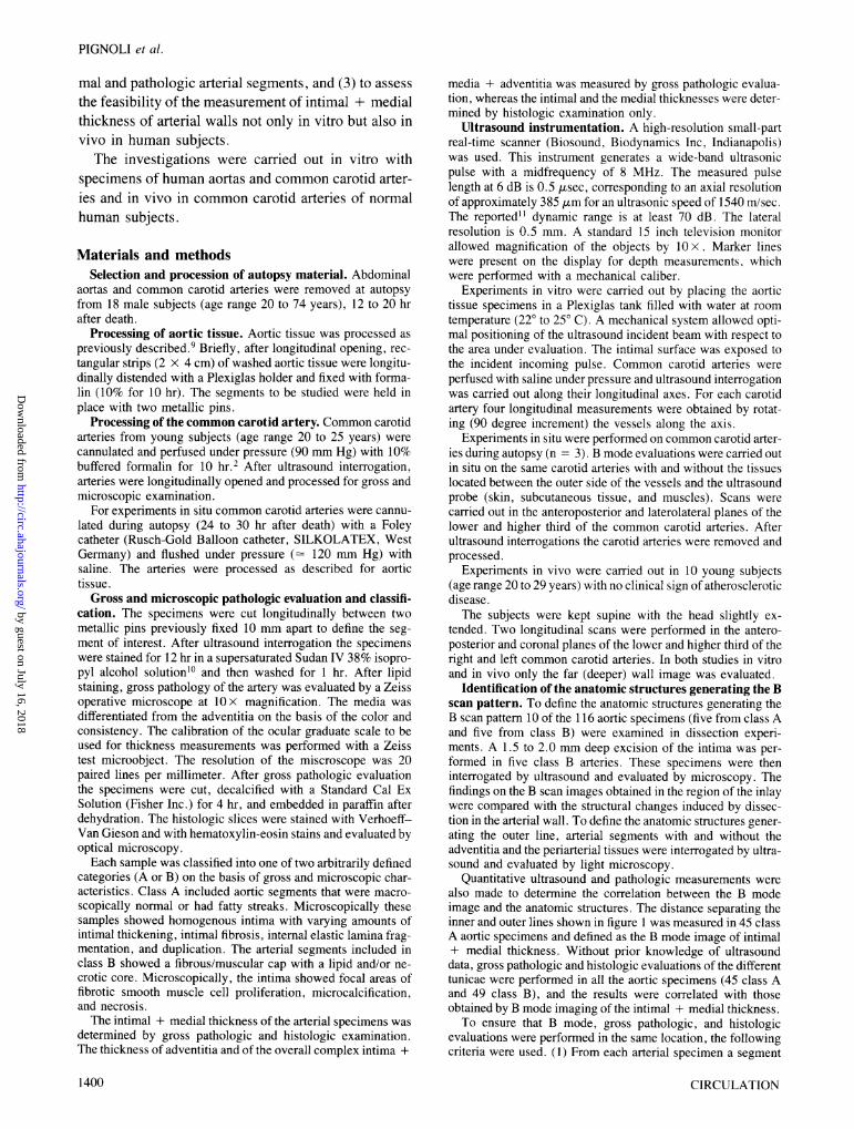
FIGURE 1. The typical B scan pattern (the double line pattem) of a normal class A arterial wall. The A to B line represents thedistance between the inner and the outer echogenic lines and corresponds to the B mode image of intimal + medial thickness.
showing homogenous thickness was selected, and a distance of10 mm was delimited with two metallic pins. This procedurewas undertaken to avoid the error that might result from makingmeasurements with the three methods at slightly differentpoints. (2) B mode and gross pathologic evaluations were car-ried out in the middle of the segment under study, which wasidentified by the metallic pins. (3) Histologic measurementswere obtained in the middle of longitudinal sections identifiedby the holes left by the pins.The overall error in the identification of the site at which the
different measurements (B mode, histologic, and gross patho-logic) were made was estimated to be +0.5 mm. For all theaortic specimens showing a characteristic B scan pattern (45class A and 49 class B), the absolute and percent differencesbetween results of B mode and gross pathologic evaluations ofintimal + medial thickness were determined.
Intimal + medial thickness values obtained in vitro in com-mon carotid arteries with B mode imaging were compared withthose determined in vivo in common carotid arteries of youngliving subjects.
Statistical analysis of the experimental data. The data areexpressed as the mean + SD. Statistical analysis was carriedout by paired two-tailed t test. Correlation coefficients werecalculated by plotting the values obtained with B mode imagingagainst those obtained by pathologic techniques.
ResultsGross pathologic and histologic evaluations of aortic
specimens and common carotid arteries. One hundred six-teen aortic segments were studied. Fifty of the 116
segments (43%) were grouped in class A (mascroscop-ically normal or with fatty streaks), whereas 66 (57%)were grouped in class B (aortic segments with athero-sclerotic lesions). Histologic evaluations of these seg-ments showed that intimal thickness of class A andclass B arteries was 0.25 + 0.12 and 1.11 ± 0.27mm, respectively, whereas intimal + medial thick-ness was 1.03 + 0.27 and 1.72 + 0.71 mm, respec-tively. Results of gross pathologic evaluations werecomparable (class A intimal + medial thickness 1. 13± 0.26 mm and class B 1.93 + 0.84 mm). The meanintimal + medial thickness measured by histologicexamination of common carotid arteries in vitro (n =
44) was 0.48 ± 0.06 mm. The gross pathologic evalu-ation resulted in comparable data (0.50 + 0.06 mm).
The typical B mode image of the arterial wall and its
prevalence in class A and class B aortic specimens. In all theclass A and in 85% of class B aortic specimens asimilar B scan pattern was found. This pattern wascharacterized by two parallel echogenic lines separatedby an hypoechoic or anechoic space (figure 1). This Bscan pattern was defined as the "double line pattern."The inner (luminal) line was generally more regular,smooth, and thin than the outer one. In 20% class B
Vol. 74, No. 6, December 1986
- - - - - - - - - - - - - - - - - - - - - - - - - - - - - - - - - - - - - - - - - - - - - - - - - - - - - - - - - - -
1401
by guest on July 16, 2018http://circ.ahajournals.org/
Dow
nloaded from
PIGNOLI et al.
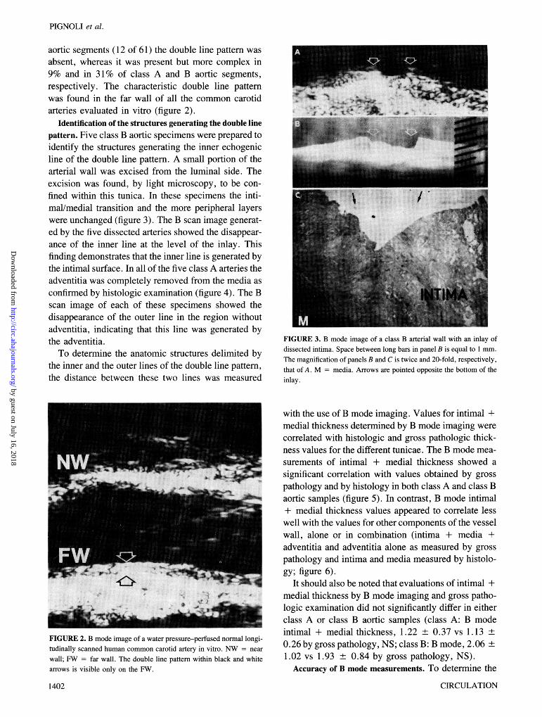
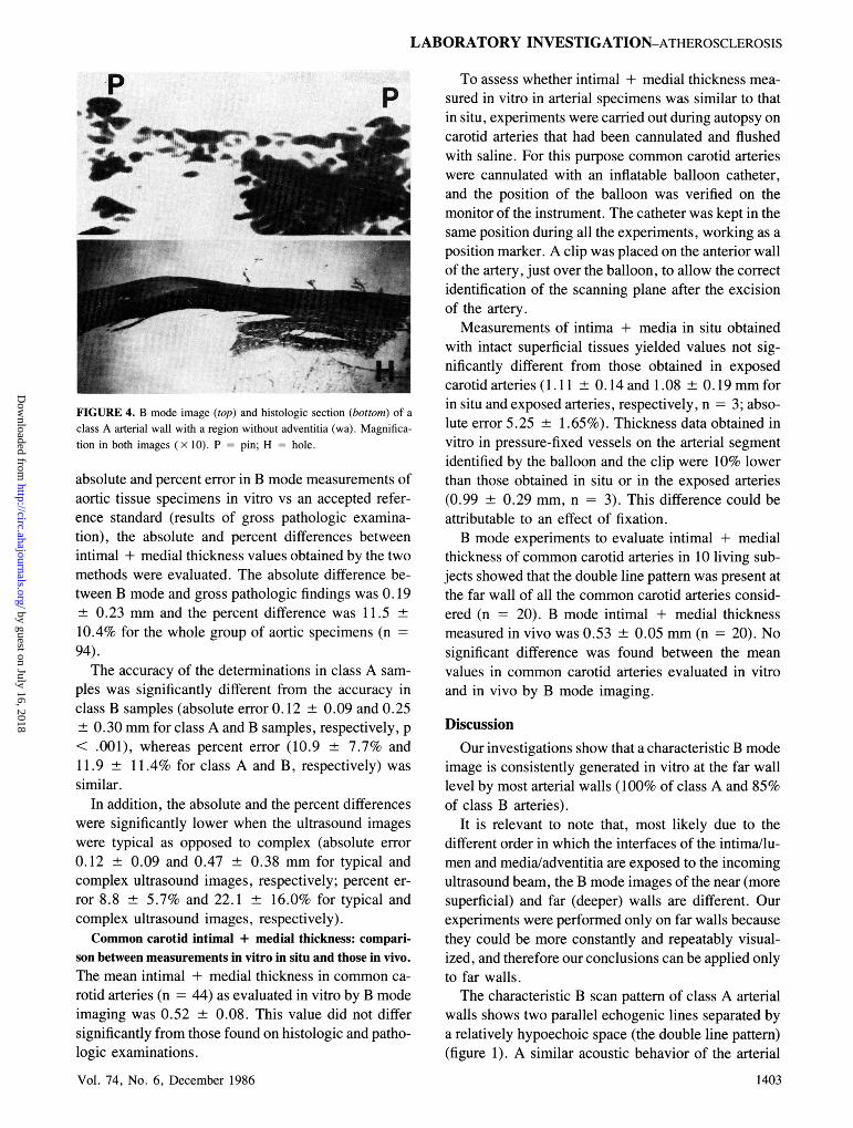
aortic segments (12 of 61) the double line pattern wasabsent, whereas it was present but more complex in9% and in 31% of class A and B aortic segments,respectively. The characteristic double line patternwas found in the far wall of all the common carotidarteries evaluated in vitro (figure 2).
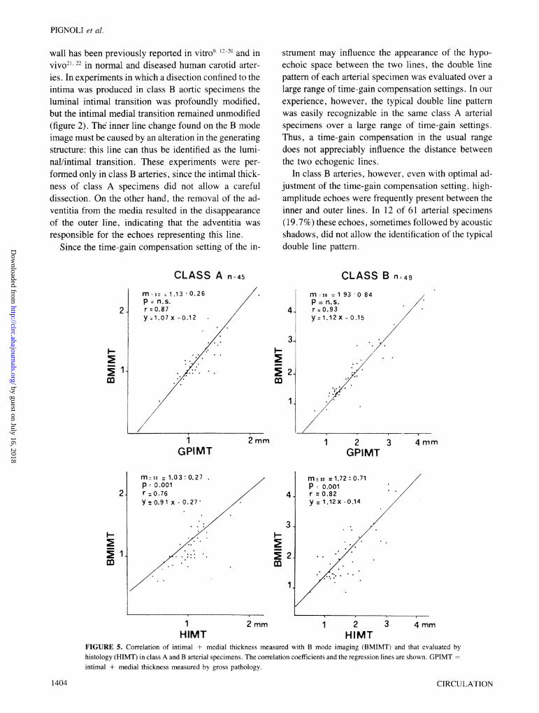
Identification of the structures generating the double linepattern. Five class B aortic specimens were prepared toidentify the structures generating the inner echogenicline of the double line pattern. A small portion of thearterial wall was excised from the luminal side. Theexcision was found, by light microscopy, to be con-fined within this tunica. In these specimens the inti-mal/medial transition and the more peripheral layerswere unchanged (figure 3). The B scan image generat-ed by the five dissected arteries showed the disappear-ance of the inner line at the level of the inlay. Thisfinding demonstrates that the inner line is generated bythe intimal surface. In all of the five class A arteries theadventitia was completely removed from the media asconfirmed by histologic examination (figure 4). The Bscan image of each of these specimens showed thedisappearance of the outer line in the region withoutadventitia, indicating that this line was generated bythe adventitia.To determine the anatomic structures delimited by
the inner and the outer lines of the double line pattern,the distance between these two lines was measured
FIGURE 2. B mode image of a water pressure-perfused normal longi-tudinally scanned human common carotid artery in vitro. NW = near
wall; FW far wall. The double line pattern within black and white
arrows is visible only on the FW.
1402
FIGURE 3. B mode image of a class B arterial wall with an inlay ofdissected intima. Space between long bars in panel B is equal to 1 mm.The magnification of panels B and C is twice and 20-fold, respectively,that of A. M = media. Arrows are pointed opposite the bottom of theinlay.
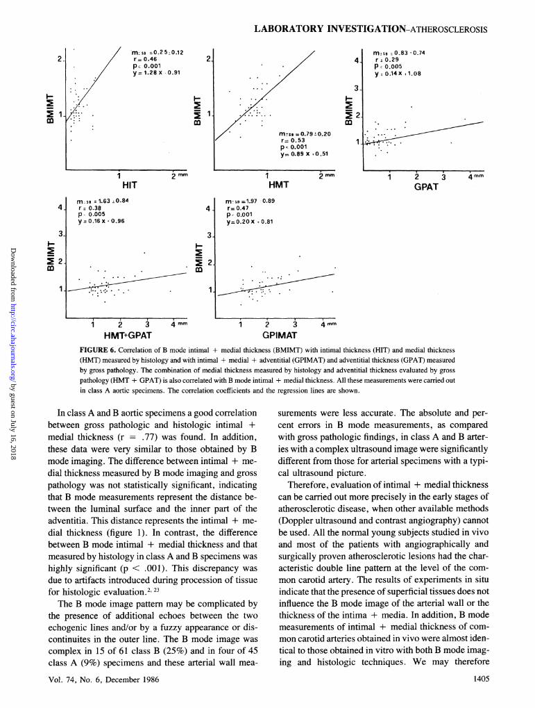
with the use of B mode imaging. Values for intimal +medial thickness determined by B mode imaging werecorrelated with histologic and gross pathologic thick-ness values for the different tunicae. The B mode mea-surements of intimal + medial thickness showed asignificant correlation with values obtained by grosspathology and by histology in both class A and class Baortic samples (figure 5). In contrast, B mode intimal+ medial thickness values appeared to correlate lesswell with the values for other components of the vesselwall, alone or in combination (intima + media +adventitia and adventitia alone as measured by grosspathology and intima and media measured by histolo-gy; figure 6).
It should also be noted that evaluations of intimal +medial thickness by B mode imaging and gross patho-logic examination did not significantly differ in eitherclass A or class B aortic samples (class A: B modeintimal + medial thickness, 1.22 ± 0.37 vs 1.13 ±0.26 by gross pathology, NS; class B: B mode, 2.06 +1.02 vs 1.93 + 0.84 by gross pathology, NS).
Accuracy of B mode measurements. To determine the
CIRCULATION
by guest on July 16, 2018http://circ.ahajournals.org/
Dow
nloaded from
LABORATORY INVESTIGATION-ATHEROSCLEROSIS
.p
* * -'ta
-~~~~~~~~~~~~4.
FIGURE 4. B mode image (top) and histologic section (bottom) of a
class A arterial wall with a region without adventitia (wa). Magnifica-tion in both images ( X 10). P = pin; H = hole.
absolute and percent error in B mode measurements ofaortic tissue specimens in vitro vs an accepted refer-ence standard (results of gross pathologic examina-tion), the absolute and percent differences betweenintimal + medial thickness values obtained by the twomethods were evaluated. The absolute difference be-tween B mode and gross pathologic findings was 0. 19
0.23 mm and the percent difference was 11.5 +
10.4% for the whole group of aortic specimens (n94).The accuracy of the determinations in class A sam-
ples was significantly different from the accuracy inclass B samples (absolute error 0.12 ± 0.09 and 0.25± 0.30 mm for class A and B samples, respectively, p
< .001), whereas percent error (10.9 ± 7.7% and11.9 ± 11.4% for class A and B, respectively) was
similar.
In addition, the absolute and the percent differenceswere significantly lower when the ultrasound imageswere typical as opposed to complex (absolute error
0.12 + 0.09 and 0.47 ± 0.38 mm for typical andcomplex ultrasound images, respectively; percent er-
ror 8.8 ± 5.7% and 22.1 ± 16.0% for typical andcomplex ultrasound images, respectively).Common carotid intimal + medial thickness: compari-
son between measurements in vitro in situ and those in vivo.The mean intimal + medial thickness in common ca-
rotid arteries (n 44) as evaluated in vitro by B mode
imaging was 0.52 + 0.08. This value did not differsignificantly from those found on histologic and patho-logic examinations.
To assess whether intimal + medial thickness mea-sured in vitro in arterial specimens was similar to thatin situ, experiments were carried out during autopsy oncarotid arteries that had been cannulated and flushedwith saline. For this purpose common carotid arterieswere cannulated with an inflatable balloon catheter,and the position of the balloon was verified on themonitor of the instrument. The catheter was kept in thesame position during all the experiments, working as aposition marker. A clip was placed on the anterior wallof the artery, just over the balloon, to allow the correctidentification of the scanning plane after the excisionof the artery.
Measurements of intima + media in situ obtainedwith intact superficial tissues yielded values not sig-nificantly different from those obtained in exposedcarotid arteries (l.1l1 + 0.14 andl.08 + 0.19 mmforin situ and exposed arteries, respectively, n - 3; abso-lute error 5.25 ± 1.65%). Thickness data obtained invitro in pressure-fixed vessels on the arterial segmentidentified by the balloon and the clip were 10% lowerthan those obtained in situ or in the exposed arteries(0.99 ± 0.29 mm, n = 3). This difference could beattributable to an effect of fixation.B mode experiments to evaluate intimal + medial
thickness of common carotid arteries in 10 living sub-jects showed that the double line pattern was present atthe far wall of all the common carotid arteries consid-ered (n = 20). B mode intimal + medial thicknessmeasured in vivo was 0.53 ± 0.05 mm (n = 20). Nosignificant difference was found between the meanvalues in common carotid arteries evaluated in vitroand in vivo by B mode imaging.
DiscussionOur investigations show that a characteristic B mode
image is consistently generated in vitro at the far walllevel by most arterial walls (100% of class A and 85%of class B arteries).
It is relevant to note that, most likely due to thedifferent order in which the interfaces of the intima/lu-men and media/adventitia are exposed to the incomingultrasound beam, the B mode images of the near (moresuperficial) and far (deeper) walls are different. Ourexperiments were performed only on far walls becausethey could be more constantly and repeatably visual-ized, and therefore our conclusions can be applied onlyto far walls.The characteristic B scan pattern of class A arterial
walls shows two parallel echogenic lines separated bya relatively hypoechoic space (the double line pattern)(figure 1). A similar acoustic behavior of the arterial
1403Vol. 74, No. 6, December 1986
by guest on July 16, 2018http://circ.ahajournals.org/
Dow
nloaded from
PIGNOLI et al.
wall has been previously reported in vitro9- 1`20 and invivo21. 22 in normal and diseased human carotid arter-
ies. In experiments in which a disection confined to theintima was produced in class B aortic specimens theluminal intimal transition was profoundly modified,but the intimal medial transition remained unmodified(figure 2). The inner line change found on the B modeimage must be caused by an alteration in the generatingstructure: this line can thus be identified as the lumi-nal/intimal transition. These experiments were per-
formed only in class B arteries, since the intimal thick-ness of class A specimens did not allow a carefuldissection. On the other hand, the removal of the ad-ventitia from the media resulted in the disappearanceof the outer line, indicating that the adventitia was
responsible for the echoes representing this line.Since the time-gain compensation setting of the in-
CLASS A n= 45
mosoD 1.1 3 0.26p= n.s.
2 r = 0.87y =1.07 x -0.12
E 1.m
2
1.
1GPIMT
strument may influence the appearance of the hypo-echoic space between the two lines, the double linepattern of each arterial specimen was evaluated over a
large range of time-gain compensation settings. In our
experience, however, the typical double line patternwas easily recognizable in the same class A arterialspecimens over a large range of time-gain settings.Thus, a time-gain compensation in the usual range
does not appreciably influence the distance betweenthe two echogenic lines.
In class B arteries, however, even with optimal ad-justment of the time-gain compensation setting, high-amplitude echoes were frequently present between theinner and outer lines. In 12 of 61 arterial specimens(19.7%) these echoes, sometimes followed by acousticshadows, did not allow the identification of the typicaldouble line pattern.
CLASS B n 49
m so = 1 93 0 84P = n.s.r = 0.93y = 1.12 x - 0.15
1 2GPIMT
3 4mm
M-so = 1.03- 0.27 msso =1.72+0.71p 0.001 p 0.001
2 r =0.76 4 r =0.82y 0.9H1 x I0-27* y 1.12 x0.14
1 2 mm 1 2 3 4 mmHIMT HIMT
FIGURE 5. Correlation of intimal + medial thickness measured with B mode imaging (BMIMT) and that evaluated byhistology (HIMT) in class A and B arterial specimens. The correlation coefficients and the regression lines are shown. GPIMTintimal + medial thickness measured by gross pathology.
1404 CIRCULATION
by guest on July 16, 2018http://circ.ahajournals.org/
Dow
nloaded from
LABORATORY INVESTIGATION-ATHEROSCLEROSIS
mrSD =0.250.12r= 0.46p- 0.001y= 1.28X .0.91
I-
CD
HIT
m so = 1.63 0.84r = 0.38p o.oosy = 0.16 x -0.96
* --
Mrsm = 0.79 ±0.20r= 0.53p - 0.001y 0.89 X :0.51
4-
3-
I-
2m
HMT
4-
3-
I-2
>-2.m
1.
1 2 3HMT+GPAT
m. S =1.97 -0.89r= 0.47P' 0.001y= 0.20X 0.81
1 2 3GPIMAT
4 mm
FIGURE 6. Correlation of B mode intimal + medial thickness (BMIMT) with intimal thickness (HIT) and medial thickness
(HMT) measured by histology and with intimal + medial + adventitial (GPIMAT) and adventitial thickness (GPAT) measured
by gross pathology. The combination of medial thickness measured by histology and adventitial thickness evaluated by gross
pathology (HMT + GPAT) is also correlated with B mode intimal + medial thickness. All these measurements were carried out
in class A aortic specimens. The correlation coefficients and the regression lines are shown.
In class A and B aortic specimens a good correlationbetween gross pathologic and histologic intimal +medial thickness (r = .77) was found. In addition,these data were very similar to those obtained by Bmode imaging. The difference between intimal + me-
dial thickness measured by B mode imaging and gross
pathology was not statistically significant, indicatingthat B mode measurements represent the distance be-tween the luminal surface and the inner part of theadventitia. This distance represents the intimal + me-
dial thickness (figure 1). In contrast, the differencebetween B mode intimal + medial thickness and thatmeasured by histology in class A and B specimens was
highly significant (p < .001). This discrepancy was
due to artifacts introduced during procession of tissuefor histologic evaluation.2'23The B mode image pattern may be complicated by
the presence of additional echoes between the two
echogenic lines and/or by a fuzzy appearance or dis-continuites in the outer line. The B mode image was
complex in 15 of 61 class B (25%) and in four of 45class A (9%) specimens and these arterial wall mea-
Vol. 74, No. 6, December 1986
surements were less accurate. The absolute and per-
cent errors in B mode measurements, as comparedwith gross pathologic findings, in class A and B arter-ies with a complex ultrasound image were significantlydifferent from those for arterial specimens with a typi-cal ultrasound picture.
Therefore, evaluation of intimal + medial thicknesscan be carried out more precisely in the early stages ofatherosclerotic disease, when other available methods(Doppler ultrasound and contrast angiography) cannotbe used. All the normal young subjects studied in vivoand most of the patients with angiographically andsurgically proven atherosclerotic lesions had the char-acteristic double line pattern at the level of the com-
mon carotid artery. The results of experiments in situindicate that the presence of superficial tissues does notinfluence the B mode image of the arterial wall or thethickness of the intima + media. In addition, B modemeasurements of intimal + medial thickness of com-mon carotid arteries obtained in vivo were almost iden-tical to those obtained in vitro with both B mode imag-ing and histologic techniques. We may therefore
1405
m.s :0.83 t0.74r- 0.29P 0.005y. 0.14X +1.08
~~~~1 *
CD2
4-
3-
m
1 2GPAT
2.
1
3 4 mm
1 .11 X v
4 mm
by guest on July 16, 2018http://circ.ahajournals.org/
Dow
nloaded from
PIGNOLI et al.
conclude that the results in vitro reported in this studycan be extended to conditions in vivo.The present study demonstrates that: (1) a character-
istic B mode image (the double line pattern) is consis-tently generated in vitro by the far walls of most of thearteries, (2) the distance between the transducer-facingedges of the inner and outer lines of the B mode imagecorrelates with and does not differ significantly fromthe intimal + medial thickness, (3) regarding the accu-racy of B mode imaging, a percent error of less than20% has been found in 77% of the arterial specimensof both classes (normal and pathologic), and (4) theaccuracy of B mode imaging depends on the presenceof a typical B scan image. In 85% of class B arterialspecimens a characteristic B mode image was foundand in 59% of specimens of the same class the imagewas typical and easy to interpret.We conclude that B mode imaging represents a use-
ful tool for the detection and monitoring of changes inintimal + medial thickness, allowing the evaluation ofchanges in the arterial wall in areas without localizedplaques. At the present time, it is difficult to determinewhether this information will be of clinical relevance.It is possible that early changes in vascular thicknesswill later result in atheroma; they could on the otherhand evolve only in a diffuse intimal thickening andhave no hemodynamic relevance. It is also not knownwhether a relationship exists between the morphologicchanges in the vessel wall detected by our meth-od (thickening) and local damage to the endotheliallayer, which can induce alterations of potential clini-cal relevance. The noninvasive nature of this newapproach is a recommendation for its use in the pre-clinical diagnosis and follow-up of patients withatherosclerosis.2425
We thank Prof. C. R. Sirtori for helpful discussion and criti-cism in preparing the manuscript. Prof. Romeo Pozzato, Chair-man and Professor of Forensic Medicine, University of Milan,Milan, and Prof. T. Longo, Professor of Surgery. University ofMilan, Milan, are also acknowledged.
References1. Bond MG, Adams MR, Bullock BC: Complicating factors in eval-
uating coronary artery atherosclerosis. Artery 9: 21, 19812. Zarins K, Zatina MA, Glagov S: Correlation of postmortem angi-
ography with pathologic anatomy: quantitation of atheroscleroticlesions. In Bond MG, Insull W, Glagov S, Chander AB, CornhillJF. editors: Clinical diagnosis of atherosclerosis. New York, 1983,Springer Verlag, p 283
3. Roberts WC: The coronary arteries in coronary heart disease: mor-phologic observations. Pathobiol Annu 5: 249, 1975
4. Wissler RW: Principles of the pathogenesis of atherosclerosis. InBraunwald E, editor: Heart disease: a textbook of cardiovascularmedicine. Philadelphia, 1984, Saunders, p 1183
5. Stegall HG: Ultrasonic measurements of organ dimensions. In Ren-eman RS, editor: Cardiovascular applications of ultrasound. Am-sterdam, 1974, North Holland Publishing Co., page 150
6. Ohkawai H, Nitta SI, Tanaka M, Dunn F: In vivo measurement ofthickness or of speed of sound in biological tissue structures. IEEETrans Sonic Ultrasonic SU-30: 231, 1983
7. Insull W Jr: Universal reference standards for measuring athero-sclerosis lesions. The quest for the "gold standard." In Bond MG,Insull W, Glagov S, Chandler AB, Cormhill JF, editors: Clinicaldiagnosis of atherosclerosis. Quantitative methods of evaluation.New York, 1983, Springer Verlag. p 283
8. Berson AS: Ultrasound B scan assessment program. Atherosclero-sis Rev 10: 169, 1983
9. Pignoli P: Ultrasound B-mode imaging for arterial wall thicknessmeasurement. Atherosclerosis Rev 12: 177, 1984
10. Sheenan DC, Hrapchak BB: Theory and practice of histotech-nology. St. Louis, 1980, The CV Mosby Co, p 206
11. Biodynamics Inc: Biosound instructions manual. Indianapolis,1981
12. Murakami R: Analytical and experimental determination of acous-tic reflecting characteristics of normal aortic walls, thesis. Univer-sity of Washington, Seattle, 1973
13. Beretsky I: Detection and characterization of atherosclerosis in ahuman arterial wall by raylographic technique, an in vitro study. InWhite D, Brown R, editors: Ultrasound in medicine. New York.1977, Plenum Press, vol 3B, p 1597
14. Barber FE: Scattering from arterial tissues by ultrasonic micro-probe. In White D, Brown R, editors: Ultrasound in medicine. NewYork, 1977, Plenum Press, vol 3B, p 1979
15. Greenleaf JF, Duck FA, Samayoa WF, Johnson SA: Ultrasonicdata acquisition and processing system for atherosclerotic tissuecharacterization. 1974 Ultrasonics Symposium Proceedings. IEEECatalog No. 74CH0896-ISU, 1974
16. Hartley DJ, Strandness DE Jr: The effects of atherosclerosis on thetransmission of ultrasound. J Surg Res 9: 575, 1969
17. Rooney JA, Gammel PM, Hestenes JD, Chin HP, BlankenhornDH: The use of ultrasonic spectroscopy to characterize calcifiedlesions. IEEE Trans Sonics Ultrasonics SU-28: 291, 1981
18. Rooney JA, Gammel PM, Hestenes JD, Chin HP, BlankenhornDH: Velocity and attenuation of sound in arterial tissue. J AcoustSoc Am 71: 462, 1982
19. Geleskie JV, Shung KK: Further studies on acoustic impedance ofmajor bovine blood vessel walls. J Acoust Soc Am 71: 467, 1982
20. Wolverson M, Bashiti H, Sundaram M, Heiberg E, Grider R:Ultrasonic tissue characterization of atheromatous plaques using ahigh resolution real time scanner. Presented at the 25th ScientificSession, American Institute of Ultrasound in Medicine, New Or-leans, Sept. 15-19, 1980
21. Zwiebel WJ: High resolution B mode and Duplex carotid sonog-raphy. In Zeiwbel WJ, editor: Introduction to vascular ultrasonog-raphy. New York, 1982, Grune & Stratton, p 103
22. James EM, Earnest IV G, Forbes DF, Houser OW, Folger WN:High resolution dynamic ultrasound imaging of the carotid bifurca-tion: a prospective evaluation. Radiology 144: 853, 1982
23. Bahr GF, Bloom G, Friberg U: Volume changes of tissues inphysiological fluids during fixation in osmium tetroxide or formal-dehyde and during subsequent treatment. Exp Cell Res 12: 342,1957
24. Bond MG, Riley WA, Barnes RW, Kaduck JM, Ball MR: Valida-tion studies of a noninvasive real time B scan imaging system. InBerson AS, Budinger IF, Ringquist I, Mock MB, Watson JT,Powel RS, editors: Noninvasive techniques for assessment ofatherosclerosis in peripheral, carotid and coronary arteries. NewYork, 1982, Raven Press, p 197
25. Arteriosclerosis 1981, Report of the Working Group on arterioscle-rosis of the National Heart, Lung and Blood Institute. U.S. Depart-ment of Health and Human Services, NIH Publication No. 81-2034, July 198 1, p 27
CIRCULATION1406
by guest on July 16, 2018http://circ.ahajournals.org/
Dow
nloaded from
P Pignoli, E Tremoli, A Poli, P Oreste and R Paolettiimaging.
Intimal plus medial thickness of the arterial wall: a direct measurement with ultrasound
Print ISSN: 0009-7322. Online ISSN: 1524-4539 Copyright © 1986 American Heart Association, Inc. All rights reserved.
is published by the American Heart Association, 7272 Greenville Avenue, Dallas, TX 75231Circulation doi: 10.1161/01.CIR.74.6.1399
1986;74:1399-1406Circulation.
http://circ.ahajournals.org/content/74/6/1399the World Wide Web at:
The online version of this article, along with updated information and services, is located on
http://circ.ahajournals.org//subscriptions/
is online at: Circulation Information about subscribing to Subscriptions:
http://www.lww.com/reprints Information about reprints can be found online at: Reprints:
document. Permissions and Rights Question and Answer information about this process is available in the
located, click Request Permissions in the middle column of the Web page under Services. FurtherEditorial Office. Once the online version of the published article for which permission is being requested is
can be obtained via RightsLink, a service of the Copyright Clearance Center, not theCirculationpublished in Requests for permissions to reproduce figures, tables, or portions of articles originallyPermissions:
by guest on July 16, 2018http://circ.ahajournals.org/
Dow
nloaded from