Embed Size (px)
Citation preview
Biomed Pap Med Fac Univ Palacky Olomouc Czech Repub. 2014 Sep; 158(3):404-411.
404
Intima media thickness measurement as a marker of subclinical atherosclerosis in SLE patient
Andrea Smrzovaa, Pavel Horaka, Martina Skacelovaa, Zuzana Hermanovab, Katerina Langovac, Josef Zadrazila, Dalibor Novotnyd
Aim. Accelerated atherosclerosis in systemic lupus erythematosus (SLE) is an important cause of morbidity and mortal-ity. The pathophysiology of accelerated atherosclerosis in SLE is mediated by factors such as inflammatory processes in the vascular wall, specific antibodies, dyslipoproteinemia, endothelial dysfunction and the high prevalence of traditional risk factors for cardiovascular diseases. In this context, we evaluated the clinical significance of ultrasound examination of the carotic arteries in the early diagnosis of atherosclerosis. Methods. The study included 63 patients with SLE (female: male 53:10, mean age 38.4±12.7 years, mean disease duration 143.0 ±82.6 months), 24 patients had lupus nephritis. The control group consisted of 24 volunteers (female: male 20:4 mean age 31.04±8.59). Intima media thickness (IMT) was measured by ultrasound on both sides. The results were correlated with markers of lipid spectrum, anti-dsDNA, antinucleosomal and anticardiolipin antibodies, lupus anticoagulant and complement components. Clinical disease activity and damage were evaluated by SLEDAI and SLICC indices. Lifestyle and other important factors were examined per protocol and by questionnaire. Results. A significant difference of IMT (P≤0.03) was found between the lupus patients and sex-age adjusted healthy controls with an in mean IMT in SLE patients of 0.569±0.11 mm, in control group 0.495±0.05 mm. A significant correla-tion between IMT and disease duration, age, positivity of lupus anticoagulant, use of ACE inhibitors, glomerular filtration and serum creatinine were found. No difference in IMT was found between patients with or without lupus nephritis.Conclusion. IMT measurement could be used as a clinical predictor of risk of accelerated atherosclerosis in lupus patients.
Key words: systemic lupus erythematodes, atherosclerosis, intima media thickness
Received: December 11, 2012; Accepted with revision: July 8, 2013; Available online: July 29, 2013http://dx.doi.org/10.5507/bp.2013.054
aDepartment of Internal Medicine III - Nephrology, Rheumatology, Endocrinology, Faculty of Medicine and Dentistry, Palacky University Olomouc, Czech RepublicbDepartment of Immunology, Faculty of Medicine and Dentistry, Palacky University OlomouccDepartment of Medical Biophysics, Faculty of Medicine and Dentistry, Palacky University OlomoucdDepartment of Cinical Biochemistry, University Hospital OlomoucCorresponding author: Andrea Smrzova, e-mail: [email protected]
INTRODUCTION
Accelerated atherosclerosis (AT) is one of the ma-jor late complications of systemic lupus erythematosus (SLE) and is recognized as a major cause of morbidity and mortality in lupus patients. The first reports of early atherosclerotic involvement appeared in 1975 (ref.1). One year later, a study described bimodal “twin-peaks” pattern of mortality rate related to SLE, the first peak being asso-ciated with active lupus, the second with acute myocardial infarction or other cardiovascular diseases10-15 years af-ter initial diagnosis2. The probability of a 10-year survival of lupus patients is now more than 90% (ref.3). The impact of previous frequent causes of death like high activity of auto inflammatory process, renal insufficiency, infection and sepsis have been diminished but not eliminated while cardiovascular diseases as a cause of lupus mortality have increased considerably. Patients with SLE have more than six times higher risk of AT than the general population4. In studies, this relative risk ranges from 5.0-10.0 depend-ing on the design of the observation and the odds ratio
lays between 4.8 and 9.8 (ref.4,5). Two retrospective stud-ies on traditional risk factors for CVD mortality showed that patients with SLE had a ten times higher incidence of nonfatal myocardial infarction and seventeen times higher mortality due to coronary heart disease indepen-dently of their traditional CVD risk factor profile6. The data from the General Practice Research Database in the UK showed 2.67 times more common acute myocardial infarction in SLE patients than in controls7. Subclinical AT is present in up to 40% of lupus patients8. The vast majority of these publications show that higher prevalence of traditional risk factor cannot be the only explanation for these alarming findings and that the complex inflam-matory character of SLE with numerous consequences plays a crucial role9.
In this study, the selected risk factors of AT were in-vestigated in the group of SLE patients followed up at the department of rheumatology and compared with ultrasound evaluation of intima media thickness of the common carotid artery.

Biomed Pap Med Fac Univ Palacky Olomouc Czech Repub. 2014 Sep; 158(3):404-411.
405
MATERIALS AND METHODS
Sixty three patients with SLE, 53 females and 10 males, all fulfilling four or more criteria of the American College of Rheumatology for the disease and who had given informed consent, were examined in the out-patient lupus clinic and the clinical and laboratory parameters were assessed in the period from September 2010 to December 2011 (ref.10). The inclusion criteria were di-agnosis of SLE with all types of organ damage. Patients with a previous history of atherosclerosis (before mani-festation of SLE) were excluded. Measurement of IMT, laboratories parameters and information from question-naires were collected in the same time in one visit. The mean age was 38.4±12.7 years (18-72), mean duration of the disease 143.0±82.6 months (0-342), age of onset of SLE was 28.0±12.0 years (median 25.0, 9 minimum, 60 maximum). The sub-group of lupus nephritis (LN) patients comprised 24 in all, but in two renal diseases was not confirmed by histology of the kidney biopsy. In the remaining two cases, the diagnosis of lupus nephritis was based merely on clinical data (proteinuria and ne-phrotic syndrome) as renal biopsy would be associated with substantial risk due to thrombocytopenia. Whereas the mean age of patient in LN group was 33.71 years and in patients without LN 41.3 years, the age matched subgroup (32.1 years) of lupus patients without LN was used for comparison of IMT values.
The clinical examination included a detailed history of the disease, hypertension, diabetes, smoking, family his-tory of premature cardiovascular disease (before the age of 55 for men or 60 for women in first-degree relatives), nutrition and sport activities or sedentary lifestyle assess-ment by simple questionnaire and further on, standard-ized measurement of systolic and diastolic blood pressure, obesity defined by body mass index and waist circumfer-ence and complete physical examination at study entry.
In each patient, the cumulative dose of glucocorticoids (equivalent of prednisone) and eventually of hydroxychlo-rochine (if ever given) were calculated. The data on use of other drugs possibly interfering with atherosclerosis such as ACEi (angiotensin converting inhibitors), statins and anticoagulation or antiaggregation were collected.
The SLE Disease Activity Index (SLEDAI) was used for evaluation of clinical disease activity and Systemic Lupus International Collaborating Clinics/American College of Rheumatology (SLICC/ACR) index for as-sessment of disease damage11,12.
The levels of C3 and C4 components of the comple-ment were measured using nephelometry (Analyzer BNII Dade Behring) with diagnostic antiserum (Orion Diagnostica Company, Espoo, Finland). The normal lim-its for the C3 component of the complement are 0.98-1.97 g/L and 0.12-0.4 g/L for the C4 component. Anti-double-stranded DNA (anti-dsDNA) and antinucleosomal anti-bodies were detected by ELISA (Organtec Diagnostika GmbH, Mainz, Germany). The producer expected normal values less than 15 IU/mL for anti-dsDNA and less than 20 UI/mL for antinucleosomal antibodies.
Anticardiolipin antibodies (ACLA IgG, IgM) were tested by ELISA (Aeskulisa Cardiolipn-GM, Wendelsheim, Germany) with normal limits between 0-10 GPL-UI/mL for ACLA IgG and 0-15 GPL-UI/mL for IgM. Lupus anticoagulant was measured by routine method with determination of lac screen ration (LSR) 0-15 for negativity. High sensitivity C reactive protein was measured by nephelometry (Roche/Hitachi 912, Modolar P analyzers, Germany) with expected normal levels of 0-5 mg/L. Complete lipid spectrum was assessed by neph-elometry (Roche/Hitachi , Germany). Normal value of cholesterol was 2.90-5.00 mmol/L, triglycerides 0.45-1.70 mmol/L, HDL 1.00-2.10 mmol/L, LDL 1.20-3.00 mmol/L, lipoprotein (a) 0-0.3 g/L, apolipoprotein A-1 1.15-1.60 g/L, apolipoprotien B 0-1.20 g/L and creatinine 64-104 μmol/L. Glomerular filtration was calculated us-ing formula of MDRD (Modification of Diet in Renal Disease).
The B-mode ultrasound scan of the common carotid artery was performed in all participants including con-trols by the standard protocol. Five scans were measured on each side and the mean was calculated. The far wall common carotid intima media thickness (IMT) measure-ments were made at a predefined site 1cm from the ca-rotid bulb free of plaques defined according to Touboul13. All measurements were performed by the same trained ultrasound operator. The intima media thickness index was measured also in the age and sex matched control group of 24 healthy volunteers (female: male 20:4, mean age 31.04±8.59 years).
Descriptive statistics, regression, correlation, Mann–Whitney, Kruskal-Wallis test, t-tests and Levene’s Test for Equality of variance and Chi-Square Tests were used for statistical evaluation of the results. ANOVA software was used for the calculations and statistic graphs.
RESULTS
The characteristic of SLE group and prevalence of cardiovascular risk factors is summarized in the Table 1, which also summarizes the correlation of these param-eters with IMT. The treated arterial hypertension was present in 25 patients (39.7%) and criteria for diabetes mellitus were met in 15 patients (23.8%). Twenty six (41.3%) patients admitted current or past cigarette smok-ing in the last five years. The positive family history of CVD was prevalent in 4.7% (3 respondents), hyperten-sion at an early age (before 55 in males and before 60 in females) in 25% (16 patients). Two third of patients (40 respondents) did not comply with a diet containing predefined amounts of vegetables14. According to the questionnaire, a sedentary life style (threshold defined as aerobic exercise 30 min 5 times a week) prevailed among the examined patients (n=43, 68.3%) (ref.14). Use of al-cohol beverages more than 20 mg per day or absolute abstinence was present in 6.3% respectively 42.9% (4 resp. 27 patients). The mean BMI was 24.79±5.1 (16.93-39.45), the waist circumference 84.95±14.1 cm (61-121 cm), hip circumference 99.11±11.9 cm (78-130 cm).

Biomed Pap Med Fac Univ Palacky Olomouc Czech Repub. 2014 Sep; 158(3):404-411.
406
Table 1. Characteristic of group of 63 lupus patients and results of IMT (intima-media thickness) measurement and statistical evaluation by sig 2 tailed.
Mean (SD) or %
RangeCorrelation coefficient
Sig. (2-tailed)
Number of patients (n) 63Women (n, %) 53 (84.1)Mean age (years) 38.38±12.7 18-72 0.666 > 0.001Duration of SLE (months) 143.14±82.6 0-292 0.329 0.008Age in diagnosis SLE (years) 28.04±12.0 9.7-60.9Hypertension (n, %) 25 (39.7) NSDiabetes (n, %) 15 (23.8) NSSmoking cigarette (n,%)** 26 (41.3) 0.07Family history of CVD or hypertension in early age**** 3.16 (4.7, 25.4) NSHealthy food (n, %) 40 (66.7) NSSedentary life style* (n, %) 43 (68.3) NSUsing of alcohol*** (n, %) 4. 27 (6.3, 42.9) NSBMI (n, %) 24.79±5.1 16.94-39.45 0.309 0.014Waist circumference (cm) 84.82±14.1 60-121 0.428 >0.001Hips circumference (cm) 99.11±11.9 78-130 0.381 0.002Cumulative dose of glucocorticoids (g) (n=61, 97%) 27.87±25.2 -0.057 NSDaily dose of glucocorticoids (mg) 14.90±18.9 NSUsing hydroxychlorochin, cumulative dose (g) (n=35, 55.5%)
387.09±632.2 -0.164 NS
Daily dose (mg) 205.69±64.8 NSUsing of ACEi (n,%) 31 (49.2) 0.003Using of statins (n,%) 13 (20.6) NSAnticoagulation therapy (n,%) 16 (25.4) NSAntiagregation therapy (n,%) 8 (12.7) NSSLICC/ACR/DI 1.17±1.4 0-5 0.093 NSSLEDAI 7.22±5.4 0-20 -0.027 NSC3 (g/L) 0.99±0.3 0.42-1.62 0.100 NSC4 (g/L) 0.15±0.1 0.05-0.49 0.223 NSAnti ds DNA (UI/mL) 98.37±105.3 3-300 0.010 NSANUC (UI/mL) 124.54±79.5 13-300 -0.084 NSPositivity ACLA IgG (n, %) 10 (15.9) 1-300 -0.187 NSPositivity ACLA IgM (n, %) 7 (11.1) 1-101 -0.164 NSLSR 1.4 0.38-4.64 -0.258 0.041hsCRP g/L 5.14±6.2 0-27 0.092 NSTC mmol/L 5.15±1.17 3.58-9.65 0.227 NSTG mmol/L 1.72±1.0 0.49-5.16 0.088 NSHDL mmol/L 1.38±0.4 0.75-2.91 0.114 NSLDL mmol/L 3.02±1.1 1.28-7.18 0.158 NSTC/HDL 3.97±1.2 1.92-6.6 0.075 NSApo lipoprotein A-1 (g/L) 1.50±0.4 0.77-2.85 0.059 NSApo lipoprotein B (g/L) 0.93±0.3 0.53-1.83 0.047 NSLipoprotein (a) (g/L) 0.39±0.5 0.03-1.76 0.113 NSCreatinine (μmol/L) 71.32±20.5 41-133 0.321 0.010Glomerular filtration rate (mL/s) 1.58±0.5 0.26-2.42 0.002
*Smoking - current smoker, ex-fumator in the last 5 years** Positive family history - for first-stepwise relative in women over 65 years in men over 55 years***Sedentery life style - aerobic exercise less than 30 min 5times a week ****Alcohol - abstainer / more than the recommended daily dose

Biomed Pap Med Fac Univ Palacky Olomouc Czech Repub. 2014 Sep; 158(3):404-411.
407
Only three patients were glucocorticoids naïve. The rest of the patients (n=61, 97%) were taking glucocor-ticoids with a mean cumulative dose of 27.87±25.2 g Prednisone or its equivalent (0.24-132 g), mean daily dose was 14.90±18.9 mg (3.1-131.3), mean duration of therapy was 97.80±7.7 months (2-285). Some patients had been taking glucocorticoids before diagnosis of SLE; the dose was included in the calculation. Thirty five patients were current or past users of hydroxychlorochine or chlorochine (55.5%) with mean cumulative dose of 387.09±632.2 g (6-3765), mean daily dose was 205.69±64.8 mg (16-500 mg) in duration of 54.97±49.2 months (1-251). Thirty one pa-tients (49.2%) were taking ACE inhibitors, 13 patients (20.6%) statins, 24 patients (38.1%) were on anticoagula-tion therapy with warfarin (n=16, 25.4%) or low molecular weight heparin (n=8, 12.7%) and 23 patients (36.5%) were taking antiaggregation therapy.
The mean SLICC/ACR damage index reached 1.17±1.4 (0-5) and disease activity assessed by SLEDAI was 7.22±5.4 (0-20).
Serum levels of C3 complement component was 0.99±0.3 g/L (0.42-1.62), C4 was 0.15±0.1 g/L (0.05-0.49). The mean concentration of anti-dsDNA antibodies was 98.37±105.3 (3-300) IU/mL and of antinucleosomal antibodies 124.54±79.5 IU/mL. Twenty patients (31.75%) were negatively tested for anti-dsDNA Ab and nine pa-tients (14.2%) were negative for antinucleosomal antibod-ies and ten patients were negative for both at the time of examination.
Anticardiolipin antibodies (ACLA) IgG were posi-tive in 10 patients (15.9%), ACLA IgM in seven patients (11.1%); lupus anticoagulant (LSR) over 1.15 was pres-ent in 23 patients (36.5%). Positivity of ACLA and lupus anticoagulants was following: Positive ACLA IgG plus positive LSR was present in eight respondents (12.7%), positive ACLA IgM plus positive LSR in four patients (6.3%), and finally three patients expressed ACLA IgM, IgG plus LSR (4.8%).
The mean serum levels of high sensitivity C-reactive protein upon exclusion of infection were 5.14±6.2 mg/L (0.3-27). The lipid profile was as follows: total choles-terol was 5.15±1.2 mmol/L (3.58-9.65), triglycerides 1.72±1.0 mmol/L (0.49-5.16), HDL 1.38±0.4 mmol/L (0.75-2.91), LDL 3.02±1.1 mmol/L (1.28-7.18), CH/HDL ration 3.97±1.2 (1.92-6.72), serum levels of apolipopro-tein A-1 were1.50±0.4 g/L (0.77-2.85 g/L), apolipopro-tein B 0.93±0.3 g/L (0.53-1.83g/L) and lipoprotein (a) 0.39±0.5 g/L (0.03-1.76).
Mean levels of serum creatinine were 71.32±20.5 μmol/L (41-133) and glomerular filtration (calculation by Modification of Diet in Renal Disease – MDRD) 1.58±0.47 mL/s (0.26-2.42).
All patients underwent ultrasound examination by high sensitivity B-mode scan IMT. The mean value of IMT on left side was 0.579±0.12 mm (0.38-1.02 mm), on right side 0.561±0.10 mm (0.39-0.95) and the calculated mean was 0.569±0.11 mm (0.39-0.95). Healthy controls had IMT on left side 0.502±0.06 mm (0.41-0.656 mm), on right side 0.489±0.05 mm (0.418-0.602 mm) and mean was 0.495±0.049 mm (0.414-0.627 mm). We prefer distri-
bution to groups with absolute number of IMT, plaques was not detected.
Lupus nephritis defined by histology was present in 24 (38.1%) patients in our group. None of the patients had minimal mesangial lupus nephritis (Class I) or scler-otizing nephritis (Class VI), while Class II (mesangial proliferative lupus nephritis) was present in 6.3%; focal segmental lupus nephritis (Class III) in 17.5%, diffuse proliferative lupus nephritis (Class IV) was detected in 7.9% and membranous lupus nephritis (Class V) in 4.8% (ref.15). In two patients the clinical manifestation of lupus nephritis with nephrotic syndrome was present but biopsy was contraindicated by clinicians. The IMT in group of patients with LN on left side was 0.548±0.052 mm (0.43-0.67 mm), on right side 0.538±0.059 mm (0.44-0.664 mm); mean was 0.538±0.054 mm (0.46-0.67 mm). IMT in all patients without LN was measured within 0.38 mm to 1.02 mm on left side with a mean of 0.601±0.15 mm and from 0.34 to 0.91 mm on the right side with a mean of 0.578±0.12 mm, and calculated mean of both sides was 0.589±0.13 mm (0.37-0.945). After adjustment for age, the IMT of lupus nephritis patients was 0.529±0.07 mm (0.383-0.73) on left, 0.518±0.07 mm (0.39-0.66) on right and calculated mean was 0.524±0.07 mm (0.387-0.661).
CONCLUSION
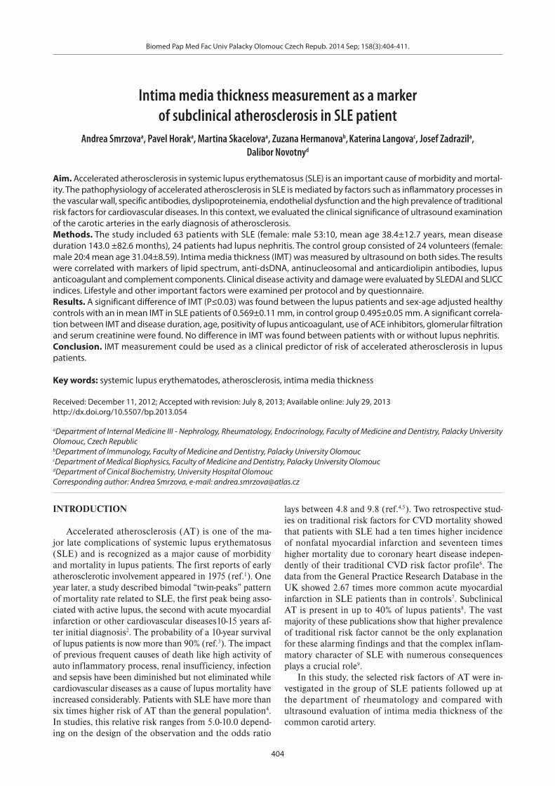
The IMT values differed significantly between the lu-pus patients and healthy controls using a t-test, P≤0.03 (Fig. 1). No significant difference was found between pa-tients with and without lupus nephritis (Fig. 2).
Fig. 1. Correlation of IMT (mm) in 63 SLE patients and 24 healthy controls.

Biomed Pap Med Fac Univ Palacky Olomouc Czech Repub. 2014 Sep; 158(3):404-411.
408
Fig. 2. Correlation of IMT(mm) in age-mached 23 patient with lupus nephritis, 23 patient without lupus nephritis and 24 healthy control respendens.
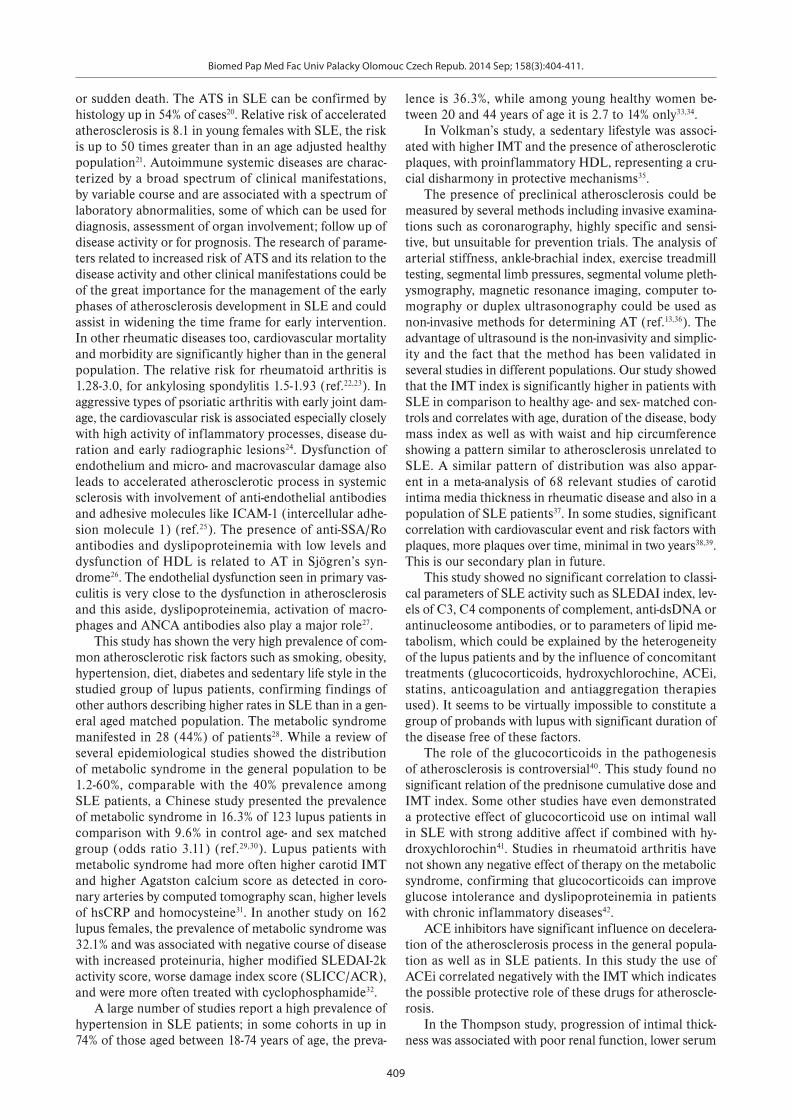
Fig. 3. Correlation of IMT (mm) and waist circumference (cm) in a group of 63 lupus patients.
Fig. 4. Correlation of IMT (mm) and glomerular filtration in a group of 63 lupus patients.
Fig. 5. Significant different of IMT (P=0.003) between pa-tients treated with ACEi (n=31, 49.2%) and without ACEi medi-cation (n=32, 50.8%).
The IMT correlated positively and significantly with age (P≤0.001), duration of disease (P=0.008), with BMI (P=0.014), waist and hip circumference (P≤0.001 reps. P=0.014, Fig. 3) and with decrease of renal function im-pacting the serum creatinine (P=0.01) or glomerular fil-tration rate calculated by MDRD (P=0.02, Fig. 4). The medication of ACEi showed a negative correlation to the IMT index (P=0.0≤03, Fig. 5). There was a trend to rela-tion between current or past smoking, levels of C3 and C4 components of complement, total cholesterol and IMT (P=0.056-0.07), however none of these factors met statisti-cal significance at P 0.05.
DISCUSSION
Advances in the diagnosis and treatment of systemic lupus erythematosus (SLE) in the last fifty years has en-abled patients to survive to the late phases of disease, which are characterized by different pattern of manifes-tation than the early disease with increase in cardiovas-cular diseases, infections, tumors and osteoporosis. The presence of AT associated with SLE seems to be even higher than in diabetes mellitus approaching the rate of Cushing’s syndrome and sharing comparable risk with other high risk disease like renal insufficiency, familiar hyperlipoproteinemia or states after organ transplanta-tion16-19.
The pathogenesis of atherosclerosis in SLE is com-plex. Atherosclerosis often presents even before the 40th year of age, commonly with angina, myocardial infarction
Biomed Pap Med Fac Univ Palacky Olomouc Czech Repub. 2014 Sep; 158(3):404-411.
409
or sudden death. The ATS in SLE can be confirmed by histology up in 54% of cases20. Relative risk of accelerated atherosclerosis is 8.1 in young females with SLE, the risk is up to 50 times greater than in an age adjusted healthy population21. Autoimmune systemic diseases are charac-terized by a broad spectrum of clinical manifestations, by variable course and are associated with a spectrum of laboratory abnormalities, some of which can be used for diagnosis, assessment of organ involvement; follow up of disease activity or for prognosis. The research of parame-ters related to increased risk of ATS and its relation to the disease activity and other clinical manifestations could be of the great importance for the management of the early phases of atherosclerosis development in SLE and could assist in widening the time frame for early intervention. In other rheumatic diseases too, cardiovascular mortality and morbidity are significantly higher than in the general population. The relative risk for rheumatoid arthritis is 1.28-3.0, for ankylosing spondylitis 1.5-1.93 (ref.22,23). In aggressive types of psoriatic arthritis with early joint dam-age, the cardiovascular risk is associated especially closely with high activity of inflammatory processes, disease du-ration and early radiographic lesions24. Dysfunction of endothelium and micro- and macrovascular damage also leads to accelerated atherosclerotic process in systemic sclerosis with involvement of anti-endothelial antibodies and adhesive molecules like ICAM-1 (intercellular adhe-sion molecule 1) (ref.25). The presence of anti-SSA/Ro antibodies and dyslipoproteinemia with low levels and dysfunction of HDL is related to AT in Sjögren’s syn-drome26. The endothelial dysfunction seen in primary vas-culitis is very close to the dysfunction in atherosclerosis and this aside, dyslipoproteinemia, activation of macro-phages and ANCA antibodies also play a major role27.
This study has shown the very high prevalence of com-mon atherosclerotic risk factors such as smoking, obesity, hypertension, diet, diabetes and sedentary life style in the studied group of lupus patients, confirming findings of other authors describing higher rates in SLE than in a gen-eral aged matched population. The metabolic syndrome manifested in 28 (44%) of patients28. While a review of several epidemiological studies showed the distribution of metabolic syndrome in the general population to be 1.2-60%, comparable with the 40% prevalence among SLE patients, a Chinese study presented the prevalence of metabolic syndrome in 16.3% of 123 lupus patients in comparison with 9.6% in control age- and sex matched group (odds ratio 3.11) (ref.29,30). Lupus patients with metabolic syndrome had more often higher carotid IMT and higher Agatston calcium score as detected in coro-nary arteries by computed tomography scan, higher levels of hsCRP and homocysteine31. In another study on 162 lupus females, the prevalence of metabolic syndrome was 32.1% and was associated with negative course of disease with increased proteinuria, higher modified SLEDAI-2k activity score, worse damage index score (SLICC/ACR), and were more often treated with cyclophosphamide32.
A large number of studies report a high prevalence of hypertension in SLE patients; in some cohorts in up in 74% of those aged between 18-74 years of age, the preva-
lence is 36.3%, while among young healthy women be-tween 20 and 44 years of age it is 2.7 to 14% only33,34.
In Volkman’s study, a sedentary lifestyle was associ-ated with higher IMT and the presence of atherosclerotic plaques, with proinflammatory HDL, representing a cru-cial disharmony in protective mechanisms35.
The presence of preclinical atherosclerosis could be measured by several methods including invasive examina-tions such as coronarography, highly specific and sensi-tive, but unsuitable for prevention trials. The analysis of arterial stiffness, ankle-brachial index, exercise treadmill testing, segmental limb pressures, segmental volume pleth-ysmography, magnetic resonance imaging, computer to-mography or duplex ultrasonography could be used as non-invasive methods for determining AT (ref.13,36). The advantage of ultrasound is the non-invasivity and simplic-ity and the fact that the method has been validated in several studies in different populations. Our study showed that the IMT index is significantly higher in patients with SLE in comparison to healthy age- and sex- matched con-trols and correlates with age, duration of the disease, body mass index as well as with waist and hip circumference showing a pattern similar to atherosclerosis unrelated to SLE. A similar pattern of distribution was also appar-ent in a meta-analysis of 68 relevant studies of carotid intima media thickness in rheumatic disease and also in a population of SLE patients37. In some studies, significant correlation with cardiovascular event and risk factors with plaques, more plaques over time, minimal in two years38,39. This is our secondary plan in future.
This study showed no significant correlation to classi-cal parameters of SLE activity such as SLEDAI index, lev-els of C3, C4 components of complement, anti-dsDNA or antinucleosome antibodies, or to parameters of lipid me-tabolism, which could be explained by the heterogeneity of the lupus patients and by the influence of concomitant treatments (glucocorticoids, hydroxychlorochine, ACEi, statins, anticoagulation and antiaggregation therapies used). It seems to be virtually impossible to constitute a group of probands with lupus with significant duration of the disease free of these factors.
The role of the glucocorticoids in the pathogenesis of atherosclerosis is controversial40. This study found no significant relation of the prednisone cumulative dose and IMT index. Some other studies have even demonstrated a protective effect of glucocorticoid use on intimal wall in SLE with strong additive affect if combined with hy-droxychlorochin41. Studies in rheumatoid arthritis have not shown any negative effect of therapy on the metabolic syndrome, confirming that glucocorticoids can improve glucose intolerance and dyslipoproteinemia in patients with chronic inflammatory diseases42.
ACE inhibitors have significant influence on decelera-tion of the atherosclerosis process in the general popula-tion as well as in SLE patients. In this study the use of ACEi correlated negatively with the IMT which indicates the possible protective role of these drugs for atheroscle-rosis.
In the Thompson study, progression of intimal thick-ness was associated with poor renal function, lower serum

Biomed Pap Med Fac Univ Palacky Olomouc Czech Repub. 2014 Sep; 158(3):404-411.
410
C3 complement levels, as well as the use of immunosup-pressive agents43. However, the present study showed no negative effect of lupus nephritis on the IMT, which could be consequence of relatively good and stable kidney func-tions in the studied group. Nevertheless, correlation be-tween the level of serum creatinine, glomerular filtration on the one hand and IMT on the other hand was also apparent in this study.
Positivity of lupus anticoagulant was associated with significantly higher IMT l indicating the importance of the antiphospholipid syndrome and maybe particularly of lupus anticoagulant in the development of atheroscle-rosis. Cardiovascular events in patients with positivity of lupus anticoagulants are more common with an odds ratio of 5.3 for IM and even 43.1 for ischemic stroke. Combination of this risk factor with smoking elevated this odds ratio to 33.7, respectively to 87 according to Urbanus44.
Accelerated atherosclerosis in patients with SLE is a very complex process. The study showed the role of measurement of IMT as the parameter of early athero-sclerosis which also correlated with other risk factors of accelerated atherosclerosis. The study highlights the ne-cessity of awareness of atherosclerosis in SLE and active evaluation of risk factors and the importance of early and systematic intervention. Care for lupus patients should also include measurement to decrease the cardiovascular risk adopting similar recommendation as for rheumatoid arthritis45. IMT measurement could be used as one of the clinical predictors of risk of accelerated atherosclerosis in lupus patients.
ACKNOWLEDGEMENTS
Authorship contributions: AS: literature search; AS, PH: manuscript writing; AS, PH, ZH: study design; AS, MS, ZH, DN, PH, JZ: data collection and analysis; AS, PH, KL: data interpretation; KL, PH, AS: statistical analy-sis and figures; PH, AS: final approval.
Conflict of interest statement: None declared.
REFERENCES
1. Bulkley BH, Roberts WC. The heart in systemic lupus erythematosus and the changes induced in it by corticosteroid therapy. A study of 36 necropsies patients. Am J Med 1975;58:243-64.
2. Urowitz MB, Bookman AA, Koehler BE, Gordon DA, Smythe HA, Ogryzlo MA. The bimodal mortality pattern of systemic lupus ery-thematosus. Am J Med 1976;60:221-5.
3. Borchers AT, Keen CL, Shoenfeld Y, Gershwin ME. Surviving the but-terfly and the wolf: mortality trends in systemic lupus erythemato-sus. Autoimmun Rev 2004;3:423-53.
4. Manzi S, Meilahn EN, Rairie JE, Conte CG, Medsger TA Jr, Jansen-McWilliams L, D'Agostino RB, Kuller LH. Age-specific incidence rates of myocardial infarction and angina in women with systemic lu-pus erythematosus: comparison with the Framingham Study. Am J Epidemiol 1997;145:408-15.
5. Asanuma Y, Oeser A, Shintani AK, Turner E, Olsen N, Fazio S, Linton MF, Raggi P, Stein CM. Premature coronary-artery atherosclerosis in systemic lupus erythematosus. N Engl J Med 2004;350:1571-5.
6. Esdaile JM, Abrahamowicz M, Grodzicky T, Li Y, Panaritis C, du Berger
R, Côte R, Grover SA, Fortin PR, Clarke AE, Senécal JL. Traditional Framingham risk factors fail to fully account for accelerated ath-erosclerosis in systemic lupus erythematosus. Arthritis Rheum 2001;44:2331-7.
7. Fischer LM, Schlienger RG, Matter C, Jick H, Meier CR. Effect of rheu-matoid arthritis or systemic lupus erythematosus on the risk of first-time acute myocardial infarction. Am J Cardiol 2004;93:198-200.
8. Bruce IN, Gladman DD, Urowitz MB. Premature atherosclerosis in sys-temic lupus erythematosus. Rheum Dis Clin North Ann 2000;26:257-78.
9. Manzi S, Wasko MC. Inflammation-mediated rheumatic diseases and atherosclerosis. Ann Rheum Dis 2000;59:321-5.
10. Tan EM, Cohen AS, Fries JF, Masi AT, McShane DJ, Rothfield NF, Schaller JG, Talal N, Winchester RJ. The 1982 revised criteria for the classification of systemic lupus erythematosus. Arthritis Rheum 1982;25:1271-7.
11. Bombardier C, Gladman DD, Urowitz MB, Caron D, Chang CH. Derivation of the SLEDAI. A disease activity index for lupus pa-tients. The Committee on Prognosis Studies in SLE. Arthritis Rheum 1992;35:630-40.
12. Gladman DD, Urowitz MB. The SLICC/ACR damage index: progress report and experience in the field. Lupus 1999;8:632-7.
13. Touboul PJ, Hennerici MG, Meairs S, Adams H, Amarenco P, Desvarieux M, Ebrahim S, Fatar M, Hernandez Hernandez R, Kownator S, Prati P, Rundek T, Taylor A, Bornstein N, Csiba L, Vicaut E, Woo KS, Zannad F. Mannheim intima - media thickness consensus. Cerebrovasc Dis 2004;18:246-9.
14. Vaverková H, Soska V, Rosolová H, Ceska R, Cífková R, Freiberger T, Pit'ha J, Poledne R, Stulc T, Urbanová Z, Vráblík M. Czech atheroscle-rosis society guidelines for the diagnosis and treatment of dyslipid-emia in adults. Cas Lek Cesk 2007;146:2-15.
15. Weening JJ, D'Agati VD, Schwartz MM, Seshan SV, Alpers CE, Appel GB, Balow JE, Bruijn JA, Cook T, Ferrario F, Fogo AB, Ginzler EM, Hebert L, Hill G, Hill P, Jennette JC, Kong NC, Lesavre P, Lockshin M, Looi LM, Makino H, Moura LA, Nagata M. International Society of Nephrology Working Group on the Classification of Lupus Nephritis, Renal Pathology Society Working Group on the Classification of Lupus Nephritis The classification of glomerulonephritis in systemic lupus erythematosus revisited. Kidney Int 2004;65:521-30.
16. Almdal T, Scharling H, Jensen JS, Vestergaard H. The independent effect of type 2 diabetes mellitus on ischemic heart disease, stroke, and death. Arch Intern Med 2004;164:1422-6.
17. Chanson P, Salenave S. Metabolic syndrome in Cushing's syndrome. Neuroendocrinology 2010;92, Suppl 1:96-101.
18. Horcicka V, Zadrazil J, Karasek D, Al Jabry S, Krejci K, Strebl P, Halenka M, Elfmark M. Significance of HLA nondependent risk factors of chronic transplant nephropathy for the development of endo-thelial dysfunction after kidney transplantation. Transplant Proc 2009;41:1599-603.
19. Dobmeyer J, Lohrmann J, Feussner G. Prevalence and association of atherosclerosis at three different arterial sites in patients with type III hyperlipoproteinemia. Atherosclerosis 1996;119:89-98.
20. Cervera R, Khamashta MA, Font J, Sebastiani GD, Gil A, Lavilla P, Mejía JC, Aydintug AO, Chwalinska-Sadowska H, de Ramón E, Fernández-Nebro A, Galeazzi M, Valen M, Mathieu A, Houssiau F, Caro N, Alba P, Ramos-Casals M, Ingelmo M, Hughes GR. Morbidity and mortality in systemic lupus erythematosus during a 10-year period: a compari-son of early and late manifestations in a cohort of 1,000 patients. Medicine (Baltimore) 2003;82:299-308.
21. Maksimowcz-McKinnon K, Manzi S. Cardiovascular Manifestations of Lupus. In: Wallace JD, Hahn BH. Dubois’ Lupus erythematosus: Philadelphia; 2007. p. 670-1.
22. Turesson C, Jarenros A, Jacobsson L. Increased incidence of cardio-vascular disease in patients with rheumatoid arthritis: results from a community based study. Ann Rheum Dis 2004;63:952-5.
23. Heeneman S, Daemen MJ. Cardiovascular risks in spondyloarthriti-des. Curr Opin Rheumatol 2007;19:358-62.
24. Kimhi O, Caspi D, Bornstein NM, Maharshak N, Gur A, Arbel Y, Comaneshter D, Paran D, Wigler I, Levartovsky D, Berliner S, Elkayam O. Prevalence and risk factors of atherosclerosis in patients with pso-riatic arthritis. Semin Arthritis Rheum 2007;36:203-9.
25. Cerinic MM, Valentini G, Sorano GG. Blood coagulation, fibrinolysis,

Biomed Pap Med Fac Univ Palacky Olomouc Czech Repub. 2014 Sep; 158(3):404-411.
411
and markers of endothelial dysfunction in systemic sclerosis. Semin Arthritis Rheum 2003;32:285-95.
26. Gerli R, Bartoloni Bocci E, Vaudo G, Marchesi S, Vitali C, Shoenfeld Y. Traditional cardiovascular risk factors in primary Sjögren's syndrome - role of dyslipidaemia. Rheumatology (Oxford) 2006;45:1580-1.
27. de Leeuw K, Sanders JS, Stegeman C, Smit A, Kallenberg CG, Bijl M. Accelerated atherosclerosis in patients with Wegener's granuloma-tosis. Ann Rheum Dis 2005;64:753-9.
28. Alberti KG, Zimmet P, Shaw J. IDF Epidemiology Task Force Consensus Group The metabolic syndrome - a new worldwide definition. Lancet 2005;366:1059-62.
29. Tailor AM, Peeters PH, Norat T, Vineis P, Romaguera D. An update on the prevalence of the metabolic syndrome in children and adoles-cents. Int J Pediatr Obes 2010;5:202-13.
30. Nascimento IS, Quaio CR, Sinicato NA, Appenzeller S, de Carvalho JF. Aspects of atherosclerosis and metabolic syndrome in lupus ery-thematosus. Acta Reumatol Port 2010;35:294-300.
31. Mok CC, Poon WL, Lai JP, Wong CK, Chiu SM, Wong CK, Lun SW, Ko GT, Lam CW, Lam CS. Metabolic syndrome, endothelial injury, and subclinical atherosclerosis in patients with systemic lupus erythe-matosus. Scand J Rheumatol 2010;39:42-9.
32. Telles R, Lanna C, Ferreira G, Rebeiro A. Metabolic syndrome in patients with systemic lupus erythematosus: association with tra-ditional risk factors for coronary heart disease and lupus character-istics. Lupus 2010;19:803-9.
33. Petri M. Detection of coronary artery disease and the role of tradi-tional risk factors in the Hopkins Lupus Cohort. Lupus 2000;9:170-5.
34. Burt VL, Cutler JA, Higgins M, Horan MJ, Labarthe D, Whelton P, Brown C, Roccella EJ. Trends in the prevalence, awareness, treat-ment, and control of hypertension in the adult US population. Data from the health examination surveys, 1960 to 1991. Hypertension 1995;26:60-9.
35. Volkmann ER, Grossman JM, Sahakian LJ, Skaggs BJ, FitzGerald J, Ragavendra N, Charles-Schoeman C, Chen W, Gorn A, Karpouzas G, Weisman M, Wallace DJ, Hahn BH, McMahon M. Low physical activity is associated with proinflammatory high-density lipoprotein and increased subclinical atherosclerosis in women with systemic lupus erythematosus. Arthritis Care Res 2010;62:258-65.
36. Hirsch AT, Haskal ZJ, Hertzer NR, Bakal CW, Creager MA, Halperin JL, Hiratzka LF, Murphy WR, Olin JW, Puschett JB, Rosenfield KA, Sacks D, Stanley JC, Taylor LM Jr, White CJ, White J, White RA, Antman EM,
Smith SC Jr, Adams CD, Anderson JL, Faxon DP, Fuster V, Gibbons RJ, Hunt SA, Jacobs AK, Nishimura R, Ornato JP, Page RL, Riegel B. ACC/AHA 2005 Practice Guidelines for the management of patients with peripheral arterial disease (lower extremity, renal, mesenteric, and abdominal aortic). Circulation 2006;113:463-558.
37. Tyrrell PN, Beyene J, Feldman BM, McCrindle BW, Silverman ED, Bradley TJ. Rheumatic disease and carotid intima-media thickness: a systematic review and meta-analysis. Arterioscler Thromb Vasc Biol 2010;30:1014-26.
38. Kiani AN, Post WS, Magder LS, Petri M. Predictors of progression in atherosclerosis over 2 years in systemic lupus erythematosus. Rheumatology (Oxford). 2011;50(11): 2071–9.
39. Belibou C, Ancuţa C, Ancuţa E, Filoş C, Chirieac R. Carotid intima-me-dia thickness and plaque as surrogate biomarkers of atherosclerosis among consecutive women with systemic lupus erythematosus. Rom J Morphol Embryol. 2012;53(1):29-34.
40. Karp I, Abrahamowicz M, Fortin PR, Pilote L, Neville C, Pineau CA, Esdaile JM. Recent corticosteroid use and recent disease activity: independent determinants of coronary heart disease risk factors in systemic lupus erythematosus? Arthritis Rheum 2008;59:169-75.
41. Rahman P, Gladman DD, Urowitz MB. The cholesterol lowering ef-fect of antimalarial drugs is enhanced in patients with lupus taking corticosteroid drugs. Rheumatol 1999;26:325-30.
42. Toms TE, Panoulas VF, Douglas KM, Griffiths HR, Kitas GD. Lack of as-sociation between glucocorticoid use and presence of the metabolic syndrome in patients with rheumatoid arthritis: a cross-sectional study. Arthritis Res Ther 2008;10:145-53.
43. Thompson T, Sutton-Tyrrell K, Wildman RP, Kao A, Fitzgerald SG, Shook B, Tracy RP, Kuller LH, Brockwell S, Manzi S. Progression of carotid intima-media thickness and plaque in women with systemic lupus erythematosus. Arthritis Rheum 2008;58:835-42.
44. Urbanus RT, Siegerink B, Roest M, Rosendaal FR, de Groot PG, Algra A. Antiphospholipid antibodies and risk of myocardial infarction and ischemic stroke in young women in the RATIO study: a case-control study. Lancet Neurol 2009;8: 971-3.
45. Peters MJ, Symmons DP, McCarey D, Dijkmans BA, Nicola P, Kvien TK, McInnes IB, Haentzschel H, Gonzalez-Gay MA, Provan S, Semb A, Sidiropoulos P, Kitas G, Smulders YM, Soubrier M, Szekanecz Z, Sattar N, Nurmohamed MT. EULAR evidence-based recommendations for cardiovascular risk management in patients with rheumatoid ar-thritis and other forms of inflammatory arthritis. Ann Rheum Dis 2010;69:325-31.