Embed Size (px)
Citation preview
Diabetologia© Springer-Verlag 2003DOI 10.1007/s00125-003-1033-8
Article
Intervening before the onset of Type 1 diabetes:baseline data from the European NicotinamideDiabetes Intervention Trial (ENDIT)The European Nicotinamide Diabetes Intervention Trial (ENDIT) Group* · E. A. M. Gale(✉)
The European Nicotinamide Diabetes Intervention Trial (ENDIT) Group*Diabetes and Metabolism, Division of Medicine, University of Bristol, Bristol, UK
E. A. M. GaleMedical School Unit, Southmead Hospital, Bristol, BS10 5NB, UK
✉ E. A. M. GaleE-mail: [email protected]
Received: 19 August 2002 / Revised: 13 November 2002 / Published online: 27 February 2003
Abstract Aims/hypothesis To set up a clinical trial to establish whether nicotinamide can prevent
or delay clinical onset of Type 1 diabetes.
Method The European Nicotinamide Diabetes Intervention Trial is a randomised, double-blind,
placebo-controlled intervention trial undertaken in 18 European countries, Canada and the USA.
Entry criteria were a first-degree family history of Type 1 diabetes, age 3–40 years, confirmed
islet cell antibody (ICA) levels greater than or equal to 20 JDF units, and a non-diabetic OGTT;
the study group was further characterised by intravenous glucose tolerance testing, measurement
of antibodies to GAD, IA-2 and insulin and HLA class II genotyping.
Results ICA screening was carried out in approximately 30,000 first-degree relatives. A total
of 1004 individuals fulfilled ICA criteria for eligibility, and 552 (288 male) were randomised to
treatment. Of these, 331 were aged less than 20 years (87% siblings and 13% offspring of the
proband with diabetes) and 221 were 20 years of age or more (76% parents, 21% siblings and 3%
offspring). Oral glucose tolerance was normal in 500 and impaired in 52 (9.4%), and first phase
1
insulin response in the IVGTT was below the 10th centile in 34%. Additional islet autoantibodies
were identified in 354 trial entrants. Diabetes-associated HLA class II haplotypes were found in
84% of the younger age group and 80% of the older group. The protective haplotype
HLA-DQA1*0102-DQB1*0602 was found in 10% overall.
Conclusions/interpretation ENDIT has shown that a trial of an intervention designed to halt
or delay progression to Type 1 diabetes can be carried out on a multinational collaborative basis,
as and when potentially safe and effective forms of intervention become available. Primary screening
with biochemically defined autoantibodies will substantially reduce the number of lower risk
individuals to be included in future intervention trials
Keywords Type 1 diabetes · ENDIT · nicotinamide · prediction · prevention · ICA · DPT-1
Abbreviations
ENDIT European Nicotinamide Diabetes Intervention Trial
ICA Islet cell autoantibodies
IA-2 Protein tyrosine phosphatase IA-2
IAA Islet autoantibodies
FPIR First phase insulin response
IDS Immunology of Diabetes Society
SSO Sequence specific oligonucleotides
NOD Non-obese diabetic
BB Bio-breed
PARP Poly (ADP)-ribose polymerase
JDF Juvenile Diabetes Federation
* For a full list of the group members see acknowledgements
An erratum to this article can be found at http://dx.doi.org/10.1007/s00125-003-1202-9
Europe includes countries with the highest incidence of Type 1 (insulin-dependent) diabetes
mellitus in the world. These rates continue to rise rapidly, and the EURODIAB TIGER Concerted
Action has shown an annual 3 to 4% increase in the incidence of childhood diabetes across our
continent [1]. In Finland, it is estimated that childhood diabetes is now four times as common as
2
it was in the 1950s [2]. There is currently no means of preventing or curing this disorder. Type 1
diabetes can, however, be predicted by screening for islet autoantibodies, and both animal and
human pilot studies suggest that prevention is possible. The US Diabetes Prevention Trial—Type
1 (DPT-1) has recently reported experience with parenteral insulin therapy in high risk relatives
of an individual with Type 1 diabetes [3]. Here we show baseline data from a trial of high-dose
oral nicotinamide, and demonstrate that a candidate preventive measure can be tested successfully
on a multinational collaborative basis.
MethodsStudy Design ENDIT is a randomised, double-blind, placebo controlled trial designed to test
whether daily oral administration of high dose nicotinamide can produce a clinically useful reduction
in the rate of progression to Type 1 diabetes in relatives at increased risk of contracting the disease.
The null hypothesis is that nicotinamide at a dose of 1.2 g/m2 cannot achieve a 35 to 40% reduction
in the rate of progression to Type 1 diabetes over a five year period. The study is designed to have
90% power to detect such a difference at the 5% significance level, based on the assumption that
non-diabetic first-degree relatives under the age of 40 with confirmed levels of islet cell antibodies
(ICA) greater than or equal to 20 JDF units have more than 40% risk of insulin therapy within 5
years. Sample size calculations showed that a minimum of 211 in each group would be needed to
achieve this.
Inclusion Criteria First-degree relatives of patients who developed Type 1 diabetes before age
20, and who were themselves aged between 3 and 40 years, were eligible for screening. At the
start of the study, the upper age limit for inclusion was 60 years, but this was lowered in 1995
following a multicentre analysis which showed that the risk associated with ICA in relatives above
age 40 was low [4]. Family members over age 40 who had already been recruited and screened
received an explanation of this revised risk estimate but were allowed to proceed to randomisation
if they so wished.
Study organisation Study co-ordination and data management were carried out centrally.
Participants have been identified via 354 local centres in 20 countries in Europe, Canada and the
USA (Table 1). Participation in each country is organised through one to two national co-ordinators,
who are responsible for all communications with local centres and for ensuring local compliance
with the protocol. The protocol was approved by the research ethics committee or equivalent in
3
each participating centre, and also by the appropriate national drug regulatory authorities. Written
informed consent was obtained from all participants.
[Table 1. will appear here. See end of document.]
Screening Eligible first-degree relatives were screened for ICA. A second sample was requested
from any relative with ICA greater than or equal to 5 JDF units on initial testing. Those with ICA
greater than or equal to 20 JDF units in at least one sample as measured in the central laboratory
and with ICA greater than or equal to 5 JDF units in the other sample were invited to undergo
further evaluation.
Other inclusion criteria Prior to randomisation a full clinical assessment was carried out with
determination of standard biochemical and haematological safety parameters, oral glucose tolerance
test and intravenous glucose tolerance test. Height and weight were measured at the outset in all
participants, and Tanner staging carried out on children; an X-ray of the left wrist was taken to
estimate bone age in all children under the age of 14.
Exclusion criteria Individuals with any chronic disease likely to affect outcome, toxicity or
compliance, women who were breast feeding, pregnant or of child bearing age and not using
effective contraception, and anyone taking vitamin preparations containing nicotinamide were
excluded from the study. Those found to have diabetes on oral glucose tolerance testing were also
excluded.
Oral glucose tolerance test (OGTT) Oral glucose (1.75 g/kg body weight, up to a maximum of
75 g) was administered following an overnight fast. Venous plasma samples collected at 0 and
120 minutes were tested for glucose in the local study centre laboratory, and diabetes and impaired
glucose tolerance were defined using WHO criteria [5].
Intravenous glucose tolerance test (IVGTT) IVGTTs were carried out according to the ICARUS
protocol [6]. A glucose dose of 0.5 g/kg, up to a maximum of 35 g, was infused over 3 min±15 s,
and blood samples collected at −5, 0, 1, 3, 5 and 10 min. First phase insulin response was calculated
as the sum of the insulin levels at +1 and +3 min.
Islet cell antibodies The initial ICA assay in all or most samples was carried out in local laboratories
in ten countries (Table 1), and all ICA testing was done in the central laboratory for the remainder.
Samples found to have ICA greater than or equal to 5 JDF units in local laboratories were sent to
the central laboratory for confirmation. All local laboratories participated in a sample exchange
and workshop programme to ensure that assay sensitivity was maintained throughout the period
of screening, but study entry was based solely upon results from the central laboratory.
4
ICA determination in the central laboratory was performed by indirect immunofluorescence
[7]. Briefly, sera were incubated on sections of human pancreas for 30 min at room temperature.
After washing, bound antibody was revealed using a sheep anti-human immunoglobulin (GAM)
FITC conjugate (TBS, Birmingham, UK). The sections were examined in a blinded fashion under
a fluorescence microscope (Leica, Wetzlar, Germany) by two observers. All samples were initially
assayed at a 1 in 2 dilution and assigned an arbitrary intensity score from 0 to 7. Samples with an
intensity score of 1 or greater were re-assayed in a quantification assay [8], while those samples
with an intensity score of less than 1 were considered negative. Five standards and two internal
controls were included in each assay. Overall assay performance was monitored by the inclusion
of 3 coded “external” quality control samples in every 100 samples assayed and the inter assay
co-efficient of variation (CV) of the ICA assay was 51% at 14 JDF units, 35% at 38 JDF units and
22% at 60 JDF units. The ICA assay achieved 81% sensitivity with 86% specificity in the First
Immunology of Diabetes Society (IDS) Combined Antibody Workshop [9]. A number of pancreas
substrates were used during the course of the study. To maintain consistency, each new substrate
was evaluated in parallel with the old substrate by measuring ICA on a panel of 140 samples.
GAD and IA-2ic autoantibodies Autoantibodies to GAD and IA-2ic were measured in radiobinding
assays in the Division of Medicine, University of Bristol, UK [10], and considered positive if
above the 97.5th centile of a control population of 2860 schoolchildren. The GAD antibody assay
achieved 91% sensitivity with 99% specificity, and the IA-2 antibody assay achieved 74% sensitivity
with 99% specificity in the first Immunology of Diabetes Society (IDS) combined antibody
workshop [9].
Insulin autoantibodies Samples were assayed for insulin autoantibodies in the Division of Medicine,
University of Bristol, UK [11], using a format similar to that used for measuring GAD and IA-2
antibodies. Immune complexes were isolated using protein A sepharose. Bound counts for each
sample were calculated after subtraction of background counts, and results were expressed in
arbitrary units derived from a standard curve. Sera with insulin binding above 0.4 units were tested
in a competition assay in which each sample was incubated with label in the presence of excess
unlabelled insulin (Humulin, Lilly, Basingstoke, Hants, UK). Specific bound counts were converted
into arbitrary units as described above. Samples assayed for insulin autoantibodies were considered
positive if they had levels above the 97.5th centile of the schoolchildren. The assay achieved 58%
sensitivity with 99% specificity on the samples included in the First IDS Combined Antibody
Workshop [9].
5
Insulin assay Plasma insulin was measured in a single laboratory at the Steno Diabetes Centre,
Gentofte, Denmark using an enzyme-linked two-site immunoassay [12]. The method uses two
murine monoclonal antibodies that bind to different epitopes on the insulin molecule and shows
a less than 1% cross-reactivity with intact human proinsulin. During the course of the study, kits
produced by two manufacturers (ELISA, Dako, Ely, UK) and (Autodelfia, Wallac Oy, Turku,
Finland) were used for insulin measurement. The kits were based on the same immunochemistry
and detailed method comparison showed results obtained by both methods to be essentially identical.
To allow comparison of IVGTT results with FPIR centiles obtained in the University of Washington,
Seattle and used for DPT-1, 105 samples spanning the range 0–500 pmol/l were assayed in both
laboratories. The regression equation of insulin concentration measured in Seattle on the insulin
concentration measured in the Steno laboratory was used to derive a correction factor to standardise
the measurement to the Seattle assay [13].
HLA genotyping HLA genotyping was carried out in the Institute of Transplantation Immunology,
National Hospital, Oslo, Norway. Typing of HLA-DQA1, -DQB1 and -DRB1*04 subtypes were
mainly done using PCR-SSO [14]. A few samples were also typed for DQA1, DQB1 and/or
DRB1*04 subtypes with an allele specific PCR kit (Olerup SSP Genovision, Oslo, Norway) and/or
with a reverse dot-blot kit (Reli-SSOP, Dynal, Oslo, Norway).
ResultsFirst-degree relatives were screened from 20 countries (Table 1). Initial ICA testing on
approximately 16 000 samples was carried out in local laboratories and 3402 of these were sent
to the central laboratory for confirmation of ICA positivity, while samples from a further 13 718
relatives were first tested for ICA in the central laboratory. Approximately 16% of those tested
were above age 40 when the first screening sample was collected.
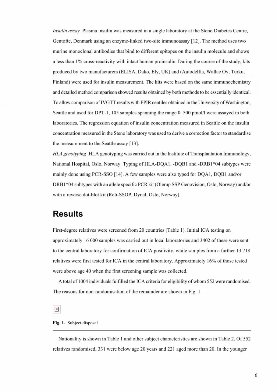
A total of 1004 individuals fulfilled the ICA criteria for eligibility of whom 552 were randomised.
The reasons for non-randomisation of the remainder are shown in Fig. 1.
Fig. 1. Subject disposal
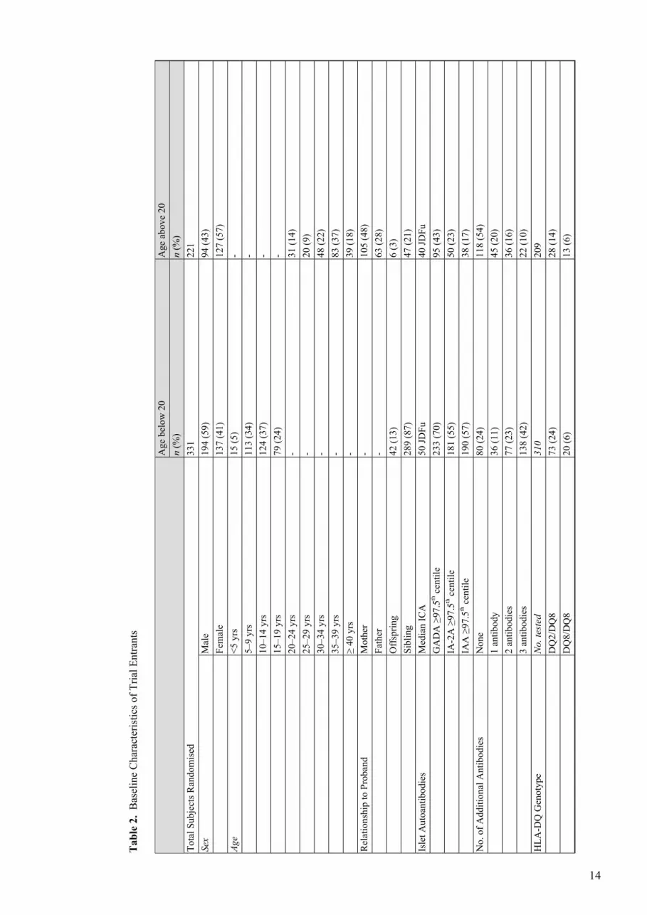
Nationality is shown in Table 1 and other subject characteristics are shown in Table 2. Of 552
relatives randomised, 331 were below age 20 years and 221 aged more than 20. In the younger
6
age group 87% were siblings of a diabetic proband and 13% were offspring. In the older age group
21% were siblings, 76% were parents and 3% were offspring.
[Table 2. will appear here. See end of document.]
Islet autoantibodies Overall, 64% of those randomised had at least one other antibody marker in
addition to ICA, representing 76% of those aged less than 20 and 46% of those above this age.
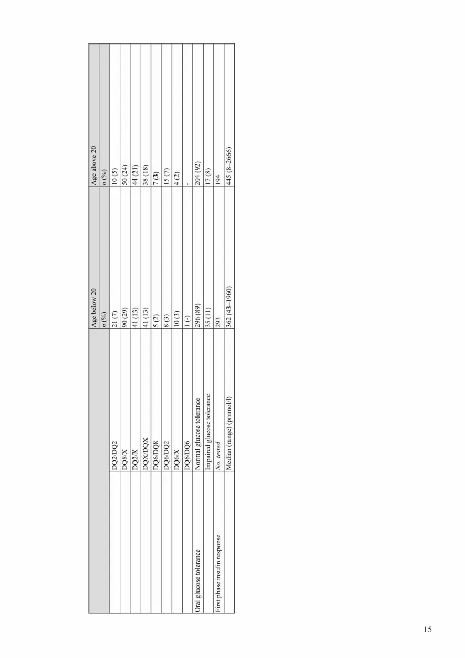
HLA class II The diabetes-associated haplotypes HLA-DQA1*03-DQB1*0302 (DQ8) and/or
HLA-DQA1*0501-DQB1*0201 (DQ2) were found in 84% of the younger age group and 80% of
the older group. The high-risk DQ2/DQ8 heterozygous genotype was found in 23% of the younger
and 14% of the older group respectively. The protective HLA-DQA1*0102-DQB1*0602 (DQ6)
was found in 8% of those below age 20 and 12% of the older group. This haplotype was associated
with ICA alone in 26 individuals, with one additional antibody in 11, with two additional antibodies
in four, and with three additional antibodies in nine individuals. For technical reasons, genotyping
could not be carried out on 33 samples (21 below age 20).
Oral glucose tolerance Overall, of 552 individuals randomised, 52 (9%) had impaired glucose
tolerance (IGT) by WHO criteria in the initial oral glucose tolerance test. Those with ICA alone
were less likely to have IGT than those with additional antibodies (4% vs. 12%, χ2=10.5, p=0.001).
Of 50 individuals with DQ6, 10% had IGT compared with 9% in the remainder. (χ2=0.04, p=0.85).
First phase insulin response The median (range) first phase insulin response, measured as 1+3 min
insulin levels in the intravenous glucose tolerance test, was 362 pmol/l (43–1960 pmol/l) in those
under age 20 years and 476 pmol/l (8–2666 pmol/l) in the older age group. Overall, 164 of 487
tested (34%) had FPIR below the equivalent of the 10th centile used as an entry criterion for the
parenteral arm of DPT-1; 100 individuals in the younger age group (34%) and 64 in the older age
group (33%). Those with ICA alone were less likely to have an FPIR below the 10th centile than
those with additional antibodies (19% vs. 42%, χ2=26.52, p<0.0001). The frequency of low FPIR
was however similar in individuals with an HLA-DQ6 haplotype and those who did not carry the
protective haplotype (27% vs. 34%, χ2=1.11, p=0.29). For technical reasons, including sample
haemolysis, FPIR was not available on 65 individuals (38 below age 20).
DiscussionThe prodromal phase preceding the onset of Type 1 diabetes is characterised by progressive
beta-cell destruction and the appearance of circulating islet autoantibody markers, thus providing
an opportunity for intervention to halt or delay the disease process. Prophylaxis of diabetes has
7
been achieved, sometimes with lasting protection, using a variety of interventions in two animal
models of spontaneous autoimmune diabetes, the NOD mouse and the BB rat. Early human studies
of immune intervention concentrated on the attempt to prolong beta-cell function in recently
diagnosed patients requiring insulin therapy. Prospective randomised placebo controlled
double-blind studies showed, for example, that Cyclosporin A can protect beta-cell function to
the extent that some 25% of patients on Cyclosporin were able to control their diabetes on diet
alone one year after diagnosis, as against 5 to 10% of controls [15,16]. Unfortunately this benefit
does not persist, and the high rate of complications has limited this approach to therapy. Since the
great majority of beta-cells seem to have been destroyed by the time of clinical presentation, it
seems reasonable to conclude that earlier intervention, at a stage when the beta-cell mass is relatively
intact, could produce a more lasting benefit.
Once autoimmunity has been activated, beta cells are destroyed by a cellular immune response
which involves cytotoxic T cells and macrophages and results in release of proinflammatory
cytokines. Impaired insulin secretion and beta-cell damage are thought to result from generation
of nitric oxide and/or oxygen free radicals within the cell [17]. Single strand DNA damage leads
to activation of the DNA repair enzyme poly(ADP-ribose) polymerase (PARP). PARP is an
abundant nuclear protein which shuttles onto and repairs DNA strand breaks. In doing so it
consumes nicotinamide adenine dinucleotide (NAD), transferring the dinucleotide moiety of NAD
to poly (ADP-ribose). Activation of PARP thus results in depletion of intracellular NAD levels,
resulting in a rapid fall in available intracellular energy levels. Excessive activation of PARP and
depletion of intracellular NAD levels are precursors of cell death. Excessive poly (ADP-ribose)
formation due to PARP activation contributes to programmed cell death through energy starvation
as the result of NAD consumption. The protective effect of nicotinamide upon murine beta cells
is thought to be mediated by this pathway [18, 19, 20,21], but it could differ in its effects upon
human beta cells [22]. Previous trials in high risk individuals have reached conflicting conclusions
[23,24].
Nicotinamide is the amide of nicotinic acid; both are components of vitamin B3. Although their
action as anti-pellagra vitamins is similar, their metabolic effects are quite different. In particular,
nicotinic acid has clinical uses as a vasodilator and lipid lowering agent, and induces insulin
resistance. These effects are not seen to any significant extent with nicotinamide, and insulin
secretion is unaffected [25] although it has also been reported to induce a milder degree of insulin
resistance [26]. Nicotinamide is water-soluble, and readily absorbed by mouth. Pharmacokinetic
studies carried out by our group have shown that human metabolism is predominantly by
8
methylation to N-methyl-2-pyridone-5-carboxamide, in which form it is excreted in the urine;
oxidation to nicotinamide oxide also occurs, but there is no direct conversion to nicotinic acid
[27,28]. At high doses, such as those used in ENDIT, peak levels of nicotinamide of 0.3–1.0 mmol/l
are seen in the circulation; the 50% inhibition concentration of nicotinamide for PARP is about
0.1 mmol/l [29]. The safety profile of high dose nicotinamide has been reviewed, and the ratio of
risk to benefit would be highly favourable if efficacy can be shown [30].
First-degree relatives of a child with Type 1 diabetes are at increased risk of progression to the
disease, and are highly motivated to participate in such studies; it is therefore logical to offer
intervention to people in this category. Although now largely superseded by measurement of other
autoantibodies, ICA for many years formed the basis of diabetes prediction, and were used to
identify high risk of progression in ENDIT, as in current US studies of intervention in pre-type 1
diabetes [3]. Family studies have shown that 2 to 2.5% of siblings and parents of a child with
diabetes have ICA greater than or equal to 20 JDF units, and that 40% of those aged less than 40
years will progress to insulin treatment within 5 years of first detection. This risk is increased if
additional autoantibodies to GAD, IA-2 or insulin are detected [31,32], or if first phase insulin
secretion is impaired [4].
The US Diabetes Prevention Trial—Type 1 (DPT-1) started shortly after ENDIT and has
screened more than 70 000 first-degree relatives for ICA. The results of intervention using parental
insulin have recently been reported [3], and a second trial using oral insulin is in progress. These
studies require very large-scale participation, and in Europe are most logically done on a
multinational basis. For this reason simple robust entry criteria were selected for ENDIT, and the
IVGTT was not used as a randomisation criterion. The parenteral arm of DPT-1 required an FPIR
below 10% of a control population, and 37% of ENDIT participants would have fallen into this
category. Since DPT-1 selected individuals with a low insulin response, it is not surprising that
33% had evidence of metabolic decompensation in the form of impaired glucose tolerance, as
against 9% of those in ENDIT.
When ENDIT was planned (the study began in 1994), risk assessment by means of autoantibodies
other than ICA had not been validated. Since then it has been shown that primary screening for
autoantibodies against biochemically defined islet antigens has the potential to provide an enriched
sample for future intervention trials [33]. In the light of present knowledge we would now have
excluded the 198 ENDIT participants who had ICA without other islet autoantibodies, and we
have confirmed a lower rate of IGT and low FPIR in this group. Other intervention studies have
also excluded patients carrying the protective haplotype HLA-DQA1*0102-DQB1*0602 (DQ6)
9
[3]. Within ENDIT, however, of 50 individuals with DQ6, 26 had ICA alone but the remainder
had more than one autoantibody; nine had all four. In addition the frequency of both low FPIR
and IGT in the subgroup with DQ6 were similar to those in the individuals who did not carry this
haplotype. Further study will be needed to establish which individuals with DQ6 should be excluded
from future intervention trials.
The outcome of ENDIT will be announced shortly, but we have learned several lessons. First,
and most important, we have established that it is feasible to launch a multinational collaborative
intervention trial involving both Europe and North America. In consequence, promising
interventions can now be tested before the onset of end-stage beta-cell failure. The corollary is,
however, that intervention at this stage of the disease must be undertaken on a continental scale,
which means that only the most promising interventions can be tested in this way. Validated
surrogate end-points for diabetes development would shorten the time scale of such interventions,
but have yet to be developed. For this reason the current emphasis is upon testing interventions
which might offer beta-cell rescue following clinical diagnosis of Type 1 diabetes, although this
carries the risk that interventions of possible value earlier in the disease pathway might be discarded
because they are ineffective in the latter stages of the disease [34]. As and when future interventions
become available however, ENDIT has shown that high quality, relatively low-budget interventions
before clinical onset can be carried out on a multinational collaborative basis, as and when
potentially safe and effective forms of intervention become available. It is to be hoped that Europe
will be able to develop funding mechanisms capable of supporting such long-term ventures.
Acknowledgements The final phase of ENDIT was funded by Juvenile Diabetes Research
Foundation grant 4-2000-943; earlier phases were funded by European Union grants PL92 0957
and PL95 0771. We are grateful to Novo Nordisk for additional financial support throughout the
period of the trial and for their help in provision of the trial tablets. We thank the many technical,
nursing and medical staff in the participating centres for all their help in running the trial.
*The ENDIT Group:
Co-ordinator: E.A.M. Gale; Deputy Co-ordinators: P.J. Bingley, M. Knip.
Study Administrators: C.L. Emmett, H. Swankie, S. Fewell, P. Kearsey.
National Co-ordinators: Austria: E. Schober; Belgium: F.K. Gorus; Canada: J. Dupre, J.
Mahon; Croatia: V. Profozic; Denmark: J.I. Reimers; T. Mandrup-Poulsen; England: P.J. Bingley;
Finland: M. Knip; France: C. Levy-Marchal; Germany: C. Jaeger; Greece: C. Bartsocas, A. Vazeou;
Hungary: M. Györko, G. Soltesz, L. Madacsy, Italy: M.R. Pastore, P. Pozzilli; Northern Ireland:
10
D.J. Carson, H. Tennett; Norway: K. Dahl-Jørgensen, G. Joner; Poland: I. Kinalska, A.
Mrozikiewicz; Russia: Y. Vaykhonsky; Scotland: K. Robertson; Spain: A. de Leiva, M.T.
Martinez-Larrad, M. Serrano-Rios; Sweden: J. Ludvigsson; Switzerland: E.J. Schoenle; Texas:
W.J. Riley; Turkey: M.T. Yilmaz.
Laboratory Support: University of Bristol:, K.M. Gillespie, H. Gillmor, W.P.T. Moore, A.
Norcross, A.J.K. Williams; Steno Diabetes Centre, Gentofte: B. Dinesen, S. Kjellberg, T.
Mandrup-Poulsen, H. Niebling; National Hospital, Oslo: H.E. Akselsen, E. Thorsby, D.E. Undlien.
Data and Safety Monitoring Committee: A.L. Drash, O. Aagenes, G. Dahlquist, A. Laupacis,
A.E.M. McLean, J. Nerup, B. Weber.
Writing Committee: E.A.M. Gale, P.J. Bingley, C.L. Emmett.
References1. EURODIAB ACE Study Group (2000) Variation and trends in incidence of childhood diabetes inEurope. Lancet 355:873–876
2. Tuomilehto J, Karvonen E, Pitkaniemi J et al. (1999) Record-high incidence of type 1 (insulin-dependent)diabetes in Finnish children. The Finnish Childhood Type 1 Diabetes Registry Group. Diabetologia42:655–660
3. Diabetes Prevention Trial-Type 1 Diabetes Study Group (2002) Effects of insulin in relatives of patientswith type 1 diabetes mellitus. New Engl J Med 346:1685–1691
4. Bingley PJ for the ICARUS Group (1996) Interactions of age, islet cell antibodies, insulin autoantibodiesand first phase insulin response in predicting risk of progression to IDDM in relatives: the ICARUSdata set. Diabetes 45: 1720–1728
5. World Health Organization (1985) Diabetes mellitus: Report of a WHO Study Group. WHO TechnicalReport Series No 727, World Health Organisation, Geneva
6. Bingley PJ, Coleman P, Eisenbarth GS et al. (1992) Standardization of NGTT to predict IDDM. DiabetesCare 15:1313–1316
7. Williams AJK, Bingley PJ, Moore WPM, Gale EAM and the ENDIT screening group (2002) Isletautoantibodies, nationality and gender: a multinational screening study in first-degree relatives ofpatients with Type I diabetes. Diabetologia 45:217–223
8. Bonifacio E, Bingley PJ, Shattock M et al. (1990) Quantification of islet cell antibodies and predictionof insulin-dependent diabetes. Lancet 335:147–149
9. Verge CF, Stenger D, Bonifacio E et al. (1998) Combined use of autoantibodies (IA-2 autoantibody,GAD autoantibody, insulin autoantibody, cytoplasmic islet cell antibodies) in type 1 diabetes:Combinatorial Islet Autoantibody Workshop. Diabetes 47:1857–1866
10. Bingley PJ, Bonifacio E, Williams AJK, Genovese S, Bottazzo GF, Gale EAM (1997) Prediction ofIDDM in the general population: strategies based on combinations of autoantibody markers. Diabetes46:1701–1710
11. Williams AJK, Bingley PJ, Bonifacio E, Palmer JP, Gale EAM (1997) A novel microassay for insulinautoantibodies. J Autoimmun 10:473–478
12. Andersen L, Dinesen B, Jørgensen PN, Poulsen F, Røder ME (1993) Enzyme immunoassay for intacthuman insulin in serum or plasma. Clin Chem 39:578–582
13. McCulloch DK, Bingley PJ, Colman R, Jackson R, Gale EAM, ICARUS group (1993) Comparison ofbolus and infusion protocols for determining acute insulin response to intravenous glucose in normalhumans. Diabetes Care 16:911–915
11
14. Undlien DE, Friede T, Rammensee HG et al (1997) HLA-encoded genetic predisposition in IDDM:DR4 subtypes may be associated with different degrees of protection. Diabetes 46:143–149
15. Feutren G, Papoz L, Assan R et al. (1986) Cyclosporin increases the rate and length of remission ininsulin-dependent diabetes of recent onset. Results of a multicentre double-blind trial. Lancet ii: 119–124
16. Canadian-European Randomized Control Trial Group (1988) Cyclosporin-induced remission of IDDMafter early intervention: association of lyr of Cyclosporin treatment with enhanced insulin secretion.Diabetes 37:1574–1582
17. Eizirik DL, Pavlovic D (1997) Is there a role for nitric oxide in beta cell dysfunction and damage inIDDM? Diabet Metab Rev 13:293–307
18. Zhang J, Dawson VL, Dawson TM, Snyder SH (1994) Nitric oxide activation of poly (ADP-ribose)synthetase in neurotoxicity. Science 263:687–689
19. Mandrup-Poulsen T, Reimers JI, Andersen HU et al. (1993) Nicotinamide treatment in the preventionof insulin-dependent diabetes mellitus. Diabetes Metab Rev 9:295–309
20. Burkart V, Wang Z-Q, Radons J et al. (1999) Mice lacking the poly (ADP-ribose) polymerase gene areresistant to pancreatic beta cell destruction and diabetes development induced by Streptozocin. NatMed 5:314–319
21. Kolb H, Burkart V (1999) Nicotinamide in type 1 diabetes. Mechanism of action revisited. DiabetesCare 22 [Suppl 2]: B16–B20
22. Hoorens A, Pipeleers D (1999) Nicotinamide protects human beta cells against chemically-inducednecrosis, but not against cytokine-induced apoptosis. Diabetologia 42:55–59
23. Elliott RB, Chase HP (1991) Prevention or delay of Type 1 (insulin-dependent) diabetes mellitus inchildren using nicotinamide. Diabetologia 34:362–365
24. Lampeter EF, Klinghammer A, Scherbaum WA et al. (1998) The Deutsche Nicotinamide InterventionStudy: an attempt to prevent Type 1 diabetes. Diabetes 47:980–984
25. Bingley PJ, Caldas C, Bonfanti R, Gale EAM (1993) Nicotinamide and insulin secretion in normalsubjects. Diabetologia 36:675–677
26. Greenbaum CJ, Sears KL, Kahn SE, Palmer JP (1996) Nicotinamide’s effect on glucose metabolismin subjects at risk for IDDM. Diabetes 45:1631–1634
27. Gillmor HA, Bolton CH, Hopton H et al. (1999) Measurement of nicotinamide andN-methyl-2-pyridone-5-carboxamide in plasma by high performance liquid chromatography. BiomedicalChromatography 13:360–362
28. Moore WPT, Bolton CH, Downs, L, Gillmor HA, Gale EAM (2000) Measurement ofn-methyl-2-pyridone-carboxamide in urine by high performance liquid chromatography. BiomedChromatogr 14:69–71
29. Pociot F, Reimers JI, Andersen HU (1993) Nicotinamide—biological actions and therapeutic potentialin diabetes prevention. IDIG Workshop, Copenhagen, Denmark, 4–5 December 1992. Diabetologia36:574–576
30. Knip M, Douek IF, Moore WPT et al. (2001) Safety of high-dose nicotinamide: a review. Diabetologia43:1337–1345
31. Bingley PJ, Christie MR, Bonifacio E et al. (1994) Combined analysis of autoantibodies improvesprediction of IDDM in islet cell antibody-positive relatives. Diabetes 43:1304–1310
32. Verge CF, Gianini R, Kawaski E et al. (1996) Prediction of type 1 diabetes in first-degree relativesusing a combination of insulin, GAD and ICA512bdc/IA-2 autoantibodies. Diabetes 45:926–933
33. Bingley PJ, Williams AJK, Gale EAM (1999) Optimized autoantibody-based risk assessment in familymembers: implications for future intervention trials. Diabetes Care 22:1796–1801
34. Kolb H, Gale EAM (2001) Does partial preservation of residual β-cell function justify immuneintervention in recent onset Type I diabetes? Diabetologia 44:1349–1353
12
Tab
le 1.
Individuals over 20 years old at randomisation
Individuals under 20 years old at randomisation
Local Centre
63
Austria
-2
Belgium
a
3439
Canadaa
1-
Croatia
1812
Denmark
2754
Finlanda
116
Francea
410
Germanya
34
Greece
820
Hungary
26
Italya
1917
Norway
2343
Poland
48
Russia
1520
Spaina
149
Swedena
38
Switzerland
64
Turkey
2937
United Kingdom
a
419
USA (Texas) a
a Initial ICA testing in local laboratory
13
Tab
le 2. Baseline Characteristics of Trial Entrants
Age above 20
Age below 20
n (%)
n (%)
221
331
Total Subjects Randomised
94 (43)
194 (59)
Male
Sex
127 (57)
137 (41)
Female
-15 (5)
<5 yrs
Age
-113 (34)
5–9 yrs
-124 (37)
10–14 yrs
-79 (24)
15–19 yrs
31 (14)
-20–24 yrs
20 (9)
-25–29 yrs
48 (22)
-30–34 yrs
83 (37)
-35–39 yrs
39 (18)
-≥ 40 yrs
105 (48)
-Mother
Relationship to Proband
63 (28)
-Father
6 (3)
42 (13)
Offspring
47 (21)
289 (87)
Sibling
40 JDFu
50 JDFu
Median ICA
Islet Autoantibodies
95 (43)
233 (70)
GADA ≥97.5th centile
50 (23)
181 (55)
IA-2A ≥97.5th centile
38 (17)
190 (57)
IAA ≥97.5th centile
118 (54)
80 (24)
None
No. of Additional Antibodies
45 (20)
36 (11)
1 antibody
36 (16)
77 (23)
2 antibodies
22 (10)
138 (42)
3 antibodies
209
310
No. tested
HLA-DQ Genotype
28 (14)
73 (24)
DQ2/DQ8
13 (6)
20 (6)
DQ8/DQ8
14
Age above 20
Age below 20
n (%)
n (%)
10 (5)
21 (7)
DQ2/DQ2
50 (24)
90 (29)
DQ8/X
44 (21)
41 (13)
DQ2/X
38 (18)
41 (13)
DQX/DQX
7 (3)
5 (2)
DQ6/DQ8
15 (7)
8 (3)
DQ6/DQ2
4 (2)
10 (3)
DQ6/X
-1 (-)
DQ6/DQ6
204 (92)
296 (89)
Normal glucose tolerance
Oral glucose tolerance
17 (8)
35 (11)
Impaired glucose tolerance
194
293
No. tested
First phase insulin response
445 (8–2666)
362 (43–1960)
Median (range) (pmmol/l)
15