Embed Size (px)
Citation preview
British Journal of Cancer (1997) 76(12), 1661-1666© 1997 Cancer Research Campaign
Interstitial pneumonia in patients receiving granulocytecolony-stimulating factor during chemotherapy: surveyin Japan 1991-96
N Niitsul, S Wk12, K Muroi3, S Motomura4, M Murakami5, H Takeyama6, A Ohsaka7 and A Urabe2
'First Department of Internal Medicine, Toho University School of Medicine, Tokyo, 143, Japan; 2Division of Hematology, Kanto Teishin Hospital, Tokyo, 141,Japan; 3Division of Hematology, Department of Medicine, Jichi Medical School, Tochigi, 329-04, Japan; 4First Department of Internal Medicine, Urafune HospitalYokohama City University School of Medicine, Yokohama, 232, Japan; 5Department of Internal Medicine, Osaka Second Police Hospital, Osaka, 567, Japan;6Department of Internal Medicine, Nagoya Ekisaikai Hospital, Aichi, 454, Japan; 7Division of Hematology, Hitachi General Hospital, Ibaraki, 317, Japan
Summary Twenty cases of interstitial pneumonia secondary to treatment with granulocyte colony-stimulating factor (G-CSF) were reviewed.Their interstitial pneumonia had the following features: (a) it occurred predominantly in patients aged 60 years or older; (b) it was prevalentamong patients with haematological malignancies, particularly non-Hodgkin's lymphoma; (c) in all patients G-CSF was given after anti-canceragents with potential to affect the lungs; (d) at the onset, many patients had symptoms such as dyspnoea and fever; and (e) the leucocyte(neutrophil) count as well as lactate dehydrogenase (LDH) and C-reactive protein (CRP) levels were usually higher than normal at the onset.These findings indicate that, when G-CSF is used in combination with pneumotoxic anti-cancer agents, respiratory function should bemonitored before and during treatment. If the leucocyte (or neutrophil) count and/or LDH and CRP increase suddenly in association withdyspnoea and fever during administration of G-CSF, interstitial pneumonia should be suspected. Accordingly, a chest radiograph andpulmonary functional tests should be performed promptly. If a diagnosis of interstitial pneumonia is made, steroid pulse therapy should becommenced immediately.
Keywords: granulocyte colony-stimulating factor; interstitial pneumonia; haematological malignancy
Granulocyte colony-stimulating factor (G-CSF) is used to treatgranulocytopenia secondary to cancer chemotherapy and bonemarrow transplantation. It is effective in reducing the occurrenceof fever and infection associated with granulocytopenia. Althoughthe well-known adverse events of G-CSF are fever, bone pain andliver dysfunction, these problems are largely transient and disap-pear after the completion of treatment (Niitsu and Umeda, 1994).Recently, compared with many other countries, interstitial pneu-monia possibly related to G-CSF administration appears to bemore frequently observed in Japan (lki et al, 1993; Katoh et al,1993; Okubo and Nakazawa 1993; Murayama et al, 1994),although the link between pneumonitis and G-CSF has not beenclearly explained. GM-CSF, another haematopoietic growthfactor, has been associated with adult respiratory distresssyndrome (ARDS) and acute respiratory insufficiency (Wiley et al,1993). Precise knowledge of the characteristics of interstitial pneu-monia due to G-CSF is necessary for early diagnosis and this mayallow us to improve the outcome. Accordingly, we reviewed casesof interstitial pneumonia secondary to G-CSF therapy reported inJapan to clarify its clinical characteristics as well as possiblemethods of diagnosis and treatment.
Received 11 June 1997Revised 22 May 1997Accepted 9th June 1997
Correspondence to: N Niitsu, First Department of Internal Medicine, TohoUniversity School of Medicine, 11-1, Ohmori-nishi 6-chome, Ohta-ku, Tokyo,143, Japan
MATERIALS AND METHODS
The subjects of this study were patients receiving either filgrastimor lenograstim and who presented symptoms consistent with thediagnosis for interstitial pneumonia between November 1991 andJanuary 1996 by the criteria shown below. The criteria for diag-nosis of interstitial pneumonia were determined as follows: (a)chest radiograph films and computerized tomography (CT) scans
that showed findings characteristic of interstitial pneumonia; (b)the PaO2 was < 70 mmHg at onset or decreased by 20 mmHg afteradministration of G-CSF; (c) infection and tumour metastasis were
excluded by bacteriological, cytological and histological examina-tion of sputum, bronchoalveolar lavage fluid and transbronchialbiopsy specimens that were used to detect bacteria, fungi, protozoaand viruses or because neither organisms nor tumour cells were
detected in any of these specimens; and (d) interstitial pneumoniadeveloped within 10 days of completion of G-CSF therapy afteradministration of anti-cancer agents. Patients with a history oflung disease were not included in this study. Twenty patients werediagnosed with interstitial pneumonia using the above criteriaand all of them were reported to the Japanese Ministry of Healthand Welfare.
RESULTS
Background data on patients
The 20 patients with concurrent interstitial pneumonia were aged63 years on average and 14 were at least 60 years old, indicatingthat it predominantly affected elderly patients. The primary
1661
brought to you by COREView metadata, citation and similar papers at core.ac.uk
provided by PubMed Central
1662 N Niitsu et al
Table 1 Characteristics of patients who developed interstitial pneumoniaduring treatment with recombinant human granulocyte colony-stimulatingfactor
Number of patientsMedian age (range) yearsSex: male/female
2063 (41-73)10/10
DiagnosisNon-Hodgkin's lymphoma
Histology (working formulation)Low grade
FollicularIntermediate grade
Diffuse largeDiffuse mixedDiffuse small cleaved
High gradeImmunoblastic
Primary/relapsePhenotypeT/B/unknown
Stage (Ann Arbor)l/lWl/IV/unknown
Acute monocytic leukaemia (M5b*)
Performance status (WHO)0/1/2/3/4/unknown
19
1061
18/1
4/10/5
4/5/8/21
1 0/1l12101314
*French-American-British classification.
disease was haematological malignancy in all 20 patients.Nineteen of them had non-Hodgkin's lymphoma (NHL) and onehad acute monocytic leukaemia (M5b: French-American-Britishclassification). NHL was of intermediate grade in 89.5% andmainly in the advanced stages according to the Ann Arbor classifi-cation (Table 1). On admission, none of the patients showed anysignificant changes in peripheral blood findings, pulmonary func-tion, immunology or coagulation parameters (Table 2).
Association of interstitial pneumonia withchemotherapy regimens, number of chemotherapycourses and response to treatment (Table 3)
All 20 patients received G-CSF as adjuvant therapy during cancer
chemotherapy.The chemotherapy regimens used always included pneumotoxic
agents, such as cyclophosphamide (CPA), bleomycin (BLM),methotrexate (MTX) and etoposide (VP-16). Interstitial pneu-monia developed most commonly (in seven patients) during thesecond course of chemotherapy and in 15 patients before thefourth course of chemotherapy. The median total doses ofpneumotoxic chemotherapy agents were CPA 1800 mg i-2(584-5250), BLM 18 mg in2 (11-55), MTX 1137 mg i-2(321-3094), VP-16 394 mg in2 (345-1859) and the numbers oftreated patients were 19 (95%), 11 (55%), 5 (25%), and 4 (20%)
Table 2 Comparisons of laboratory findings between baseline and at onset of interstitial pneumonia during treatment withrecombinant human granulocyte colony-stimulating factor
Baseline At onset of Interstitial pneumonia
Haematological examinationsWBC (rd)Neutrophil (j.)Platelet (x 104 jl-1)RBC (x 104 gl-1)Haemoglobin (g dl-')LDH (IU I-')CRP (mg dl-1)
Immunological examinationsIgA (mg dl-')IgG (mg dl-1)IgM (mg dl-')T cell (%)B cell (%)CD4 (%)
CD8 (%)
CD4/CD8 ratio
Pulmonary function testpaO2 (kPa)PaCO2 (kPa)AadO2 (kPa)%DLCO
Coagulation testPT (second)APTT (second)FDP (igg ml-')Fibrinogen (mg dl-')D-dimer (ng ml-')
5100(1700-11300)2438 (816-8995)23.8 (2-46)
429.5 (302-470)12.4 (8-14)331 (212-646)a0.3 (0-3.44)a
291 (191-438)1841 (1460-5174)171 (84-722)92 (82.1-98)2 (1-8)
28.6 (27.2-48.3)21.4 (7.6-48.4)1.41 (0.68-6.36)
11.5 (10.0-13.4)5.3 (4.6-5.5)2.0 (0.1-4.4)93 (82-98)
11.4 (10.0-11.9)33.4 (26.2-43.6)<10384(126-561)<100
10000(3800-41500)8000 (2700-20925)18.5 (0.9-162)332 (13.4 448)10.4 (7-13)607 (248-1072)3.2 (0-26.2)
186 (129-371)1620 (591-2090)124 (50-268)92 (87-98)3 (1-8)
30.1 (28.6-34.5)25.9 (14.9-38.1)1.32 (0.85-2.01)
7.0 (4.3-9.7)4.5 (3.6-6-1)7.4 (3.0-10.4)
11.4 (10.1-11.8)36.0 (27.4-43)<10403 (145-555)<100
British Journal of Cancer (1997) 76(12), 1661-1666
Values are given as median (range). aDuring chemotherapy before G-CSF administration and before onset of interstitialpneumonia episode. Abbreviations: WBC, white blood cell; RBC, red blood cell; PaO2, partial pressure of arterial oxygen,PaCO2, partial pressure of arterial carbon dioxide; AaDO2, arterial-alveolar difference of oxygen; DLCO, pulmonary diffusingcapacity for carbon monoxide; PT, prothrombin time; APTT, activated partial thromboplastin time; FDP, fibrinogen degradationproducts.
0 Cancer Research Campaign 1997
Interstitial pneumonia in patients receiving G-CSF 1663
Table 3 Chemotherapy regimens, clinical response and total dose of anti-cancer agents and G-CSF until onset of interstitial pneumonia in the 20 patients
Anticancer agentsa G-CSFb
Number Sex Age Diagnosis Chemotherapy Course Clinical CPA BLM MTX VP-16 (gg per Durationsregimen at onset response (mg m-2) (mg m-2) (mg m-2) (mg m-2) body) (day)
1 M 41 NHL Pro-MACE 2 CR 2652 3094 442 1650 122 M 56 NHL COP-BLAMIII 1 PR 782 28 600 43 F 63 NHL COPP/CHOP 2 PR 1242 375 54 F 65 NHL COP-BLAMIII 4 CR 1655 29 750 105 F 62 NHL COP-BLAMIII 7 CR 1972 55 450 66 F 66 NHL COP-BLAM 2 CR 584 15 300 47 M 73 AMoL AraC + VP-16 4 PR 1859 1280 88 M 71 NHL ProMACE-CytaBOM 4 CR 1728 14 321 346 450 69 F 68 NHL CHOP 4 PR 2786 1790 1910 F 66 NHL CHOP 2 CR 800 570 511 F 47 NHL CHOP 4 CR 4000 400 412 M 69 NHL CHOP 2 PD 706 700 713 F 64 NHL DXR+VP-16+CPA+VCR+BLM+PSL 6 CR 3500 17 345 345 500 514 F 55 NHL COP-BLAM 2 CR 1200 15 600 615 M 48 NHL COP-BLAM 3 PR 1800 1 1 700 616 F 63 NHL COP-BLAM 3 CR 1800 21 600 617 M 49 NHL MACOP-B 12 weeks Unknown 2160 30 1240 600 518 M 62 NHL MACOP-B 11 weeks Unknown 1989 18 1137 1750 1419 M 62 NHL CHOP 7 CR 5250 1100 1220 M 67 NHL CHOP 2 CR 1500 1100 12
aTotal dose administered until onset of interstitial pneumonia episode. bTotal dose administered only course at onset of interstitial pneumonia episode.Abbreviations: CHOP, cyclophosphamide, doxorubicin, vincristine, prednisolone; COPP, cyclophosphamide, vincristine, procarbazine, prednisolone; ProMACE-CytaBOM, prednisolone, methotrexate, doxorubicin, cyclophosphamide, etoposide, cytarabine, bleomycin, vincristine; COP-BLAM, COP-BLAMIII,cyclophosphamide, vincristine, prednisolone, bleomycin, doxorubicin, procarbazine; MACOP-B, methotrexate, doxorubicin, cyclophosphamide, vincristine,prednisolone, bleomycin; CR, complete remission; PR, partial remission; PD, progressive disease.
respectively. Out of the 18 patients whose clinical response wasreported and was available, 17 (94%) achieved complete remissionor partial remission. Interstitial pneumonia developed mostfrequently (in 12 patients) during the administration of G-CSF andin three other patients within 3 days of completion of administra-tion of G-CSF. G-CSF was given for a median duration of 6 days(range 4-19 days). And the median total dose of G-CSF was600 jig per body (range 300-1790 jg per body). There was nocorrelation between total dose of G-CSF and interstitial pneu-monia. One patient received concurrent G-CSF and bleomycinchemotherapy. The G-CSF preparation used was filgrastim ineight patients and lenograstim in twelve. Patients did not receiveany other medication, apart from anti-cancer agents, which mighthave induced interstitial pneumonia.
Clinical characteristics at the onset of interstitialpneumonia (Tables 4, 5 and 6)
SymptomsThe most common symptom of interstitial pneumonia wasdyspnoea (11 patients, 55%) followed by fever (ten patients, 50%).
Leucocyte and neutrophil counts and serum levels of LDHand CRPAt the onset of interstitial pneumonia the leucocyte count was2 10 000 il-l in ten patients and the neutrophil count was . 5000 jl-'in 11 patients. Interstitial pneumonia most frequently developed6 days after the leucocyte (neutrophil) nadir or after rapid recoveryof the leucocyte count. At the onset of interstitial pneumonia, lactatedehydrogenase (LDH) increased in 12 patients and C-reactiveprotein (CRP) increased in 14 patients.
Imaging findingsThe most common finding on chest radiograph films was a gran-ular or reticular pattern throughout the lung fields, which wasobserved in 12 patients, followed by a similar pattern involving thelower fields of both lungs. CT scans of the chest showed a granularor reticular pattern extending throughout both lungs in 77.8% ofpatients.
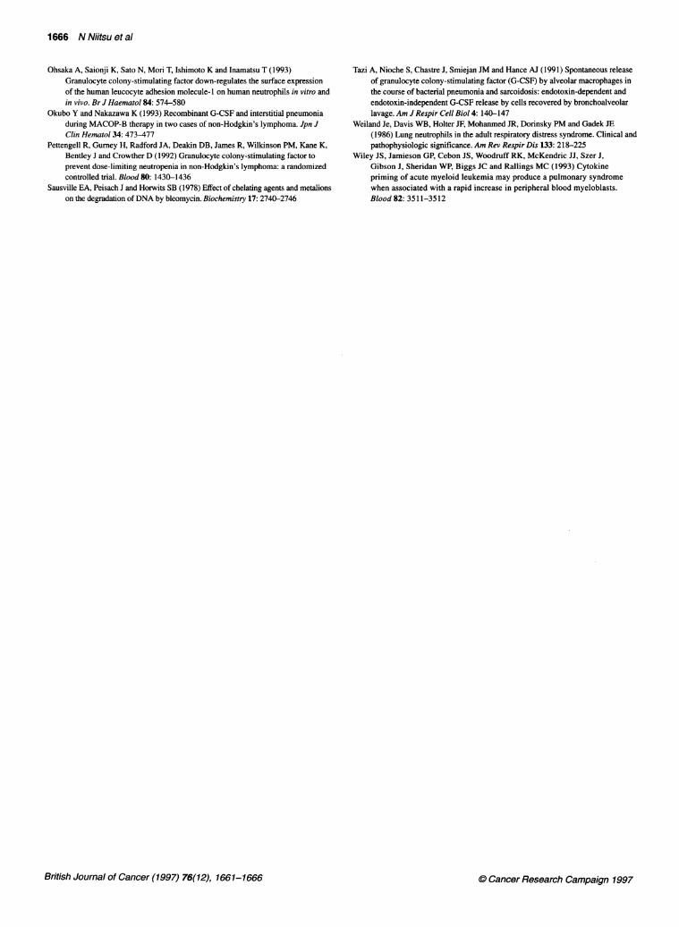
Findings on bronchoalveolar lavage (BAL) andtransbronchial lung biopsy (TBLB)BAL was performed in seven patients and the total cell count of allpatients excluding one patient was measured. It was unmeasurablein one patient. The total cell count increased in all six patients. Thepercentage of lymphocytes and neutrophils increased in four andtwo patients respectively. The CD4/CD8 ratio decreased in fivepatients and it was less than 1. TBLB performed in three patientsshows typical histological changes of interstitial pneumonia.Figure IA and B shows the changes in the same patient and FigureIC shows the changes in another patient. These figure of typicalhistological changes are similar to interstitial pneumonia, with theapparent thickening of alveolar walls and infiltration of smallround cells. Also, a small amount of intra-alveolar exudative pneu-monia was observed, as well as granuloma formation in two cases.
Treatment and outcome
Of the 20 patients, 19 received steroid pulse therapy; the remainingpatient received 02 administration. A total of 17 patients who weretreated in the earliest part of the study period recovered, but threepatients in this group eventually died because of respiratory insuf-ficiency and multiple organ failure.
British Journal of Cancer (1997) 76(12), 1661-16660 Cancer Research Campaign 1997

1664 N Niitsu et al
Table 4 Clinical symptoms at onset of interstitial pneumonia during treatmentwith recombinant human granulocyte colony-stimulating factor
Clinical symptoms Number of patients (%)
Dyspnoea 11(55)Fever (> 380C) 10 (50)Shortness of breath 2 (10)Cough 2 (10)General fatigue 2 (10)Dull headache 1 (5)
Table 5 Chest radiograph, chest CT and bronchoalveolar lavage findings atonset of interstitial pneumonia during treatment with recombinant humangranulocyte colony-stimulating factor
Chest radiograph examinationNumber of examined patients 19Region of diffuse granular and reticular shadow
Bilateral whole lung fields 12 (63.2%)Bilateral lower lung fields 5 (26.3%)Right middle and lower lung field 1 (5.3%)Right lower lung field 1 (5.3%)
Chest-CT examinationNumber of examined patients 9Region of diffuse granular and reticular shadow
Bilateral lung fields 7 (77.8%)Right lung field 2 (22.2%)
Table 6 Bronchoalveolar lavage
Case Total number of cells Macrophage Neutrophil Lymphocyte Eosinophil CD4/CD8(x 106 cells ml-1) (%) (%) (%) (%) ratio
1 14.0 19.0 52.0 21.0 8.0 1.712 2.5 30.8 6.9 62.3 0.0 0.413 14.3 27.4 56.8 15.3 0.0 0.84 10.2 32.4 9.0 58.6 0.0 0.55 14.0 17.0 8.0 74.0 1.0 0.536 13.9 18.0 4.0 74.0 2.0 0.897 ND* ND ND ND ND 1.16
*ND, not done.
DISCUSSION
G-CSF has been used to treat various forms of neutropenia. Thisdrug is believed to increase the neutrophil count and enhanceneutrophil function (Pettengell et al, 1992). The activation ofneutrophils is called a priming effect by which G-CSF enhancesneutrophil phagocytosis and migration as well as superoxideproduction (Laver and Moore, 1989). These are important biolog-ical defence mechanisms that primarily act to prevent bacterialinvasion but simultaneously enhance inflammatory reactions thatcan injure host tissues (Weiland et al, 1986). It has been reportedthat neutrophils are involved in the progression of ARDS. In BALfluid from patients with ARDS, neutrophils are increased innumber and there is an increase in neutrophil elastase activity(Idell et al, 1985). In BAL fluid from patients with interstitialpneumonia, the G-CSF production by alveolar macrophages isincreased significantly, suggesting involvement of G-CSF in thepathogenesis of this condition (Tazi et al, 1991).
Interstitial pneumonia related to the use of G-CSF has occasion-ally been reported and many mechanisms have been proposed forits aetiology. Matthews (1993) suggested that G-CSF mightaugment the pneumotropic toxicity of BLM because pneumotoxi-city occurred in three out of five patients with NHL who receivedABVD (doxorubicin, bleomycin, vinblastine and dacarbazin) plusG-CSF. Dirix et al (1994) considered that BLM pneumonia mightbe made worse by a rapid increase in the number and activity ofneutrophils because of G-CSF. BLM is thought to react withFe2+ to produce superoxide (02-), which damages DNA moleculesand gives rise to pulmonary dysfunction (Sausville et al, 1978).According to Bastion et al (1994), who conducted two randomizedplacebo-controlled trials in NHL patients to assess the effectiveness
of G-CSF, chemotherapy including BLM given in combination withG-CSF did not augment the pneumotropic toxicity of bleomycin.Another clinical study assessed pulmonary disease in patients withaggressive NHL who received BACOP (bleomycin, doxorubicin,cyclophosphamide, vincristine, prednisolone) therapy with orwithout G-CSF. Pneumonia occurred in 33% of patients receivingBACOP with G-CSF and in 4% of the control patients. The authorsrecommended that G-CSF should be used carefully when combinedwith chemotherapy regimens that involve repeated BLM adminis-tration over a long period (Lei et al, 1994). The mechanism bywhich G-CSF when combined with certain anti-cancer agents givesrise to pneumonia remains to be clarified. G-CSF has not beenreported to cause pneumonia in patients receiving it alone and itonly causes pneumonia in patients receiving combined therapy withanti-cancer agents. This suggests that G-CSF increases the numberof activated neutrophils that exert a deleterious effects on sub-clinical lung damage produced by anti-cancer agents and results inthe manifestation of pulmonary dysfunction.
In addition to the effect on haemopoiesis, G-CSF enhancesmature neutrophil functions both in vitro and in vivo. Previousstudies indicated that G-CSF administration enhances superoxiderelease in neutrophils from patients with maligant lymphoma(Ohsaka et al, 1989). And Ohsaka et al (1993) reported that G-CSFinversely regulates the surface expression of cellular adhesionmolecules on human neutrophils, that is G-CSF down-regulatesthe expression of L-selectin and up-regulates the expression ofCDllb/CD18 leucocyte integrin on neutrophils. These findingssuggest that G-CSF may enhance host defence and participate inthe inflammatory process through the neutrophil-endothelial cellinteractions. However, neutrophil-derived oxygen metabolites andproteinases have also been implicated in the pathogenesis of tissue
British Journal of Cancer (1997) 76(12), 1661-1666 0 Cancer Research Campaign 1997

Interstitial pneumonia in patients receiving G-CSF 1665
A~~~~~~A.k ......... ..................~~~~~~~~~~~~~~~~~~~~~~~~~~~~~~~~~~.......
.................................,: .:.:i.r
~~~~~~~~~~~~.... ...(..X ee*'"...'
............
* : _*: P :e~ ~ * ~
B .~~~~~~~~~~~~~~~~~~~~~~~~~~~~~~~~~~~~~~~~~~~~~~~~~~~~.^.,.......... . ..... . i.........~~~~~~ I
.. ... ....ew'::
Figure 1 (A) (B) Thickening of alveolar walls with small round cellsinfiltration, granuloma formation and small amount of exudate are seen(haematoxylin-eosin staining, HE x 100, elastica-Masson staining x 100).(C) Thickening of alveolar walls with slight degree of small round cellsinfiltration, swelling alveolar epithelium cells, granuloma-like histiocyticagglutination and small amount of exudate are seen (HE x 67)
injury. Although the aetiology of interstitial pneumonia afterG-CSF application remains to be elucidated, activated or primedneutrophils may participate in the development of the disease.The present study showed that interstitial pneumonia secondary
to G-CSF has the following characteristics: (a) it occurred predom-inantly in older patients (>60 years); (b) it was prevalent among
patients with haematological malignancies, particularly NHL; (c)all patients received G-CSF after administration of BLM, MTX,CPA, VP-16 or other pneumotoxic anti-cancer agents; (d) the onsetof interstitial pneumonia occurred during administration of G-CSFin 12 out of 20 patients; (e) the earliest symptoms of interstitialpneumonia were usually dyspnoea and a temperature 2 38°C; (f)the onset was associated with an increase in the leucocyte(neutrophil) count in many cases; (g) initial elevation of the LDHand CRP levels was observed in 12 and 14 patients respectively; (h)on chest radiograph films, changes appeared first in the lower lungfields and spread gradually over the entire lungs; (i) in manypatients, the total cell count in BAL fluid was increased; and (j)alleviation of interstitial pneumonia was achieved in patientstreated by steroid pulse therapy soon after onset. In brief, whenG-CSF is given to patients who have previously received pneumo-toxic anti-cancer agents, such as bleomycin and methotrexate,pulmonary function should be monitored by measuring PaO2 and% DLCO before and during G-CSF therapy.
Interstitial pneumonia should be suspected if dyspnoea andfever are associated with rapidly increasing leucocyte andneutrophil counts, as well as an elevation ofLDH and CRP duringadministration of G-CSF. The diagnosis should be confirmed bypulmonary function tests and chest radiograph examination. It isimportant to start steroid pulse therapy as early as possible whenthe diagnosis is made.
ACKNOWLEDGEMENT
We express our appreciation to Dr M Mochizuki at Kanto TeishinHospital for technical support in TBLB and cytopathological inter-pretation of specimens.
REFERENCES
Bastion Y, Reyes F, Bosly A, Gisselbrecht C, Yver A, Gilles E, Maral J and CoiffierB (1994) Possible toxicity with the association of G-CSF and bleomycin.Lancet 343: 1221-1222
Dirix LY, Schrijvers D, Druwe P, Van Den Brande J, Verhoeven D and Van OoostermAT (1994) Pulmonary toxicity and bleomycin. Lancet 344: 56
Idell S, Kucich U, Fein A, Kueppers F, James HL, Walsh PN, Weinbaum G, ColmnnRW and Cohen AB (1985) Neutrophil elastase-releasing factors inbronchoalveolar lavage from patients with adult respiratory distress syndrome.Am Rev Respir Dis 132: 1098-1105
Iki S, Yoshinaga K, Ohbayashi Y and Urabe A (1993) Cytotoxic drug-inducedpneumonia and possible augmentation by G-CSF-clinical attention (letter). AnnHematol 66: 217-218
Katoh M, Shikoshi K, Takada M, Umeda M, Tsukahara T, Kitagawa S and Shirai T(1993) Development of interstitial pneumonitis during treatment withgranulocyte colony-stimulating factor. Ann Hematol 67: 201-202
Laver J and Moore MAS (1989) Clinical use of recombinant human hematopoieticgrowth factors. J Natl Cancer Inst 81: 1370-1382
Lei Kik, Leung WT and Johnson PJ (1994) Serious pulmonary complications inpatients receiving recombinant granulocyte colony-stimulating factor duringBACOP chemotherapy for aggressive non-Hodgkin's lymphoma. Br J Cancer70: 1009-1013
Matthews JH (1993) Pulmonary toxicity ofABVD chemotherapy and G-CSF inHodgkin's disease: possible synergy (letter). Lancet 342: 988
Murayama J, Kawakami T and Togawa S (1994) Two cases of malignant lymphomapatient who developed interstitial pneumonia after CHOP-G therapy. Med JIbaraki Prefecture Hospital 12: 121-129
Niitsu N and Umeda M (1994) The effects of chemotherapy and G-CSF in patientswith non-Hodgkin's lymphoma. Chemotherapy 42: 346-350
Ohsaka A, Kitagawa S, Sakamoto S, Miura Y, Takanashi N, Takaku F and Saito M(1989) In vivo activation of human neutrophil functions by administration ofrecombinant human granulocyte colony-stimulating factor in patients withmalignant Iymphoma. Blood 74: 2743-2748
C Cancer Research Campaign 1997 British Journal of Cancer (1997) 76(12), 1661-1666
1666 N Niitsu et al
Ohsaka A, Saionji K, Sato N, Mori T, Ishimoto K and Inamatsu T (1993)Granulocyte colony-stimulating factor down-regulates the surface expressionof the human leucocyte adhesion molecule- I on human neutrophils in vitro andin vivo. Br J Haematol 84: 574-580
Okubo Y and Nakazawa K (1993) Recombinant G-CSF and interstitial pneumoniaduring MACOP-B therapy in two cases of non-Hodgkin's lymphoma. Jpn JClin Hematol 34: 473-477
Pettengell R, Gurney H, Radford JA, Deakin DB, James R, Wilkinson PM, Kane K,Bentley J and Crowther D (1992) Granulocyte colony-stimulating factor toprevent dose-limiting neutropenia in non-Hodgkin's lymphoma: a randomizedcontrolled trial. Blood 80: 1430-1436
Sausville EA, Peisach J and Horwits SB (1978) Effect of chelating agents and metalionson the degradation ofDNA by bleomycin. Biochemistry 17: 2740-2746
Tazi A, Nioche S, Chastre J, Smiejan JM and Hance AJ (1991) Spontaneous releaseof granulocyte colony-stimulating factor (G-CSF) by alveolar macrophages inthe course of bacterial pneumonia and sarcoidosis: endotoxin-dependent andendotoxin-independent G-CSF release by cells recovered by bronchoalveolarlavage. Am J Respir Cell Biol 4: 140-147
Weiland Je, Davis WB, Holter JF, Mohanmed JR, Dorinsky PM and Gadek JE(1986) Lung neutrophils in the adult respiratory distress syndrome. Clinical andpathophysiologic significance. Am Rev Respir Dis 133: 218-225
Wiley JS, Jamieson GP, Cebon JS, Woodruff RK, McKendric JJ, Szer J,Gibson J, Sheridan WP, Biggs JC and Rallings MC (1993) Cytokinepriming of acute myeloid leukemia may produce a pulmonary syndromewhen associated with a rapid increase in peripheral blood myeloblasts.Blood 82: 3511-3512
British Journal of Cancer (1997) 76(12), 1661-1666 0 Cancer Research Campaign 1997