Embed Size (px)
Citation preview
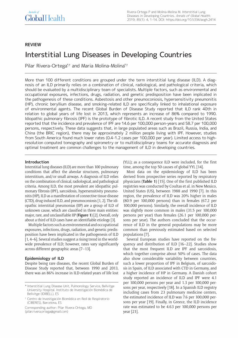
IntroductionInterstitial lung diseases (ILD) are more than 100 pulmonary conditions that affect the alveolar structures, pulmonary interstitium, and/or small airways. A diagnosis of ILD relies on the combination of clinical, radiological, and pathological criteria. Among ILD, the most prevalent are idiopathic pul-monary fibrosis (IPF), sarcoidosis, hypersensitivity pneumo-nitis (HP), ILD as a manifestation of connective tissue disease (CTD), drug-induced ILD, and pneumoconiosis [1, 2]. The idi-opathic interstitial pneumonias (IIP) are a group of ILD of unknown cause, which are classified in three main entities: major, rare, and unclassifiable IIP (Figure 1) [2]. Overall, only about a third of ILD cases have an identifiable etiology [3].
Multiple factors such as environmental and occupational exposures, infections, drugs, radiation, and genetic predis-position have been implicated in the pathogenesis of ILD [1, 4–6]. Several studies suggest a rising trend in the world-wide prevalence of ILD; however, rates vary significantly across different geographic areas [7–13].
Epidemiology of ILDDespite being rare diseases, the recent Global Burden of Disease Study reported that, between 1990 and 2013, there was an 86% increase in ILD-related years of life lost
(YLL); as a consequence ILD were included, for the first time, among the top 50 causes of global YYL [14].
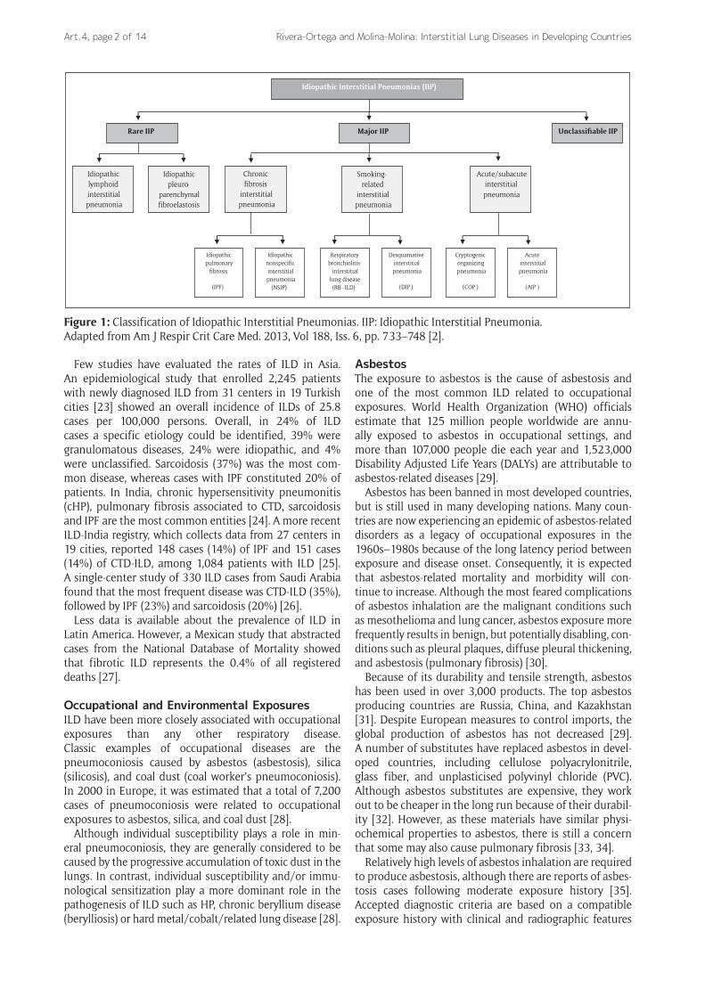
Most data on the epidemiology of ILD has been derived from prospective series reported by respiratory physicians (Table 1) [15]. One of the first published ILD registries was conducted by Coultas et al. in New Mexico, United States (US), between 1988 and 1990 [7]. In this region, the prevalence of ILD was 20% higher in males (80.9 per 100,000 persons) than in females (67.2 per 100,000 persons). Similarly, the overall incidence of ILD was slightly more common in males (31.5 per 100,000 persons per year) than females (26.1 per 100,000 per-sons per year). The authors concluded that the occur-rence of ILD in the general populations may be more common than previously estimated based on selected populations [7].
Several European studies have reported on the fre-quency and distribution of ILD [16–22]. Studies show that the most frequent ILD are IPF and sarcoidosis, which together comprise about 50% of cases. The data also show considerable variability between countries, such a lower proportion of IPF in Belgium, of sarcoido-sis in Spain, of ILD associated with CTD in Germany, and a higher incidence of HP in Germany. A Danish cohort study reported an incidence of ILD and IPF were 4.1 per 100,000 persons per year and 1.3 per 100,000 per-sons per year, respectively [18]. In a Spanish ILD registry including cases from 23 pulmonary medicine centers, the estimated incidence of ILD was 7.6 per 100,000 per-sons per year [19]. Finally, in Greece, the ILD incidence rate was estimated to be 4.63 per 100,000 persons per year [21].
Rivera-Ortega P and Molina-Molina M. Interstitial Lung Diseases in Developing Countries. Annals of Global Health. 2019; 85(1): 4, 1–14. DOI: https://doi.org/10.5334/aogh.2414
* Interstitial Lung Disease Unit, Pulmonology Service, Bellvitge University Hospital. Instituto de Investigación Biomédica de Bellvitge (IDIBELL), ES
† Centro de Investigación Biomédica en Red de Respiratorio (CIBERES), Barcelona, ES
Corresponding author: Pilar Rivera-Ortega, MD ([email protected])
REVIEW
Interstitial Lung Diseases in Developing CountriesPilar Rivera-Ortega*,† and Maria Molina-Molina*,†
More than 100 different conditions are grouped under the term interstitial lung disease (ILD). A diag-nosis of an ILD primarily relies on a combination of clinical, radiological, and pathological criteria, which should be evaluated by a multidisciplinary team of specialists. Multiple factors, such as environmental and occupational exposures, infections, drugs, radiation, and genetic predisposition have been implicated in the pathogenesis of these conditions. Asbestosis and other pneumoconiosis, hypersensitivity pneumonitis (HP), chronic beryllium disease, and smoking-related ILD are specifically linked to inhalational exposure of environmental agents. The recent Global Burden of Disease Study reported that ILD rank 40th in relation to global years of life lost in 2013, which represents an increase of 86% compared to 1990. Idiopathic pulmonary fibrosis (IPF) is the prototype of fibrotic ILD. A recent study from the United States reported that the incidence and prevalence of IPF are 14.6 per 100,000 person-years and 58.7 per 100,000 persons, respectively. These data suggests that, in large populated areas such as Brazil, Russia, India, and China (the BRIC region), there may be approximately 2 million people living with IPF. However, studies from South America found much lower rates (0.4–1.2 cases per 100,000 per year). Limited access to high-resolution computed tomography and spirometry or to multidisciplinary teams for accurate diagnosis and optimal treatment are common challenges to the management of ILD in developing countries.
Rivera-Ortega and Molina-Molina: Interstitial Lung Diseases in Developing CountriesArt. 4, page 2 of 14
Few studies have evaluated the rates of ILD in Asia. An epidemiological study that enrolled 2,245 patients with newly diagnosed ILD from 31 centers in 19 Turkish cities [23] showed an overall incidence of ILDs of 25.8 cases per 100,000 persons. Overall, in 24% of ILD cases a specific etiology could be identified, 39% were granulomatous diseases, 24% were idiopathic, and 4% were unclassified. Sarcoidosis (37%) was the most com-mon disease, whereas cases with IPF constituted 20% of patients. In India, chronic hypersensitivity pneumonitis (cHP), pulmonary fibrosis associated to CTD, sarcoidosis and IPF are the most common entities [24]. A more recent ILD-India registry, which collects data from 27 centers in 19 cities, reported 148 cases (14%) of IPF and 151 cases (14%) of CTD-ILD, among 1,084 patients with ILD [25]. A single-center study of 330 ILD cases from Saudi Arabia found that the most frequent disease was CTD-ILD (35%), followed by IPF (23%) and sarcoidosis (20%) [26].
Less data is available about the prevalence of ILD in Latin America. However, a Mexican study that abstracted cases from the National Database of Mortality showed that fibrotic ILD represents the 0.4% of all registered deaths [27].
Occupational and Environmental ExposuresILD have been more closely associated with occupational exposures than any other respiratory disease. Classic examples of occupational diseases are the pneumoconiosis caused by asbestos (asbestosis), silica (silicosis), and coal dust (coal worker’s pneumoconiosis). In 2000 in Europe, it was estimated that a total of 7,200 cases of pneumoconiosis were related to occupational exposures to asbestos, silica, and coal dust [28].
Although individual susceptibility plays a role in min-eral pneumoconiosis, they are generally considered to be caused by the progressive accumulation of toxic dust in the lungs. In contrast, individual susceptibility and/or immu-nological sensitization play a more dominant role in the pathogenesis of ILD such as HP, chronic beryllium disease (berylliosis) or hard metal/cobalt/related lung disease [28].
AsbestosThe exposure to asbestos is the cause of asbestosis and one of the most common ILD related to occupational exposures. World Health Organization (WHO) officials estimate that 125 million people worldwide are annu-ally exposed to asbestos in occupational settings, and more than 107,000 people die each year and 1,523,000 Disability Adjusted Life Years (DALYs) are attributable to asbestos-related diseases [29].
Asbestos has been banned in most developed countries, but is still used in many developing nations. Many coun-tries are now experiencing an epidemic of asbestos-related disorders as a legacy of occupational exposures in the 1960s–1980s because of the long latency period between exposure and disease onset. Consequently, it is expected that asbestos-related mortality and morbidity will con-tinue to increase. Although the most feared complications of asbestos inhalation are the malignant conditions such as mesothelioma and lung cancer, asbestos exposure more frequently results in benign, but potentially disabling, con-ditions such as pleural plaques, diffuse pleural thickening, and asbestosis (pulmonary fibrosis) [30].
Because of its durability and tensile strength, asbestos has been used in over 3,000 products. The top asbestos producing countries are Russia, China, and Kazakhstan [31]. Despite European measures to control imports, the global production of asbestos has not decreased [29]. A number of substitutes have replaced asbestos in devel-oped countries, including cellulose polyacrylonitrile, glass fiber, and unplasticised polyvinyl chloride (PVC). Although asbestos substitutes are expensive, they work out to be cheaper in the long run because of their durabil-ity [32]. However, as these materials have similar physi-ochemical properties to asbestos, there is still a concern that some may also cause pulmonary fibrosis [33, 34].
Relatively high levels of asbestos inhalation are required to produce asbestosis, although there are reports of asbes-tosis cases following moderate exposure history [35]. Accepted diagnostic criteria are based on a compatible exposure history with clinical and radiographic features
Figure 1: Classification of Idiopathic Interstitial Pneumonias. IIP: Idiopathic Interstitial Pneumonia.Adapted from Am J Respir Crit Care Med. 2013, Vol 188, Iss. 6, pp. 733–748 [2].
Idiopathic Interstitial Pneumonias (IIP)
Rare IIP Major IIP Unclassifiable IIP
Idiopathic lymphoid interstitial pneumonia
Idiopathic pleuro-
parenchymal fibroelastosis
Chronic fibrosis
interstitial pneumonia
Smoking-related
interstitial pneumonia
Acute/subacute interstitial pneumonia
Idiopathic pulmonary
fibrosis
(IPF)
Idiopathic nonspecific interstitial pneumonia
(NSIP)
Respiratory bronchiolitis-
interstitial lung disease (RB -ILD)
Desquamative interstitial pneumonia
(DIP )
Cryptogenic organizing pneumonia
(COP )
Acute interstitial pneumonia
(AIP )
Rivera-Ortega and Molina-Molina: Interstitial Lung Diseases in Developing Countries Art. 4, page 3 of 14
Tabl
e 1
: Pre
vale
nce
and
Inci
denc
e of
Inte
rsti
tial
Lun
g D
isea
ses
in D
evel
oped
and
Dev
elop
ing
Coun
trie
s.
Dev
elop
ed c
ount
ries
Dev
elop
ing
coun
trie
s
Euro
peA
mer
ica
Asi
aA
sia
Euro
pe/
Asi
a
Flan
ders
(B
elgi
um)
199
2–1
99
6
Ger
man
y19
95
Ital
y19
97
–19
99
Spai
n/RE
NIA
19
98
–20
00
Spai
n/SE
PAR
20
00
–20
01
Gre
ece
20
04
Den
mar
k2
00
3–2
00
9EX
CITI
NG
-ILD
(G
erm
any)
2
014
–201
6
New
Mex
ico
(U
nite
d St
ates
of
Am
eric
a)
198
8–1
99
0
Saud
i A
rabi
a 2
00
8–2
011
Indi
a19
97
Indi
a Re
gist
ry
201
2–2
015
Turk
ey2
00
7–2
00
9
Prev
alen
t ca
ses
Inci
dent
ca
ses
Inci
dent
ca
ses
Prev
alen
t ca
ses
Inci
dent
case
sIn
cide
nt
case
sPr
eval
ent
case
sIn
cide
nt
case
sIn
cide
nt
case
sIn
cide
nt
case
sPr
eval
ent
case
sIn
cide
nt
case
sIn
cide
nt
case
sIn
cide
nt
case
sIn
cide
nt
case
sIn
cide
nt
case
s
Subj
ects
36
226
423
411
3874
451
196
725
443
120
125
820
233
026
010
8422
45
Unk
now
n et
iolo
gy
Sarc
oido
sis
112
(31)
69 (2
6)83
(35)
344
(30)
87 (1
2)76
(15)
330
(34)
60 (2
3)–
46 (2
3)30
(11.
6)16
(7.8
)67
(20)
140
(53.
8)85
(7.8
)77
1 (3
7.6)
IPF/
IIP*
62 (1
7)50
(19)
76 (3
2)41
7 (3
7)28
7 (3
9)21
5 (4
2)23
4 (2
4)66
(25)
121
(28)
/186
(4
3)64
(32)
/82
(41)
58 (2
2.5)
63 (3
1.2)
77 (2
3.3)
/ 10
8 (3
2.3)
79 (3
0.4)
148
(13.
7)40
8 (1
9.9)
/ 53
2 (2
6)
COP-
BOO
P10
(2.3
)9
(3.4
)16
(6.8
)57
(5)
38 (5
.1)
53 (1
0)51
(5.3
)18
(7)
10 (3
)4
(2)
–1
(0.5
)7
(2.1
)–
–58
(2.8
)
(C)E
P9
(2.2
)7
(2.7
)–
27 (2
.3)
––
21 (2
.2)
7 (2
.7)
4 (1
)–
3 (1
.2)
1 (0
.5)
1 (0
.3)
––
19 (1
)
CTD
27 (7
.5)
19 (7
.2)
5 (2
.1)
–69
(9.3
)51
(19)
120
(12)
30 (1
2)54
(13)
12 (6
)33
(12.
8)18
(9)
115
(34.
8)35
(13.
5)15
1 (1
3.9)
201
(9.8
)
Vasc
uliti
s#5
(1.4
)4
(1.5
)2
(0.8
)25
(2.2
)–
–14
(1.5
)6
(2.3
)–
–2
(1.2
)8
(4)
––
–42
(2)
EG-H
X13
(3.6
)7
(2.7
)–
73 (7
.2)
6 (0
.8)
15 (3
)37
(3.8
)7
(2.7
)8
(2)
–2
(0.8
)–
1 (0
.3)
––
28 (1
.3)
Exog
enou
s etio
logy
EAA
(HP)
47 (1
3)32
(12)
25 (1
1)50
(4.3
)38
(5.1
)34
(7)
25 (2
.6)
7 (2
.7)
32 (7
)36
(18)
–3
(1.5
)21
(6.4
)–
513
(47.
3)82
(4)
Dru
g¶12
(3.3
)12
(5)
6 (2
.6)
21 (1
.8)
–21
(4)
17 (1
.8)
4 (1
.5)
20 (5
)4
(2)
6 (2
.3)
10 (5
)4
(1.2
)3
(1.2
)–
71 (3
.5)
Pneu
moc
o-ni
osis
°19
(5)
18 (6
.8)
6 (2
.6)
–55
(7.4
)–
20 (2
)8
(3.1
)–
–36
(13.
9)21
(10.
4)–
3 (1
.2)
–24
1 (1
1.8)
Vari
able
etio
logy
Non
spec
ific
fi
bros
is33
(9.1
)27
(10)
12 (5
.1)
–69
(9.3
)–
82 (8
.5)
40 (1
5)62
(14)
12 (6
)43
(16.
7)28
(13.
9)6
(1.8
)–
––
Oth
ers
13 (3
.8)
10 (3
.8)
–12
4 (1
1)76
(10)
9 (2
)15
(1.5
)6
(2.3
)10
1 (2
5)9
(4)
44 (1
7)33
(16.
2)5
(1.5
)–
187
(17.
3)58
(2.7
)
n: n
umbe
r of
sub
ject
s. D
ata
are
pres
ente
d as
n (%
), un
less
oth
erw
ise
stat
ed.
REN
IA: R
egis
try
of I
nter
stit
ial P
neum
opat
hies
of
And
alus
ia; S
EPA
R: S
ocie
dad
Espa
ñola
de
Neu
mol
ogía
y C
irug
ía T
orác
ica;
EXC
ITIN
G-IL
D: E
xplo
ring
Clin
ical
and
Epi
dem
iolo
gica
l Cha
ract
eris
tics
of
Inte
rsti
tial
Lun
g D
isea
ses;
IPF
: id
iopa
thic
pul
mon
ary
fibr
osis
; IIP
: idi
opat
hic
inte
rsti
tial
pne
umon
ia; C
OP:
cry
ptog
enic
org
aniz
ing
pneu
mon
ia; B
OO
P: b
ronc
hiol
itis
obl
iter
ans
orga
nizi
ng p
neum
onia
(not
nec
essa
rily
cry
ptog
enic
); (C
)EP:
(chr
onic
) eos
inop
hilic
pn
eum
onia
; CTD
: con
nect
ive
tiss
ue d
isea
se; E
G: e
osin
ophi
lic g
ranu
lom
a; H
X: h
isti
ocyt
osis
X; E
AA
(HP)
: ext
rins
ic a
llerg
ic a
lveo
litis
(hyp
erse
nsit
ivit
y pn
eum
onit
is).
* If
ther
e is
dat
a av
aila
ble
from
IIP,
it w
ill b
e sh
own
sepa
rate
ly, a
fter
the
IPF
data
. The
IPF
is p
art o
f the
IIP.
# G
oodp
astu
re’s
, gra
nulo
mat
osis
wit
h po
lyan
giit
is (W
egen
er’s
), Ch
rug-
Stra
uss,
etc
.¶ R
adia
tion
was
als
o in
clud
ed in
the
Ital
ian,
SEP
AR,
US,
Indi
a, a
nd T
urke
y re
gist
ries
.° C
oal w
orke
r’s p
neum
ocon
iosi
s w
as e
xclu
ded
in th
e Fl
emis
h, It
alia
n an
d SE
PAR
regi
stri
es. T
he A
mer
ican
and
Tur
kish
regi
stri
es in
clud
e oc
cupa
tion
al e
xpos
itio
n. T
he In
dian
stu
dy (1
997)
incl
udes
onl
y si
licos
is.

Rivera-Ortega and Molina-Molina: Interstitial Lung Diseases in Developing CountriesArt. 4, page 4 of 14
characteristic of asbestosis [36]. Unfortunately, a firm diagnosis may be difficult to establish, as asbestosis resembles a variety of other inflammatory and fibrotic lung diseases such as pneumoconiosis, IPF, respiratory bronchiolitis, and sarcoidosis [30]. The phenomenon of para-occupational or “take home” asbestos exposure due to dust accumulated on the worker’s clothing or hair has been recognized for over 50 years [30]. Multiple ARD cases of ARD caused by para-occupational exposure have been reported in the literature [37–39]. However, the vast majority of the cases occurred among family members of workers in industries characterized by high exposures and nearly always to amphibole fibers.
Other Occupational ExposuresInorganic dusts are an important cause of pulmonary fibrosis, respiratory disability, and death. Silicosis is a pul-monary disease resulting from the inhalation and accu-mulation of inorganic silica dust in the lung. The risk of disease is related to lifetime cumulative exposure and to amount of inhaled crystalline silica, which depends on the concentration and the size of breathable particles (<5 um) and on individual susceptibility [40]. Silicosis has a rela-tively high prevalence among workers involved in mica mining, silica and fire clay brick making, iron and steel foundries, metal casting, grinding, boiler-scaling, and polishing and manufacturing of glass, paints, and rubber [24, 41]. Special attention is required for new construc-tion materials, such as “quartz conglomerates,” which contains a high proportion of silica to increase the stiff-ness and may be inhaled when cutting or polishing it [40]. Although prevention efforts have been in place for many decades, silicosis is a serious problem worldwide, particu-larly in developing countries, where the burden is often under-reported because of inadequate surveillance [42]. In the Brazilian gold-mining area in Minas Gerais, more than 4,500 workers were reported to have had silicosis between 1978 and 1998 [43]. Of gold miners in South Africa dying from accidents (e.g., injuries, burns, poison-ing, and drowning), proportions with silicosis identified at autopsy increased from 3% to 32% for black miners and from 18% to 22% for white miners between 1975 and 2007 [44]. Most recently, exposure to silica in the textile sector has been reported as a novel and unusual source of silicosis in Turkey between 1991 and 2006, as a result of sandblasting denim; in this study, of 145 evaluated work-ers, 53% were diagnosed with silicosis [45].
Silicosis is also an occupational health concern in devel-oped countries; according to the carcinogen exposure report (CAREX) released in 2000, 3.2 million European workers were exposed to crystalline silica [46]. China has the highest number of cases of silicosis, with more than 500,000 cases recorded between 1991 and 1995, and more than 24,000 deaths annually [42, 47]. In the United States, more than 121,000 workers were exposed to breathable crystalline silica in 1993 [48], and 3,600–7,300 silicosis cases occurred annually from 1987 to 1996 [49].
Byssinosis is a chronic respiratory disease observed among workers exposed to cotton, flax, and soft hemp dust. Cotton processing employs many workers through-out the world and carries the maximum risk of byssinosis
among those involved in the initial processes of yarn man-ufacture [50]. At the beginning of the 1990s, byssinosis rates declined in developed countries due to the introduc-tion of dust control measures in the textile mills; however, similar patterns have not yet been observed in developing areas. For example, in India, studies have shown a high prevalence of byssinosis in textile mills [51–53]. A study from South Africa that examined 2,411 textile workers showed that the prevalence of byssinosis was highest (44%) among bale opening and blowroom workers [54]. In a study conducted in a textile factory in Cameroon, the overall prevalence of byssinosis was 28% [55]. A study from Ethiopia showed that the prevalence of byssinosis was 43% among blowing workers and 38% in carding workers [56]. Similarly, two studies from Sudan showed a high prevalence of byssinosis (67% and 40%, respectively) in workers in the blowing and carding sections [57, 58]. A strong correlation between textile factory site and risk of byssinosis was reported in a study from Egypt, which showed disease in 21% of workers in opening and clean-ing sections, 13% of workers in the carding and combing rooms, compared to <3% in other workers [59]. A more recent study in a cotton factory in Benin, found that the prevalence of byssinosis was 21% in exposed compared to 8% in unexposed workers (p = 0.006) [60].
HPHP due to organic dust exposures is common and some cases may progress to pulmonary fibrosis. Agents capable of inducing HP are found in the workplace, home, and rec-reational environments. HP-inducing antigens are com-monly classified in five broad categories represented by disease prototypes: bacteria, fungus, mycobacteria, pro-teins, and chemical products (Table 2) [61, 62]. The list of antigens implicated in HP shows the broad spectrum of possible causes and the difficulties to abrogate exposures. The mechanisms leading to acute versus chronic forms of HP after antigen exposure is an unresolved question that has important management and prognostic implications. Chronic HP, which seems to be the consequence of long-term low-level exposure, can clinically resemble IPF and have a similar long-term outcome. Conversely, acute HP, which is usually a consequence of short exposure to high concentrations of an antigen, usually presents an inflam-matory pulmonary response.
Exposure to Air PollutionAir pollution is a well-established risk factor for airway dis-eases and lung cancer. However, few studies have inves-tigated the relationship between air pollution and ILD [63]. Ambient air pollution includes chemical, biologic, and particulate materials released into the atmosphere. Of the air pollutants regulated by the United Sates Envi-ronmental Protection Agency (particulate matter [PM], ozone [O3], nitrogen dioxide [NO2], sulfur dioxide, carbon monoxide, and lead), PM, ground-level O3, and NO2 have been most strongly associated with adverse respiratory outcomes. PM is a uniquely complex mixture that may include solid particles, liquids, and vapors. Sources of PM include geologic formations (e.g., sand, salt), metals, and fossil fuel combustion (e.g., diesel exhaust particles,

Rivera-Ortega and Molina-Molina: Interstitial Lung Diseases in Developing Countries Art. 4, page 5 of 14
black carbon). PM is typically defined by size, such as PM ≤ 10 um or ≤ 2.5 um in aerodynamic diameter (PM10 and PM2.5, respectively); however, its toxicity varies depending on factors like particle weight and composition, as well as host factors determining the location and density of depo-sition in the respiratory tract [64]. Organic components of PM may trigger abnormal immune responses leading to inflammation, epithelial damage, and over time, fibrosis [63]. NO2 is emitted whenever fossil fuels are combusted; it is a good marker of traffic-related air pollution and is an indicator for the larger group of nitrogen oxides (NOx). NOx combine with other compounds, such ammonia and moisture, to form small particles capable of penetrating deep into the lung. Tropospheric O3 exists within 10 km of the Earth’s surface and is photochemically produced through the reactions of sunlight with other pollutants like volatile organic compounds and NOx. In both human and animal studies, O3 has been found to induce airway hyperreactivity and airway inflammation, as well as to modify the cell/surface phenotypic expression of immu-noregulatory proteins [63, 65–68].
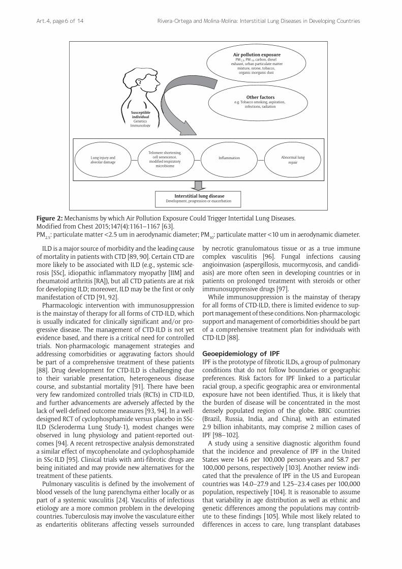
Air pollution could be associated with the development, progression, or exacerbation of ILD via mechanisms of lung injury-alveolar damage [69, 70], telomere shortening [71–74], cell senescence [75], changes in the respiratory
microbiome [76–79], inflammation [80–82], and/or abnormal lung repair (Figure 2) [63, 83–86]. Additionally, individual genetic or epigenetic factors may impact the phenotypic expression of ILD resulting from environmen-tal exposures, and future research is necessary to deline-ate these mechanisms [63].
According to the recent WHO report, air pollution levels in urban areas increased during 2008 and 2013 by 8%. High-income regions of the Americas, Europe, and the Western Pacific demonstrate decreasing air pollution, while the other developing countries had increasing levels [87].
ILD Associated with CTDThe intersection of CTD and the ILD is complex. Although often considered as a single entity, “CTD-ILD” actually reflects a heterogeneous spectrum of diverse CTD and a variety of patterns of interstitial pneumonia. The evalu-ation of patients with CTD that develop ILD, or the assessment for underlying CTD in those presenting with presumed “idiopathic” ILD can be challenging and should be optimized by rational immunological testing. When a diagnosis of CTD-ILD is confirmed, careful assessments to determine extra- versus intra-thoracic disease activity, and degrees of impairment are needed [88].
Table 2: Common Types of Hypersensitivity Pneumonitis According to Major Classes of Antigens.
Class of antigens Specific antigens Sources Type of disease
Organic particulate matters
Microbes
Bacteria Saccharopolyspora rectivirgula, Thermoactinomyces vulgaris
Moldy hay, grain Farmer’s lung
Fungus Aspergillus species Moldy hay, grainMoldy compost and mushrooms
Farmer’s lungMushrooms worker’s lung
Trichosporon cutaneum Contaminated houses Japanese summer-type HP
Penicillium species Moldy corkMoldy cheese or cheese casings
SuberosisCheese washer’s lung
Alternaria species Contaminated wood pulp or dust Woodworker’s lung
Mycobacteria Mycobacterium avium-intracellulare
Mold on ceiling, tub waterMist from pool water, sprays and fountains
Hot tub lungSwimming pool lung
Proteins
Animal proteins Proteins in avian droppings and serum and on feathers
Parakeets, budgerigars, pigeons, parrots, cockatiels, ducks
Pigeon breeder’s lung, bird fancier’s lung
Avian proteins Feather beds, pillow, duvets Feather duvet lung
Silkworm proteins Dust from silkworm larvae and cocoons Silk production HP
Plant’s proteins Grain flour (wheat, rye, oats, maize)
Flour dust Flour dust alveolitis
Legumes (soy) Legumes (soy), flour dust Soya dust alveolitis
Wood (cabreuva, cedar, mahag-ony, pine, ramin, umbrella pine)
Wood particles Wood fiber alveolitis
Inorganic particulate matters
Chemicals products Diisocyanates, trimellitic anhydride
Polyurethane foams, spray paints, dyes, glues
Chemical worker’s lung
HP: hypersensitivity pneumonitis.
Rivera-Ortega and Molina-Molina: Interstitial Lung Diseases in Developing CountriesArt. 4, page 6 of 14
ILD is a major source of morbidity and the leading cause of mortality in patients with CTD [89, 90]. Certain CTD are more likely to be associated with ILD (e.g., systemic scle-rosis [SSc], idiopathic inflammatory myopathy [IIM] and rheumatoid arthritis [RA]), but all CTD patients are at risk for developing ILD; moreover, ILD may be the first or only manifestation of CTD [91, 92].
Pharmacologic intervention with immunosuppression is the mainstay of therapy for all forms of CTD-ILD, which is usually indicated for clinically significant and/or pro-gressive disease. The management of CTD-ILD is not yet evidence based, and there is a critical need for controlled trials. Non-pharmacologic management strategies and addressing comorbidities or aggravating factors should be part of a comprehensive treatment of these patients [88]. Drug development for CTD-ILD is challenging due to their variable presentation, heterogeneous disease course, and substantial mortality [91]. There have been very few randomized controlled trials (RCTs) in CTD-ILD, and further advancements are adversely affected by the lack of well-defined outcome measures [93, 94]. In a well-designed RCT of cyclophosphamide versus placebo in SSc-ILD (Scleroderma Lung Study-1), modest changes were observed in lung physiology and patient-reported out-comes [94]. A recent retrospective analysis demonstrated a similar effect of mycophenolate and cyclophosphamide in SSc-ILD [95]. Clinical trials with anti-fibrotic drugs are being initiated and may provide new alternatives for the treatment of these patients.
Pulmonary vasculitis is defined by the involvement of blood vessels of the lung parenchyma either locally or as part of a systemic vasculitis [24]. Vasculitis of infectious etiology are a more common problem in the developing countries. Tuberculosis may involve the vasculature either as endarteritis obliterans affecting vessels surrounded
by necrotic granulomatous tissue or as a true immune complex vasculitis [96]. Fungal infections causing angioinvasion (aspergillosis, mucormycosis, and candidi-asis) are more often seen in developing countries or in patients on prolonged treatment with steroids or other immunosuppressive drugs [97].
While immunosuppression is the mainstay of therapy for all forms of CTD-ILD, there is limited evidence to sup-port management of these conditions. Non-pharmacologic support and management of comorbidities should be part of a comprehensive treatment plan for individuals with CTD-ILD [88].
Geoepidemiology of IPFIPF is the prototype of fibrotic ILDs, a group of pulmonary conditions that do not follow boundaries or geographic preferences. Risk factors for IPF linked to a particular racial group, a specific geographic area or environmental exposure have not been identified. Thus, it is likely that the burden of disease will be concentrated in the most densely populated region of the globe. BRIC countries (Brazil, Russia, India, and China), with an estimated 2.9 billion inhabitants, may comprise 2 million cases of IPF [98–102].
A study using a sensitive diagnostic algorithm found that the incidence and prevalence of IPF in the United States were 14.6 per 100,000 person-years and 58.7 per 100,000 persons, respectively [103]. Another review indi-cated that the prevalence of IPF in the US and European countries was 14.0–27.9 and 1.25–23.4 cases per 100,000 population, respectively [104]. It is reasonable to assume that variability in age distribution as well as ethnic and genetic differences among the populations may contrib-ute to these findings [105]. While most likely related to differences in access to care, lung transplant databases
Figure 2: Mechanisms by which Air Pollution Exposure Could Trigger Intertidal Lung Diseases.Modified from Chest 2015;147(4):1161–1167 [63].PM2.5: particulate matter <2.5 um in aerodynamic diameter; PM10: particulate matter <10 um in aerodynamic diameter.
Interstitial lung disease Development, progression or exacerbation
Air pollution exposure PM 2.5, PM 10, carbon, diesel
exhaust, urban particulate matter mixture, ozone, tobacco, organic-inorganic dust
Other factors e.g. Tobacco smoking, aspiration,
infections, radiation
Telomere shortening, cell senescence,
modified respiratory microbiome
Lung injury and alveolar damage
Inflammation Abnormal lung repair
Susceptible individual Genetics
Immunology
Rivera-Ortega and Molina-Molina: Interstitial Lung Diseases in Developing Countries Art. 4, page 7 of 14
in 2006 showed that blacks and Hispanics with IPF had a lower survival from time of listing compared with whites [106, 107].
Recent data suggest an increasing prevalence and a stable or increasing incidence of IPF in western countries [108–113, 121–124]. Incidence and mortality studies from South America suggest a low incidence (0.4–1.2 cases per 100,000 people per year) [114, 115]. In a large database Brazilian study, the incidence of IPF was estimated at 0.26 cases per 100,000 persons per year in 1996, rising to 0.48 per 100,000 persons per year in 2010 [114]. The lower incidence in South America may be due to under-diagno-sis or under-reporting on death certificates.
There have been few epidemiologic studies in Asian communities. Insurance claims-based studies from East Asia showed a low incidence (1.2–3.8 per 100,000 per year) [116–118], although mortality statistics from Japan suggested a higher incidence. In East Asia, the higher severity of disease in study subjects from insur-ance datasets likely reflects exclusion of milder cases and may explain the lower incidence compared to west-ern countries [116, 119]. Adjusted IPF mortality statis-tics from Oceania ranged from 5.08–6.49 per 100,000 population [120].
Environment, Smoking, and Diet in IPFAlthough “idiopathic” by definition, potential etiological factors have been implicated in the development of IPF [125–127]. IPF has been associated with industrial and production-based jobs as well as metal and wood dust occupational exposures [63]. The most well-established environmental risk factor for IPF is tobacco smoking (odds ratio for ever smokers of 1.6, 95% confidence interval [CI]: 1.1–2.4) [128–130].
Organic components of PM may trigger abnormal immune responses leading to inflammation, epithelial damage, and over time, fibrosis. There is a small but grow-ing body of evidence suggesting a potential relationship between exposure to air pollution exposure and ILD exac-erbations [63]. In a study of 325 patients with IPF, ambient air pollution was found to modify longitudinal changes in lung function, suggesting that pollutants may differen-tially alter the immunomodulatory pathways associated with IPF [131]. Similarly, a study of a well-defined cohort of patients with IPF found O3 and NO2 exposure to be asso-ciated with an increased risk of acute exacerbation and mortality [132].
Evidence linking diet to IPF is limited. Lungs from patients with IPF appear to be deficient in glutathione [127], suggesting suboptimal antioxidant defenses. High intake of vegetables, green tea, and fish has been associ-ated with a decreased risk for IPF, possibly due to their anti-oxidant properties [133]. Further studies are needed to clarify these findings. Other etiologies may also be implicated in the development of IPF including viral infec-tions, especially hepatitis C and the Epstein-Barr virus. Britton and colleagues demonstrated an increased risk of IPF with the use of antidepressant medications [107, 127]; further studies in animal models are necessary to better understand this possible relationship.
Gender and IPFClinical studies in IPF have enrolled a larger proportion of men than women; few studies explicitly report that IPF is more common in men. A study assessing IPF and chronic obstructive pulmonary disease (COPD) showed a signifi-cant association with male gender and increased preva-lence of combined pulmonary fibrosis and emphysema (CPFE). The OR for male gender having CPFE was 18 (95% CI: 3–773), and subjects with CPFE had a lower median survival time compared to IPF subjects, though this appears to be related to presence of pulmonary hyperten-sion or more severe restrictive lung physiology [134].
Genetics of Pulmonary FibrosisMany clinical disorders that are associated with pulmonary fibrosis have been linked to specific inherited gene muta-tions and polymorphisms [135–137]. Early studies that iden-tified evidence of inherited risk for developing pulmonary fibrosis focused on familial cases, including variants such as genes coding for mucin 5B surfactant proteins [136] or those involved in telomere homeostasis and function [73]. Studies that have focused particularly on genome-wide linkage analyses have identified numerous gene polymor-phisms that are associated with increased risk for pulmo-nary fibrosis [135, 138–140]. However, not all races have been evaluated, even for widely studied genetic mutations that have demonstrated their association with pulmonary fibrosis. Therefore, global collaboration for genetic studies is a priority to better understand the disease.
Comorbidities of IPFIPF is associated with pulmonary or extrapulmonary comorbidities. Pulmonary comorbidities include pulmo-nary hypertension, emphysema, and lung cancer, while non-pulmonary conditions include venous thromboem-bolism, coronary artery disease, congestive heart failure, sleep-disordered breathing, gastro-oesophageal reflux dis-ease, and anxiety or depression. Although some of these comorbid conditions share risk factors with IPF, the risk in patients with IPF is still greater than expected by chance. This might indicate that IPF fosters an environment for the development or perpetuation of comorbid condi-tions, or alternatively that they share unknown causative factors. Optimal management of IPF therefore requires a comprehensive approach, including the identification and treatment of comorbid conditions to optimize patient outcomes [141].
Current Diagnosis Criteria and Treatment of IPFIn 2011, American Thoracic Society (ATS), European Res-piratory Society (ERS), the Japanese Respiratory Society (JRS), and the Latin-American Thoracic Society (ALAT) jointly published an evidence-based statement for the diagnosis and management of IPF [128]. This document provided an update of the diagnosis criteria: [4] 1) exclu-sion of other known causes of ILD (e.g., domestic and occupational environmental exposures, CTD and drug toxicity); 2) presence of an usual interstitial pneumonia (UIP) pattern on chest high-resolution computed tomog-
Rivera-Ortega and Molina-Molina: Interstitial Lung Diseases in Developing CountriesArt. 4, page 8 of 14
raphy (HRCT); and 3) specific combinations of HRCT and biopsy UIP patterns in individuals undergoing surgical lung biopsy (SLB) [108]. The criteria originated from the evidence that in an appropriate clinical setting, the pres-ence of a classical UIP pattern on the HRCT has a very high positive predictive value (90% to 100%) for a histological diagnosis of UIP [142, 143].
In 2015, recommendations for the treatment of IPF were updated based of new scientific evidence [128, 144]. This was a major milestone, as for the first time a thera-peutic recommendation with a high level of evidence was established for two antifibrotic drugs: Pirfenidone and Nintedanib [145]. These new drugs provide benefits in terms of a significant reduction in mortality, position-ing IPF as one of the few areas in respiratory medicine in which treatment could provide such clinically significant improvements [146].
ConclusionsILDs are an heterogeneous group of relatively uncommon diseases. Few data are available on ILD epidemiology, especially in developing countries, although the preva-lence and incidence seem to be increasing in many areas. IPF is the most common and studied of the idiopathic ILDs, with updated guidelines for diagnosis and new treatment options. The involvement of centers in developing coun-tries should be encouraged, for example through the ILD global registries and/or increased access to expert multi-disciplinary team consensus, as it would help to obtain an accurate and prompt diagnosis and health access to treatment. Additionally, these strategies would allow understanding racial and environmental risk factors, and therefore, provide insights in the pathogenesis of ILDs.
Competing InterestsThe authors have no competing interests to declare.
References 1. ATS/ERS. American Thoracic Society/European
Respiratory Society International multidisciplinary consensus classification of the idiopathic intersti-tial pneumonias. Am J Respir Crit Care Med. 2002; 165(2): 277–304. DOI: https://doi.org/10.1164/ajrccm.165.2.ats01
2. Travis WD, Costabel U, Hansell DM, et al. An official American Thoracic Society/European Res-piratory Society statement: Update of the inter-national multidisciplinary classification of the idiopathic interstitial pneumonias. Am J Respir Crit Care Med. 2013; 188(6): 733–748. DOI: https://doi.org/10.1164/rccm.201308-1483ST
3. Valeyre D, Duchemann B, Nunes H, et al. Intersti-tial lung diseases. ERS Monograph. 2014; chapter 6, 65: 79–87.
4. American Thoracic Society. Idiopathic pulmonary fibrosis: Diagnosis and treatment. International consensus statement. American Thoracic Society (ATS) and the European Respiratory Society (ERS). Am J Respir Crit Care Med. 2000; 161(2): 646–664. DOI: https://doi.org/10.1164/ajrccm.161.2.ats3-00
5. Bouros D. Current classification of idiopathic inter-stitial pneumonias. Monaldi Arch Chest Dis. 2000; 55(6): 450–454.
6. Verleden GM, du Bois RM, Bouros D, et al. Genetic predisposition and pathogenetic mechanisms of interstitial lung diseases of unknown origin. Eur Respir J. 2001; 18(Suppl 32): 17s–29s.
7. Coultas DB, Zumwalt RE, Black WC, et al. The epi-demiology of interstitial lung diseases. Am J Respir Crit Care Med. 1994; 150(4): 967–972. DOI: https://doi.org/10.1164/ajrccm.150.4.7921471
8. Schweisfurth H. Report by the scientific working group for therapy of lung diseases: German fibrosis register with initial results. Pneumologie. 1996; 50(12): 899–901.
9. Schweisfurth H, Kieslich C, Satake N, et al. How are interstitial lung diseases diagnosed in Germany? Results of the scientific registry for the explora-tion of interstitial lung diseases (“Fibrosis registry”) of the WATL. Pneumologie. 2003; 57(7): 373–382. DOI: https://doi.org/10.1055/s-2003-40557
10. Thomeer M, Demedts M, Vandeurzen K and VRGT Working Group on Interstitial Lung Diseases. Registration of interstitial lung diseases by 20 centres of respiratory medicine in Flanders. Acta Clin Belg. 2001; 56(3): 163–172. DOI: https://doi.org/10.1179/acb.2001.026
11. Roelandt M, Demedts M, Callebaut W, et al. Epidemiology of interstitial lung disease (ILD) in flanders: Registration by pneumologists in 1992–1994. Working group on ILD, VRGT. Acta Clin Belg. 1995; 50(5): 260–268. DOI: https://doi.org/10.1080/17843286.1995.11718459
12. Agostini C, Albera C, Bariffi F, et al. First report of the Italian register for diffuse infiltrative lung dis-orders (RIPID). Monaldi Arch Chest Dis. 2001; 56(4): 364–368.
13. Tinelli C, De Silvestri A, Richeldi L, et al. The Italian register for diffuse infiltrative lung disorders (RIPID): A four-year report. Sarcoidosis Vasc Diffuse Lung Dis. 2005; 22(Suppl 1): S4–S8.
14. GBD 2013 Mortality and Causes of Death Collaborators. Global, regional, and national age-sex specific all-cause and cause-specific mortality for 240 causes of death, 1990–2013: A systematic anal-ysis for the Global Burden of Disease Study 2013. Lancet. 2015; 385(9963): 117–171. DOI: https://doi.org/10.1016/S0140-6736(14)61682-2
15. ERS. Interstitial lung diseases, In: ERS European Lung White-book. Chapter 22. ERS; 2015. http://www.erswhitebook.org/chapters/interstitial-lung-diseases/. Access date: September 15, 2016.
16. Kreuter M, Herth FJF, Wacker M, et al. Exploring clinical and epidemiological characteristics of inter-stitial lung diseases: Rationale, aims, and design of a nationwide prospective registry—The EXCITING-ILD Registry. Biomed Res Int. 2015; Epub 2015 Nov 10. DOI: https://doi.org/10.1155/2015/123876
17. Kreuter M, Herth FJF, Wacker M, et al. Interims analysis of the EXCITING-ILD registry (registry

Rivera-Ortega and Molina-Molina: Interstitial Lung Diseases in Developing Countries Art. 4, page 9 of 14
for exploring clinical and epidemiological char-acteristics of interstitial lung diseases). Eur Resp J. 2016; 48(Suppl 60): PA3905. DOI: https://doi.org/10.1183/13993003.congress-2016.PA3905
18. Hyldgaard C. A cohort study of Danish patients with interstitial lung diseases: Burden, severity, treatment and survival. Dan Med J. 2015; 62(4): B5069.
19. Xaubet A, Ancochea J, Morell F, et al. Report on the incidence of interstitial lung diseases in Spain. Sarcoidosis Vasc Diffuse Lung Dis. 2004; 21(1): 64–70.
20. Lopez-Campos JL, Rodríguez-Becerra E and Neumosur Task Group, Registry of Inter-stitial Lung Diseases. Incidence of inter-stitial lung diseases in the south of Spain 1998–2000: The RENIA study. Eur J Epide-miol. 2004; 19(2): 155–161. DOI: https://doi.org/10.1023/B:EJEP.0000017660.18541.83
21. Karakatsani A, Papakosta D, Rapti A, et al. Epidemiology of interstitial lung diseases in Greece. Respir Med. 2009; 103(8): 1122–1129. DOI: https://doi.org/10.1016/j.rmed.2009.03.001
22. Thomeer MJ, Costabel U, Rizzato G, et al. Comparison of registries of interstitial lung diseases in three European countries. Eur Respir J. 2001; 18(Suppl 32): 114s–118s.
23. Musellim B, Okumus G, Uzaslan E, et al. Epidemiology and distribution of interstitial lung diseases in Turkey. Clin Respir J. 2014; 8(1): 55–62. DOI: https://doi.org/10.1111/crj.12035
24. Jindal SK and Gupta D. Incidence and recognition of interstitial pulmonary fibrosis in developing countries. Curr Opin Pulm Med. 1997; 3(5): 378–383. DOI: https://doi.org/10.1097/00063198-199709000-00011
25. Collins B, Singh S, Joshi J, et al. ILD-India regis-try: Idiopathic pulmonary fibrosis (IPF) and connec-tive tissue disease (CTD) associated interstitial lung disease (CTD-ILD). Eur Resp J. 2016; 48(Suppl 60): PA812. DOI: https://doi.org/10.1183/13993003.congress-2016.PA812
26. Alhamad EH. Interstitial lung diseases in Saudi Arabia: A single-center study. Ann Thorac Med. 2013; 8(1): 33–37. DOI: https://doi.org/10.4103/1817-1737.105717
27. Martínez-Briceño D, García-Sancho C, Fernández-Plata R, et al. Tendencia de la mortalidad por enfermedades intersticiales en México, período 2000–2010. Neumol Cir Torax. 2014; 73(3): 179–184.
28. ERS. Occupational lung diseases. In: ERS European Lung White-book. Chapter 24. ERS; 2015. http://www.erswhitebook.org/chapters/occupational-lung-diseases/. Access date: September 15, 2016.
29. The World Health Organization. Asbestos: Elimination of Asbestos-related Diseases. Fact sheet Nº343; 2010. Available at: http://www.cancer-researchuk.org/cancer-info/cancerstats/types/Meso-thelioma/incidence/#source6. Access date: March 7, 2016.
30. Prazakova S, Thomas PS, Sandrini A, et al. Asbestos and the lung in the 21st century: An update. Clin Respir J. 2014; 8: 1–10. DOI: https://doi.org/10.1111/crj.12028
31. Baas P and Burgers S. ASIA: Asbestos stop in Asia. Respirology. 2015; 20(4): 521. DOI: https://doi.org/10.1111/resp.12533
32. Subramanian V and Madhavan N. Asbestos problem in India. Lung Cancer. 2005; 49(Suppl 1): S9–S12. DOI: https://doi.org/10.1016/j.lungcan.2005.03.003
33. Baron PA. Measurement of airborne fibers: A review. Ind Health. 2001; 39(2): 39–50. DOI: https://doi.org/10.2486/indhealth.39.39
34. Warheit DB, Reed KL and Webb TR. Man-made respirable-sized organic fibers: What do we know about their toxicological profiles? Ind Health. 2001; 39(2): 119–125. DOI: https://doi.org/10.2486/indhealth.39.119
35. Roggli VL, Gibbs AR, Attanoos R, et al. Pathology of asbestosis—An update of the diagnosis criteria: Report of the asbestosis committee of the college of American pathologists and pulmonary pathology society. Arch Pathol Lab Med. 2010; 134(3): 462–480.
36. American Thoracic Society. A diagnosis and ini-tial management of nonmalignant diseases related to asbestos. Am J Respir Crit Care Med. 2004; 170(6): 691–715. DOI: https://doi.org/10.1164/rccm.200310-1436ST
37. Donovan EP, Donovan BL, McKinley MA, et al. Evaluation of take home (para-occupational) exposure to asbestos and disease: A review of the literature. Crit Rev Toxicol. 2012; 42(9): 703–731. DOI: https://doi.org/10.3109/10408444.2012.709821
38. Rake C, Gilham C, Hatch J, et al. Occupational, domestic and environmental mesothelioma risks in the British population: A case-control study. Br J Cancer. 2009; 100(7): 1175–1183. DOI: https://doi.org/10.1038/sj.bjc.6604879
39. Ferrante D, Bertolotti M, Todesco A, et al. Cancer mortality and incidence of mesothelioma in a cohort of wives of asbestos workers in Casale Monferrato, Italy. Environ Health Perspect. 2007; 115(10): 1401–1405. DOI: https://doi.org/10.1289/ehp.10195
40. Pérez-Alonso A, Córdoba-Doña JA, Millares- Lorenzo JL, et al. Outbreak of silicosis in Spanish quartz conglomerate workers. Int J Occup Environ Health. 2014; 20(1): 26–32. DOI: https://doi.org/10.1179/2049396713Y.0000000049
41. Grewal KS, Arora VK and Gupta SP. Industrial lung diseases. Progress in Clinical Medicine in India. 3rd series. Ahuja MMS (ed.). 1979; 420–438. New Delhi: Arnold Heinemann.
42. Leung CC, Yu IT and Chen W. Silicosis. Lancet. 2012; 379(9830): 2008–2018. DOI: https://doi.org/10.1016/S0140-6736(12)60235-9
43. Carneiro APS, Barreto SM, Siqueira AL, et al. Con-tinued exposure to silica after diagnosis of silicosis

Rivera-Ortega and Molina-Molina: Interstitial Lung Diseases in Developing CountriesArt. 4, page 10 of 14
in Brazilian gold miners. Am J Ind Med. 2006; 49: 811–818. DOI: https://doi.org/10.1002/ajim.20379
44. Nelson G, Girdler-Brown B, Ndlovu N, et al. Three decades of silicosis: Disease trends at autopsy in South African gold miners. Environ Health Perspect. 2010; 118: 421–426. DOI: https://doi.org/10.1289/ehp.0900918
45. Akgun M, Araz O, Akkurt I, et al. An epidemic of silicosis among former denim sandblasters. Eur Respir J. 2008; 32: 1295–1303. DOI: https://doi.org/10.1183/09031936.00093507
46. Kauppinen T, Toikkanen J, Pedersen D, et al. Occupational exposure to carcinogens in the European Union. Occup Environ Med. 2000; 57: 10–18. DOI: https://doi.org/10.1136/oem.57.1.10
47. The World Health Organization. Silicosis. Fact sheet Nº238; 2000. Available at: http://web.archive.org/web/20070510005843/, http://www.who.int/mediacentre/factsheets/fs238/en/. Access date: March 7, 2016.
48. Linch KD, Miller WE, Althouse RB, et al. Surveillance of respirable crystalline silica dust using OSHA com-pliance data (1979–1995). Am J Ind Med. 1998; 34: 547–558. DOI: https://doi.org/10.1002/(SICI)1097-0274(199812)34:6<547::AID-AJIM2>3.0.CO;2-B
49. Rosenman KD, Reilly MJ and Henneberger PK. Estimating the total number of newly-recognized silicosis cases in the United States. Am J Ind Med. 2003; 44: 141–147. DOI: https://doi.org/10.1002/ajim.10243
50. Parikh JR. Byssinosis in developing countries. Br J Ind Med. 1992; 49(4): 217–219. DOI: https://doi.org/10.1136/oem.49.4.217
51. Parikh JR, Bhagia LJ, Majumdar PK, et al. Prevale nce of byssinosis in textile mills at Ahmedabad, India. Br J Ind Med. 1989; 46(11): 787–790. DOI: https://doi.org/10.1136/oem.46.11.787
52. Gupta S and Gupta BK. A study of byssinosis and associated respiratory disorders in cotton mill workers. Indian J Chest Dis Allied Sci. 1988; 28(4): 183–188.
53. Barjatiya MK, Mathur RN and Swaroop A. Byssinosis in cotton textile workers of Kishangarh. Indian J Chest Dis Allied Sci. 1990; 32(4): 215–223.
54. White NW. Byssinosis in South Africa. A survey of 2411 textile workers. S Afr Med J. 1989; 75(9): 435–442.
55. Takam J and Nemery B. Byssinosis in a textile factory in Cameroon: a preliminary study. Br J Ind Med. 1988; 45(12): 803–809. DOI: https://doi.org/10.1136/oem.45.12.803
56. Woldeyohannes M, Bergevin Y, Mgeni AY, et al. Respiratory problems among cotton textile mill workers in Ethiopia. Br J Ind Med. 1991; 48: 110–115. DOI: https://doi.org/10.1136/oem.48.2.110
57. Awad el Karim MA, Osman Y and el Haimi YA. Byssinosis: environmental and respiratory symptoms among textile workers in Sudan. Int Arch Occup Environ Health. 1986; 57(2): 101–108. DOI: https://doi.org/10.1007/BF00381377
58. Awad el Karim MA and Onsa SH. Prevalence of byssinosis and respiratory symptoms among spin-ners in Sudanese cotton mills. Am J Ind Med. 1987; 12(3): 281–289. DOI: https://doi.org/10.1002/ajim.4700120305
59. Noweir MH, Noweir KH, Osman HA, et al. An environmental and medical study of byssinosis and other respiratory conditions in the cotton textile industry in Egypt. Am J Ind Med. 1984; 6(3): 173–183. DOI: https://doi.org/10.1002/ajim.4700060303
60. Hinson AV, Schlünssen V, Agodokpessi G, et al. The prevalence of byssinosis among cotton workers in the north of Benin. Int J Occup Environ Med. 2014; 5: 194–200.
61. Lacasse Y, Assayag E and Cormier Y. Myths and controversies in hypersensitivity pneumonitis. Semin Respir Crit Care Med. 2008; 29(6): 631–642. DOI: https://doi.org/10.1055/s-0028-1101273
62. Spagnolo P, Rossi G, Cavazza A, et al. Hypersensitivity pneumonitis: A comprehensive review. J Investig Allergol Clin Immunol. 2015; 25(4): 237–250.
63. Johannson KA, Balmes JR and Collard HR. Air pollution exposure: A novel environmental risk factor for interstitial lung disease? Chest. 2015; 147(4): 1161–1167. DOI: https://doi.org/10.1378/chest.14-1299
64. Pinkerton KE. A Critical Review of the Particu-late Matter Toxicology Literature for Senate Bill 25 Review of the Particulate Matter Standard. Sacramento, CA: California Environmental Protec-tion Agency, Air Resources Board, Research Division. 2002; 1–93.
65. Alexis NE, Lay JC, Hazucha M, et al. Low-level ozone exposure induces airways inflammation and modifies cell surface phenotypes in healthy humans. Inhal Toxicol. 2010; 22(7): 593–600. DOI: https://doi.org/10.3109/08958371003596587
66. Scannell C, Chen L, Aris RM, et al. Greater ozone-induced inflammatory responses in subjects with asthma. Am J Respir Crit Care Med. 1996; 154(1): 24–29. DOI: https://doi.org/10.1164/ajrccm.154.1.8680687
67. Song H, Tan W and Zhang X. Ozone induces inflammation in bronchial epithelial cells. J Asthma. 2011; 48(1): 79–83. DOI: https://doi.org/10.3109/02770903.2010.529224
68. Larsen ST, Matsubara S, McConville G, et al. Ozone increases airway hyperreactivity and mucus hyperproduction in mice previously exposed to allergen. J Toxicol Environ Health A. 2010; 73(11): 738–747. DOI: https://doi.org/10.1080/15287391003614034
69. Adamson IY and Hedgecock C. Patterns of particle deposition and retention after instillation to mouse lung during acute injury and fibrotic repair. Exp Lung Res. 1995; 21(5): 695–709. DOI: https://doi.org/10.3109/01902149509050837
70. Beeh KM, Beier J, Haas IC, et al. Glutathione deficiency of the lower respiratory tract in patients

Rivera-Ortega and Molina-Molina: Interstitial Lung Diseases in Developing Countries Art. 4, page 11 of 14
with idiopathic pulmonary fibrosis. Eur Respir J. 2002; 19(6): 1119–1123. DOI: https://doi.org/10.1183/09031936.02.00262402
71. Grahame TJ and Schlesinger RB. Oxidative stress-induced telomeric erosion as a mechanism underlying airborne particulate matter-related car-diovascular disease. Part Fibre Toxicol. 2012; 9: 21. DOI: https://doi.org/10.1186/1743-8977-9-21
72. Hou L, Wang S, Dou C, et al. Air pollution expo sure and telomere length in highly exposed subjects in Beijing, China: A repeated-measure study. Environ Int. 2012; 48: 71–77. DOI: https://doi.org/10.1016/j.envint.2012.06.020
73. Garcia CK. Idiopathic pulmonary fibrosis: Update on genetic discoveries. Proc Am Thorac Soc. 2011; 8(2): 158–162. DOI: https://doi.org/10.1513/pats.201008-056MS
74. Alder JK, Chen JJ, Lancaster L, et al. Short tel-omeres are a risk factor for idiopathic pulmonary fibrosis. Proc Natl Acad Sci USA. 2008; 105(35): 13051–13056. DOI: https://doi.org/10.1073/pnas.0804280105
75. Adamson IY, Vincent R and Bjarnason SG. Cell injury and interstitial inflammation in rat lung after inhalation of ozone and urban particulates. Am J Respir Cell Mol Biol. 1999; 20(5): 1067–1072. DOI: https://doi.org/10.1165/ajrcmb.20.5.3468
76. Adar SD, Huffnagle GB and Curtis JL. The respiratory microbiome: An underappreciated player in the human response to inhaled pollutants? Ann Epidemiol. 2016; 26(5): 355–359. DOI: https://doi.org/10.1016/j.annepidem.2016.03.010
77. Han MK, Zhou Y, Murray S, et al. Lung micro biome and disease progression in idiopathic pulmonary fibrosis: An analysis of the COMET study. Lancet Respir Med. 2014; 2: 548–556. DOI: https://doi.org/10.1016/S2213-2600(14)70069-4
78. Molyneaux PL and Maher TM. The role of infection in the pathogenesis of idiopathic pulmonary fibro-sis. Eur Respir Rev. 2013; 22: 376–381. DOI: https://doi.org/10.1183/09059180.00000713
79. Faner R, Sibila O, Agustí A, et al. The microbiome in respiratory medicine: Current challenges and future perspectives. Eur Respir J. 2017; 49(4): 1602086. DOI: https://doi.org/10.1183/13993003.02086-2016
80. Devlin RB, Duncan KE, Jardim M, et al. Controlled exposure of healthy young volunteers to ozone causes cardiovascular effects. Circulation. 2012; 126(1): 104–111. DOI: https://doi.org/10.1161/CIRCULATIONAHA.112.094359
81. Neophytou AM, Hart JE, Cavallari JM, et al. Traffic-related exposures and biomarkers of systemic inflammation, endothelial activation and oxidative stress: A panel study in the US trucking industry. Environ Health. 2013; 12: 105. DOI: https://doi.org/10.1186/1476-069X-12-105
82. Patel MM, Chillrud SN, Deepti KC, et al. Traffic-related air pollutants and exhaled mark-ers of airway inflammation and oxidative stress
in New York City adolescents. Environ Res. 2013; 121: 71–78. DOI: https://doi.org/10.1016/j.envres.2012.10.012
83. Last JA, Reiser KM, Tyler WS, et al. Long-term consequences of exposure to ozone. I. Lung collagen content. Toxicol Appl Pharmacol. 1984; 72(1): 111–118. DOI: https://doi.org/10.1016/0041-008X(84)90254-0
84. Reiser KM, Tyler WS, Hennessy SM, et al. Long- term consequences of exposure to ozone. II. Struc-tural alterations in lung collagen of monkeys. Toxicol Appl Pharmacol. 1987; 89(3): 314–322. DOI: https://doi.org/10.1016/0041-008X(87)90151-7
85. Li YJ, Shimizu T, Hirata Y, et al. Diesel exhaust particle induce epithelial-to-mesenchymal transition by oxidative stress in human bronchial epithelial cell. Eur Respir J. 2013; 42(suppl 57): P3896.
86. Líbalová H, Uhlířová K, Kléma J, et al. Global gene expression changes in human embryonic lung fibroblasts induced by organic extracts from respirable air particles. Part Fibre Toxicol. 2012; 9: 1. DOI: https://doi.org/10.1186/1743-8977-9-1
87. The World Health Organization. Ambient Air Pollution: A Global Assessment of Exposure and Burden of Disease; 2016. http://www.who.int/phe/publications/air-pollution-global-assessment/en/. Access date: September 15, 2016.
88. Fischer A and Chartrand S. Assessment and management of connective tissue disease-asso-ciated interstitial lung disease. Sarcoidosis Vasc Diffuse Lung Dis. 2015; 32(1): 2–21.
89. Winstone TA, Assayag D, Wilcox PG, et al. Predictors of mortality and progression in scleroderma-associated interstitial lung diseases: A systematic review. Chest. 2014; 146(2): 422–436. DOI: https://doi.org/10.1378/chest.13-2626
90. Kim EJ, Collard HR and King TE, Jr. Rheumatoid arthritis-associated interstitial lung disease: The rel-evance of histopathologic and radiographic pattern. Chest. 2009; 136(5): 1397–1405. DOI: https://doi.org/10.1378/chest.09-0444
91. Bryson T, Sundaram B, Khanna D, et al. Connective tissue disease-associated interstitial pneumonia and idiopathic interstitial pneumonia: Similarity and difference. Semin Ultrasound CT MR. 2014; 35(1): 29–38. DOI: https://doi.org/10.1053/j.sult.2013.10.010
92. Nana AM, Ngnie C, Wandji A, et al. Non-infectious lung manifestations of autoimmune diseases in Cameroon. Afr J Respir Med. 2012; 8(1): 12–14.
93. Seibold JR, Denton CP, Furst DE, et al. Randomized, prospective, placebo-controlled trial of bosentan in interstitial lung disease secondary to systemic sclerosis. Arthritis Rheum. 2010; 62(7): 2101–2108. DOI: https://doi.org/10.1002/art.27466
94. Tashkin DP, Elashoff R, Clements PJ, et al. Cyclophosphamide versus placebo in scleroderma lung disease. N Engl J Med. 2006; 354(25): 2655–2666. DOI: https://doi.org/10.1056/NEJMoa055120
Rivera-Ortega and Molina-Molina: Interstitial Lung Diseases in Developing CountriesArt. 4, page 12 of 14
95. Shenoy PD, Bavaliya M, Sashidharan S, et al. Cyclophosphamide versus mycophenolate mofetil in scleroderma interstitial lung disease (SSc-ILD) as induction therapy: A single-centre, retrospec-tive analysis. Arthritis Res Ther. 2016; 18(1): 123. DOI: https://doi.org/10.1186/s13075-016-1015-0
96. Strauss I, Liebermann KV and Churg J. Pulmo nary vasculitis. In: Fishman AP (ed.), Pulmonary Diseases and Disorders. 2nd ed. 1988; 1127–1156. New York: McGraw Hill Book Company.
97. Udwadia FE. Pulmonary eosinophilic syndrome. In: Ahuja MMS (ed.), Progress in Clinical Medicine in India. 2nd series. 1978; 453–475. New Delhi: Arnold Heinemann.
98. Richeldi L, Rubin AS, Avdeev S, et al. Idiopathic pulmonary fibrosis in BRIC countries: The cases of Brazil, Russia, India, and China. BMC Med. 2015; 13: 237. DOI: https://doi.org/10.1186/s12916-015-0495-0
99. Avdeev SN. Idiopathic pulmonary fibrosis: Current concepts and diagnostic approaches. Practical Pulmonology. 2014; 4: 16–23.
100. Gribbin J, Hubbard RB, Le Jeune I, et al. Incidence and mortality of idiopathic pulmonary fibrosis and sarcoidosis in the UK. Thorax. 2006; 61(11): 980–985. DOI: https://doi.org/10.1136/thx.2006.062836
101. Richeldi L, du Bois RM, Raghu G, et al. Efficacy and safety of nintedanib in idiopathic pulmonary fibrosis. N Engl J Med. 2014; 370(22): 2071–2082. DOI: https://doi.org/10.1056/NEJMoa1402584
102. Wei LQ, Peng SC, Cao J, et al. Clinical features and diagnosis and treatment of diffuse interstitial lung disease: A multi-center study. Chinese General Practice. 2012; 15: 2521–2524.
103. Exposito DB, Lanes S, Donneyong M, et al. Idiopathic pulmonary fibrosis in United States automated claims. Incidence, prevalence, and algo-rithm validation. Am J Respir Crit Care Med. 2015; 192(10): 1200–1207. DOI: https://doi.org/10.1164/rccm.201504-0818OC
104. Nalysnyk L, Cid-Ruzafa J, Rotella P, et al. Incidence and prevalence of idiopathic pulmo-nary fibrosis: Review of the literature. Eur Respir Rev. 2012; 21(126): 355–361. DOI: https://doi.org/10.1183/09059180.00002512
105. Baddini-Martinez J and Pereira CA. How many patients with idiopathic pulmonary fibro-sis are there in Brazil? J Bras Pneumol. 2015; 41(6): 560–561. DOI: https://doi.org/10.1590/s1806-37562015000000165
106. Lederer DJ, Arcasoy SM, Barr RG, et al. Racial and ethnic disparities in idiopathic pulmonary fibrosis: A UNOS/OPTN database analysis. Am J Trans-plant. 2006; 6(10): 2436–2442. DOI: https://doi.org/10.1111/j.1600-6143.2006.01480.x
107. Zeki AA, Schivo M, Chan AL, et al. Geoepidemiol-ogy of COPD and idiopathic pulmonary fibrosis. J Autoimmun. 2010; 34(3): J327–J338. DOI: https://doi.org/10.1016/j.jaut.2009.11.004
108. Sgalla G, Biffi A and Richeldi L. Idiopathic pulmonary fibrosis: Diagnosis, epidemiology and natural history. Respirology. 2016; 21(3): 427–437. DOI: https://doi.org/10.1111/resp.12683
109. Hutchinson J, Fogarty A, Hubbard R, et al. Global incidence and mortality of idiopathic pulmonary fibrosis: A systematic review. Eur Respir J. 2015; 46(3): 795–806. DOI: https://doi.org/10.1183/09031936.00185114
110. Navaratnam V, Fleming KM, West J, et al. The rising incidence of idiopathic pulmonary fibrosis in the U.K. Thorax. 2011; 66(6): 462–467. DOI: https://doi.org/10.1136/thx.2010.148031
111. Maher TM, Strongman H, Boggon R, et al. Idiopathic pulmonary fibrosis survival has not improved in the 21st century: Analysis of CPRD gold primary care data. Thorax. 2013; 68(Suppl 3): A82–A83. DOI: https://doi.org/10.1136/thoraxjnl-2013-204457.168
112. Kornum JB, Christensen S, Grijota M, et al. The incidence of interstitial lung disease 1995–2005: A Danish nationwide population-based study. BMC Pulm Med. 2008; 8: 24. DOI: https://doi.org/10.1186/1471-2466-8-24
113. von Plessen C, Grinde O and Gulsvik A. Incidence and prevalence of cryptogenic fibrosing alveoli-tis in a Norwegian community. Respir Med. 2003; 97(4): 428–435. DOI: https://doi.org/10.1053/rmed.2002.1466
114. Rufino RL, Costa CH, Accar J, et al. Incidence and mortality of interstitial pulmonary fibrosis in Brazil. Am J Respir Crit Care Med. 2013; 187: A1458.
115. Fortuna FP, Perin C, Cunha L, et al. Mortality caused by idiopathic pulmonary fibrosis in the state of Rio Grande do Sul (Brazil). J Pneumologia. 2003; 29: 121–124. DOI: https://doi.org/10.1590/S0102-35862003000300002
116. Lai CC, Wang CY, Lu HM, et al. Idiopathic pulmo nary fibrosis in Taiwan: A population-based study. Respir Med. 2012; 106(11): 1566–1574. DOI: https://doi.org/10.1016/j.rmed.2012.07.012
117. Han S, Mok Y, Jee SH and Danoff SK. Incidence and mortality of idiopathic pulmonary fibrosis in South Korea. Am J Respir Crit Care Med. ATS. 2013; A1460. https://www.atsjournals.org/doi/abs/10.1164/ajrccm-conference.2013.187.1_MeetingAbstracts.A1460.
118. Munakata M, Asakawa M, Hamma Y, et al. Pre-sent status of idiopathic interstitial pneumonia-from epidemiology to etiology. Nihon Kyobu Shikkan Gakkai Zasshi. 1994; 32: 187–192.
119. Natsuizaka M, Chiba H, Kuronuma K, et al. Epidemiologic survey of Japanese patients with idiopathic pulmonary fibrosis and investigation of ethnic differences. Am J Respir Crit Care Med. 2014; 190(7): 773–779. DOI: https://doi.org/10.1164/rccm.201403-0566OC
120. Hutchinson JP, McKeever TM, Fogarty AW, et al. Increasing global mortality from idiopathic pulmo-nary fibrosis in the twenty-first century. Ann Am

Rivera-Ortega and Molina-Molina: Interstitial Lung Diseases in Developing Countries Art. 4, page 13 of 14
Thorac Soc. 2014; 11(8): 1176–1185. DOI: https://doi.org/10.1513/AnnalsATS.201404-145OC
121. Raghu G, Chen SY, Hou Q, et al. Incidence and prevalence of idiopathic pulmonary fibro-sis in US adults 18–64 years old. Eur Respir J. 2016; 48(1): 179–186. DOI: https://doi.org/10.1183/13993003.01653-2015
122. Hyldgaard C, Hilberg O, Muller A, et al. A cohort study of interstitial lung diseases in central Denmark. Respir Med. 2014; 108(5): 793–799. DOI: https://doi.org/10.1016/j.rmed.2013.09.002
123. Agabiti N, Porretta MA, Bauleo L, et al. Idiopathic Pulmonary Fibrosis (IPF) incidence and prevalence in Italy. Sarcoidosis Vasc Diffuse Lung Dis. 2014; 31(3): 191–197.
124. Jo H, Glaspole I, Moodley Y, et al. Disease progression in early idiopathic pulmonary fibro-sis: Insights from the Australian IPF registry. Eur Resp J. 2016; 48(Suppl 60). DOI: https://doi.org/10.1183/13993003.congress-2016.PA2100
125. Miyake Y, Sasaki S, Yokoyama T, et al. Occupat ional and environmental factors and idiopathic pulmonary fibrosis in Japan. Ann Occup Hyg. 2005; 49(3): 259–265.
126. Baumgartner KB, Samet JM, Coultas DB, et al. Occupational and environmental risk factors for idiopathic pulmonary fibrosis: A multicenter case-control study. Collaborating Centers. Am J Epide-miol. 2000; 152(4): 307–315. DOI: https://doi.org/10.1093/aje/152.4.307
127. Britton J and Hubbard R. Recent advances in the aetiology of cryptogenic fibrosing alveolitis. Histopathology. 2000; 37(5): 387–392. DOI: https://doi.org/10.1046/j.1365-2559.2000.01098.x
128. Raghu G, Collard HR, Egan JJ, et al. An offi-cial ATS/ERS/JRS/ALAT statement: Idiopathic pulmonary fibrosis: Evidence-based guidelines for diagnosis and management. Am J Respir Crit Care Med. 2011; 183(6): 788–824. DOI: https://doi.org/10.1164/rccm.2009-040GL
129. Baumgartner KB, Samet JM, Stidley CA, et al. Cigarette smoking: A risk factor for idiopathic pul-monary fibrosis. Am J Respir Crit Care Med. 1997; 155(1): 242–248. DOI: https://doi.org/10.1164/ajrccm.155.1.9001319
130. Antoniou KM, Hansell DM, Rubens MB, et al. Idiopathic pulmonary fibrosis: Outcome in relation to smoking status. Am J Respir Crit Care Med. 2008; 177(2): 190–194. DOI: https://doi.org/10.1164/rccm.200612-1759OC
131. Richards TJ, Kuhlengel TK, Choi J, et al. Does ambient air pollution exposure modify longitudi-nal disease outcome in a cohort of patients with idiopathic pulmonary fibrosis? Am J Respir Crit Care Med. ATS. 2011; A5433. DOI: https://doi.org/10.1164/ajrccm-conference.2011.183.1_Meet-ingAbstracts.A5433
132. Johannson KA, Vittinghoff E, Lee K, et al. Acute exacerbation of idiopathic pulmonary fibrosis
associated with air pollution exposure. Eur Respir J. 2014; 43(4): 1124–1131. DOI: https://doi.org/10.1183/09031936.00122213
133. Iwai K, Mori T, Yamada N, et al. Idiopathic pulmonary fibrosis. Epidemiologic approaches to occupational exposure. Am J Respir Crit Care Med. 1994; 150(3): 670–675. DOI: https://doi.org/10.1164/ajrccm.150.3.8087336
134. Mejia M, Carillo G, Rojas-Serrano J, et al. Idiopathic pulmonary fibrosis and emphysema: Decreased survival associated with severe pulmonary arterial hypertension. Chest. 2009; 136(1): 10–15. DOI: https://doi.org/10.1378/chest.08-2306
135. Mathai SK, Yang IV, Schwarz MI, et al. Incorporating genetics into the identification and treatment of Idiopathic Pulmonary Fibro-sis. BMC Med. 2015; 13(1): 191. DOI: https://doi.org/10.1186/s12916-015-0434-0
136. Whitsett JA, Wert SE, Weaver TE. Diseases of pulmonary surfactant homeostasis. Annu Rev Pathol. 2015; 10: 371–93. DOI: https://doi.org/10.1146/annurev-pathol-012513-104644
137. Newton CA, Batra K, Torrealba J, et al. Telomere-related lung fibrosis is diagnostically heterogeneous but uniformly progressive. Eur Respir J. 2016; 48(6): 1710–1720. DOI: https://doi.org/10.1183/13993003.00308-2016
138. Seibold MA, Wise AL, Speer MC, et al. A common MUC5B promoter polymorphism and pulmonary fibrosis. N Engl J Med. 2011; 364(16): 1503–12. DOI: https://doi.org/10.1056/NEJMoa1013660
139. Fingerlin TE, Murphy E, Zhang W, et al. Genome-wide association study identifies multiple susceptibility loci for pulmonary fibrosis. Nat Genet. 2013; 45(6): 613–20. DOI: https://doi.org/10.1038/ng.2609
140. Meyer KC. Pulmonary fibrosis, part I: Epidemio logy, pathogenesis, and diagnosis. Expert Rev Res pir Med; 2017. DOI: https://doi.org/10.1080/17476348.2017.1312346
141. King CS and Nathan SD. Idiopathic pulmonary fibrosis: Effects and optimal management of comorbidities. Lancet Respir Med. 2017; 5(1): 72–84. DOI: https://doi.org/10.1016/S2213-2600(16)30222-3
142. Sundaram B, Gross BH, Martinez FJ, et al. Accuracy of high-resolution CT in the diagnosis of diffuse lung disease: Effect of predominance and distribution of findings. Am J Roentgenol. 2008; 191: 1032–1039. DOI: https://doi.org/10.2214/AJR.07.3177
143. Brownell R, Moua T, Henry TS, et al. The use of pretest probability increases the value of high-reso-lution CT in diagnosing usual interstitial pneumonia. Thorax. 2017; 72(5): 424–429. DOI: https://doi.org/10.1136/thoraxjnl-2016-209671
144. Raghu G, Rochwerg B, Zhang Y, et al. An official ATS/ERS/JRS/ALAT clinical practice

Rivera-Ortega and Molina-Molina: Interstitial Lung Diseases in Developing CountriesArt. 4, page 14 of 14
guideline: Treatment of idiopathic pulmonary fibrosis. An update of the 2011 clinical prac-tice guideline. Am J Respir Crit Care Med. 2015; 192: e3–19. DOI: https://doi.org/10.1164/rccm.201506-1063ST
145. Schünemann HJ, Oxman AD, Brozek J, et al. GRADE Working Group. Grading quality of evi-dence and strength of recommendations for
diagnostic tests and strategies. Br Med J. 2008; 17: 1106–10. DOI: https://doi.org/10.1136/bmj.39500.677199.AE
146. Xaubet A, Molina-Molina M, Acosta O, et al. Guidelines for the medical treatment of idiopathic pulmonary fibrosis. Arch Bronconeumol. 2017; 53(5): 263–269. DOI: https://doi.org/10.1016/j.arbres.2016.12.011
How to cite this article: Rivera-Ortega P and Molina-Molina M. Interstitial Lung Diseases in Developing Countries. Annals of Global Health. 2019; 85(1): 4, 1–14. DOI: https://doi.org/10.5334/aogh.2414
Published: 22 January 2019
Copyright: © 2019 The Author(s). This is an open-access article distributed under the terms of the Creative Commons Attribution 4.0 International License (CC-BY 4.0), which permits unrestricted use, distribution, and reproduction in any medium, provided the original author and source are credited. See http://creativecommons.org/licenses/by/4.0/.
Annals of Global Health is a peer-reviewed open access journal published by Ubiquity Press. OPEN ACCESS