Embed Size (px)
Citation preview
1521-0081/65/1/500–543$25.00 http://dx.doi.org/10.1124/pr.111.005223PHARMACOLOGICAL REVIEWS Pharmacol Rev 65:500–543, January 2013Copyright © 2013 by The American Society for Pharmacology and Experimental Therapeutics
International Union of Basic and Clinical Pharmacology.LXXXVII. Complement Peptide C5a, C4a,
and C3a ReceptorsAndreas Klos, Elisabeth Wende, Kathryn J. Wareham, and Peter N. Monk
Department for Medical Microbiology, Medical School Hannover, Hannover, Germany (A.K., E.W.); and the Departmentof Infection and Immunity, University of Sheffield Medical School, Sheffield, United Kingdom (K.J.W., P.N.M.)
Abstract. . . . . . . . . . . . . . . . . . . . . . . . . . . . . . . . . . . . . . . . . . . . . . . . . . . . . . . . . . . . . . . . . . . . . . . . . . . . . . . . . . . . . 502I. Introduction. . . . . . . . . . . . . . . . . . . . . . . . . . . . . . . . . . . . . . . . . . . . . . . . . . . . . . . . . . . . . . . . . . . . . . . . . . . . . . . . . 503
A. Production of Complement Peptides. . . . . . . . . . . . . . . . . . . . . . . . . . . . . . . . . . . . . . . . . . . . . . . . . . . . . . 503B. Concentrations of Complement Peptides in Health and Disease . . . . . . . . . . . . . . . . . . . . . . . . . . . 503C. C3a and C5a Generation outside the Complement Cascade . . . . . . . . . . . . . . . . . . . . . . . . . . . . . . . 504D. Deactivation of Complement Peptides . . . . . . . . . . . . . . . . . . . . . . . . . . . . . . . . . . . . . . . . . . . . . . . . . . . . 505
II. The Role of Complement Peptides in Pathophysiology . . . . . . . . . . . . . . . . . . . . . . . . . . . . . . . . . . . . . . . . 505A. Complement Peptides Are Important Biomarkers of Disease. . . . . . . . . . . . . . . . . . . . . . . . . . . . . . 505B. Functions of the Complement Peptides beyond Innate Immunity . . . . . . . . . . . . . . . . . . . . . . . . . 506
1. Cell Migration and Homing. . . . . . . . . . . . . . . . . . . . . . . . . . . . . . . . . . . . . . . . . . . . . . . . . . . . . . . . . . . 5062. Adaptive Immunity. . . . . . . . . . . . . . . . . . . . . . . . . . . . . . . . . . . . . . . . . . . . . . . . . . . . . . . . . . . . . . . . . . . 5073. Hemopoiesis. . . . . . . . . . . . . . . . . . . . . . . . . . . . . . . . . . . . . . . . . . . . . . . . . . . . . . . . . . . . . . . . . . . . . . . . . . 5084. Regeneration and Other Functions. . . . . . . . . . . . . . . . . . . . . . . . . . . . . . . . . . . . . . . . . . . . . . . . . . . . 508
III. Structure of Complement Peptides. . . . . . . . . . . . . . . . . . . . . . . . . . . . . . . . . . . . . . . . . . . . . . . . . . . . . . . . . . . 508A. C3a . . . . . . . . . . . . . . . . . . . . . . . . . . . . . . . . . . . . . . . . . . . . . . . . . . . . . . . . . . . . . . . . . . . . . . . . . . . . . . . . . . . . . 508
1. Sequence. . . . . . . . . . . . . . . . . . . . . . . . . . . . . . . . . . . . . . . . . . . . . . . . . . . . . . . . . . . . . . . . . . . . . . . . . . . . . 5082. Structure. . . . . . . . . . . . . . . . . . . . . . . . . . . . . . . . . . . . . . . . . . . . . . . . . . . . . . . . . . . . . . . . . . . . . . . . . . . . . 5093. Peptide Analogs of C3a . . . . . . . . . . . . . . . . . . . . . . . . . . . . . . . . . . . . . . . . . . . . . . . . . . . . . . . . . . . . . . . 5094. Peptidomimetic Analogs of C3a . . . . . . . . . . . . . . . . . . . . . . . . . . . . . . . . . . . . . . . . . . . . . . . . . . . . . . . 510
B. C4a . . . . . . . . . . . . . . . . . . . . . . . . . . . . . . . . . . . . . . . . . . . . . . . . . . . . . . . . . . . . . . . . . . . . . . . . . . . . . . . . . . . . . 5111. Sequence. . . . . . . . . . . . . . . . . . . . . . . . . . . . . . . . . . . . . . . . . . . . . . . . . . . . . . . . . . . . . . . . . . . . . . . . . . . . . 511
C. C5a . . . . . . . . . . . . . . . . . . . . . . . . . . . . . . . . . . . . . . . . . . . . . . . . . . . . . . . . . . . . . . . . . . . . . . . . . . . . . . . . . . . . . 5111. Sequence. . . . . . . . . . . . . . . . . . . . . . . . . . . . . . . . . . . . . . . . . . . . . . . . . . . . . . . . . . . . . . . . . . . . . . . . . . . . . 5112. Structure. . . . . . . . . . . . . . . . . . . . . . . . . . . . . . . . . . . . . . . . . . . . . . . . . . . . . . . . . . . . . . . . . . . . . . . . . . . . . 5113. Receptor Interacting Residues of C5a . . . . . . . . . . . . . . . . . . . . . . . . . . . . . . . . . . . . . . . . . . . . . . . . . 5114. Peptide Analogs of C5a . . . . . . . . . . . . . . . . . . . . . . . . . . . . . . . . . . . . . . . . . . . . . . . . . . . . . . . . . . . . . . . 512
a. Agonists . . . . . . . . . . . . . . . . . . . . . . . . . . . . . . . . . . . . . . . . . . . . . . . . . . . . . . . . . . . . . . . . . . . . . . . . . . 512b. Antagonists . . . . . . . . . . . . . . . . . . . . . . . . . . . . . . . . . . . . . . . . . . . . . . . . . . . . . . . . . . . . . . . . . . . . . . . 512
5. Naturally Occurring Non-Complement-Derived Analogs of C5a . . . . . . . . . . . . . . . . . . . . . . . . 5136. Nonpeptidic Analogs of C5a . . . . . . . . . . . . . . . . . . . . . . . . . . . . . . . . . . . . . . . . . . . . . . . . . . . . . . . . . . 514
IV. Receptors . . . . . . . . . . . . . . . . . . . . . . . . . . . . . . . . . . . . . . . . . . . . . . . . . . . . . . . . . . . . . . . . . . . . . . . . . . . . . . . . . . . 515A. Introduction . . . . . . . . . . . . . . . . . . . . . . . . . . . . . . . . . . . . . . . . . . . . . . . . . . . . . . . . . . . . . . . . . . . . . . . . . . . . . 515B. C3a Receptor . . . . . . . . . . . . . . . . . . . . . . . . . . . . . . . . . . . . . . . . . . . . . . . . . . . . . . . . . . . . . . . . . . . . . . . . . . . . 516
1. Sequence and Genetics . . . . . . . . . . . . . . . . . . . . . . . . . . . . . . . . . . . . . . . . . . . . . . . . . . . . . . . . . . . . . . . 5162. Post-translational Modifications . . . . . . . . . . . . . . . . . . . . . . . . . . . . . . . . . . . . . . . . . . . . . . . . . . . . . . 516
a. Glycosylation . . . . . . . . . . . . . . . . . . . . . . . . . . . . . . . . . . . . . . . . . . . . . . . . . . . . . . . . . . . . . . . . . . . . . 516b. Tyrosine Sulfation . . . . . . . . . . . . . . . . . . . . . . . . . . . . . . . . . . . . . . . . . . . . . . . . . . . . . . . . . . . . . . . . 516
This study was funded in part by the British Heart Foundation [Project Grant PG/09/018/25279] (to P.N.M., for K.J.W.); by the HannoverBiomedical Research School (HBRS/ZIB) (to E.W.); the Helmholtz International Research School for Infection Biology (HIRSIB) (to E.W.); andBMBF Verbund Zoonotischer Chlamydien, TP9 [Project Grants 01 Kl 1011F] (to E.W.), SFB587, TP A16 (to A.K.)].
Address correspondence to: Peter N. Monk, Department of Infection and Immunity, University of SheffieldMedical School, Beech Hill Road,Sheffield S10 2RX, United Kingdom. E-mail: [email protected]
dx.doi.org/10.1124/pr.111.005223.
500
by guest on January 24, 2022D
ownloaded from
/content/suppl/2014/09/18/65.1.500.DC1.html Supplemental Material can be found at:
/content/66/2/466.full.pdfAn erratum has been published:
c. Phosphorylation. . . . . . . . . . . . . . . . . . . . . . . . . . . . . . . . . . . . . . . . . . . . . . . . . . . . . . . . . . . . . . . . . . . 516d. S-acylation. . . . . . . . . . . . . . . . . . . . . . . . . . . . . . . . . . . . . . . . . . . . . . . . . . . . . . . . . . . . . . . . . . . . . . . . 516
3. Expression . . . . . . . . . . . . . . . . . . . . . . . . . . . . . . . . . . . . . . . . . . . . . . . . . . . . . . . . . . . . . . . . . . . . . . . . . . . 5174. Ligand Binding by C3a Receptor. . . . . . . . . . . . . . . . . . . . . . . . . . . . . . . . . . . . . . . . . . . . . . . . . . . . . . 5175. C3a Receptor Signaling. . . . . . . . . . . . . . . . . . . . . . . . . . . . . . . . . . . . . . . . . . . . . . . . . . . . . . . . . . . . . . . 517
a. G protein mediated . . . . . . . . . . . . . . . . . . . . . . . . . . . . . . . . . . . . . . . . . . . . . . . . . . . . . . . . . . . . . . . 517b. Arrestin mediated. . . . . . . . . . . . . . . . . . . . . . . . . . . . . . . . . . . . . . . . . . . . . . . . . . . . . . . . . . . . . . . . . 517
C. C5a1 Receptor . . . . . . . . . . . . . . . . . . . . . . . . . . . . . . . . . . . . . . . . . . . . . . . . . . . . . . . . . . . . . . . . . . . . . . . . . . . 5181. Sequence and Genetics . . . . . . . . . . . . . . . . . . . . . . . . . . . . . . . . . . . . . . . . . . . . . . . . . . . . . . . . . . . . . . . 5182. Post-translational Modifications . . . . . . . . . . . . . . . . . . . . . . . . . . . . . . . . . . . . . . . . . . . . . . . . . . . . . . 518
a. Glycosylation . . . . . . . . . . . . . . . . . . . . . . . . . . . . . . . . . . . . . . . . . . . . . . . . . . . . . . . . . . . . . . . . . . . . . 518b. Tyrosine sulfation. . . . . . . . . . . . . . . . . . . . . . . . . . . . . . . . . . . . . . . . . . . . . . . . . . . . . . . . . . . . . . . . . 518c. Phosphorylation. . . . . . . . . . . . . . . . . . . . . . . . . . . . . . . . . . . . . . . . . . . . . . . . . . . . . . . . . . . . . . . . . . . 518
3. Expression . . . . . . . . . . . . . . . . . . . . . . . . . . . . . . . . . . . . . . . . . . . . . . . . . . . . . . . . . . . . . . . . . . . . . . . . . . . 5194. Ligand Binding by C5a1 Receptor . . . . . . . . . . . . . . . . . . . . . . . . . . . . . . . . . . . . . . . . . . . . . . . . . . . . . 519
a. C5a1 receptor binds both C5a and C5a des-Arg . . . . . . . . . . . . . . . . . . . . . . . . . . . . . . . . . . . . 5195. Two-Site Binding Paradigm . . . . . . . . . . . . . . . . . . . . . . . . . . . . . . . . . . . . . . . . . . . . . . . . . . . . . . . . . . 519
a. Binding site one: C5a1 receptor N terminus. . . . . . . . . . . . . . . . . . . . . . . . . . . . . . . . . . . . . . . . 519b. Binding site two: transmembrane/extracellular loop regions of C5a1 receptor . . . . . . . 520c. Comparison with C5a2 receptor . . . . . . . . . . . . . . . . . . . . . . . . . . . . . . . . . . . . . . . . . . . . . . . . . . . 521
6. C5a1 Receptor Signaling . . . . . . . . . . . . . . . . . . . . . . . . . . . . . . . . . . . . . . . . . . . . . . . . . . . . . . . . . . . . . . 522a. G protein mediated . . . . . . . . . . . . . . . . . . . . . . . . . . . . . . . . . . . . . . . . . . . . . . . . . . . . . . . . . . . . . . . 522b. Arrestin signaling. . . . . . . . . . . . . . . . . . . . . . . . . . . . . . . . . . . . . . . . . . . . . . . . . . . . . . . . . . . . . . . . . 522
D. C5a2 Receptor as a C5a and C5a des-Arg Receptor . . . . . . . . . . . . . . . . . . . . . . . . . . . . . . . . . . . . . . . 5221. Sequence and Genetics . . . . . . . . . . . . . . . . . . . . . . . . . . . . . . . . . . . . . . . . . . . . . . . . . . . . . . . . . . . . . . . 5222. Post-translational Modification . . . . . . . . . . . . . . . . . . . . . . . . . . . . . . . . . . . . . . . . . . . . . . . . . . . . . . . 523
a. Glycosylation . . . . . . . . . . . . . . . . . . . . . . . . . . . . . . . . . . . . . . . . . . . . . . . . . . . . . . . . . . . . . . . . . . . . . 523b. Tyrosine sulfation. . . . . . . . . . . . . . . . . . . . . . . . . . . . . . . . . . . . . . . . . . . . . . . . . . . . . . . . . . . . . . . . . 523c. Phosphorylation. . . . . . . . . . . . . . . . . . . . . . . . . . . . . . . . . . . . . . . . . . . . . . . . . . . . . . . . . . . . . . . . . . . 523d. S-acylation. . . . . . . . . . . . . . . . . . . . . . . . . . . . . . . . . . . . . . . . . . . . . . . . . . . . . . . . . . . . . . . . . . . . . . . . 523
3. C5a2 Receptor Expression . . . . . . . . . . . . . . . . . . . . . . . . . . . . . . . . . . . . . . . . . . . . . . . . . . . . . . . . . . . . 523a. Intracellular or extracellular localization for C5a2 receptor . . . . . . . . . . . . . . . . . . . . . . . . . 523b. Regulation of expression . . . . . . . . . . . . . . . . . . . . . . . . . . . . . . . . . . . . . . . . . . . . . . . . . . . . . . . . . . 523
4. Ligand Binding by C5a2 Receptor . . . . . . . . . . . . . . . . . . . . . . . . . . . . . . . . . . . . . . . . . . . . . . . . . . . . . 524a. C5a and C5a des-Arg . . . . . . . . . . . . . . . . . . . . . . . . . . . . . . . . . . . . . . . . . . . . . . . . . . . . . . . . . . . . . 524b. Other complement peptides . . . . . . . . . . . . . . . . . . . . . . . . . . . . . . . . . . . . . . . . . . . . . . . . . . . . . . . 524c. Comparison with C5a1 Receptor ligand binding . . . . . . . . . . . . . . . . . . . . . . . . . . . . . . . . . . . . 524d. Modulation of ligand binding . . . . . . . . . . . . . . . . . . . . . . . . . . . . . . . . . . . . . . . . . . . . . . . . . . . . . . 524
5. C5a2 Receptor Signaling . . . . . . . . . . . . . . . . . . . . . . . . . . . . . . . . . . . . . . . . . . . . . . . . . . . . . . . . . . . . . . 524a. G protein mediated . . . . . . . . . . . . . . . . . . . . . . . . . . . . . . . . . . . . . . . . . . . . . . . . . . . . . . . . . . . . . . . 524
ABBREVIATIONS: ALS, amyotrophic lateral sclerosis; AMD, age-related macular degeneration; AMPK, adenosine monophosphate-activated protein kinase; AP-1, activator protein 1; apoB, apolipoprotein B; apoE, apolipoprotein E; ASP, acylation-stimulating protein; AU-rich, adenylate-uridylate-rich; BMI, body mass index; Cha, cyclohexylalanine; CHIPS, chemotaxis inhibitory protein of Staphylococcus aureus;CHO, Chinese hamster ovary; CLP, cecal ligation and puncture; CPB, carboxypeptidase B; CPN, carboxypeptidase N; CPR, carboxypeptidaseR; CRP, C-reactive protein; DC, dendritic cell; DMSO, dimethyl sulfoxide; EC, extracellular domain; EDTA, ethylenediaminetetraacetic acid;ELISA, enzyme-linked immunosorbent assay; ERK, extracellular signal-regulated kinase; FEV1, forced expiratory volume in 1 second; FL,fluorescence labeled; FPR, formyl peptide receptor; GPCR, G-protein-coupled receptor; HEK, human embryonic kidney; HMGB1, high-mobility group box 1; Hoo, hydroxyorotic acid; HSF, human skin fibroblast; IC, inhibitory concentration; IFN, interferon; IL, interleukin; JPE-1375, Hoo-Phe-Orn-Pro-(d-HLeu)-Phe4F-Phe; LDL, low-density lipoprotein; LPL, lipoprotein lipase; LPS, lipopolysaccharide; MAC,membrane attack complex; MAPK, mitogen-activated protein kinase; MeLeu, methyl-leucine; NDT 9513727, N,N-bis(1,3-benzodioxol-5-ylmethyl)-1-butyl-2,4-diphenyl-1H-imidazole-5-methanamine; NF-kB, nuclear factor kB; NMR, nuclear magnetic resonance; MSC,mesenchymal stem cells; NDT 9513727, N,N-bis(1,3-benzodioxol-5-ylmethyl)-1-butyl-2,4-diphenyl-1H-imidazole-5-methanamine; OmpH,outer-membrane protein H; OVA, ovalbumin; PBMC, peripheral blood mononuclear cell; PLC, phospholipase C; PMN, polymorphonuclearneutrophilic granulocyte; PMX53, (3D53) (Ac)Phe-[Orn-Pro-dCha-Trp-Arg]; PT, pertussis toxin; RBL, rat basophilic leukemia; RSM, randomsaturation mutagenesis; S-1-P, sphingosine-1-phosphate; SB290157, N(2)-[(2,2-diphenylethoxy)acetyl]-L-arginine; SLE, systemic lupuserythematosus; SNP, single-nucleotide polymorphism; TAFI, thrombin-activatable fibrinolysis inhibitor; TG, triglyceride; TGC, thioglycolate;TLR, Toll-like receptor, TM, transmembrane domain; TNF-a, tumor-necrosis factor a; UTR, untranslated region; VLDL, very low-densitylipoprotein; W54011, N-[(4-dimethylaminophenyl)methyl]-N-(4-isopropylphenyl)-7-methoxy-1,2,3,4-tetrahydronaphthalen-1-carboxamidehydrochloride.
Complement Peptide Receptors 501
b. Internalization of C5a2 receptor . . . . . . . . . . . . . . . . . . . . . . . . . . . . . . . . . . . . . . . . . . . . . . . . . . . 525c. Arrestin-mediated signaling . . . . . . . . . . . . . . . . . . . . . . . . . . . . . . . . . . . . . . . . . . . . . . . . . . . . . . . 526d. Dimerization. . . . . . . . . . . . . . . . . . . . . . . . . . . . . . . . . . . . . . . . . . . . . . . . . . . . . . . . . . . . . . . . . . . . . . 526e. Cross-talk with Toll-like receptors . . . . . . . . . . . . . . . . . . . . . . . . . . . . . . . . . . . . . . . . . . . . . . . . . 526
6. Pro- and Anti-inflammatory Effects of C5a2 Receptor . . . . . . . . . . . . . . . . . . . . . . . . . . . . . . . . . . 527E. C5a2 Receptor as a Possible Receptor for C3a des-Arg /Acylation Stimulating Protein . . . . . 527
1. Mediators of Metabolic Activity . . . . . . . . . . . . . . . . . . . . . . . . . . . . . . . . . . . . . . . . . . . . . . . . . . . . . . . 5272. Acylation-Stimulating Protein . . . . . . . . . . . . . . . . . . . . . . . . . . . . . . . . . . . . . . . . . . . . . . . . . . . . . . . . 5283. The Challenges in Designing Studies Involving Acylation-Stimulating Protein . . . . . . . . . 528
a. Lack of an acylation-stimulating protein knockout model. . . . . . . . . . . . . . . . . . . . . . . . . . . 528b. Problems with C3 knockout animals. . . . . . . . . . . . . . . . . . . . . . . . . . . . . . . . . . . . . . . . . . . . . . . 528c. Receptor for acylation-stimulating protein . . . . . . . . . . . . . . . . . . . . . . . . . . . . . . . . . . . . . . . . . 528d. C3a des-Arg regarded as inert. . . . . . . . . . . . . . . . . . . . . . . . . . . . . . . . . . . . . . . . . . . . . . . . . . . . . 528e. Difficulties working with C3a/C3a des-Arg . . . . . . . . . . . . . . . . . . . . . . . . . . . . . . . . . . . . . . . . . 528f. Simulation of specific binding . . . . . . . . . . . . . . . . . . . . . . . . . . . . . . . . . . . . . . . . . . . . . . . . . . . . . 528g. Purification of acylation-stimulating protein . . . . . . . . . . . . . . . . . . . . . . . . . . . . . . . . . . . . . . . 529h. Recombinant acylation-stimulating protein . . . . . . . . . . . . . . . . . . . . . . . . . . . . . . . . . . . . . . . . 529
4. Review of Evidence for C5a2 Receptor as the Receptor for Acylation-StimulatingProtein . . . . . . . . . . . . . . . . . . . . . . . . . . . . . . . . . . . . . . . . . . . . . . . . . . . . . . . . . . . . . . . . . . . . . . . . . . . . . . . 529a. Introduction . . . . . . . . . . . . . . . . . . . . . . . . . . . . . . . . . . . . . . . . . . . . . . . . . . . . . . . . . . . . . . . . . . . . . . 529b. Changes after application of an oral fat load in diets. . . . . . . . . . . . . . . . . . . . . . . . . . . . . . . 529c. Modified acylation-stimulating protein levels in various diseases and during a
modified hormonal status . . . . . . . . . . . . . . . . . . . . . . . . . . . . . . . . . . . . . . . . . . . . . . . . . . . . . . . . . 529d. Animal models, complement factor C3, acylation-stimulating protein, and
energy metabolism . . . . . . . . . . . . . . . . . . . . . . . . . . . . . . . . . . . . . . . . . . . . . . . . . . . . . . . . . . . . . . . . 530e. C5ar22/2 mice and blockade of C5a2 receptor by antibodies . . . . . . . . . . . . . . . . . . . . . . . . 530f. Systemic application of purified acylation-stimulating protein or recombinant
acylation-stimulating protein . . . . . . . . . . . . . . . . . . . . . . . . . . . . . . . . . . . . . . . . . . . . . . . . . . . . . . 530g. In vitro data using purified acylation-stimulating protein, synthetic C3a-related
peptides, or recombinant acylation-stimulating protein . . . . . . . . . . . . . . . . . . . . . . . . . . . . . 531h. Summary and assessment of the role of C3a des-Arg /ASP and C5a2 receptor . . . . . . 532
V. Complement Peptide Functions Not Mediated by the Three Known Receptors . . . . . . . . . . . . . . . . 532A. C4a . . . . . . . . . . . . . . . . . . . . . . . . . . . . . . . . . . . . . . . . . . . . . . . . . . . . . . . . . . . . . . . . . . . . . . . . . . . . . . . . . . . . . 532B. C3a . . . . . . . . . . . . . . . . . . . . . . . . . . . . . . . . . . . . . . . . . . . . . . . . . . . . . . . . . . . . . . . . . . . . . . . . . . . . . . . . . . . . . 532C. C5a . . . . . . . . . . . . . . . . . . . . . . . . . . . . . . . . . . . . . . . . . . . . . . . . . . . . . . . . . . . . . . . . . . . . . . . . . . . . . . . . . . . . . 533D. Conclusions, Future Directions. . . . . . . . . . . . . . . . . . . . . . . . . . . . . . . . . . . . . . . . . . . . . . . . . . . . . . . . . . . 533
1. Nomenclature . . . . . . . . . . . . . . . . . . . . . . . . . . . . . . . . . . . . . . . . . . . . . . . . . . . . . . . . . . . . . . . . . . . . . . . . 5332. Drugs That Act at Complement Peptide Receptors . . . . . . . . . . . . . . . . . . . . . . . . . . . . . . . . . . . . 5333. Signaling by Complement Peptide Receptors . . . . . . . . . . . . . . . . . . . . . . . . . . . . . . . . . . . . . . . . . . 533
References . . . . . . . . . . . . . . . . . . . . . . . . . . . . . . . . . . . . . . . . . . . . . . . . . . . . . . . . . . . . . . . . . . . . . . . . . . . . . . . . . . 533
Abstract——The activation of the complement cas-cade, a cornerstone of the innate immune response,produces a number of small (74–77 amino acid) frag-ments, originally termed anaphylatoxins, that arepotent chemoattractants and secretagogues that acton a wide variety of cell types. These fragments, C5a,C4a, and C3a, participate at all levels of the immuneresponse and are also involved in other processes suchas neural development and organ regeneration. Theirprimary function, however, is in inflammation, so theyare important targets for the development of anti-inflammatory therapies. Only three receptors forcomplement peptides have been found, but there are
no satisfactory antagonists as yet, despite intensiveinvestigation. In humans, there is a single receptor forC3a (C3a receptor), no known receptor for C4a, and tworeceptors for C5a (C5a1 receptor and C5a2 receptor).The most recently characterized receptor, the C5a2receptor (previously known as C5L2 or GPR77), hasbeen regarded as a passive binding protein, butsignaling activities are now ascribed to it, so wepropose that it be formally identified as a receptorand be given a name to reflect this. Here, we describethe complex biology of the complement peptides,introduce a new suggested nomenclature, and reviewour current knowledge of receptor pharmacology.
502 Klos et al.
I. Introduction
A. Production of Complement Peptides
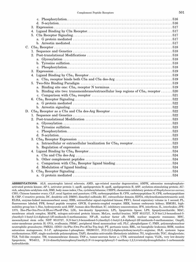
Complement is a vital part of the host defensesystem, capable of reacting to foreign material anddamaged or altered host tissues (Carroll and Sim,2011). Complement-like genes are present in organ-isms that diverged more than 1.3 billion years ago(reviewed in Pinto et al., 2007); animals that areincapable of mounting an adaptive immune responsepossess complement, including the sea-squirt Cionaintestinalis (Pinto et al., 2003) and the “living fossil”horseshoe crab, Carcinoscorpius rotundica (Zarkadiset al., 2001). The complement system consists ofa network of soluble and cell-surface proteins thatcan recognize potential threats and then undergo anamplification phase to produce a response of sufficientproportions to neutralize the perceived danger. Theoverall response is a balance between positive andnegative regulators that allows very fine control ofwhat is a potentially hazardous system: misdirected orexcessive activation of complement can be rapidlylethal to the host.There are three major pathways for the initiation of
a complement response (Fig. 1). The first or classicpathway usually requires the formation of immunecomplexes of IgM or IgG1 antibodies. The closelyrelated second or lectin pathway relies on the directrecognition of foreign material by a series of solublepattern-recognition receptors. The third or alternativepathway depends on the continual turnover of onecomponent, C3. When complement activation occurs inthe fluid phase or proximal to host tissues, inhibitoryfactors such as CD59 or decay-accelerating factorensure that no further complement response happens.However, on a receptive surface lacking the appropri-ate control factors (microbes or xenografts, for exam-ple), complement activation is greatly magnified tobecome a full-blown response even in the absence ofany positive triggers.The complement cascade is based on a series of
proteolytic events, with inactive complement proteinssuccessively cleaved to form the next active protease inthe chain. The terminal event is the formation of themembrane attack complex (MAC), a lytic pore in themembrane that can cause the death of some cell types.The MAC is inefficient at the cytolysis of nucleatedcells, primarily because of defense mechanisms thatprevent MAC formation or remove MAC from the cellsurface (Tegla et al., 2011). Sublytic MAC, however, isa proinflammatory stimulus for some cell types(reviewed in Dobrina et al., 2002) that has also beenlinked with the control of hemopoiesis (Ratajczak et al.,2010). The large pool of precursor complement proteinsallows a rapid and sizeable response to detectedthreats; conversely, any failure to control the comple-ment system can lead to inflammatory disease. As
a result of proteolysis, a series of small, biologicallyactive protein fragments are produced: C3a and C5afrom all three pathways and C4a primarily from theclassic pathway. These protein fragments are rapidlymetabolized by carboxypeptidases (Section I.D), form-ing des-arginated fragments (Burger and Zilow, 1993).Other fragments—for example, derived from thecleavage of C2 or factor B—are also produced, butthese are structurally unrelated to C3a, C4a, and C5a(Krishnan et al., 2009) and are outside the scope of thisreview.
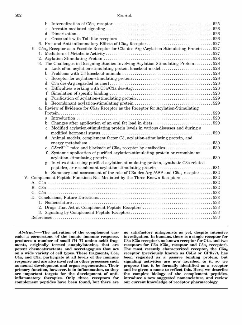
B. Concentrations of Complement Peptides in Healthand Disease
Determining the “physiologic” levels of complementpeptides is problematic, with wide variations in thereported values (Table 1). This is due in part to thedifferent assay techniques and in part to the nature ofthe biologic sample tested. Serum, obtained afterclotting of plasma, generally contains higher levels of
Fig. 1. Pathways for the production of complement peptides. The threemajor routes of complement activation and the extrinsic pathways for thedirect production of complement peptides by protease activities areshown.
Complement Peptide Receptors 503
the fragments due to the actions of the proteases in theclotting cascade on C3, C4, and C5 (Amara et al., 2008)(see Section I.C). Thus, plasma is a more reliableindicator of circulating complement peptide levels,particularly where EDTA has been used, effectivelyblocking all three major routes of complement activa-tion. Plasma concentrations in healthy human subjectshave been reported to be 119, 219, and 5.2 ng/ml forC3a, C4a, and C5a, respectively (Table 1). It is likelythat all these studies are reporting the des-arginatedforms of the fragments because the assay methodscannot discriminate between the two forms. In addi-tion, neoepitope-specific antibodies, which can discrim-inate between C3 and its cleavage product, detectusually the des-arginated form of C3a with highersensitivity. Complement peptide levels are clearlyelevated in inflammatory diseases and even in preg-nancy (Table 1). Elevation of C3a but not C4a suggestsactivation of the alternative pathway, which does notinvolve the proteolysis of C4, whereas very high levelsof both C4a and C3a suggest that both the classic/lectinand alternative pathways are constantly in operation.Both C5a and C5a des-Arg can be rapidly cleared byreceptor-mediated endocytosis (Oppermann and Gotze,1994), unlike C4a des-Arg and C3a des-Arg, whichtherefore accumulate to much higher levels.
C. C3a and C5a Generation outside theComplement Cascade
Although the production of complement peptides isusually linked to the activation of the complementcascade, biologically relevant amounts are also gener-ated by the so-called extrinsic pathways. This was first
reported in 1968, when proteolysis of C5 by trypsin wasobserved to produce anaphylatoxic activity (Cochraneand Muller-Eberhard, 1968). This type of activation isnow known to be widespread. Gingipain-1, a cysteineproteinase from Porphyromonas gingivalis, cleaves C3and C5 to produce leukocyte chemoattractant activitiesthat strongly resemble C3a and C5a (Wingrove et al.,1992), albeit with higher molecular weights thanpredicted for these fragments. Extrinsic pathwayactivation may be a common feature of many patho-genic bacteria, being exploited by unrelated organismssuch as Tanarella forsythia (Jusko et al., 2012) andAeromonas sobria (Nitta et al., 2008). For P. gingivalis,the production of C5a has been proposed to inhibitproduction mediated by Toll-like receptor 2 (TLR2)interleukin-12 (IL-12), allowing escape from immuneclearance (Liang et al., 2011). The feces of the housedust mite Dermatophagoides farinae contain a pro-tease, DerP1, which can generate active complementpeptides from C3 and C5 (Maruo et al., 1997). Asbestosand silica can also cause the cleavage of C5 (Governaet al., 2000, 2002, 2005). More recently, cross-talkbetween the complement and the coagulation cascadeshas been demonstrated to result in the robustgeneration of C3a and C5a, through the actions offactors Xa/XIa, plasmin, and thrombin (Amara et al.,2010). Factor VII–activating protease can also activateC3 and C5 (Kanse et al., 2012), producing C3a identicalto that formed by complement cascade activation butwith the N-terminal 4 residues missing from C5a.Enzymes released by activated or damaged host cellscan also cause C3a and C5a generation: the pro-apoptotic aspartic acid protease cathepsin D, elevated
TABLE 1Complement peptide concentrations in normal and diseased states
Reported concentrations of complement peptides are shown as nanogram per milliliter.
SourcePeptide
ReferenceC3a C4a C5a
NormalPlasma 98 342 ND Lee et al., 2006bSerum 4800 1000 ;0 Langone et al., 1984Pregnancy, plasma 2393 7827 20 Richani et al., 2005Plasma 125 160 ND Terui et al., 1987Serum 10,000 2000 110 Ohta et al., 2011Vitreous humor 98 77 ;0 Mondino et al., 1988Plasma/serum 152/1597 155/580 5.4/312 Wagner and Hugli, 1984Plasma 86 ND ND Stove et al., 1996Plasma 156 ND 5 Bengtson et al., 1987Plasma 20 40 5 Strey et al., 2009
DiseaseAspirin-induced asthma, plasma 148 815 ND Lee et al., 2006bPsoriasis, plasma 253 428 ND Terui et al., 1987Colorectal carcinoma, serum 43,646 ND ND Habermann et al., 2006Influenza serum 24,000 10,300 129 Ohta et al., 2011Sepsis, plasma 976 ND ND Stove et al., 1996Sepsis, serum, nonsurviving 1092 2385 10 Nakae et al., 1996Sepsis, serum, surviving 629 631 12.3 Nakae et al., 1996Acute ischemia, plasma 630 — 12.1 Bengtson et al., 1987Liver resection, plasma 42 34 6 Strey et al., 2009Chronic hepatitis C virus infection, serum — 12,200 — Imakiire et al., 2012
ND, not determined.
504 Klos et al.
in trauma (Huber-Lang et al., 2012); b-tryptase,secreted by mast cells (Fukuoka et al., 2008); andgranzyme B, produced by leukocytes (Perl et al., 2012).The relative importance of these fragment-generationsystems that lie outside the complement cascade is notyet clear. The increasing availability of inhibitors ofcomplement peptide generation (Qu et al., 2009;Woodruff et al., 2011) that inhibit either commonpathways (e.g., compstatin, which acts on C3) or morespecific pathways (e.g., eculizumab, which acts on C5proteolysis) will help to answer this question.
D. Deactivation of Complement Peptides
Human serum contains a potent deactivator ofcomplement peptides, shown to be a carboxypepti-dase B–like activity that removes the C-terminalArg residues of C3a and C5a (Bokisch et al., 1969;Bokisch and Muller-Eberhard, 1970). Inhibition bya carboxypeptidase inhibitor, DL-2-mercaptomethyl-3-guanidinoethylthiopropanoic acid, made administra-tion of ordinarily survivable doses of cobra venomfactor (CVF) or C3a lethal (Huey et al., 1983). Twomajor carboxypeptidases control the activity of thefragments. The zinc metalloprotease carboxypeptidaseN (CPN) is released in an active form (Levin et al.,1982), and carboxypeptidase R (CPR) is an acute-phaseprotein, up-regulated in inflammation and secreted inan inactive form, proCPR (reviewed in Campbell et al.,2001). ProCPR is bound to plasminogen and activatedby plasmin or thrombin (Wang et al., 1994; Sato et al.,2000; Nishimura et al., 2007; Leung et al., 2008). Itpreferentially degrades C5a over C3a (Campbell et al.,2002) and can remove Lys residues from fibrin clots,preventing plasminogen binding (Bajzar et al., 1995).CPR is also known as plasma carboxypeptidase B,activated thrombin-activatable fibrinolysis inhibitor(TAFI), and carboxypeptidase U (Campbell et al.,2001). CPB is a more active carboxypeptidase thanCPN and also acts on bradykinin and osteopontin assubstrates. TAFI carboxypeptidase is protective inallergic asthma (Fujiwara et al., 2012) and down-regulates inflammation in rheumatoid arthritis (RA)(Song et al., 2011). Both CPR and CPN can alsoinactivate C3a and C5a octapeptides (Section III.C.4)(Campbell et al., 2002).Defense against host responses by microorganisms
can also involve complement peptide degradation.Brugia malayi and Trichinella spiralis, parasiticnematodes, release a metallocarboxypeptidase thatinactivates C5a (Rees-Roberts et al., 2010), presumablyto prevent eosinophil chemotaxis and activation.Streptococcus pyogenes produces a C5a peptidase(Wexler et al., 1985) that cleaves between His67 andLys68 (Cleary et al., 1992). This peptidase, ScpA, isa cell wall–anchored serine protease that is importantfor virulence (Husmann et al., 1997). The virulence ofthe enterobacterium Serratia marcescens, depends in
part on the activity of a 56-kDa protease that caninactivate C5a, thus inhibiting neutrophil influx (Odaet al., 1990).
II. The Role of Complement Peptidesin Pathophysiology
There is a very fine balance that must be maintainedwhenever the complement cascade is activated becauseof the destructive nature of the effector systems: toolittle may result in incomplete clearance of immunecomplexes, leading to autoimmune diseases such assystemic lupus erythematosus (SLE) and failure tocontrol infections, but too much causes damage tohealthy tissues (Carroll and Sim, 2011). The uncon-trolled or inappropriate production of complementpeptides has been implicated in many inflammatorydisorders, and, although it is beyond the scope of thisreview to discuss the roles of C3a and C5a in anydetail, a list of these disorders is shown in Tables 2 and3. It should also be noted that C3a and C5a do notalways act as partners in crime, both promotinginflammation. In the development of asthma, thegeneration of C5a during sensitization appears toinhibit the further development of lung diseasewhereas C3a promotes disease progression, throughskewing of the subsequent T-cell response. However,both fragments have deleterious effects at later stages(reviewed in Wills-Karp, 2007). C4a has no known rolein human disease.
A. Complement Peptides Are Important Biomarkersof Disease
The stability of C3a des-Arg and C4a des-Arg hasmade these complement peptides in particular poten-tially useful as biomarkers in a number of disorders,even those not traditionally seen as inflammatory innature such as cancer. Examples are the increasedserum levels of C3a that indicate the presence ofcolorectal tumors (Habermann et al., 2006), hepatitis Cvirus–related hepatocellular carcinoma (Lee et al.,2006a; Kanmura et al., 2010), benign prostatic hyper-plasia (Xie et al., 2011), and ductal carcinoma in situ ofthe breast (Solassol et al., 2010). C4a has recently beenshown to be elevated in patients with chronic hepatitisC infection and active disease but is even higher ininfected but asymptomatic individuals (Imakiire et al.,2012). Both C4a and C3a levels are predictive of theresponses of esophageal cancer patients to chemo-radiation (Maher et al., 2011). In more obviouslyinflammatory disorders, C3a (and to a lesser extentC4a) have been shown to be potentially useful markersin dermatomyositis (Campo et al., 2007), aneurysmalsubarachnoid hemorrhage (Mack et al., 2007), acuteLyme disease in tick-bite patients (Shoemaker et al.,2008; Stricker et al., 2009), the exudative form of age-related macular degeneration (Machalinska et al.,
Complement Peptide Receptors 505
2009), endometriosis (Fassbender et al., 2009), adversepregnancy outcomes (Lynch et al., 2011), chronicobstructive pulmonary disease (Marc et al., 2004;Zhang et al., 2011), cryptogenic and large-vesseldisease subtypes of stroke (Stokowska et al., 2011),heart failure (Gombos et al., 2012), cerebral arteriove-nous malformations (Haque et al., 2011), asthma (Jokset al., 2008), gestational diabetes mellitus (Lappas,2011), SLE (Wild et al., 1990), acute relapses inmultiple sclerosis (Ingram et al., 2010), IgA nephrop-athy/Henoch–Schonlein nephritis (Abou-Ragheb et al.,1992) and impaired renal function (Abou-Ragheb et al.,1991), atopic dermatitis (Sergeev Iu et al., 1989),psoriasis (Takematsu et al., 1986) and psoriaticarthritis (Muto et al., 1991), idiopathic pulmonaryarterial hypertension (Abdul-Salam et al., 2006), post-exercise malaise in myalgic encephalomyelitis/chronicfatigue syndrome (Nijs et al., 2010), AIDS-associatedretinitis (Mondino et al., 1990), and grafted corneas(Mondino and Sumner, 1990). In addition, C4a and C5alevels decrease after liver resection whereas C3a levelsincrease (Strey et al., 2009); and C3a and C4a areelevated in liver transplant recipients (Pfeifer et al.,2000). Finally, elevated C3a and C4a in sepsis are
associated with a fatal outcome, whereas C5a levelsare not correlated (Hack et al., 1989).
B. Functions of the Complement Peptides beyondInnate Immunity
The complement system has evolved a number ofnonimmunologic functions. Homeostasis is maintainedby the action of complement on cellular debris, which isimportant in the prevention of autoimmune responses.In development, complement has roles in bone metab-olism, hemopoiesis, angiogenesis, and tissue repair.Complement is also involved in liver and lens re-generation (reviewed in Rutkowski et al., 2010a). Thisrange of activities makes the complement system andits fragments important players in neoplasia (Rutkow-ski et al., 2010b) and a wide variety of other functions,some of which we will detail.
1. Cell Migration and Homing. In the centralnervous system, C3a (and to a lesser extent C5a) isactive during the development of the rat cerebellum(Benard et al., 2008) and the in vitro differentiationand migration of neural progenitor cells (Shinjyo et al.,2009). C3a is known to be the coattractant thatorganizes neural crest cells when they migrate during
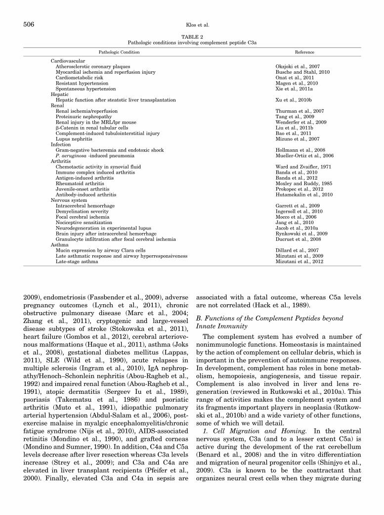
TABLE 2Pathologic conditions involving complement peptide C3a
Pathologic Condition Reference
CardiovascularAtherosclerotic coronary plaques Oksjoki et al., 2007Myocardial ischemia and reperfusion injury Busche and Stahl, 2010Cardiometabolic risk Onat et al., 2011Resistant hypertension Magen et al., 2010Spontaneous hypertension Xie et al., 2011a
HepaticHepatic function after steatotic liver transplantation Xu et al., 2010b
RenalRenal ischemia/reperfusion Thurman et al., 2007Proteinuric nephropathy Tang et al., 2009Renal injury in the MRL/lpr mouse Wenderfer et al., 2009b-Catenin in renal tubular cells Liu et al., 2011bComplement-induced tubulointerstitial injury Bao et al., 2011Lupus nephritis Mizuno et al., 2007
InfectionGram-negative bacteremia and endotoxic shock Hollmann et al., 2008P. aeruginosa -induced pneumonia Mueller-Ortiz et al., 2006
ArthritisChemotactic activity in synovial fluid Ward and Zvaifler, 1971Immune complex induced arthritis Banda et al., 2010Antigen-induced arthritis Banda et al., 2012Rheumatoid arthritis Moxley and Ruddy, 1985Juvenile-onset arthritis Prokopec et al., 2012Antibody-induced arthritis Hutamekalin et al., 2010
Nervous systemIntracerebral hemorrhage Garrett et al., 2009Demyelination severity Ingersoll et al., 2010Focal cerebral ischemia Mocco et al., 2006Nociceptive sensitization Jang et al., 2010Neurodegeneration in experimental lupus Jacob et al., 2010aBrain injury after intracerebral hemorrhage Rynkowski et al., 2009Granulocyte infiltration after focal cerebral ischemia Ducruet et al., 2008
AsthmaMucin expression by airway Clara cells Dillard et al., 2007Late asthmatic response and airway hyperresponsiveness Mizutani et al., 2009Late-stage asthma Mizutani et al., 2012
506 Klos et al.
early development (Carmona-Fontaine et al., 2011).The C3a receptor (C3a receptor, also known as C3aR;see Section IV.B) is a key mediator of insulin resistanceand functions by modulating macrophage infiltrationand activation in adipose tissue (Mamane et al., 2009).The homing of hemopoietic stem and progenitor cells tobone marrow also depends on the C3a-C3a receptoraxis (Reca et al., 2003).2. Adaptive Immunity. In addition to well-known
roles in innate immunity, the complement peptidesalso act to modulate adaptive immunity (Chenowethet al., 1982; Ottonello et al., 1999). The presence ofcomplement peptide receptors on certain subsets of Band T cells has been reported (Martin et al., 1997;
Werfel et al., 2000). Expression levels are much higheron antigen-presenting cells (Sacks, 2010). C3a and C5aproduced by dendritic cells (DC) are required foroptimal CD4 T-cell help for CD8 T cells in murineallograft rejection (Vieyra et al., 2011). Similarly,locally produced C5a and C3a provide costimulatoryand survival signals for naive CD4+ T cells (Strainicet al., 2008), and gdT-cell function in sepsis can bemodulated by C5a (Han et al., 2011) due to increasedproduction of IL-17 after blockade of DC C5a receptor(C5a1 receptor, also known as C5R1, CD88; Section IV.C) (Xu et al., 2010a). Conversely, preventing C5astimulation of DC by ablation of the C5a1 receptor orwith a receptor antagonist induces Treg and TH17 cells
TABLE 3Pathologic conditions involving complement peptide C5a
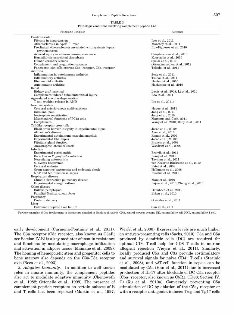
Pathologic Condition Reference
CardiovascularFibrosis in hypertension Iyer et al., 2011Atherosclerosis in ApoE2/2 mice Manthey et al., 2011Preclinical atherosclerosis associated with systemic lupus
erythematosusRua-Figueroa et al., 2010
Arterial injury in atherosclerosis-prone mice Shagdarsuren et al., 2010Hemodialysis-associated thrombosis Kourtzelis et al., 2010Human coronary lesions Speidl et al., 2011Complement and coagulation cascades Oikonomopoulou et al., 2012Pancreatic islet cells express C5a1 receptor, C5a2 receptor Tokodai et al., 2011
ArthritisInflammation in autoimmune arthritis Song et al., 2011Inflammatory arthritis Tsuboi et al., 2011Rheumatoid arthritis Hueber et al., 2010Autoimmune arthritis Hashimoto et al., 2010
RenalKidney graft survival Lewis et al., 2008; Li et al., 2010Complement-induced tubulointerstitial injury Bao et al., 2011
Age-related macular degenerationT-cell cytokine release in AMD Liu et al., 2011a
Nervous systemCerebral arteriovenous malformations Haque et al., 2011Incisional pain Jang et al., 2011Nociceptive sensitization Jang et al., 2010Mitochondrial functions of PC12 cells Martinus and Cook, 2011Complement-
Toll-like receptor cross-talkWang et al., 2010; Raby et al., 2011
Blood-brain barrier integrity in experimental lupus Jacob et al., 2010cAlzheimer’s disease Ager et al., 2010Experimental autoimmune encephalomyelitis Ramos et al., 2009Experimental CNS lupus Jacob et al., 2010bPituitary gland function Francis et al., 2008Amyotrophic lateral sclerosis Woodruff et al., 2008
InfectionExperimental periodontitis Breivik et al., 2011Bone loss in P. gingivalis infection Liang et al., 2011Necrotizing enterocolitis Tayman et al., 2011S. aureus bacteremia von Köckritz-Blickwede et al., 2010Cerebral malaria Patel et al., 2008Gram-negative bacteremia and endotoxic shock Hollmann et al., 2008NKT and NK function in sepsis Fusakio et al., 2011
Respiratory diseaseChronic obstructive pulmonary disease Marc et al., 2010Experimental allergic asthma Lajoie et al., 2010; Zhang et al., 2010
Other diseaseBullous pemphigoid Heimbach et al., 2011Familial Mediterranean fever Erken et al., 2010
PregnancyPreterm delivery Gonzalez et al., 2011
LiverFulminant hepatic liver failure Sun et al., 2011
Further examples of C5a involvement in disease are detailed in Monk et al. (2007). CNS, central nervous system; NK, natural killer cell; NKT, natural killer T cell.
Complement Peptide Receptors 507
by increasing the production of transforming growthfactor-b (TGF-b) (Weaver et al., 2010). In experimentalautoimmune encephalomyelitis, a model of multiplesclerosis, interferon-g (IFN-g) and IL-17 production inautoreactive T cells depends on local production of C3aand C5a (Liu et al., 2008). C5a-mediated TH17 differ-entiation has been proposed to underlie some autoim-mune and inflammatory disorders such as autoimmunearthritis (Hashimoto et al., 2010), and blockade of thispathway may be beneficial for the control of thesediseases. However, C5a can negatively regulate TH17cell differentiation in asthma (Lajoie et al., 2010), so thistherapeutic strategy may be of limited use. In contrastto the many reports of DC activation by complementpeptides, T-cell-expressed C5a1 receptor is required forenhanced T-cell expansion, as a result of inhibition ofapoptosis (Lalli et al., 2008).3. Hemopoiesis. In contrast to the effects of C3a on
stem-cell homing to bone marrow as mentioned pre-viously, C5a and C5a des-Arg disrupt the CXCL12(SDF-1a)/CXCR4 axis and increase the mobilization ofhemopoietic stem and progenitor cells (Jalili et al.,2010). Mobilization of these cells is also impaired inC5-deficient mice (Lee et al., 2009). The roles ofcomplement peptides in stem-cell mobilization havebeen reviewed elsewhere (Ratajczak et al., 2012).4. Regeneration and Other Functions. Human mes-
enchymal stem cells (MSC), which are involved in therepair of various tissues, express complement peptidereceptors and are chemoattracted to C3a and C5a(Schraufstatter et al., 2009). Interestingly, MSC havebeen found to modulate innate immunity by activatingthe complement cascade (Moll et al., 2011). Comple-ment C3a and C5a can modulate bone biology ininflammation (Ignatius et al., 2011b). C5a1 receptorhas been shown to control osteoblast migration duringfracture healing (Ignatius et al., 2011a), and efficientosteoclast differentiation requires local complementactivation (Ignatius et al., 2011b). Normal heartfunction appears to depend on the C5a/C5a1 receptoraxis, with C5 deficiency and receptor knockout orblockade causing a “state of distress” (Mullick et al.,2011). C3a also has positive effects on food intakeregulation, with the administration of C3a to thecentral nervous system leading to suppression ofappetite (Ohinata et al., 2002, 2007, 2009; Ohinataand Yoshikawa, 2008).
III. Structure of Complement Peptides
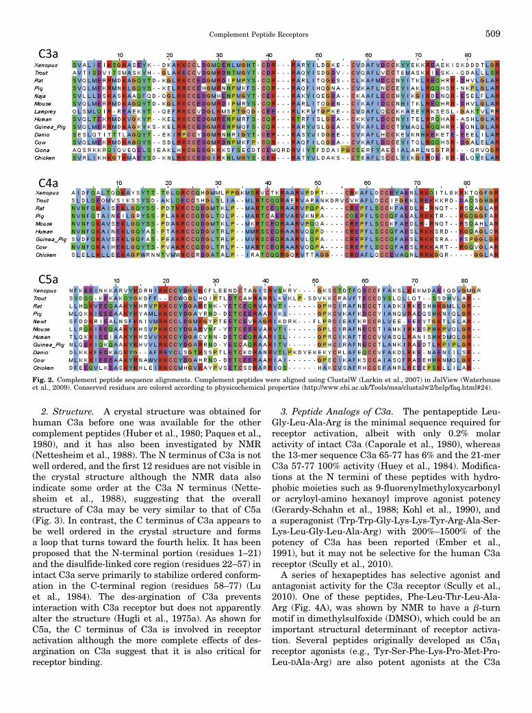
Complement components C3, C4, and C5 are thoughtto have arisen by gene duplication events predating theemergence of cartilaginous fish (Terado et al., 2003). Itis therefore not surprising that the complementpeptides derived from these proteins all have similarprimary structures, with 74 to 79 amino acids (Fig. 2,A–C) and largely conserved Cys residues that suggest
similar patterns of disulfide bridges in the tertiarystructures. Nuclear magnetic resonance (NMR) andX-ray crystallographic studies have confirmed that thethree-dimensional structures of C3a (Nettesheim et al.,1988) and C5a (Zuiderweg et al., 1988) are very similaroverall.
A. C3a
1. Sequence. All C3a sequences found to date havesix Cys residues (numbered 23, 24, 37, 50, 57, and 58 inguinea pig C3a) (Gerard et al., 1988), apart from thedeuterostome, C. intestinalis, where the outermost pairof Cys residues are missing, implying that it has onlytwo disulfide bridges (Fig. 2A). It is noteworthy thatthe disulfide bridges in human C3a appear to beunusually labile (Chang et al., 2008), a phenomenonthat may provide additional regulation of the activityof C3a in vivo. Guinea pig C3a has 70% identity withrat (Jacobs et al., 1978), human, porcine (Corbin andHugli, 1976), and mouse (Hugli et al., 1975b), and C3afrom these species have identical activity in smoothmuscle contraction, histamine release, and vascularpermeability assays. Among all C3a sequences, theoverall arrangement of basic residues is also highlyconserved, with a ubiquitous C-terminal Arg. There isan almost invariant C-terminal Leu-Gly-Leu-Ala-Argsequence across species, again not wholly conserved inC. intestinalis. Despite these differences, Ciona C3adoes bind to a specific receptor, which has significanthomology to the mammalian C3a receptor (Melilloet al., 2006). The Ciona C3a-C3a receptor axis isresponsible for the chemotaxis of hemocytes, suggest-ing a role for C3a in inflammation in this organism.
Interestingly, the des-Arg form of Ciona C3a alsostimulates chemotaxis of hemocytes (Pinto et al., 2003)whereas C3a des-Arg is inactive in the majority ofassays across other species. However, C3a and C3ades-Arg are equipotent in the killing of Gram-positive(e.g., Enterococcus faecalis) and Gram-negative (e.g.,Pseudomonas aeruginosa) organisms and are actuallymore potent than the classic antimicrobial peptideLL37 (Nordahl et al., 2004). It has been suggested thatC3a (and C4a) evolved initially as antimicrobialpeptides (Pasupuleti et al., 2007), with chemoattrac-tant activity emerging later. Antifungal activity of C3aand C3a des-Arg has also been demonstrated forCandida albicans (Sonesson et al., 2007). The mecha-nism appears to be the disruption of the plasmamembrane and is dependent on Arg residues althoughthe C-terminal Arg is dispensable, demonstrating thatthe chemotactic and antimicrobial activities of C3a areseparate. Heparin can also bind to the same regions ofC3a (Andersson et al., 2004) and inhibits antimicrobialactivity (Nordahl et al., 2004). Structure-activitystudies show that the formation of an amphipathichelix with a high net positive charge is critical forantimicrobial activity (Pasupuleti et al., 2008).
508 Klos et al.
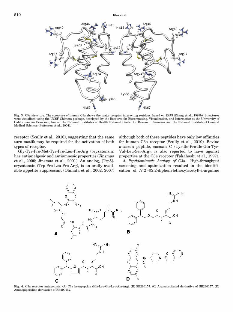
2. Structure. A crystal structure was obtained forhuman C3a before one was available for the othercomplement peptides (Huber et al., 1980; Paques et al.,1980), and it has also been investigated by NMR(Nettesheim et al., 1988). The N terminus of C3a is notwell ordered, and the first 12 residues are not visible inthe crystal structure although the NMR data alsoindicate some order at the C3a N terminus (Nette-sheim et al., 1988), suggesting that the overallstructure of C3a may be very similar to that of C5a(Fig. 3). In contrast, the C terminus of C3a appears tobe well ordered in the crystal structure and formsa loop that turns toward the fourth helix. It has beenproposed that the N-terminal portion (residues 1–21)and the disulfide-linked core region (residues 22–57) inintact C3a serve primarily to stabilize ordered conform-ation in the C-terminal region (residues 58–77) (Luet al., 1984). The des-argination of C3a preventsinteraction with C3a receptor but does not apparentlyalter the structure (Hugli et al., 1975a). As shown forC5a, the C terminus of C3a is involved in receptoractivation although the more complete effects of des-argination on C3a suggest that it is also critical forreceptor binding.
3. Peptide Analogs of C3a. The pentapeptide Leu-Gly-Leu-Ala-Arg is the minimal sequence required forreceptor activation, albeit with only 0.2% molaractivity of intact C3a (Caporale et al., 1980), whereasthe 13-mer sequence C3a 65-77 has 6% and the 21-merC3a 57-77 100% activity (Huey et al., 1984). Modifica-tions at the N termini of these peptides with hydro-phobic moieties such as 9-fluorenylmethyloxycarbonylor acryloyl-amino hexanoyl improve agonist potency(Gerardy-Schahn et al., 1988; Kohl et al., 1990), anda superagonist (Trp-Trp-Gly-Lys-Lys-Tyr-Arg-Ala-Ser-Lys-Leu-Gly-Leu-Ala-Arg) with 200%–1500% of thepotency of C3a has been reported (Ember et al.,1991), but it may not be selective for the human C3areceptor (Scully et al., 2010).
A series of hexapeptides has selective agonist andantagonist activity for the C3a receptor (Scully et al.,2010). One of these peptides, Phe-Leu-Thr-Leu-Ala-Arg (Fig. 4A), was shown by NMR to have a b-turnmotif in dimethylsulfoxide (DMSO), which could be animportant structural determinant of receptor activa-tion. Several peptides originally developed as C5a1receptor agonists (e.g., Tyr-Ser-Phe-Lys-Pro-Met-Pro-Leu-DAla-Arg) are also potent agonists at the C3a
Fig. 2. Complement peptide sequence alignments. Complement peptides were aligned using ClustalW (Larkin et al., 2007) in JalView (Waterhouseet al., 2009). Conserved residues are colored according to physicochemical properties (http://www.ebi.ac.uk/Tools/msa/clustalw2/help/faq.html#24).
Complement Peptide Receptors 509
receptor (Scully et al., 2010), suggesting that the sameturn motifs may be required for the activation of bothtypes of receptor.Gly-Tyr-Pro-Met-Tyr-Pro-Leu-Pro-Arg (oryzatensin)
has antianalgesic and antiamnesic properties (Jinsmaaet al., 2000; Jinsmaa et al., 2001). An analog, [Trp5]-oryzatensin (Trp-Pro-Leu-Pro-Arg), is an orally avail-able appetite suppressant (Ohinata et al., 2002, 2007)
although both of these peptides have only low affinitiesfor human C3a receptor (Scully et al., 2010). Bovinek-casein peptide, casoxin C (Tyr-Ile-Pro-Ile-Gln-Tyr-Val-Leu-Ser-Arg), is also reported to have agonistproperties at the C3a receptor (Takahashi et al., 1997).
4. Peptidomimetic Analogs of C3a. High-throughputscreening and optimization resulted in the identifi-cation of N(2)-[(2,2-diphenylethoxy)acetyl]-L-arginine
Fig. 3. C5a structure. The structure of human C5a shows the major receptor interacting residues, based on 1KJS (Zhang et al., 1997b). Structureswere visualized using the UCSF Chimera package, developed by the Resource for Biocomputing, Visualization, and Informatics at the University ofCalifornia–San Francisco, funded the National Institutes of Health National Center for Research Resources and the National Institute of GeneralMedical Sciences (Pettersen et al., 2004).
Fig. 4. C3a receptor antagonists. (A) C3a hexapeptide (His-Leu-Gly-Leu-Ala-Arg). (B) SB290157. (C) Arg-substituted derivative of SB290157. (D)Aminopiperidine derivative of SB290157.
510 Klos et al.
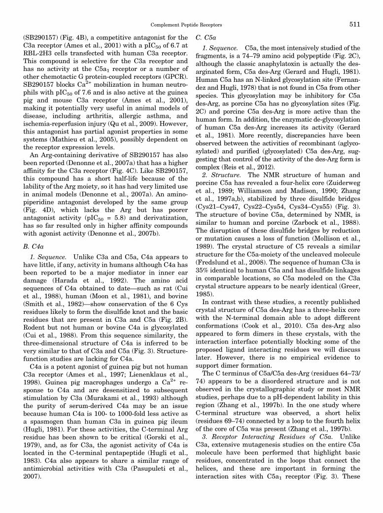
(SB290157) (Fig. 4B), a competitive antagonist for theC3a receptor (Ames et al., 2001) with a pIC50 of 6.7 atRBL-2H3 cells transfected with human C3a receptor.This compound is selective for the C3a receptor andhas no activity at the C5a1 receptor or a number ofother chemotactic G protein-coupled receptors (GPCR).SB290157 blocks Ca2+ mobilization in human neutro-phils with pIC50 of 7.6 and is also active at the guineapig and mouse C3a receptor (Ames et al., 2001),making it potentially very useful in animal models ofdisease, including arthritis, allergic asthma, andischemia-reperfusion injury (Qu et al., 2009). However,this antagonist has partial agonist properties in somesystems (Mathieu et al., 2005), possibly dependent onthe receptor expression levels.An Arg-containing derivative of SB290157 has also
been reported (Denonne et al., 2007a) that has a higheraffinity for the C3a receptor (Fig. 4C). Like SB290157,this compound has a short half-life because of thelability of the Arg moiety, so it has had very limited usein animal models (Denonne et al., 2007a). An amino-piperidine antagonist developed by the same group(Fig. 4D), which lacks the Arg but has poorerantagonist activity (pIC50 = 5.8) and derivatization,has so far resulted only in higher affinity compoundswith agonist activity (Denonne et al., 2007b).
B. C4a
1. Sequence. Unlike C3a and C5a, C4a appears tohave little, if any, activity in humans although C4a hasbeen reported to be a major mediator in inner eardamage (Harada et al., 1992). The amino acidsequences of C4a obtained to date—such as rat (Cuiet al., 1988), human (Moon et al., 1981), and bovine(Smith et al., 1982)—show conservation of the 6 Cysresidues likely to form the disulfide knot and the basicresidues that are present in C3a and C5a (Fig. 2B).Rodent but not human or bovine C4a is glycosylated(Cui et al., 1988). From this sequence similarity, thethree-dimensional structure of C4a is inferred to bevery similar to that of C3a and C5a (Fig. 3). Structure-function studies are lacking for C4a.C4a is a potent agonist of guinea pig but not human
C3a receptor (Ames et al., 1997; Lienenklaus et al.,1998). Guinea pig macrophages undergo a Ca2+ re-sponse to C4a and are desensitized to subsequentstimulation by C3a (Murakami et al., 1993) althoughthe purity of serum-derived C4a may be an issuebecause human C4a is 100- to 1000-fold less active asa spasmogen than human C3a in guinea pig ileum(Hugli, 1981). For these activities, the C-terminal Argresidue has been shown to be critical (Gorski et al.,1979), and, as for C3a, the agonist activity of C4a islocated in the C-terminal pentapeptide (Hugli et al.,1983). C4a also appears to share a similar range ofantimicrobial activities with C3a (Pasupuleti et al.,2007).
C. C5a
1. Sequence. C5a, the most intensively studied of thefragments, is a 74–79 amino acid polypeptide (Fig. 2C),although the classic anaphylatoxin is actually the des-arginated form, C5a des-Arg (Gerard and Hugli, 1981).Human C5a has an N-linked glycosylation site (Fernan-dez and Hugli, 1978) that is not found in C5a from otherspecies. This glycosylation may be inhibitory for C5ades-Arg, as porcine C5a has no glycosylation sites (Fig.2C) and porcine C5a des-Arg is more active than thehuman form. In addition, the enzymatic de-glycosylationof human C5a des-Arg increases its activity (Gerardet al., 1981). More recently, discrepancies have beenobserved between the activities of recombinant (aglyco-sylated) and purified (glycosylated) C5a des-Arg, sug-gesting that control of the activity of the des-Arg form iscomplex (Reis et al., 2012).
2. Structure. The NMR structure of human andporcine C5a has revealed a four-helix core (Zuiderweget al., 1989; Williamson and Madison, 1990; Zhanget al., 1997a,b), stabilized by three disulfide bridges(Cys21–Cys47, Cys22–Cys54, Cys34–Cys55) (Fig. 3).The structure of bovine C5a, determined by NMR, issimilar to human and porcine (Zarbock et al., 1988).The disruption of these disulfide bridges by reductionor mutation causes a loss of function (Mollison et al.,1989). The crystal structure of C5 reveals a similarstructure for the C5a-moiety of the uncleaved molecule(Fredslund et al., 2008). The sequence of human C3a is35% identical to human C5a and has disulfide linkagesin comparable locations, so C5a modeled on the C3acrystal structure appears to be nearly identical (Greer,1985).
In contrast with these studies, a recently publishedcrystal structure of C5a des-Arg has a three-helix corewith the N-terminal domain able to adopt differentconformations (Cook et al., 2010). C5a des-Arg alsoappeared to form dimers in these crystals, with theinteraction interface potentially blocking some of theproposed ligand interacting residues we will discusslater. However, there is no empirical evidence tosupport dimer formation.
The C terminus of C5a/C5a des-Arg (residues 64–73/74) appears to be a disordered structure and is notobserved in the crystallographic study or most NMRstudies, perhaps due to a pH-dependent lability in thisregion (Zhang et al., 1997b). In the one study whereC-terminal structure was observed, a short helix(residues 69–74) connected by a loop to the fourth helixof the core of C5a was present (Zhang et al., 1997b).
3. Receptor Interacting Residues of C5a. UnlikeC3a, extensive mutagenesis studies on the entire C5amolecule have been performed that highlight basicresidues, concentrated in the loops that connect thehelices, and these are important in forming theinteraction sites with C5a1 receptor (Fig. 3). These
Complement Peptide Receptors 511
can be divided into three separate clusters (Mollisonet al., 1989; Huber-Lang et al., 2003). Site 1 is formed byresidues 12–20, including His15, Lys19 and 20, andArg46. Site 2 includes Asp24, Arg37, Arg40, andpossibly Lys49. Site 3 comprises residues 67–74, in-cluding His67, Lys68, Arg69, and the C-terminal Arg74(Mollison et al., 1989; Bubeck et al., 1994; Toth et al.,1994; Vlattas et al., 1994). Other important residues arehydrophobic: Val18, Leu43, Met70, and Leu72; thelatter residue when mutated to either Lys or Asp causesbig loss in binding affinity (Mollison et al., 1989).The unpaired Cys27 is unique to human C5, and
mutation to Arg has been found to be necessary todisplay C5a on phage (Cain et al., 2003; Heller et al.,1999), presumably by preventing aberrant cross-linking. However, this residue has been exploited tohelp map the ligand-binding site on the C5a1 receptor(Section IV.C.4), and the data suggest that this part ofC5a lies close to the receptor N terminus.Human C5a (1–69), lacking the C-terminal loop-helix
structure, can still bind to cell-surface receptors, albeitwith considerably lower affinity than intact C5a, butcannot activate the receptor (Chenoweth et al., 1982).The substitution of the C-terminal pentapeptidesequence of human C3a (Leu-Gly-Leu-Ala-Arg) to C5a(1–69) (Bautsch et al., 1992) gives a ligand that bindsto both the C5a1 receptor and C3a receptor, whereasadding a modified C5a C-terminal sequence througha new disulfide linkage makes a C5a1 receptorantagonist (Zhang et al., 1997a). These observationssuggest that C5a (1–69) provides a recognition site forthe receptor whereas the C terminus provides theactivation signal.4. Peptide Analogs of C5aa. Agonists. Although the pentapeptide Met-Gln-
Leu-Gly-Arg that is analogous to the C terminus of C5ais inactive at the C5a1 receptor (Chenoweth and Hugli,1980), the discovery that the C-terminal octapeptide ofhuman C5a, His-Lys-Asp-Met-Gln-Leu-Gly-Arg, hasweak agonist activity (Kawai et al., 1991, 1992) hasprovoked intense research into the mechanism ofreceptor activation and the development of antagonists.A series of decapeptide analogs first reported by
Ember et al. (1992), substituting Phe for His67, hadincreased potency over the native sequence. Thesewere later developed into a large series of constrainedpeptides. In some of these, the insertion of Pro into thesequence has resulted in greater agonist activity(Sanderson et al., 1994) but also in a loss of selectivity(Scully et al., 2010). EP-54 (Tyr-Ser-Phe-Lys-Pro-Met-Pro-Leu-DAla-Arg) is an agonist at both the C5a1receptor and C3a receptor but has little or no activityat the second C5a receptor C5a2 receptor (also knownas C5R2, C5L2, GPR77) (Scola et al., 2007). In contrast,EP-67 (Tyr-Ser-Phe-Lys-Asp-Met-Pro-(Me)Leu-DAla-Arg) is a weak agonist at both the C5a1 receptor andthe C3a receptor but may have greater activity at the
C5a2 receptor (Kawatsu et al., 1996; Short et al., 1999;Taylor et al., 2001; Vogen et al., 1999a, 1999b).
Peptides such as EP-54 and EP-67 have beendescribed as “response selective,” producing differentresponses at the C5a1 receptor depending on the celltype under study (reviewed in Taylor et al., 2001).However, a more likely explanation is the nonselectiv-ity of these peptides, with varying affinities for two ormore of the known complement peptide receptors.Peptide ligands have been found to have conservedturn structures (Tyndall et al., 2005), and C5a and itsanalogs have a b/g turn motif that has also beenobserved in bradykinin, enkephalin, and vasopressin.The conservation of the turn structure may be linked tothe mechanism of receptor activation because a two-step binding mechanism is a common feature ofsecretin family GPCR (Tyndall et al., 2005).
b. Antagonists. Further development of C-terminalpeptides resulted in a family of hexapeptide analogs ofthe form N(Me)-Phe-Lys-Pro-DCha-Xxx-DArg, whichwere potent agonists and antagonists depending onthe nature of the fifth residue (Konteatis et al., 1994).Increasing aromaticity at this position was found toenhance antagonist activity, with Trp providing com-plete antagonism. From these studies, a cyclic peptideantagonist (PMX53; 3D53, (Ac)Phe-[Orn-Pro-DCha-Trp-Arg]) has been developed that shows improvedaffinity (Wong et al., 1999) (Fig. 5A) and has a b-turnstructure similar to the C5a C terminus, as determinedby NMR (Zhang et al., 2008).
PMX53 (pIC50 = 7.05 in human neutrophil mem-branes) is an insurmountable antagonist for C5a1receptor (Strachan et al., 2000, 2001) with no discernibleactivity at the C5a2 receptor (Scola et al., 2007). Likemany peptides, PMX53 has low oral bioavailability, butits long-lasting effect means that even once-daily oraldosing is sufficient to maintain circulating levels in therat (Morgan et al., 2008). The development of PMX53has been discontinued (http://www.evaluatepharma.com/Universal/View.aspx?type=Story&id=178099), al-though PMX205, a more stable derivative (DelisleMilton et al., 2011) with a hydrocinnamic acid moietyat the N terminus (Fig. 5B), is now being used in animalmodels of disease, including rat and mouse models ofneurologic diseases such as Huntington’s disease,Alzheimer’s disease, and amyotrophic lateral sclerosis(ALS) (Fonseca et al., 2009; Woodruff et al., 2006, 2008).However, PMX205 has been reported as unlikely to beof benefit in ALS patients (ALS-TDI http://www.als.net/ALS-Research/PMX205/ALS-Topics/).
PMX53 also has limited selectivity, in common withthe related compound JPE-1375 (Hoo-Phe-Orn-Pro-(d-HLeu)-Phe4F-Phe) (Fig. 5C), and it also binds to otherreceptors such as NK2 and Mas-related gene 2 receptor(Schnatbaum et al., 2006; Subramanian et al., 2011).Despite this, PMX53 (and, to a lesser extent, JPE-1375) has been of immense value in defining the role of
512 Klos et al.
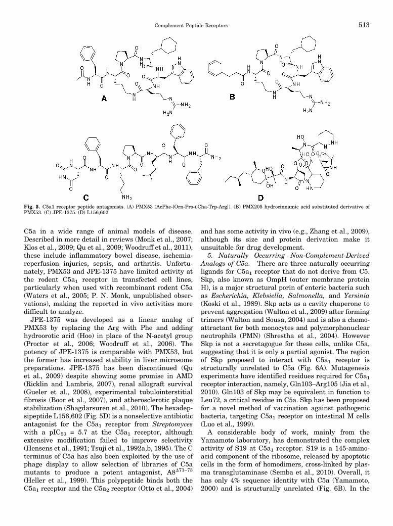
C5a in a wide range of animal models of disease.Described in more detail in reviews (Monk et al., 2007;Klos et al., 2009; Qu et al., 2009; Woodruff et al., 2011),these include inflammatory bowel disease, ischemia-reperfusion injuries, sepsis, and arthritis. Unfortu-nately, PMX53 and JPE-1375 have limited activity atthe rodent C5a1 receptor in transfected cell lines,particularly when used with recombinant rodent C5a(Waters et al., 2005; P. N. Monk, unpublished obser-vations), making the reported in vivo activities moredifficult to analyze.JPE-1375 was developed as a linear analog of
PMX53 by replacing the Arg with Phe and addinghydroorotic acid (Hoo) in place of the N-acetyl group(Proctor et al., 2006; Woodruff et al., 2006). Thepotency of JPE-1375 is comparable with PMX53, butthe former has increased stability in liver microsomepreparations. JPE-1375 has been discontinued (Quet al., 2009) despite showing some promise in AMD(Ricklin and Lambris, 2007), renal allograft survival(Gueler et al., 2008), experimental tubulointerstitialfibrosis (Boor et al., 2007), and atherosclerotic plaquestabilization (Shagdarsuren et al., 2010). The hexadep-sipeptide L156,602 (Fig. 5D) is a nonselective antibioticantagonist for the C5a1 receptor from Streptomyceswith a pIC50 = 5.7 at the C5a1 receptor, althoughextensive modification failed to improve selectivity(Hensens et al., 1991; Tsuji et al., 1992a,b, 1995). The Cterminus of C5a has also been exploited by the use ofphage display to allow selection of libraries of C5amutants to produce a potent antagonist, A8D71–73
(Heller et al., 1999). This polypeptide binds both theC5a1 receptor and the C5a2 receptor (Otto et al., 2004)
and has some activity in vivo (e.g., Zhang et al., 2009),although its size and protein derivation make itunsuitable for drug development.
5. Naturally Occurring Non-Complement-DerivedAnalogs of C5a. There are three naturally occurringligands for C5a1 receptor that do not derive from C5.Skp, also known as OmpH (outer membrane proteinH), is a major structural porin of enteric bacteria suchas Escherichia, Klebsiella, Salmonella, and Yersinia(Koski et al., 1989). Skp acts as a cavity chaperone toprevent aggregation (Walton et al., 2009) after formingtrimers (Walton and Sousa, 2004) and is also a chemo-attractant for both monocytes and polymorphonuclearneutrophils (PMN) (Shrestha et al., 2004). HoweverSkp is not a secretagogue for these cells, unlike C5a,suggesting that it is only a partial agonist. The regionof Skp proposed to interact with C5a1 receptor isstructurally unrelated to C5a (Fig. 6A). Mutagenesisexperiments have identified residues required for C5a1receptor interaction, namely, Gln103–Arg105 (Jia et al.,2010). Gln103 of Skp may be equivalent in function toLeu72, a critical residue in C5a. Skp has been proposedfor a novel method of vaccination against pathogenicbacteria, targeting C5a1 receptor on intestinal M cells(Luo et al., 1999).
A considerable body of work, mainly from theYamamoto laboratory, has demonstrated the complexactivity of S19 at C5a1 receptor. S19 is a 145-amino-acid component of the ribosome, released by apoptoticcells in the form of homodimers, cross-linked by plas-ma transglutaminase (Semba et al., 2010). Overall, ithas only 4% sequence identity with C5a (Yamamoto,2000) and is structurally unrelated (Fig. 6B). In the
Fig. 5. C5a1 receptor peptide antagonists. (A) PMX53 (AcPhe-[Orn-Pro-DCha-Trp-Arg]). (B) PMX205 hydrocinnamic acid substituted derivative ofPMX53. (C) JPE-1375. (D) L156,602.
Complement Peptide Receptors 513
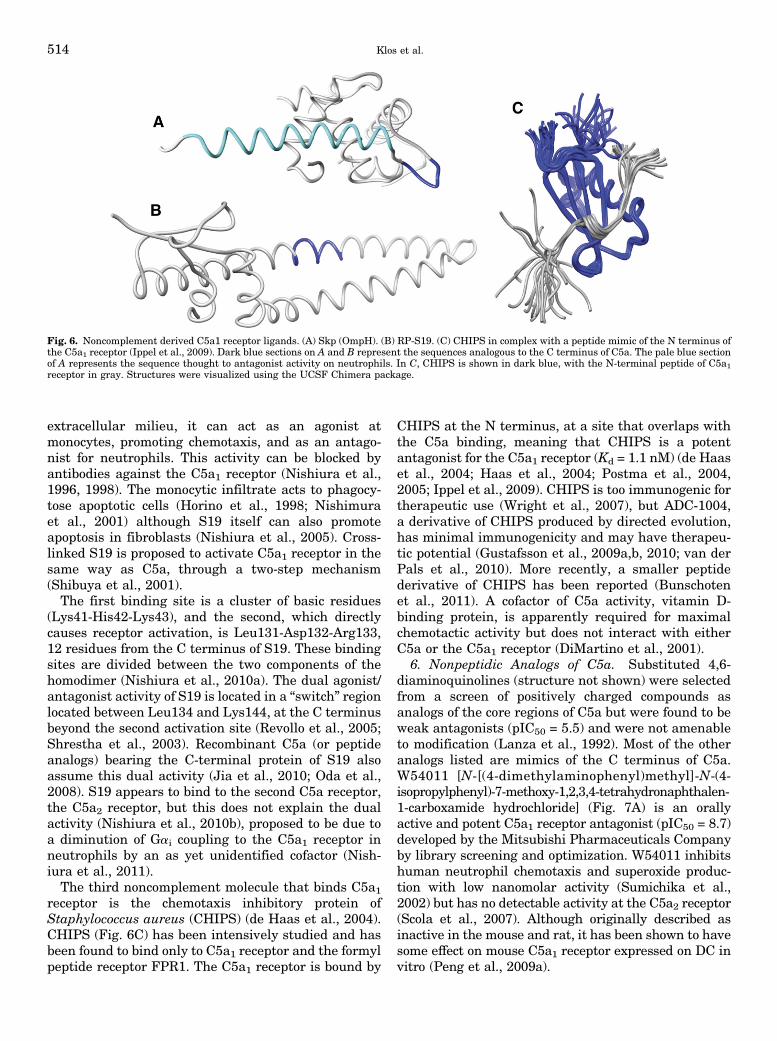
extracellular milieu, it can act as an agonist atmonocytes, promoting chemotaxis, and as an antago-nist for neutrophils. This activity can be blocked byantibodies against the C5a1 receptor (Nishiura et al.,1996, 1998). The monocytic infiltrate acts to phagocy-tose apoptotic cells (Horino et al., 1998; Nishimuraet al., 2001) although S19 itself can also promoteapoptosis in fibroblasts (Nishiura et al., 2005). Cross-linked S19 is proposed to activate C5a1 receptor in thesame way as C5a, through a two-step mechanism(Shibuya et al., 2001).The first binding site is a cluster of basic residues
(Lys41-His42-Lys43), and the second, which directlycauses receptor activation, is Leu131-Asp132-Arg133,12 residues from the C terminus of S19. These bindingsites are divided between the two components of thehomodimer (Nishiura et al., 2010a). The dual agonist/antagonist activity of S19 is located in a “switch” regionlocated between Leu134 and Lys144, at the C terminusbeyond the second activation site (Revollo et al., 2005;Shrestha et al., 2003). Recombinant C5a (or peptideanalogs) bearing the C-terminal protein of S19 alsoassume this dual activity (Jia et al., 2010; Oda et al.,2008). S19 appears to bind to the second C5a receptor,the C5a2 receptor, but this does not explain the dualactivity (Nishiura et al., 2010b), proposed to be due toa diminution of Gai coupling to the C5a1 receptor inneutrophils by an as yet unidentified cofactor (Nish-iura et al., 2011).The third noncomplement molecule that binds C5a1
receptor is the chemotaxis inhibitory protein ofStaphylococcus aureus (CHIPS) (de Haas et al., 2004).CHIPS (Fig. 6C) has been intensively studied and hasbeen found to bind only to C5a1 receptor and the formylpeptide receptor FPR1. The C5a1 receptor is bound by
CHIPS at the N terminus, at a site that overlaps withthe C5a binding, meaning that CHIPS is a potentantagonist for the C5a1 receptor (Kd = 1.1 nM) (de Haaset al., 2004; Haas et al., 2004; Postma et al., 2004,2005; Ippel et al., 2009). CHIPS is too immunogenic fortherapeutic use (Wright et al., 2007), but ADC-1004,a derivative of CHIPS produced by directed evolution,has minimal immunogenicity and may have therapeu-tic potential (Gustafsson et al., 2009a,b, 2010; van derPals et al., 2010). More recently, a smaller peptidederivative of CHIPS has been reported (Bunschotenet al., 2011). A cofactor of C5a activity, vitamin D-binding protein, is apparently required for maximalchemotactic activity but does not interact with eitherC5a or the C5a1 receptor (DiMartino et al., 2001).
6. Nonpeptidic Analogs of C5a. Substituted 4,6-diaminoquinolines (structure not shown) were selectedfrom a screen of positively charged compounds asanalogs of the core regions of C5a but were found to beweak antagonists (pIC50 = 5.5) and were not amenableto modification (Lanza et al., 1992). Most of the otheranalogs listed are mimics of the C terminus of C5a.W54011 [N-[(4-dimethylaminophenyl)methyl]-N-(4-isopropylphenyl)-7-methoxy-1,2,3,4-tetrahydronaphthalen-1-carboxamide hydrochloride] (Fig. 7A) is an orallyactive and potent C5a1 receptor antagonist (pIC50 = 8.7)developed by the Mitsubishi Pharmaceuticals Companyby library screening and optimization. W54011 inhibitshuman neutrophil chemotaxis and superoxide produc-tion with low nanomolar activity (Sumichika et al.,2002) but has no detectable activity at the C5a2 receptor(Scola et al., 2007). Although originally described asinactive in the mouse and rat, it has been shown to havesome effect on mouse C5a1 receptor expressed on DC invitro (Peng et al., 2009a).
Fig. 6. Noncomplement derived C5a1 receptor ligands. (A) Skp (OmpH). (B) RP-S19. (C) CHIPS in complex with a peptide mimic of the N terminus ofthe C5a1 receptor (Ippel et al., 2009). Dark blue sections on A and B represent the sequences analogous to the C terminus of C5a. The pale blue sectionof A represents the sequence thought to antagonist activity on neutrophils. In C, CHIPS is shown in dark blue, with the N-terminal peptide of C5a1receptor in gray. Structures were visualized using the UCSF Chimera package.
514 Klos et al.
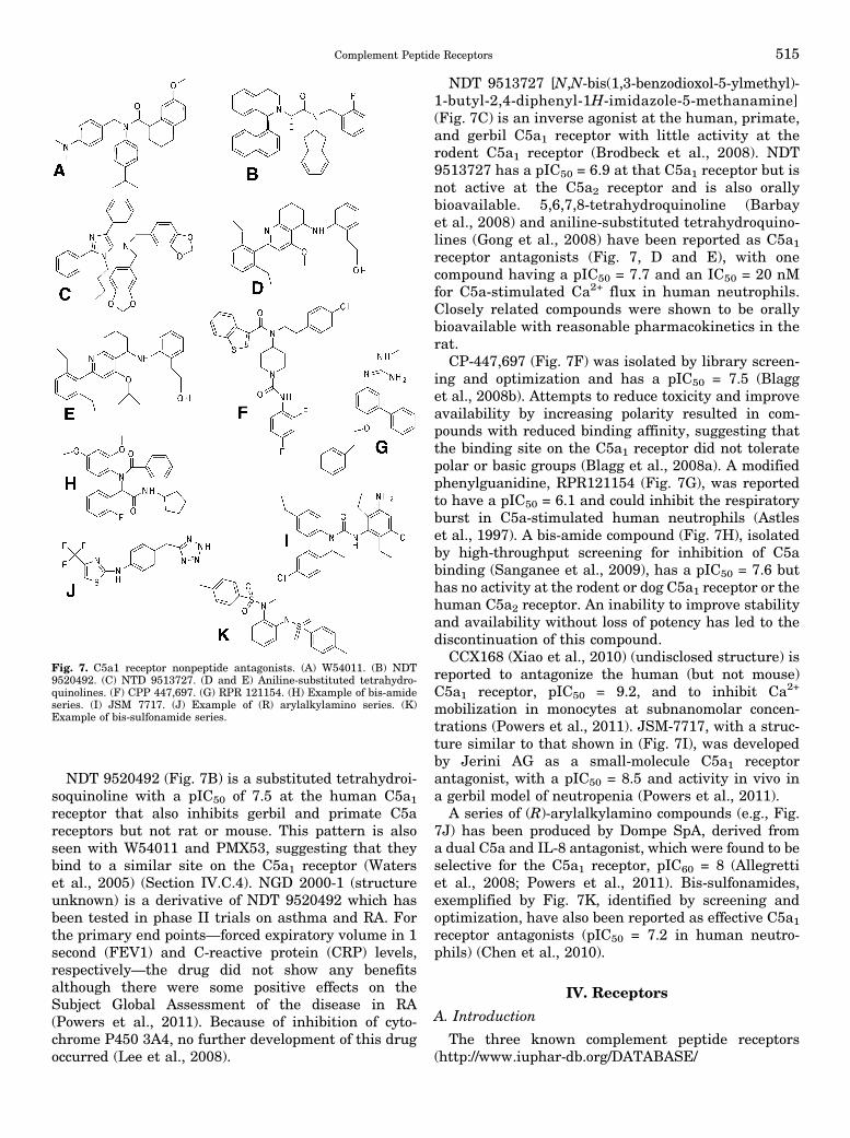
NDT 9520492 (Fig. 7B) is a substituted tetrahydroi-soquinoline with a pIC50 of 7.5 at the human C5a1receptor that also inhibits gerbil and primate C5areceptors but not rat or mouse. This pattern is alsoseen with W54011 and PMX53, suggesting that theybind to a similar site on the C5a1 receptor (Waterset al., 2005) (Section IV.C.4). NGD 2000-1 (structureunknown) is a derivative of NDT 9520492 which hasbeen tested in phase II trials on asthma and RA. Forthe primary end points—forced expiratory volume in 1second (FEV1) and C-reactive protein (CRP) levels,respectively—the drug did not show any benefitsalthough there were some positive effects on theSubject Global Assessment of the disease in RA(Powers et al., 2011). Because of inhibition of cyto-chrome P450 3A4, no further development of this drugoccurred (Lee et al., 2008).
NDT 9513727 [N,N-bis(1,3-benzodioxol-5-ylmethyl)-1-butyl-2,4-diphenyl-1H-imidazole-5-methanamine](Fig. 7C) is an inverse agonist at the human, primate,and gerbil C5a1 receptor with little activity at therodent C5a1 receptor (Brodbeck et al., 2008). NDT9513727 has a pIC50 = 6.9 at that C5a1 receptor but isnot active at the C5a2 receptor and is also orallybioavailable. 5,6,7,8-tetrahydroquinoline (Barbayet al., 2008) and aniline-substituted tetrahydroquino-lines (Gong et al., 2008) have been reported as C5a1receptor antagonists (Fig. 7, D and E), with onecompound having a pIC50 = 7.7 and an IC50 = 20 nMfor C5a-stimulated Ca2+ flux in human neutrophils.Closely related compounds were shown to be orallybioavailable with reasonable pharmacokinetics in therat.
CP-447,697 (Fig. 7F) was isolated by library screen-ing and optimization and has a pIC50 = 7.5 (Blagget al., 2008b). Attempts to reduce toxicity and improveavailability by increasing polarity resulted in com-pounds with reduced binding affinity, suggesting thatthe binding site on the C5a1 receptor did not toleratepolar or basic groups (Blagg et al., 2008a). A modifiedphenylguanidine, RPR121154 (Fig. 7G), was reportedto have a pIC50 = 6.1 and could inhibit the respiratoryburst in C5a-stimulated human neutrophils (Astleset al., 1997). A bis-amide compound (Fig. 7H), isolatedby high-throughput screening for inhibition of C5abinding (Sanganee et al., 2009), has a pIC50 = 7.6 buthas no activity at the rodent or dog C5a1 receptor or thehuman C5a2 receptor. An inability to improve stabilityand availability without loss of potency has led to thediscontinuation of this compound.
CCX168 (Xiao et al., 2010) (undisclosed structure) isreported to antagonize the human (but not mouse)C5a1 receptor, pIC50 = 9.2, and to inhibit Ca2+
mobilization in monocytes at subnanomolar concen-trations (Powers et al., 2011). JSM-7717, with a struc-ture similar to that shown in (Fig. 7I), was developedby Jerini AG as a small-molecule C5a1 receptorantagonist, with a pIC50 = 8.5 and activity in vivo ina gerbil model of neutropenia (Powers et al., 2011).
A series of (R)-arylalkylamino compounds (e.g., Fig.7J) has been produced by Dompe SpA, derived froma dual C5a and IL-8 antagonist, which were found to beselective for the C5a1 receptor, pIC60 = 8 (Allegrettiet al., 2008; Powers et al., 2011). Bis-sulfonamides,exemplified by Fig. 7K, identified by screening andoptimization, have also been reported as effective C5a1receptor antagonists (pIC50 = 7.2 in human neutro-phils) (Chen et al., 2010).
IV. Receptors
A. Introduction
The three known complement peptide receptors(http://www.iuphar-db.org/DATABASE/
Fig. 7. C5a1 receptor nonpeptide antagonists. (A) W54011. (B) NDT9520492. (C) NTD 9513727. (D and E) Aniline-substituted tetrahydro-quinolines. (F) CPP 447,697. (G) RPR 121154. (H) Example of bis-amideseries. (I) JSM 7717. (J) Example of (R) arylalkylamino series. (K)Example of bis-sulfonamide series.
Complement Peptide Receptors 515
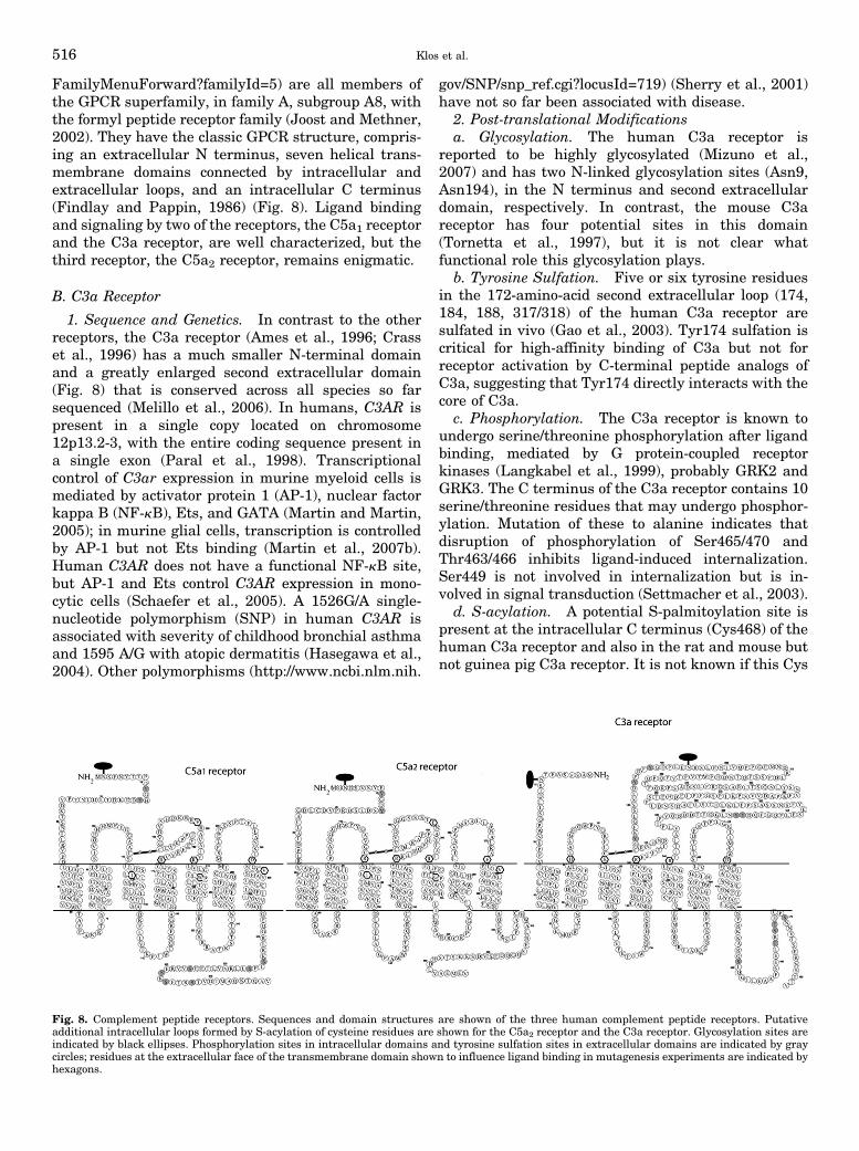
FamilyMenuForward?familyId=5) are all members ofthe GPCR superfamily, in family A, subgroup A8, withthe formyl peptide receptor family (Joost and Methner,2002). They have the classic GPCR structure, compris-ing an extracellular N terminus, seven helical trans-membrane domains connected by intracellular andextracellular loops, and an intracellular C terminus(Findlay and Pappin, 1986) (Fig. 8). Ligand bindingand signaling by two of the receptors, the C5a1 receptorand the C3a receptor, are well characterized, but thethird receptor, the C5a2 receptor, remains enigmatic.
B. C3a Receptor
1. Sequence and Genetics. In contrast to the otherreceptors, the C3a receptor (Ames et al., 1996; Crasset al., 1996) has a much smaller N-terminal domainand a greatly enlarged second extracellular domain(Fig. 8) that is conserved across all species so farsequenced (Melillo et al., 2006). In humans, C3AR ispresent in a single copy located on chromosome12p13.2-3, with the entire coding sequence present ina single exon (Paral et al., 1998). Transcriptionalcontrol of C3ar expression in murine myeloid cells ismediated by activator protein 1 (AP-1), nuclear factorkappa B (NF-kB), Ets, and GATA (Martin and Martin,2005); in murine glial cells, transcription is controlledby AP-1 but not Ets binding (Martin et al., 2007b).Human C3AR does not have a functional NF-kB site,but AP-1 and Ets control C3AR expression in mono-cytic cells (Schaefer et al., 2005). A 1526G/A single-nucleotide polymorphism (SNP) in human C3AR isassociated with severity of childhood bronchial asthmaand 1595 A/G with atopic dermatitis (Hasegawa et al.,2004). Other polymorphisms (http://www.ncbi.nlm.nih.
gov/SNP/snp_ref.cgi?locusId=719) (Sherry et al., 2001)have not so far been associated with disease.
2. Post-translational Modificationsa. Glycosylation. The human C3a receptor is
reported to be highly glycosylated (Mizuno et al.,2007) and has two N-linked glycosylation sites (Asn9,Asn194), in the N terminus and second extracellulardomain, respectively. In contrast, the mouse C3areceptor has four potential sites in this domain(Tornetta et al., 1997), but it is not clear whatfunctional role this glycosylation plays.
b. Tyrosine Sulfation. Five or six tyrosine residuesin the 172-amino-acid second extracellular loop (174,184, 188, 317/318) of the human C3a receptor aresulfated in vivo (Gao et al., 2003). Tyr174 sulfation iscritical for high-affinity binding of C3a but not forreceptor activation by C-terminal peptide analogs ofC3a, suggesting that Tyr174 directly interacts with thecore of C3a.
c. Phosphorylation. The C3a receptor is known toundergo serine/threonine phosphorylation after ligandbinding, mediated by G protein-coupled receptorkinases (Langkabel et al., 1999), probably GRK2 andGRK3. The C terminus of the C3a receptor contains 10serine/threonine residues that may undergo phosphor-ylation. Mutation of these to alanine indicates thatdisruption of phosphorylation of Ser465/470 andThr463/466 inhibits ligand-induced internalization.Ser449 is not involved in internalization but is in-volved in signal transduction (Settmacher et al., 2003).
d. S-acylation. A potential S-palmitoylation site ispresent at the intracellular C terminus (Cys468) of thehuman C3a receptor and also in the rat and mouse butnot guinea pig C3a receptor. It is not known if this Cys
Fig. 8. Complement peptide receptors. Sequences and domain structures are shown of the three human complement peptide receptors. Putativeadditional intracellular loops formed by S-acylation of cysteine residues are shown for the C5a2 receptor and the C3a receptor. Glycosylation sites areindicated by black ellipses. Phosphorylation sites in intracellular domains and tyrosine sulfation sites in extracellular domains are indicated by graycircles; residues at the extracellular face of the transmembrane domain shown to influence ligand binding in mutagenesis experiments are indicated byhexagons.
516 Klos et al.
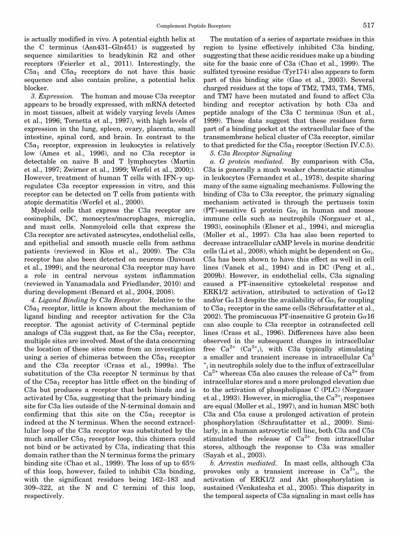
is actually modified in vivo. A potential eighth helix atthe C terminus (Asn431–Gln451) is suggested bysequence similarities to bradykinin R2 and otherreceptors (Feierler et al., 2011). Interestingly, theC5a1 and C5a2 receptors do not have this basicsequence and also contain proline, a potential helixblocker.3. Expression. The human and mouse C3a receptor
appears to be broadly expressed, with mRNA detectedin most tissues, albeit at widely varying levels (Ameset al., 1996; Tornetta et al., 1997), with high levels ofexpression in the lung, spleen, ovary, placenta, smallintestine, spinal cord, and brain. In contrast to theC5a1 receptor, expression in leukocytes is relativelylow (Ames et al., 1996), and no C3a receptor isdetectable on naive B and T lymphocytes (Martinet al., 1997; Zwirner et al., 1999; Werfel et al., 2000;).However, treatment of human T cells with IFN-g up-regulates C3a receptor expression in vitro, and thisreceptor can be detected on T cells from patients withatopic dermatitis (Werfel et al., 2000).Myeloid cells that express the C3a receptor are
eosinophils, DC, monocytes/macrophages, microglia,and mast cells. Nonmyeloid cells that express theC3a receptor are activated astrocytes, endothelial cells,and epithelial and smooth muscle cells from asthmapatients (reviewed in Klos et al., 2009). The C3areceptor has also been detected on neurons (Davoustet al., 1999), and the neuronal C3a receptor may havea role in central nervous system inflammation(reviewed in Yanamadala and Friedlander, 2010) andduring development (Benard et al., 2004, 2008).4. Ligand Binding by C3a Receptor. Relative to the
C5a1 receptor, little is known about the mechanism ofligand binding and receptor activation for the C3areceptor. The agonist activity of C-terminal peptideanalogs of C3a suggest that, as for the C5a1 receptor,multiple sites are involved. Most of the data concerningthe location of these sites come from an investigationusing a series of chimeras between the C5a1 receptorand the C3a receptor (Crass et al., 1999a). Thesubstitution of the C3a receptor N terminus by thatof the C5a1 receptor has little effect on the binding ofC3a but produces a receptor that both binds and isactivated by C5a, suggesting that the primary bindingsite for C3a lies outside of the N-terminal domain andconfirming that this site on the C5a1 receptor isindeed at the N terminus. When the second extracel-lular loop of the C3a receptor was substituted by themuch smaller C5a1 receptor loop, this chimera couldnot bind or be activated by C3a, indicating that thisdomain rather than the N terminus forms the primarybinding site (Chao et al., 1999). The loss of up to 65%of this loop, however, failed to inhibit C3a binding,with the significant residues being 162–183 and309–322, at the N and C termini of this loop,respectively.
The mutation of a series of aspartate residues in thisregion to lysine effectively inhibited C3a binding,suggesting that these acidic residues make up a bindingsite for the basic core of C3a (Chao et al., 1999). Thesulfated tyrosine residue (Tyr174) also appears to formpart of this binding site (Gao et al., 2003). Severalcharged residues at the tops of TM2, TM3, TM4, TM5,and TM7 have been mutated and found to affect C3abinding and receptor activation by both C3a andpeptide analogs of the C3a C terminus (Sun et al.,1999). These data suggest that these residues formpart of a binding pocket at the extracellular face of thetransmembrane helical cluster of C3a receptor, similarto that predicted for the C5a1 receptor (Section IV.C.5).
5. C3a Receptor Signalinga. G protein mediated. By comparison with C5a,
C3a is generally a much weaker chemotactic stimulusin leukocytes (Fernandez et al., 1978), despite sharingmany of the same signaling mechanisms. Following thebinding of C3a to C3a receptor, the primary signalingmechanism activated is through the pertussis toxin(PT)-sensitive G protein Gai in human and mouseimmune cells such as neutrophils (Norgauer et al.,1993), eosinophils (Elsner et al., 1994), and microglia(Moller et al., 1997). C3a has also been reported todecrease intracellular cAMP levels in murine dendriticcells (Li et al., 2008), which might be dependent on Gai.C5a has been shown to have this effect as well in celllines (Vanek et al., 1994) and in DC (Peng et al.,2009b). However, in endothelial cells, C3a signalingcaused a PT-insensitive cytoskeletal response andERK1/2 activation, attributed to activation of Ga12and/or Ga13 despite the availability of Gai for couplingto C5a1 receptor in the same cells (Schraufstatter et al.,2002). The promiscuous PT-insensitive G protein Ga16can also couple to C3a receptor in cotransfected celllines (Crass et al., 1996). Differences have also beenobserved in the subsequent changes in intracellularfree Ca2+ (Ca2+i), with C3a typically stimulatinga smaller and transient increase in intracellular Ca2+i in neutrophils solely due to the influx of extracellularCa2+ whereas C5a also causes the release of Ca2+ fromintracellular stores and a more prolonged elevation dueto the activation of phospholipase C (PLC) (Norgaueret al., 1993). However, in microglia, the Ca2+i responsesare equal (Moller et al., 1997), and in human MSC bothC3a and C5a cause a prolonged activation of proteinphosphorylation (Schraufstatter et al., 2009). Simi-larly, in a human astrocytic cell line, both C3a and C5astimulated the release of Ca2+ from intracellularstores, although the response to C3a was smaller(Sayah et al., 2003).
b. Arrestin mediated. In mast cells, although C3aprovokes only a transient increase in Ca2+i, theactivation of ERK1/2 and Akt phosphorylation issustained (Venkatesha et al., 2005). This disparity inthe temporal aspects of C3a signaling in mast cells has
Complement Peptide Receptors 517
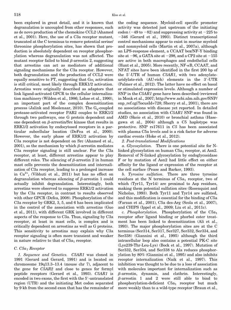
been explored in great detail, and it is known thatdegranulation is uncoupled from other responses, suchas de novo production of the chemokine CCL2 (Ahamedet al., 2001). Here, the use of a C3a receptor mutant,truncated at the C terminus to remove potential serine/threonine phosphorylation sites, has shown that pro-duction is absolutely dependent on receptor phosphor-ylation whereas degranulation was not affected. Themutant receptor failed to bind b-arrestin 2, suggestingthat arrestins can act as mediators of additionalsignaling mechanisms for the C3a receptor. However,both degranulation and the production of CCL2 wereequally sensitive to PT, suggesting that Gai activationis still critical, most likely through ERK1/2 activation.Arrestins were originally described as adaptors thatlink ligand-activated GPCR to the cellular internaliza-tion machinery (Wilden et al., 1986; Lohse et al., 1990),an important part of the complex desensitizationprocess (Jalink and Moolenaar, 2010). The Gq-coupledprotease-activated receptor PAR2 couples to ERK1/2through two pathways, one G protein dependent andone dependent on b-arrestin/Src kinase that results inERK1/2 activation by endocytosed receptors in a par-ticular subcellular location (DeFea et al., 2000).However, the early phase of ERK1/2 activation byC3a receptor is not dependent on Src (Ahamed et al.,2001), so the mechanism by which b-arrestin mediatesC3a receptor signaling is still unclear. For the C3areceptor, at least, different arrestins appear to playdifferent roles. The silencing of b-arrestin 2 in humanmast cells prevents the desensitization and internali-zation of C3a receptor, leading to a prolonged increasein Ca2+i (Vibhuti et al., 2011) but has no effect ondegranulation whereas silencing of b-arrestin 1 couldactually inhibit degranulation. Interestingly, botharrestins were observed to suppress ERK1/2 activationby the C3a receptor, in contrast to results observedwith other GPCR (Defea, 2008). Phosphorylation of theC3a receptor by GRK2, 3, 5, and 6 has been implicatedin the control of the association with arrestins (Guoet al., 2011), with different GRK involved in differentaspects of the response to C3a. Thus, signaling by C3areceptor, at least in mast cells, is complex and iscritically dependent on arrestins as well as G proteins.This sensitivity to arrestins may explain why C3areceptor signaling is often more transient and weakerin nature relative to that of C5a1 receptor.
C. C5a1 Receptor
1. Sequence and Genetics. C5AR1 was cloned in1991 (Gerard and Gerard, 1991) and is located onchromosome 19q13.3–13.4 (mouse chr 7), adjacent tothe gene for C5AR2 and close to genes for formylpeptide receptors (Gerard et al., 1993). C5AR1 isencoded in two exons, the first with the 59-untranslatedregion (UTR) and the initiating Met codon separatedby 9 kb from the second exon that has the remainder of
the coding sequence. Myeloid-cell specific promoteractivity was detected just upstream of the initiatingcodon (249 to282) and suppressing activity at2225 to2346 (Gerard et al., 1993). Distinct transcriptionalcontrol mechanisms appear to exist in murine myeloidand nonmyeloid cells (Martin et al., 2007a), althoughan LPS-response element, a CCAAT box/NF-Y bindingsite at296, a GATA site at2298, and a CP2 site at2155are active in both macrophages and endothelial cells(Hunt et al., 2005). More recently, NF-kB, CCAAT, andNFAT sites have been identified in the first 200 bp ofthe 59-UTR of human C5AR1, with two adenylate-uridylate-rich (AU-rich) elements in the 39-UTR(Palmer et al., 2012). The latter have no effect on basalor stimulated expression levels. Although a number ofSNP in the C5AR1 gene have been described (reviewedin Monk et al., 2007; http://www.ncbi.nlm.nih.gov/SNP/snp_ref.cgi?locusId=728; Sherry et al., 2001), there areno associations with disease yet reported. In detailedstudies, no association with C5AR1 SNP was found inAMD (Skeie et al., 2010) or bronchial asthma (Hase-gawa et al., 2004) although a C5 haplotype wasprotective. SNP rs17611 in C5 has been associatedwith plasma C5a levels and is a risk factor for adversecardiac events (Hoke et al., 2012).
2. Post-translational Modificationsa. Glycosylation. There is one potential site for N-
linked glycosylation on human C5a1 receptor, at Asn2.Removal of N-linked glycosylation by endoglycosidaseF or by mutation of Asn2 had little effect on eitheraffinity for the ligand or expression of the receptor atthe cell surface (Pease and Barker, 1993).
b. Tyrosine sulfation. There are three tyrosineresidues at the N terminus of C5a1 receptor, two ofwhich (Tyr11, Tyr14) are proximal to Asp residues,making them potential sulfation sites (Rosenquist andNicholas, 1993). Both Tyr11 and Tyr14 are sulfated,and this modification is essential for the binding of C5a(Farzan et al., 2001), C5a des-Arg (Scola et al., 2007),and CHIPS (Ippel et al., 2009; Liu et al., 2011c).
c. Phosphorylation. Phosphorylation of the C5a1receptor after ligand binding or phorbol ester treat-ment has been linked to desensitization (Ali et al.,1993). The major phosphorylation sites are at the Cterminus (Ser314, Ser317, Ser327, Ser332, Ser334, andSer338) (Giannini et al., 1995) although the thirdintracellular loop also contains a potential PK-C site(Lys239-Thr-Leu-Lys) (Bock et al., 1997). Mutation ofSer332, Ser334, and Ser338 to Ala reduces phosphor-ylation by 80% (Giannini et al., 1995) and also inhibitsreceptor internalization (Naik et al., 1997). Thisinhibition was thought to be due to a loss of associationwith molecules important for internalization such asb-arrestin, dynamin, and clathrin. Interestingly,b-arrestin 1 and 2 were still able to bind tophosphorylation-deficient C5a1 receptor but muchmore weakly than to a wild-type receptor (Braun et al.,
518 Klos et al.
2003). However, internalization of the receptor, onemechanism for desensitization, was found to be de-pendent only on amino acids 335–350 in a series oftruncated C5a1 receptor mutants, beyond the majorityof the phosphorylation sites (Bock et al., 1997).Similarly, another study found that although Ser334,Ser327, Ser332, and Ser338 could be phosphorylatedby PK-Cb, these sites were functionally redundant forinternalization and desensitization and even that, athigh concentrations of C5a, b-arrestin binding to the Cterminus could actually inhibit phosphorylation(Pollok-Kopp et al., 2007). Thus, the role of phosphor-ylation in the control of receptor function remainsunclear.3. Expression. Although typically expressed at high
levels on cells of myeloid origin, the C5a1 receptor isexpressed at low-to-moderate levels on a wide varietyof cell types (reviewed in Monk et al., 2007). Expressionon lymphocytes remains controversial, with somereports showing expression on inactivated B andT cells with others failing to find this or to showexpression restricted to small subsets (reviewed inKlos et al., 2009). Reports of expression in epithelialcells have been shown to be probably artifactual (Kloset al., 2009).4. Ligand Binding by C5a1 Receptor.a. C5a1 receptor binds both C5a and C5a des-Arg.
Determining the locations and mechanisms of interac-tion between C5a1 receptor and its ligands is vital tothe production of effective therapeutics. As for mostmembers of the GPCR family, there is no availablecrystal structure for the C5a1 receptor, so structuralknowledge of the receptor has been based on extensiveligand binding, receptor chimera, and mutagenesisstudies. Molecular modeling of the receptor now playsa major role in our understanding, with a recent modelof a ligand-bound C5a1 receptor providing novelinsights into the mechanisms of ligand and receptorinteraction (Nikiforovich et al., 2008). The mostextensively studied interaction to date is between thereceptor and the full-length natural ligand C5a. Invivo, C5a is rapidly converted into C5a des-Arg bycarboxypeptidase enzymes, which remove the terminalarginine residue (Bokisch and Muller-Eberhard, 1970).Experimentally, C5a binds the C5a1 receptor with a Kd
of 1 nM, whereas the truncated form has a bindingaffinity which is 10- to 100-fold lower than that of C5aand is a partial agonist at the C5a1 receptor (Higgin-bottom et al., 2005). Interactions with this truncatedform will also be considered here, where data areavailable.5. Two-Site Binding Paradigm. It is now widely
accepted that there are at least two sites of interactionbetween the C5a and C5a1 receptors, a bindingparadigm common to members of the GPCR familythat bind large macromolecular ligands (Kristiansen,2004). The first interaction is between acidic residues
in the N terminus of the C5a1 receptor and basicresidues in the core of C5a; a second binding in-teraction is thought to occur between the C terminus ofC5a and the transmembrane domains and chargedresidues at the base of the C5a1 receptor extracellularloops. This binding model mainly stems from muta-genesis studies, the creation of chimeric receptors, andinteractions with C5a analogs. Initially, experimentsshowing the C-terminal octapeptide of C5a could alonebind C5a1 receptor led to the proposal of a binding sitein the C terminus of C5a (Kawai et al., 1991) (SectionIII.C.4). It was also shown that C-terminal peptideanalogs could act as full agonists of the C5a1 receptor(Ember et al., 1992), leading to the hypothesis that notonly does the C terminus of C5a possess a receptor-binding site, but it is also critical for activation of thereceptor. However, while removing the terminal pen-tapeptide of C5a reduced receptor activity, the bindingof ligand to the receptor was unaltered, indicatingthe existence of a second binding site. This site wasidentified by the demonstration that antibodiesdirected against the N-terminal domain of the C5a1receptor reduced binding and activation in responseto C5a (Oppermann et al., 1993). In a similar way,deletion of the first 22 residues of the receptordramatically reduced binding of intact C5a, but themutated receptor could still be activated by peptideanalogs of the C5a C terminus, thus confirming thepresence of two distinct binding sites (DeMartinoet al., 1994). Critical residues and sequences withinthese general binding sites have been more challeng-ing to identify, as presented here for each bindingsite.
a. Binding site one: C5a1 receptor N terminus.Yeast random saturation mutagenesis (RSM) andNMR studies on the N terminus of the C5a1 receptorhave indicated the importance of the many acidicresidues in C5a binding (Chen et al., 1998; Hagemannet al., 2006). In general, single/double mutations ofthese have had little effect on binding whereas variousmultiple mutations within the N terminus of the C5a1receptor have significantly reduced the affinity of theC5a1 receptor for C5a. For example, the C5a1 receptor(D15,16,18,21N) reduced C5a affinity 40-fold, and theC5a1 receptor(D10,15,16,18,21N) reduced affinity by133-fold (DeMartino et al., 1994), whereas the C5a1receptor(D10N), C5a1 receptor(D27N), and C5a1 re-ceptor(D21,27N) had no effect on C5a binding (Meryand Boulay, 1994). The lack of importance of individualresidues in forming the binding site was supported bythe yeast RSM study, which concluded that no singleresidues were essential for ligand activation of theC5a1 receptor (Hagemann et al., 2006). More recently,site-specific disulfide-trapping experiments performedin yeast have identified several potential specificpoints of contact between C5a and the N terminus ofthe C5a1 receptor (Hagemann et al., 2008).
Complement Peptide Receptors 519
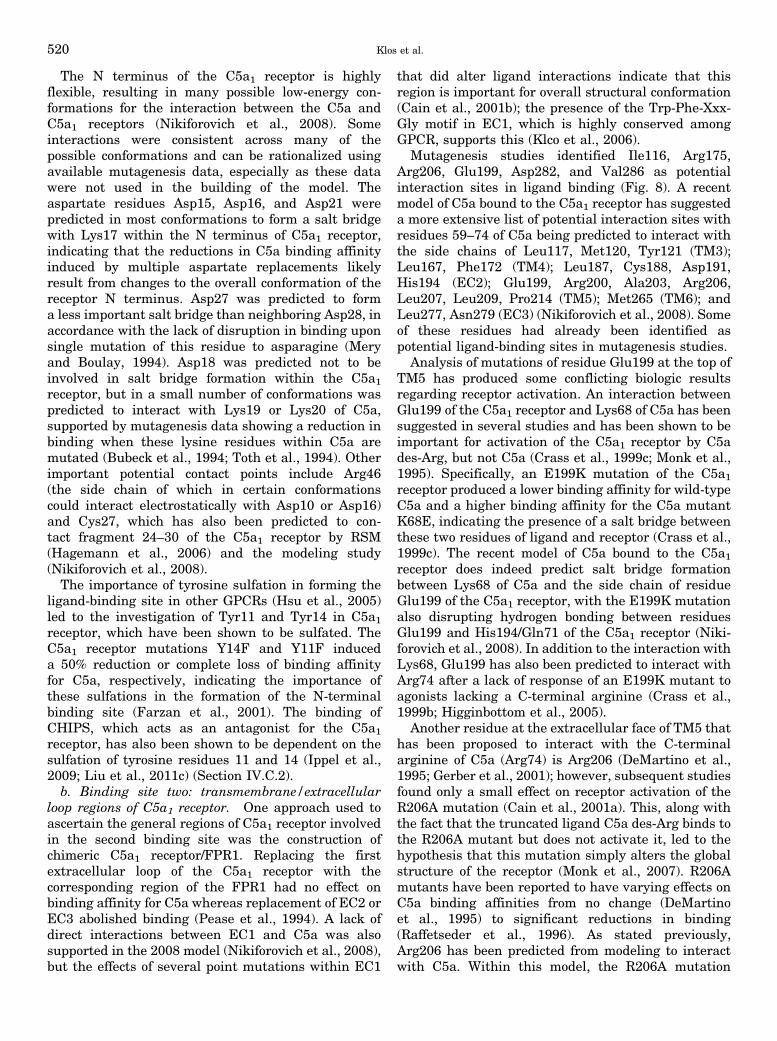
The N terminus of the C5a1 receptor is highlyflexible, resulting in many possible low-energy con-formations for the interaction between the C5a andC5a1 receptors (Nikiforovich et al., 2008). Someinteractions were consistent across many of thepossible conformations and can be rationalized usingavailable mutagenesis data, especially as these datawere not used in the building of the model. Theaspartate residues Asp15, Asp16, and Asp21 werepredicted in most conformations to form a salt bridgewith Lys17 within the N terminus of C5a1 receptor,indicating that the reductions in C5a binding affinityinduced by multiple aspartate replacements likelyresult from changes to the overall conformation of thereceptor N terminus. Asp27 was predicted to forma less important salt bridge than neighboring Asp28, inaccordance with the lack of disruption in binding uponsingle mutation of this residue to asparagine (Meryand Boulay, 1994). Asp18 was predicted not to beinvolved in salt bridge formation within the C5a1receptor, but in a small number of conformations waspredicted to interact with Lys19 or Lys20 of C5a,supported by mutagenesis data showing a reduction inbinding when these lysine residues within C5a aremutated (Bubeck et al., 1994; Toth et al., 1994). Otherimportant potential contact points include Arg46(the side chain of which in certain conformationscould interact electrostatically with Asp10 or Asp16)and Cys27, which has also been predicted to con-tact fragment 24–30 of the C5a1 receptor by RSM(Hagemann et al., 2006) and the modeling study(Nikiforovich et al., 2008).The importance of tyrosine sulfation in forming the
ligand-binding site in other GPCRs (Hsu et al., 2005)led to the investigation of Tyr11 and Tyr14 in C5a1receptor, which have been shown to be sulfated. TheC5a1 receptor mutations Y14F and Y11F induceda 50% reduction or complete loss of binding affinityfor C5a, respectively, indicating the importance ofthese sulfations in the formation of the N-terminalbinding site (Farzan et al., 2001). The binding ofCHIPS, which acts as an antagonist for the C5a1receptor, has also been shown to be dependent on thesulfation of tyrosine residues 11 and 14 (Ippel et al.,2009; Liu et al., 2011c) (Section IV.C.2).b. Binding site two: transmembrane/extracellular
loop regions of C5a1 receptor. One approach used toascertain the general regions of C5a1 receptor involvedin the second binding site was the construction ofchimeric C5a1 receptor/FPR1. Replacing the firstextracellular loop of the C5a1 receptor with thecorresponding region of the FPR1 had no effect onbinding affinity for C5a whereas replacement of EC2 orEC3 abolished binding (Pease et al., 1994). A lack ofdirect interactions between EC1 and C5a was alsosupported in the 2008 model (Nikiforovich et al., 2008),but the effects of several point mutations within EC1
that did alter ligand interactions indicate that thisregion is important for overall structural conformation(Cain et al., 2001b); the presence of the Trp-Phe-Xxx-Gly motif in EC1, which is highly conserved amongGPCR, supports this (Klco et al., 2006).
Mutagenesis studies identified Ile116, Arg175,Arg206, Glu199, Asp282, and Val286 as potentialinteraction sites in ligand binding (Fig. 8). A recentmodel of C5a bound to the C5a1 receptor has suggesteda more extensive list of potential interaction sites withresidues 59–74 of C5a being predicted to interact withthe side chains of Leu117, Met120, Tyr121 (TM3);Leu167, Phe172 (TM4); Leu187, Cys188, Asp191,His194 (EC2); Glu199, Arg200, Ala203, Arg206,Leu207, Leu209, Pro214 (TM5); Met265 (TM6); andLeu277, Asn279 (EC3) (Nikiforovich et al., 2008). Someof these residues had already been identified aspotential ligand-binding sites in mutagenesis studies.
Analysis of mutations of residue Glu199 at the top ofTM5 has produced some conflicting biologic resultsregarding receptor activation. An interaction betweenGlu199 of the C5a1 receptor and Lys68 of C5a has beensuggested in several studies and has been shown to beimportant for activation of the C5a1 receptor by C5ades-Arg, but not C5a (Crass et al., 1999c; Monk et al.,1995). Specifically, an E199K mutation of the C5a1receptor produced a lower binding affinity for wild-typeC5a and a higher binding affinity for the C5a mutantK68E, indicating the presence of a salt bridge betweenthese two residues of ligand and receptor (Crass et al.,1999c). The recent model of C5a bound to the C5a1receptor does indeed predict salt bridge formationbetween Lys68 of C5a and the side chain of residueGlu199 of the C5a1 receptor, with the E199K mutationalso disrupting hydrogen bonding between residuesGlu199 and His194/Gln71 of the C5a1 receptor (Niki-forovich et al., 2008). In addition to the interaction withLys68, Glu199 has also been predicted to interact withArg74 after a lack of response of an E199K mutant toagonists lacking a C-terminal arginine (Crass et al.,1999b; Higginbottom et al., 2005).
Another residue at the extracellular face of TM5 thathas been proposed to interact with the C-terminalarginine of C5a (Arg74) is Arg206 (DeMartino et al.,1995; Gerber et al., 2001); however, subsequent studiesfound only a small effect on receptor activation of theR206A mutation (Cain et al., 2001a). This, along withthe fact that the truncated ligand C5a des-Arg binds tothe R206A mutant but does not activate it, led to thehypothesis that this mutation simply alters the globalstructure of the receptor (Monk et al., 2007). R206Amutants have been reported to have varying effects onC5a binding affinities from no change (DeMartinoet al., 1995) to significant reductions in binding(Raffetseder et al., 1996). As stated previously,Arg206 has been predicted from modeling to interactwith C5a. Within this model, the R206A mutation
520 Klos et al.
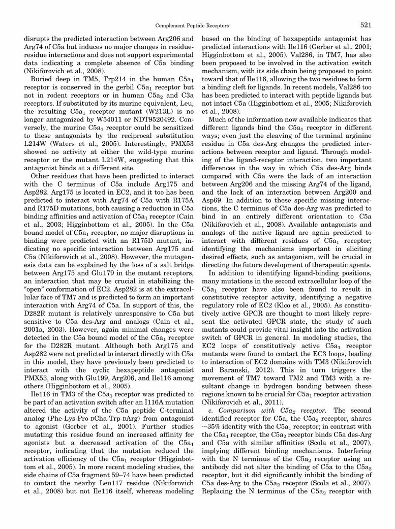
disrupts the predicted interaction between Arg206 andArg74 of C5a but induces no major changes in residue-residue interactions and does not support experimentaldata indicating a complete absence of C5a binding(Nikiforovich et al., 2008).Buried deep in TM5, Trp214 in the human C5a1
receptor is conserved in the gerbil C5a1 receptor butnot in rodent receptors or in human C5a2 and C3areceptors. If substituted by its murine equivalent, Leu,the resulting C5a1 receptor mutant (W213L) is nolonger antagonized by W54011 or NDT9520492. Con-versely, the murine C5a1 receptor could be sensitizedto these antagonists by the reciprocal substitutionL214W (Waters et al., 2005). Interestingly, PMX53showed no activity at either the wild-type murinereceptor or the mutant L214W, suggesting that thisantagonist binds at a different site.Other residues that have been predicted to interact
with the C terminus of C5a include Arg175 andAsp282. Arg175 is located in EC2, and it too has beenpredicted to interact with Arg74 of C5a with R175Aand R175D mutations, both causing a reduction in C5abinding affinities and activation of C5a1 receptor (Cainet al., 2003; Higginbottom et al., 2005). In the C5abound model of C5a1 receptor, no major disruptions inbinding were predicted with an R175D mutant, in-dicating no specific interaction between Arg175 andC5a (Nikiforovich et al., 2008). However, the mutagen-esis data can be explained by the loss of a salt bridgebetween Arg175 and Glu179 in the mutant receptors,an interaction that may be crucial in stabilizing the“open” conformation of EC2. Asp282 is at the extracel-lular face of TM7 and is predicted to form an importantinteraction with Arg74 of C5a. In support of this, theD282R mutant is relatively unresponsive to C5a butsensitive to C5a des-Arg and analogs (Cain et al.,2001a, 2003). However, again minimal changes weredetected in the C5a bound model of the C5a1 receptorfor the D282R mutant. Although both Arg175 andAsp282 were not predicted to interact directly with C5ain this model, they have previously been predicted tointeract with the cyclic hexapeptide antagonistPMX53, along with Glu199, Arg206, and Ile116 amongothers (Higginbottom et al., 2005).Ile116 in TM3 of the C5a1 receptor was predicted to
be part of an activation switch after an I116A mutationaltered the activity of the C5a peptide C-terminalanalog (Phe-Lys-Pro-DCha-Trp-DArg) from antagonistto agonist (Gerber et al., 2001). Further studiesmutating this residue found an increased affinity foragonists but a decreased activation of the C5a1receptor, indicating that the mutation reduced theactivation efficiency of the C5a1 receptor (Higginbot-tom et al., 2005). In more recent modeling studies, theside chains of C5a fragment 59–74 have been predictedto contact the nearby Leu117 residue (Nikiforovichet al., 2008) but not Ile116 itself, whereas modeling
based on the binding of hexapeptide antagonist haspredicted interactions with Ile116 (Gerber et al., 2001;Higginbottom et al., 2005). Val286, in TM7, has alsobeen proposed to be involved in the activation switchmechanism, with its side chain being proposed to pointtoward that of Ile116, allowing the two residues to forma binding cleft for ligands. In recent models, Val286 toohas been predicted to interact with peptide ligands butnot intact C5a (Higginbottom et al., 2005; Nikiforovichet al., 2008).
Much of the information now available indicates thatdifferent ligands bind the C5a1 receptor in differentways; even just the cleaving of the terminal arginineresidue in C5a des-Arg changes the predicted inter-actions between receptor and ligand. Through model-ing of the ligand-receptor interaction, two importantdifferences in the way in which C5a des-Arg bindscompared with C5a were the lack of an interactionbetween Arg206 and the missing Arg74 of the ligand,and the lack of an interaction between Arg200 andAsp69. In addition to these specific missing interac-tions, the C terminus of C5a des-Arg was predicted tobind in an entirely different orientation to C5a(Nikiforovich et al., 2008). Available antagonists andanalogs of the native ligand are again predicted tointeract with different residues of C5a1 receptor;identifying the mechanisms important in elicitingdesired effects, such as antagonism, will be crucial indirecting the future development of therapeutic agents.
In addition to identifying ligand-binding positions,many mutations in the second extracellular loop of theC5a1 receptor have also been found to result inconstitutive receptor activity, identifying a negativeregulatory role of EC2 (Klco et al., 2005). As constitu-tively active GPCR are thought to most likely repre-sent the activated GPCR state, the study of suchmutants could provide vital insight into the activationswitch of GPCR in general. In modeling studies, theEC2 loops of constitutively active C5a1 receptormutants were found to contact the EC3 loops, leadingto interaction of EC2 domains with TM3 (Nikiforovichand Baranski, 2012). This in turn triggers themovement of TM7 toward TM2 and TM3 with a re-sultant change in hydrogen bonding between theseregions known to be crucial for C5a1 receptor activation(Nikiforovich et al., 2011).
c. Comparison with C5a2 receptor. The secondidentified receptor for C5a, the C5a2 receptor, shares;35% identity with the C5a1 receptor; in contrast withthe C5a1 receptor, the C5a2 receptor binds C5a des-Argand C5a with similar affinities (Scola et al., 2007),implying different binding mechanisms. Interferingwith the N terminus of the C5a2 receptor using anantibody did not alter the binding of C5a to the C5a2receptor, but it did significantly inhibit the binding ofC5a des-Arg to the C5a2 receptor (Scola et al., 2007).Replacing the N terminus of the C5a2 receptor with
Complement Peptide Receptors 521
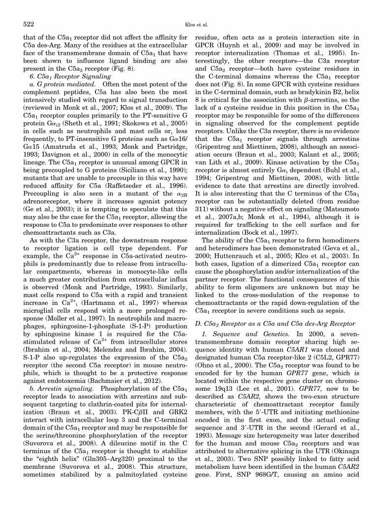
that of the C5a1 receptor did not affect the affinity forC5a des-Arg. Many of the residues at the extracellularface of the transmembrane domain of C5a1 that havebeen shown to influence ligand binding are alsopresent in the C5a2 receptor (Fig. 8).6. C5a1 Receptor Signalinga. G protein mediated. Often the most potent of the
complement peptides, C5a has also been the mostintensively studied with regard to signal transduction(reviewed in Monk et al., 2007; Klos et al., 2009). TheC5a1 receptor couples primarily to the PT-sensitive Gprotein Gai2 (Sheth et al., 1991; Skokowa et al., 2005)in cells such as neutrophils and mast cells or, lessfrequently, to PT-insensitive G proteins such as Ga16/Ga15 (Amatruda et al., 1993; Monk and Partridge,1993; Davignon et al., 2000) in cells of the monocyticlineage. The C5a1 receptor is unusual among GPCR inbeing precoupled to G proteins (Siciliano et al., 1990);mutants that are unable to precouple in this way havereduced affinity for C5a (Raffetseder et al., 1996).Precoupling is also seen in a mutant of the a2B
adrenoreceptor, where it increases agonist potency(Ge et al., 2003); it is tempting to speculate that thismay also be the case for the C5a1 receptor, allowing theresponse to C5a to predominate over responses to otherchemoattractants such as C3a.As with the C3a receptor, the downstream response
to receptor ligation is cell type dependent. Forexample, the Ca2+ response in C5a-activated neutro-phils is predominantly due to release from intracellu-lar compartments, whereas in monocyte-like cellsa much greater contribution from extracellular influxis observed (Monk and Partridge, 1993). Similarly,mast cells respond to C5a with a rapid and transientincrease in Ca2+i (Hartmann et al., 1997) whereasmicroglial cells respond with a more prolonged re-sponse (Moller et al., 1997). In neutrophils and macro-phages, sphingosine-1-phosphate (S-1-P) productionby sphingosine kinase 1 is required for the C5a-stimulated release of Ca2+ from intracellular stores(Ibrahim et al., 2004; Melendez and Ibrahim, 2004).S-1-P also up-regulates the expression of the C5a2receptor (the second C5a receptor) in mouse neutro-phils, which is thought to be a protective responseagainst endotoxemia (Bachmaier et al., 2012).b. Arrestin signaling. Phosphorylation of the C5a1
receptor leads to association with arrestins and sub-sequent targeting to clathrin-coated pits for internal-ization (Braun et al., 2003). PK-CbII and GRK2interact with intracellular loop 3 and the C-terminaldomain of the C5a1 receptor and may be responsible forthe serine/threonine phosphorylation of the receptor(Suvorova et al., 2008). A dileucine motif in the Cterminus of the C5a1 receptor is thought to stabilizethe “eighth helix” (Gln305–Arg320) proximal to themembrane (Suvorova et al., 2008). This structure,sometimes stabilized by a palmitoylated cysteine
residue, often acts as a protein interaction site inGPCR (Huynh et al., 2009) and may be involved inreceptor internalization (Thomas et al., 1995). In-terestingly, the other receptors—the C3a receptorand C5a2 receptor—both have cysteine residues inthe C-terminal domains whereas the C5a1 receptordoes not (Fig. 8). In some GPCR with cysteine residuesin the C-terminal domain, such as bradykinin B2, helix8 is critical for the association with b-arrestins, so thelack of a cysteine residue in this position in the C5a1receptor may be responsible for some of the differencesin signaling observed for the complement peptidereceptors. Unlike the C3a receptor, there is no evidencethat the C5a1 receptor signals through arrestins(Gripentrog and Miettinen, 2008), although an associ-ation occurs (Braun et al., 2003; Kalant et al., 2005;van Lith et al., 2009). Kinase activation by the C5a1receptor is almost entirely Gai dependent (Buhl et al.,1994; Gripentrog and Miettinen, 2008), with littleevidence to date that arrestins are directly involved.It is also interesting that the C terminus of the C5a1receptor can be substantially deleted (from residue311) without a negative effect on signaling (Matsumotoet al., 2007a,b; Monk et al., 1994), although it isrequired for trafficking to the cell surface and forinternalization (Bock et al., 1997).
The ability of the C5a1 receptor to form homodimersand heterodimers has been demonstrated (Geva et al.,2000; Huttenrauch et al., 2005; Klco et al., 2003). Inboth cases, ligation of a dimerized C5a1 receptor cancause the phosphorylation and/or internalization of thepartner receptor. The functional consequences of thisability to form oligomers are unknown but may belinked to the cross-modulation of the response tochemoattractants or the rapid down-regulation of theC5a1 receptor in severe conditions such as sepsis.
D. C5a2 Receptor as a C5a and C5a des-Arg Receptor
1. Sequence and Genetics. In 2000, a seven-transmembrane domain receptor sharing high se-quence identity with human C5AR1 was cloned anddesignated human C5a receptor-like 2 (C5L2, GPR77)(Ohno et al., 2000). The C5a2 receptor was found to beencoded for by the human GPR77 gene, which islocated within the respective gene cluster on chromo-some 19q13 (Lee et al., 2001). GPR77, now to bedescribed as C5AR2, shows the two-exon structurecharacteristic of chemoattractant receptor familymembers, with the 59-UTR and initiating methionineencoded in the first exon, and the actual codingsequence and 39-UTR in the second (Gerard et al.,1993). Message size heterogeneity was later describedfor the human and mouse C5a2 receptors and wasattributed to alternative splicing in the UTR (Okinagaet al., 2003). Two SNP possibly linked to fatty acidmetabolism have been identified in the human C5AR2gene. First, SNP 968G/T, causing an amino acid
522 Klos et al.
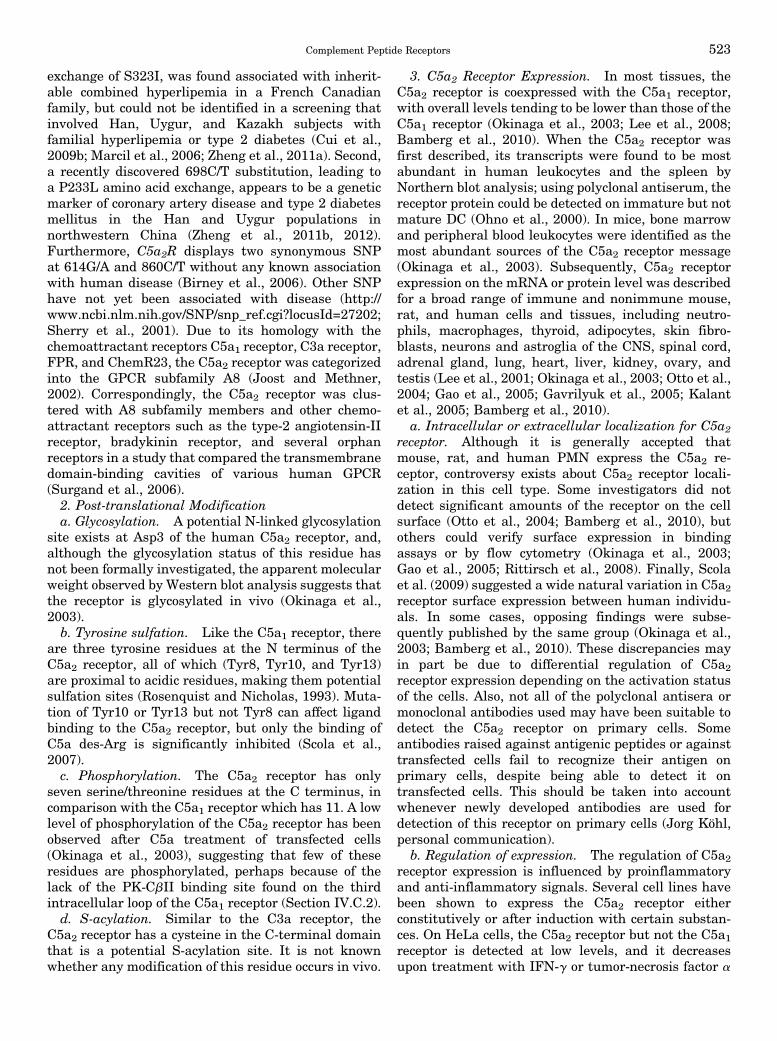
exchange of S323I, was found associated with inherit-able combined hyperlipemia in a French Canadianfamily, but could not be identified in a screening thatinvolved Han, Uygur, and Kazakh subjects withfamilial hyperlipemia or type 2 diabetes (Cui et al.,2009b; Marcil et al., 2006; Zheng et al., 2011a). Second,a recently discovered 698C/T substitution, leading toa P233L amino acid exchange, appears to be a geneticmarker of coronary artery disease and type 2 diabetesmellitus in the Han and Uygur populations innorthwestern China (Zheng et al., 2011b, 2012).Furthermore, C5a2R displays two synonymous SNPat 614G/A and 860C/T without any known associationwith human disease (Birney et al., 2006). Other SNPhave not yet been associated with disease (http://www.ncbi.nlm.nih.gov/SNP/snp_ref.cgi?locusId=27202;Sherry et al., 2001). Due to its homology with thechemoattractant receptors C5a1 receptor, C3a receptor,FPR, and ChemR23, the C5a2 receptor was categorizedinto the GPCR subfamily A8 (Joost and Methner,2002). Correspondingly, the C5a2 receptor was clus-tered with A8 subfamily members and other chemo-attractant receptors such as the type-2 angiotensin-IIreceptor, bradykinin receptor, and several orphanreceptors in a study that compared the transmembranedomain-binding cavities of various human GPCR(Surgand et al., 2006).2. Post-translational Modificationa. Glycosylation. A potential N-linked glycosylation
site exists at Asp3 of the human C5a2 receptor, and,although the glycosylation status of this residue hasnot been formally investigated, the apparent molecularweight observed by Western blot analysis suggests thatthe receptor is glycosylated in vivo (Okinaga et al.,2003).b. Tyrosine sulfation. Like the C5a1 receptor, there
are three tyrosine residues at the N terminus of theC5a2 receptor, all of which (Tyr8, Tyr10, and Tyr13)are proximal to acidic residues, making them potentialsulfation sites (Rosenquist and Nicholas, 1993). Muta-tion of Tyr10 or Tyr13 but not Tyr8 can affect ligandbinding to the C5a2 receptor, but only the binding ofC5a des-Arg is significantly inhibited (Scola et al.,2007).c. Phosphorylation. The C5a2 receptor has only
seven serine/threonine residues at the C terminus, incomparison with the C5a1 receptor which has 11. A lowlevel of phosphorylation of the C5a2 receptor has beenobserved after C5a treatment of transfected cells(Okinaga et al., 2003), suggesting that few of theseresidues are phosphorylated, perhaps because of thelack of the PK-CbII binding site found on the thirdintracellular loop of the C5a1 receptor (Section IV.C.2).d. S-acylation. Similar to the C3a receptor, the
C5a2 receptor has a cysteine in the C-terminal domainthat is a potential S-acylation site. It is not knownwhether any modification of this residue occurs in vivo.
3. C5a2 Receptor Expression. In most tissues, theC5a2 receptor is coexpressed with the C5a1 receptor,with overall levels tending to be lower than those of theC5a1 receptor (Okinaga et al., 2003; Lee et al., 2008;Bamberg et al., 2010). When the C5a2 receptor wasfirst described, its transcripts were found to be mostabundant in human leukocytes and the spleen byNorthern blot analysis; using polyclonal antiserum, thereceptor protein could be detected on immature but notmature DC (Ohno et al., 2000). In mice, bone marrowand peripheral blood leukocytes were identified as themost abundant sources of the C5a2 receptor message(Okinaga et al., 2003). Subsequently, C5a2 receptorexpression on the mRNA or protein level was describedfor a broad range of immune and nonimmune mouse,rat, and human cells and tissues, including neutro-phils, macrophages, thyroid, adipocytes, skin fibro-blasts, neurons and astroglia of the CNS, spinal cord,adrenal gland, lung, heart, liver, kidney, ovary, andtestis (Lee et al., 2001; Okinaga et al., 2003; Otto et al.,2004; Gao et al., 2005; Gavrilyuk et al., 2005; Kalantet al., 2005; Bamberg et al., 2010).
a. Intracellular or extracellular localization for C5a2receptor. Although it is generally accepted thatmouse, rat, and human PMN express the C5a2 re-ceptor, controversy exists about C5a2 receptor locali-zation in this cell type. Some investigators did notdetect significant amounts of the receptor on the cellsurface (Otto et al., 2004; Bamberg et al., 2010), butothers could verify surface expression in bindingassays or by flow cytometry (Okinaga et al., 2003;Gao et al., 2005; Rittirsch et al., 2008). Finally, Scolaet al. (2009) suggested a wide natural variation in C5a2receptor surface expression between human individu-als. In some cases, opposing findings were subse-quently published by the same group (Okinaga et al.,2003; Bamberg et al., 2010). These discrepancies mayin part be due to differential regulation of C5a2receptor expression depending on the activation statusof the cells. Also, not all of the polyclonal antisera ormonoclonal antibodies used may have been suitable todetect the C5a2 receptor on primary cells. Someantibodies raised against antigenic peptides or againsttransfected cells fail to recognize their antigen onprimary cells, despite being able to detect it ontransfected cells. This should be taken into accountwhenever newly developed antibodies are used fordetection of this receptor on primary cells (Jorg Köhl,personal communication).
b. Regulation of expression. The regulation of C5a2receptor expression is influenced by proinflammatoryand anti-inflammatory signals. Several cell lines havebeen shown to express the C5a2 receptor eitherconstitutively or after induction with certain substan-ces. On HeLa cells, the C5a2 receptor but not the C5a1receptor is detected at low levels, and it decreasesupon treatment with IFN-g or tumor-necrosis factor a
Complement Peptide Receptors 523
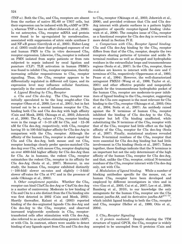
(TNF-a). Both the C5a1 and C5a2 receptors are absentfrom the surface of native HL-60 or U937 cells, buttheir expression can be induced with Bt2-cAMP or IFN-g, whereas TNF-a has no effect (Johswich et al., 2006).In rat astrocytes, C5a2 receptor mRNA and proteinwere found to be up-regulated by noradrenaline,associated with suppression of nitric oxide synthase 2and NF-kB genes (Gavrilyuk et al., 2005). Huber-Langet al. (2005) could show that prolonged exposure of ratand human PMN to C5a in vitro decreased C5a2receptor expression. Likewise, C5a2 receptor is reducedon PMN isolated from septic patients or from ratssubjected to sepsis induced by cecal ligation andpuncture (CLP). TLR activation in human PBMCswas found to inhibit C5a2 receptor expression, therebyincreasing cellular responsiveness to C5a1 receptorsignaling. Thus, the C5a2 receptor appears to bedifferentially regulated in different cell types, and itsexpression level may influence cellular functions,especially in the context of inflammation.4. Ligand Binding by C5a2 Receptora. C5a and C5a des-Arg. The C5a2 receptor was
initially speculated to be the long sought-after C4areceptor (Ohno et al., 2000; Lee et al., 2001), but in factturned out to be a second human receptor for C5a,binding both C5a and C5a des-Arg with high affinity(Cain and Monk, 2002; Okinaga et al., 2003; Johswichet al., 2006). The Kd values of C5a2 receptor bindingwere in the range of ;3–10 nM for C5a, and ;12–36nM for C5a des-Arg, respectively, with C5a2 receptorhaving 10- to 100-fold higher affinity for C5a des-Arg incomparison with the C5a1 receptor. Although theaffinity of the human C5a2 receptor is about equal forC5a and C5a des-Arg, both mouse and rat C5a2receptor homologs clearly prefer species-matched C5ades-Arg over C5a, with mouse C5a2 receptor displayingan over 4000-fold higher affinity for C5a des-Arg thanfor C5a. As in humans, the rodent C5a2 receptoroutmatches the rodent C5a1 receptor in its affinity forC5a des-Arg (Scola et al., 2007). Moreover, in onestudy, the human C5a2 receptor exhibited distinctly(;100-fold) slower on-rates and slightly (;3-fold)slower off-rates for C5a at 0°C and in the presence ofazide (Okinaga et al., 2003).b. Other complement peptides. Whether the C5a2
receptor can bind C3a/C3a des-Arg or C4a/C4a des-Argis a matter of controversy. Moderate to low binding ofC3a and C4a to a site distinct from the C5a binding sitewas first detected in 2002 (Cain and Monk, 2002).Shortly thereafter, Kalant et al. (2003) reportedbinding of the des-arginated ligands C3a des-Arg andC4a des-Arg to the C5a2 receptor, and observedincreased triglyceride synthesis in C5a2 receptor-transfected cells after stimulation with C3a des-Arg,also referred to as acylation-stimulating protein (ASP),or with C3a. In contrast, others were unable to detectbinding of any ligands apart from C5a and C5a des-Arg
to C5a2 receptor (Okinaga et al., 2003; Johswich et al.,2006), and provided evidence that C3a and C3a des-Arg interact with plastic surfaces in a pattern highlysuggestive of specific receptor-ligand binding (Johs-wich et al., 2006). The complex issue of C5a2 receptoras a functional receptor for C3a des-Arg is reviewed inmore detail in Section IV.E.
c. Comparison with C5a1 Receptor ligand binding.C5a and C5a des-Arg binding by the C5a2 receptordiffers from that of the C5a1 receptor, despite the tworeceptors sharing patterns of tyrosine and acidic N-terminal residues as well as charged and hydrophobicresidues in the extracellular loops and transmembraneregions (Scola et al., 2007). In the C5a1 receptor, theseresidues are known to interact with the core and Cterminus of C5a, respectively (Oppermann et al., 1993;Pease et al., 1994). However, the well-characterizedantagonist PMX53 (Wong et al., 1999; Taylor et al.,2001) and other effective peptidic and nonpeptidicligands for the transmembrane hydrophobic pocket ofthe human C5a1 receptor are moderate-to-poor inhib-itors of ligand binding to the C5a2 receptor, suggestingthat the C5a core segment is the most relevant for C5abinding to the C5a2 receptor (Okinaga et al., 2003; Ottoet al., 2004; Scola et al., 2007). An antibody raisedagainst the N terminus of human C5a2 receptorinhibited the binding of C5a des-Arg to the C5a2receptor but left C5a binding unaffected, whilesubstituting the N terminus of the C5a2 receptor withthat of the C5a1 receptor had little effect on the highaffinity of the C5a2 receptor for C5a des-Arg (Scolaet al., 2007). Finally, mutational analyses revealedthree N-terminal residues of the C5a2 receptor thatwere essential for C5a des-Arg binding but had littleinvolvement in C5a binding (Scola et al., 2007). Takentogether, these findings indicate that the N terminus isan important but not the only determinant of the highaffinity of the human C5a2 receptor for C5a des-Arg,and that, unlike the C5a1 receptor, critical N-terminalresidues of the C5a2 receptor interact with C5a des-Argonly, not with C5a.
d. Modulation of ligand binding. While a number ofblocking antibodies specific for the mouse, rat, orhuman C5a2 receptor have been developed andemployed by different investigators in vitro and invivo (Gao et al., 2005; Cui et al., 2007; Lee et al., 2008;Bamberg et al., 2010), to our knowledge the onlyantagonists for the human C5a2 receptor are the C5amutant jun/fos-A8, and its derivative jun/fos-A8D71–73,which inhibit ligand binding to both the C5a1 receptorand C5a2 receptor (Heller et al., 1999; Otto et al.,2004).
5. C5a2 Receptor Signalinga. G protein mediated. Despite sharing the 7TM
structure of typical GPCR, the C5a2 receptor is widelyaccepted to be uncoupled from G proteins (Cain and
524 Klos et al.
Monk, 2002; Kalant et al., 2003; Okinaga et al., 2003;Johswich et al., 2006; Scola et al., 2009). This isbelieved to be due to changes in conserved sequencesshared by most functional chemoattractant or chemo-kine receptors, including the C5a1 receptor. Primarily,both mouse and human C5a2 receptors lack the highlyconserved “DRX”motif usually found at the end of TM3(Oliveira et al., 1994; Ballesteros et al., 1998), actuallyAsp-Arg-Phe in the human C5a1 receptor. In the C5a2receptor, it reads Asp-Leu-Cys, thus lacking thearginine residue that is most important for theinteraction with G proteins (Savarese and Fraser,1992; Scheer et al., 1996; Cain and Monk, 2002;Okinaga et al., 2003). Mutating the respective argininein FPR, CCR5, histamine H2 receptor, and otherreceptors greatly decreases or altogether abolishestheir ability to couple to G proteins (Prossnitz et al.,1999; Alewijnse et al., 2000; Lagane et al., 2005; Rovatiet al., 2007). Conversely, C5a binding to a C5a2receptor in which Asp-Leu-Cys is mutated to Asp-Arg-Cys induces a low level of intracellular Ca2+
mobilization in HEK293 cells cotransfected withGa16, indicative of a partial gain-of-function incoupling to this rather promiscuous G protein subtype(Okinaga et al., 2003). However, RBL-2H3 cells trans-fected with the C5a2 receptor mutated to resemble theC5a1 receptor at this motif did not respond to C5a orC5a des-Arg in an intracellular free Ca2+ assay (Scolaet al., 2009). Given that rat basophilic leukemia (RBL)cells do not endogenously express Ga16, variations inthe amount and selectivity of the two G proteins mayexplain these differing results.Further residues in the mouse and human C5a2
receptors that differ from conserved sequences in theC5a1 receptor and other GPCR are in the “NPXXY”motif (Asn-Pro-Leu-Met-Phe in C5a2 receptor) in TM7,and deletions of serine/threonine residues and a basicregion (239Lys-Thr-Leu-Lys in C5a1 receptor) thattruncate the third intracellular domain (Cain andMonk, 2002; Scola et al., 2009). Substitution of theanalogous serine/threonine residues with alanine inthe C5a1 receptor does not interfere with ligandbinding but abolishes downstream signaling, suggest-ing that this region is also involved in G proteincoupling (Bock et al., 1997). Different mutations of theNPXXY motif have been shown to inhibit the activa-tion of Gq by the cholecystokinin B receptor and todisturb internalization, ligand binding, or coupling toselected downstream pathways in the case of FPR(Gales et al., 2000).In an effort to further elucidate the regions re-
sponsible for uncoupling the C5a2 receptor from Gproteins, a recent study compared the C5a1 receptorand the C5a2 receptor with four substitution mutantsof the human C5a2 receptor: DRX (Asp-Arg-Phe),NPXXY (Asn-Pro-Met-Leu-Tyr), DRX/NPXXY doublemutant, a chimeric receptor carrying the third
intracellular loop of C5a2 receptor, and a triple mutantcontaining all of the mentioned modifications. Unlikethe C5a1 receptor, none of the mutants exhibitedligand-induced internalization, Ca2+ mobilization, ortranslocation of endogenous b-arrestin 1 in RBL-2H3cells, and neither did the wild-type C5a2 receptor(Scola et al., 2009). Taken together, multiple structuralfeatures contribute to effectively restrict G protein-coupled signaling by the C5a2 receptor.
In accordance, several groups have consistentlyfound that binding of C5a, C3a, C4a, or their des-arginated derivatives to the wild-type C5a2 receptor invitro does not support G protein-mediated cellularresponses such as Ca2+ mobilization, degranulation,chemotaxis, or activation of the mitogen-activatedprotein kinase (MAPK) pathway, in striking contrastto the diverse effects induced by the C5a1 receptor(Cain and Monk, 2002; Kalant et al., 2003; Okinagaet al., 2003; Johswich et al., 2006; Scola et al., 2009).Pretreatment of human C5a2 receptor-expressingRBL-2H3 cells with C5a, C5a des-Arg, C3a, or C4acould facilitate Fc«RI-mediated secretory responses(Cain and Monk, 2002), implying that, at the most, theC5a2 receptor exhibits very limited capacities for signaltransduction under particular conditions.
b. Internalization of C5a2 receptor. The C5a2 re-ceptor has been suggested to act as a decoy receptor,limiting the availability of C5a or C5a des-Arg tocompeting receptors, especially the C5a1 receptor(Okinaga et al., 2003; Scola et al., 2009). This conceptwas first described in 1993 for IL-1RII (Colotta et al.,1993), and has since then been extended to GPCRincluding D6, DARC, US28, CCX CKR (CCRL-1),CRAM-B (CCRL-2), and CXCR7 (Nibbs et al., 1997;Gosling et al., 2000; Randolph-Habecker et al., 2002;Lee et al., 2003; Burns et al., 2006; Hartmann et al.,2008; Leick et al., 2010). By sequestering ligands andtargeting them for degradation, these receptors nega-tively regulate cellular responses to inflammatorymediators. Decoy receptors are commonly uncoupledfrom G proteins, most of them due to lack of the DRXmotif. Unlike signaling receptors, they do not undergoligand-induced internalization, but some of themrecycle constitutively between the cell surface andintracellular compartments, thus efficiently removingligands from the extracellular space. This may lead toa relatively high proportion of intracellular receptors,as demonstrated for D6 (Weber et al., 2004).
It has been demonstrated by the use of human PMNor transfected cell lines, including RBL-2H3, thatsignificant amounts of C5a2 receptor are locatedintracellularly, and that human C5a2 receptor isneither phosphorylated nor internalized upon bindingof C5a or C5a des-Arg (Cain and Monk, 2002; Okinagaet al., 2003; Scola et al., 2009; Bamberg et al., 2010).Instead, clathrin-dependent constitutive internaliza-tion occurs, resulting in net transport of ligand into the
Complement Peptide Receptors 525
cell and in ligand degradation (Scola et al., 2009). Thisappeared to be more efficient for C5a des-Arg than forC5a, suggesting that the C5a2 receptor might beresponsible for removing the more abundant des-arginated form from the circulation (Scola et al.,2009). Interestingly, a predominantly intracellularlocation and constitutive internalization were recentlyreported for FPR3, which, despite being capable ofeliciting cellular responses, does not seem to activate Gproteins (Rabiet et al., 2011). In the respective study,the N-terminal extracellular region and TM1 wereidentified to contain the structural determinants ofthis unusual recycling behavior, which appears to beclathrin- and b-arrestin-independent. Based on theseresults, the investigators hypothesized that FPR3 mayserve a ligand scavenging or intracellular receptorfunction, in analogy to what has been described for theC5a2 receptor (Scola et al., 2009; Bamberg et al., 2010).Again, contrasting data have come from studies in-vestigating the C5a2 receptor as a receptor for C3a des-Arg /ASP: here, ligand-induced clathrin-dependentreceptor endocytosis and recycling were observed inHEK293 cells, and were impaired by an S323Imutation, indicating that this residue is vital for C3ades-Arg /ASP-induced C5a2 receptor activation (Cuiet al., 2009b).c. Arrestin-mediated signaling. Because there is no
evidence for the involvement of G proteins in theobserved C5a2 receptor-mediated effects, the ideaemerged that the C5a2 receptor uses G protein-independent signaling, such as pathways targeted byb-arrestins (reviewed in Defea, 2008). A b-arrestin1-GFP fusion protein was translocated to the humanC5a2 receptor in transfected HEK293 cells afterstimulation with C5a, C5a des-Arg, C3a, or C3a des-Arg (Kalant et al., 2005). The presence of largeamounts of exogenous protein or varying preferencesof the C5a2 receptor for b-arrestin subtypes dependingon the cell type may account for the divergencebetween these and other results (Scola et al., 2009).Later, two independent studies of C5a2 receptor-transfected cells concordantly reported C5a-inducedtranslocation of b-arrestin 2 (Cui et al., 2009b; van Lithet al., 2009). In addition, the role attributed to the C5a2receptor as a functional receptor for C3a des-Arg (ASP)in lipid metabolism seems to be associated withrecruitment of b-arrestin 2-GFP (Cui et al., 2009a).Furthermore, it was recently shown in the humanmast cell line HMC-1 that G protein-mediated ERK1/2activation downstream of the C3a receptor may benegatively regulated by b-arrestins (Vibhuti et al.,2011). Colocalization of b-arrestin 1 with the C5a1 andC5a2 receptors occurs in endosomal vesicles of C5a-stimulated human PMNs and was found to be de-pendent on C5a1 receptor activation (Bamberg et al.,2010). Assuming a regulatory interplay of the C5a2receptor with the C5a1 receptor, as they had already
hypothesized earlier (Gerard et al., 2005), the inves-tigators showed that two putative signaling eventsdownstream of b-arrestin 1, ERK1/2 activation andchemotaxis, are enhanced after antibody blockade of theC5a2 receptor. However, it remains to be confirmed thatb-arrestin 1 mediates C5a1 receptor-induced functions inhuman primary cells or transfected cells, as they may aswell result from G protein activation (Buhl et al., 1994;Nilsson et al., 1996; Haribabu et al., 1999; Schraufstatteret al., 2009; Nishiura et al., 2010b;). In summary, thereare credible indications of an active signaling function ofthe C5a2 receptor involving b-arrestin, but furtherconfirmation should be aimed at elucidating how thismodulates the cellular response to C5a.
d. Dimerization. For the C5a1 receptor, homodime-rization as well as heterodimerization with CCR5 havebeen observed, and have been shown to lead tohomologous or heterologous co-internalization ofphosphorylation-deficient C5a1 receptor or of CCR5,respectively, upon C5a binding to the wild-type C5a1receptor (Huttenrauch et al., 2005; Rabiet et al., 2008).Interestingly, the chemokine receptor CXCR7, which isknown to associate with Gai but does not elicit anyintracellular signaling (Levoye et al., 2009), has beenfound to heterodimerize with CXCR4. The dimers wereconstitutively associated with b-arrestin and concom-itantly decoupled from Gai, resulting in potentiatedCXCL12-induced signaling via MAPK pathways topromote cell migration (Decaillot et al., 2011). It istempting to speculate that a similar interaction couldtake place between the C5a1 receptor and the C5a2receptor, shifting or switching downstream signalingfrom G protein-dependent to b-arrestin-mediatedevents, and thus providing at least some explanationfor the unresolved controversies about the functions ofthe C5a2 receptor. This possibility has been considered,but no evidence was seen in mouse cells (Chen et al.,2007), and it was not reported in human PMN, evenwhen modulation of C5a1 receptor signaling wasobserved (Bamberg et al., 2010). The investigatorsconsidered differential regulation of downstream path-ways rather than dimerization as the underlyingmechanism (Chen et al., 2007; Bamberg et al., 2010).However, more recent evidence points to the formationof hetero- and homodimers by C5a1 and C5a2 (Crokeret al., 2012; Poursharifi et al., 2013), although thefunctional significance is still unclear.
e. Cross-talk with Toll-like receptors. Apparently,nonsignaling 7TM receptors may also act as chemokinechaperones or transporters (Middleton et al., 1997;Nibbs et al., 2003), but to date, none of these functionshave been attributed to the C5a2 receptor. Lately,a cross-talk between TLR and the C5a1 receptor/C5a2receptor has been described, in which TLR activationreduces C5a2 receptor activity, thereby releasing theC5a1 receptor from negative modulation and enhancingthe inflammatory response to C5a (Raby et al., 2011).
526 Klos et al.
This study suggests that in the course of everyinflammatory reaction, a balance between TLR-induced cellular hypersensitivity to C5a and the C5a2receptor counteracting this effect has to be established,and that the net outcome of the process variesconsiderably among individuals.6. Pro- and Anti-inflammatory Effects of C5a2
Receptor. The human C5a2 receptor shares manyfeatures with the well-characterized chemokine scav-enging receptor D6, clearly implying they have anal-ogous functions. The C5a2 receptor should thus beexpected to have anti-inflammatory effects in vivo andto negatively regulate C5a1 receptor-mediatedresponses (Locati et al., 2005; Borroni et al., 2006). Infact, when compared with wild-type animals, mice witha targeted deletion of C5ar2 mounted an exaggeratedinflammatory reaction, with a 2- to 3-fold increasedneutrophil influx and higher levels of IL-6 and TNF-ain a model induced by ovalbumin (OVA) of pulmonaryimmune complex injury; the chemotaxis of murineC5ar22/2 bone marrow cells was also enhanced invitro (Gerard et al., 2005). Up-regulation of the C5a2receptor in rat astrocytes is associated with suppres-sion of proinflammatory genes (Gavrilyuk et al., 2005);upon stimulation with C5a and LPS, rat neutrophilspretreated with a C5a2 receptor-blocking antibodyproduced higher amounts of IL-6 than untreatedcontrol cells (Gao et al., 2005). Similarly, a study ofhuman septic patients found that low C5a2 receptorexpression on PMN correlated with multiorgan failureand nonsurvival, and corresponding results wereobtained from rats subjected to CLP-induced sepsis(Huber-Lang et al., 2005).The decoy receptor hypothesis has been contrasted
by a recent report that the C5a2 receptor can positivelymodulate C5a1 receptor- and C3a receptor-inducedinflammatory responses (Chen et al., 2007). TheC5ar22/2 mouse strains on C57BL/6 and BALB/cbackground used here were generated independentlyof the animals used for the aforementioned studies(Gerard et al., 2005), and several disease models wereinvestigated. In comparison with wild-type mice,C5ar22/2 animals showed reduced macrophage in-filtration in peritonitis induced by thioglycolate (TGC),and less pronounced cellular infiltration and activationafter injection of C5a and TGC in a dorsal air-pouchmodel of inflammation (Chen et al., 2007). C5ar22/2
mice were also less affected by OVA-induced airwayhyperresponsiveness than wild-type mice, as charac-terized by one lung function parameter and histologicevaluation of the inflammatory infiltrate. After in vitrostimulation with C3a or C5a, up-regulation of cell-surface markers and activation of downstream MAPKpathways were diminished in neutrophils and macro-phages from C5ar22/2 animals. Then again, deletionof C5ar2 seems to render mice more susceptible toLPS-induced shock, as indicated by elevated IL-1b
serum levels and high mortality after i.p. injection ofLPS (Chen et al., 2007).
Likewise, Rittirsch et al. (2008) support a proinflam-matory role for the C5a2 receptor: according to theirobservations in C5ar12/2 and C5ar22/2 mice subjectedto CLP-induced sepsis, and treated or not with blockingantibodies, the synergistic action of both receptors isrequired for the development of full-blown sepsis. Onlya combined pretreatment with both anti-C5a1 receptorand anti-C5a2 receptor antibodies could lower theplasma levels of proinflammatory mediators and reducelethality, with the role of the C5a2 receptor being linkedto the release of HMGB1 (Rittirsch et al., 2008). It maybe assumed that the animals used in this study wereprovided by the Gerard group, although this was notexplicitly specified.
Recently, a complex role for the C5a2 receptor hasemerged in the pathogenesis of experimental allergicasthma in mice. Here, the C5a2 receptor acts at themDC/T cell interface to suppress TH1 and TH17polarization, while concomitantly driving TH2 cytokineproduction in other pulmonary cells (Zhang et al.,2010).
E. C5a2 Receptor as a Possible Receptor for C3a des-Arg /Acylation Stimulating Protein
1. Mediators of Metabolic Activity. The adiposetissue is an active endocrine organ that secretesmediators such as adiponectin, leptin, or resistin,which are prototypical adipokines (or adipocytokines)(Kershaw and Flier, 2004). This term describes a groupof factors secreted by the white adipose tissue.Adiponectin is a member of the pattern-recognitionfamily of defense collagens, and it interacts witha number of target molecules, including surfacestructures of damaged endothelium and apoptotic cells.It can bind to complement factor C1q, thus activatingthe classic pathway of the complement system (Peakeet al., 2008), and it is regulated by factor H, a solublecomplement inhibitor (Peake and Shen, 2010). Otheradipokines include cytokines and chemokines such asIL-1, IL-6, IL-8, IL-18, TNF-a, MCP-1 (CCL2), CXCL5,and proteins involved in the acute phase response suchas PAI-1 (Kershaw and Flier, 2004; Ouchi et al., 2011).Adipsin, also known as factor D of the alternativepathway, was one of the first recognized adipokines(Cook et al., 1987; White et al., 1992). Adipokinesinfluence energy metabolism, appetite, body weight,and insulin resistance. Most of them stimulate in-flammation, but some (adiponectin and SFRP5) areanti-inflammatory. They play an important role inobesity-related metabolic dysfunction, the pathogene-sis of which implies chronic low-grade inflammation(Ouchi et al., 2011), as well as in classic inflammatorydiseases such as sepsis (Hillenbrand et al., 2012).Hence, there is a growing interest in adipokines inbiomedical sciences.
Complement Peptide Receptors 527
2. Acylation-Stimulating Protein. ASP has beenproposed mainly by the Sniderman and Cianflonegroup as a potent mediator of triglyceride synthesis,simultaneously increasing glucose transport, and asan adipokine with both metabolic and immunefunctions. Purification from human serum and se-quence analysis have revealed that ASP is identical tothe des-arginated form of C3a: C3a des-Arg (Cian-flone et al., 1987, 1989b, 2003). Functional impair-ment of ASP has been suggested as major cause offamilial hyperapobetalipoproteinemia (Snidermanet al., 1985; Cianflone et al., 1988, 1990; Snidermanand Cianflone, 1994). Moreover, it has been claimedthat the C5a2 receptor serves as a signaling receptorfor ASP on adipocytes and other cell types (Baldoet al., 1993; Kalant et al., 2003; Kalant et al., 2005;Maslowska et al., 2005; MacLaren et al., 2008).Although dozens of elaborate studies have alreadybeen published, these issues remain highly controversial.3. The Challenges in Designing Studies Involving
Acylation-Stimulating Protein. Due to the complexityof this topic, the involved research groups face a varietyof challenges with study design and data interpreta-tion. The following points must be considered tocritically assess the respective publications and ourknowledge of ASP.a. Lack of an acylation-stimulating protein knockout
model. C3a des-Arg /ASP as a cleavage product of C3is generated in all situations where the complementcascade is activated by the main activation routes.Unfortunately, it is impossible to generate a knockoutmouse with an isolated deletion of C3a des-Arg /ASPwithout interfering with other functions of the comple-ment system.b. Problems with C3 knockout animals. Although
mice deficient in the precursor molecule C3 haveerroneously been described as “ASP knock-out mice” or“ASP-deficient mice” (Cianflone et al., 1999), beyondlacking C3a des-Arg /ASP (Murray et al., 1999c,d, 2000;Xia et al., 2002, 2004) these animals are deficient inmain complement effector functions downstream of C3—that is, opsonization due to C3b, inflammation andimmune modulation mediated by C3a and C5a, sublyticcell activation, and cell lysis of bystander cells caused byMAC. Thus, C3 knockout mice cannot be considered anadequate model to specifically investigate the role ofC3a des-Arg (ASP).c. Receptor for acylation-stimulating protein. As
described previously, the C5a2 receptor is widelyaccepted as a receptor for C5a and C5a des-Arg. Itsrole as a signaling receptor for C3a des-Arg /ASP,however, is controversial (Kalant et al., 2005; Johswichet al., 2006; Johswich and Klos, 2007; Klos et al., 2009).Thus, mice with a C5ar2 gene knockout might be moreappropriate for the analysis of ASP-related issues thanC3 knockout mice, the caveat being that it remains to
be proven that the C5a2 receptor is indeed a functionalreceptor for ASP.
d. C3a des-Arg regarded as inert. Traditionally,a large number of researchers working in the comple-ment field consider C3a des-Arg as a relatively stablemetabolite without any biologic potential, making ita useful marker for complement activation (Burgeret al., 1988; Klos et al., 1988; Hartmann et al., 1993;Stove et al., 1995). Although C5a des-Arg retains someaffinity for its receptors, most complement researchersassume that C3a des-Arg does not interact with theC3a receptor (Klos et al., 1992; Ames et al., 1996; Crasset al., 1996; Johswich et al., 2006). It is thus neitherinternalized nor degraded, remaining detectable in thebloodstream. In line with this point of view, correla-tions of an increase in C3a des-Arg /ASP in body fluidsof humans or animals with a certain genotype ora disease suggest a role of complement activation andinflammation in general, and not necessarily a role ofthis particular complement cleavage product and itsassumed function. This is further stressed by the factthat unbalanced obesity and insulin resistance arethought to be linked to chronic inflammation in adiposetissue (Ouchi et al., 2011), and that complementactivation is a typical early component of an inflam-matory response (Manabe, 2011).
e. Difficulties working with C3a/C3a des-Arg.Partially due to their charge, C3a and C3a des-Arg aredifficult to handle, more difficult than C5a and C5ades-Arg (Hugli, 1975). C3a and C3a des-Arg /ASP showa significantly higher nonspecific binding to cells andsurfaces (Burg et al., 1996). For this reason, plasticmaterials have to be coated with silicone, albumin, orother inert proteins when handling C3a or C3a des-Arg/ASP. Synthetic C-terminal peptides of C3a with highpurity and more favorable physiochemical propertieshave been used to study binding and activation of C3areceptor by C3a (Ames et al., 1996; Ember et al., 1991;Hugli and Erickson, 1977; Kretzschmar et al., 1992).However, such peptides have rarely been used to studythe interaction between the C5a2 receptor and C3a des-Arg, and they mimic the effects of ASP only partially(Murray et al., 1999a).
f. Simulation of specific binding. In addition, aneasily misleading, rare phenomenon can simulatespecific binding in the absence of any cellular receptor.This occurs when binding studies with adherent cellsare performed in cell culture dishes or 96-well plates,or when membranes instead of a sucrose gradient areused to separate free from cell-bound labeled ligands.This binding of 125I-C3a or 125I-C3a des-Arg (ASP) tothe surface of materials occurs despite the presence ofalbumin or dry milk powder (but because of albumin:Cui et al., 2009a), and can be effectively and dose-dependently competed with nonlabeled C3a or C3ades-Arg, thereby imitating specific binding with a Kd
of the “plastic C3a/C3a des-Arg receptor” in the
528 Klos et al.
nanomolar range (EC50 of ;20 nM). This phenomenonis not observed using 125I-C5a or C5a des-Arg. Toprevent this pseudo-specific nonreceptor binding of125I-C3a and 125I-C3a des-Arg /ASP, all plates andmaterials have to be preincubated with protaminesulfate (or histones) in addition to albumin (Johswichet al., 2006). To address this easily misleading featureof C3a or C3a des-Arg /ASP in binding studies, the useof control cells that do not express the ASP receptor inquestion are essential. Likewise, when investigatingprimary cells, the experiment must also be performedin the absence of any cells.g. Purification of acylation-stimulating protein.
The purity of plasma-derived C3a des-Arg (pASP) isanother critical issue to be considered, especially in thecontext of the high concentrations used to show ASP-mediated functions. Human C5a and C5a des-Arg bindto and activate the C5a1 receptor with an EC50 of ;1nM (Burg et al., 1995); the EC50 of C3a needed for C3areceptor activation is only slightly higher (Klos et al.,1992; Settmacher et al., 2003). The first biologic effectsof these fragments can even be observed at ;0.1 nM.Of note, in experiments on metabolic responses, pASPhas been used at concentrations ranging from 30 nMto 10 mM, with an EC50 of one to several hundrednanomolars. As C3a/C3a des-Arg and C5a/C5a des-Arg are related cationic peptides sharing commonfeatures like molecular weight, isoelectric point, oracid stability (Hugli et al., 1975b, 1981a; Paques et al.,1980; Daumy et al., 1988), it is rather difficult toobtain highly pure preparations of C3a des-Arg fromplasma without low contaminations of other frag-ments still being present. Thus, one must excludeeven minor contaminations of C5a/C5a des-Arg or C3ato rule out the possibility of a (still intriguing) effect ofthese polypeptides on lipid and glucose metabolism.Additionally, when observed more than 20 years ago,mast-cell activation by C3a and C3a des-Arg atconcentrations exceeding 10 mM was proposed to bebased on “a nonspecific mechanism similar to that ofother polybasic compounds” (Fukuoka and Hugli,1990).h. Recombinant acylation-stimulating protein.
The use of recombinant C3a des-Arg (rASP) could solvethis problem. However, recombinant purified proteinhas been consequently applied only in some experi-ments of more recent publications (Cui et al., 2009b;Paglialunga et al., 2010). Formerly, it seemed to havebeen difficult to produce in large amounts (K. Cian-flone, personal communication) and was thereforereserved for a few key experiments that revealedrASP-induced effects in the micromolar range (Murrayet al., 1997). In any case, adequate controls are neededto exclude contaminations such as LPS, which mightinterfere with biologic responses in cell culture or inanimal models, when rASP is prepared from Escher-ichia coli (Roy et al., 2011).
4. Review of Evidence for C5a2 Receptor as theReceptor for Acylation-Stimulating Protein
a. Introduction. Based on these considerations, andto identify the studies providing the best evidence forthe proposed role of C3a des-Arg /ASP and the C5a2receptor, this review will address the followingquestions:
a. How credible is the evidence that ASP is identicalwith C3a des-Arg, and that it influences energymetabolism?
b. How indisputably has it been demonstrated thatthe C5a2 receptor plays a role in energymetabolism?
c. How convincing is the evidence that C3a des-Arg/ASP binds to and signals via the C5a2 receptor?
d. Can the C3a/C3a receptor or the C5a/C5a des-Arg/C5a1 receptor be ruled out as confoundingmediators and signaling components?
e. Do the effects observed and the concentrations ofASP used in cell culture represent a physiologicsituation in vivo?
f. Is the effect on lipid metabolism a main phys-iologic function of the complement systemand ASP? Or should it be considered as onecomponent of “inflammatory” diseases includingobesity?
b. Changes after application of an oral fat load indiets. Using a competitive enzyme-linked immuno-sorbent assay (ELISA), it was shown in 1989 that theserum levels of C3a/C3a des-Arg increased signifi-cantly within hours after an oral fat load, but not afterchallenge with glucose (Cianflone et al., 1989a, 1992).However, this was not affirmed by others (Charles-worth et al., 1998; van Oostrom et al., 2004). Inmoderately hypercholesterolemic women, ASP levelswere altered after hydrogenated fat consumption(Matthan et al., 2001). Plasma ASP concentrationand adipose tissue C3 mRNA expression were higherin obese than in lean men, and fasting plasma ASPconcentration and C3 mRNA expression negativelycorrelated with insulin sensitivity (Koistinen et al.,2001). Moderate weight loss of obese women ora prolonged fast led to decreased ASP plasma levels(Sniderman et al., 1991; Cianflone et al., 1995). Aftergastric bypass surgery in morbidly obese subjects, bodyweight, ASP, and leptin decreased and adiponectinincreased, while plasma lipids and insulin resistanceimproved (Faraj et al., 2003). Thus, diet and obesitycan influence ASP concentrations in plasma.
c. Modified acylation-stimulating protein levels invarious diseases and during a modified hormonalstatus. Plasma ASP was significantly higher inpatients with coronary artery disease, and there wasan association with plasma triglyceride, very low-density lipoprotein (VLDL) cholesterol, VLDL,
Complement Peptide Receptors 529
apolipoprotein B (apoB), and certain apolipoprotein E(apoE) phenotypes (Cianflone et al., 1997). Childrenwith Prader-Willi syndrome frequently suffer fromhigh ASP levels and dyslipidemia (de Lind vanWijngaarden et al., 2010). Increased levels of plasmaASP, CRP, insulin, cholesterol, and nonesterified fattyacids are present in women with polycystic ovarysyndrome compared with controls (Wu et al., 2009). Inpregnant women, ASP levels were markedly elevated.They correlated with elevated levels of triglycerides,apoB, low-density lipoprotein cholesterol, and bodymass index (BMI) (Saleh et al., 2007). In neonates, cordblood ASP showed a positive correlation with fetalbirth weight above average, and with maternal plasmaTG (Saleh et al., 2008).For a complementologist, an increase of C3a des-Arg
/ASP in body fluids indicates that an activation of thecomplement system has taken place. One can assumethat besides this (surrogate) marker, all effectormolecules that are released during activation of thecomplement cascade, such as C3a, C5a, C5a des-Arg,C3b, or the MAC, were at least transiently present aswell. A direct conclusion regarding a biologic functionof C3a des-Arg should thus not be drawn. In any case,it is remarkable that a correlation of complementactivation and lipid metabolism has been observed invarious settings. This supports the concept that un-balanced obesity is linked to chronic low-grade in-flammation, and suggests that drastic fat uptake canrapidly trigger a low inflammatory response.d. Animal models, complement factor C3, acylation-
stimulating protein, and energy metabolism. Serumfrom obese rats was found to contain almost twiceas much C3 as serum from lean rats (Boggs et al.,1998). In C32/2 mice, which lack significant parts ofthe complement system, including ASP, a markeddelay in postprandial TG clearance, reduced body fat,and lower leptin levels were observed in comparisonwith wild-type animals. Also, postprandial nonesteri-fied fatty acids were increased in serum/plasma in-dependently of low or high fat diets, and modestchanges in insulin/glucose metabolism implied in-creased insulin sensitivity (Murray et al., 1999d). Incontrast and of note, the Wetsel group found nosignificant differences in the plasma TG, cholesterol,or free fatty acid concentrations of fasted C32/2 micecompared with their wild-type litter mates. Plasmalipoprotein analyses indicated no significant differ-ences in various lipoproteins and triglycerol in theabsence of ASP. The C32/2 animals were not impairedin their ability to clear TG and free fatty acids fromtheir circulation after an oral fat load (Wetsel et al.,1999). These contradicting results might be caused bydifferences in the C32/2 and wild-type mouse strainsbred and used by the two research groups in differentfacilities. It suggests that the genetic background ofthese animals should be checked thoroughly (e.g., by
SNP analysis) to prove sufficient and effectivebackcrossing.
A third research group cross-bred C32/2 mice to micedeficient in both apoE and the low-density lipoproteinreceptor (Apoe2/2 Ldlr2/2 mice). Compared with con-trols, mice additionally lacking C3 had higher serumTG levels and a more proatherogenic lipoproteinprofile, and lower body weight and fat content. Incontrast, there were no differences between factor B-deficient mice, being additionally analyzed wherecomplement activation of the alternative pathway ishampered, and wild-type animals. Moreover, the sizeof lesions in the aorta was significantly higher in C32/2
mice than in controls. The investigators concluded thatcomplement activation by the classic or lectin pathwayexerts atheroprotective effects, possibly through theregulation of lipid metabolism (Persson et al., 2004).
e. C5ar22/2 mice and blockade of C5a2 receptor byantibodies. In studies conducted by or in cooperationwith the Cianflone group, C5ar22/2 mice also showedchanges in their metabolism as compared with wild-type controls: these mice are hyperphagic on a low-fatdiet. Nevertheless, their body weight and adiposetissue mass did not differ from that of wild-type littermates. On a high-fat diet, average adipocyte size andadipose tissue triglyceride/DNA content were signifi-cantly reduced. Postprandial clearance of TG wasdelayed in C5ar22/2animals, and the synthesis ofadipose tissue triglycerides, lipolysis, and fatty acidre-esterification were significantly reduced. Indirectcalorimetry measurements suggested preferential fattyacid utilization over carbohydrate (Paglialunga et al.,2007).
In vivo, application of anti-ASP and anti-C5a2receptor antibodies did not alter body weight, adiposetissue mass, food intake, or levels of insulin, leptin, oradiponectin. However, the blockade particularly of theanti-C5a2 receptor induced a significant delay in TGand nonesterified fatty acids clearance and decreasedperirenal, hepatic, and muscle TG mass. Adenosinemonophosphate-activated protein kinase (AMPK) andlipoprotein lipase (LPL) activity were increased by thistreatment. Thus, the ASP/C5a2 receptor neutralizingantibodies that effectively block the ASP-C5a2 receptorinteraction altered lipid distribution and energy utili-zation. Moreover, continuous administration of anti-ASP antibodies as compared with an IgG negativecontrol had effects on energy expenditure and glucosestorage, increasing in vivo whole-body energy utiliza-tion (Cui et al., 2007; Paglialunga et al., 2010).
f. Systemic application of purified acylation-stimu-lating protein or recombinant acylation-stimulatingprotein. Injection of 500 mg i.p. of ASP acceleratedTG clearance after a fat load in vivo in C57BL/6mice, and reduced plasma glucose within a fewhours (Murray et al., 1999b). Untreated male andfemale C32/2 mice had elevated oxygen consumption,
530 Klos et al.
increased fatty acid oxidation in liver and muscle, andincreased inguinal fat and muscle mRNA expression.Fatty acid incorporation into lipids was decreased butcould be normalized by administration of ASP togetherwith oral fat intake (Xia et al., 2004). However,assuming complete resorption of 500 mg of ASP fromthe peritoneum, its redistribution into an estimated 2ml of total blood volume should lead to ;25 mM ASP inthe circulation. For pASP, a purity of 99% wasascertained by mass spectrophotometry, potentiallypermitting up to 250 nM of contaminating proteinswith a molecular weight similar to ASP in thebloodstream. Of note, this impedes interpretation ofdata from the corresponding experiments because;10–50 nM C3a, C5a, or C5a des-Arg would alreadyresult in maximal cellular responses mediated by theC3a receptor or C5a1 receptor. Thus, when applying500 mg of pASP, it cannot be excluded that contami-nating factors could be responsible for the observedeffects.Yet there are more convincing studies with a contin-
uous administration of recombinant instead of purifiedASP over up to 4 weeks that clearly demonstratedecreased fasting levels of nonesterified fatty acids anddecreased energy expenditure in C32/2 mice, but onlysmall (if any) changes in body weight or food uptake(Paglialunga et al., 2010). As a negative control, salineis applied instead of an inert recombinant proteinpurified from E. coli in parallel with the rASP. Indifferentiated adipocytes, 100 nM rASP resulted ina drastic increase in fatty acid uptake in vitro. Thiswas blocked in vitro by incubation with an anti-ASPantibody, excluding the possibility that contamina-tions, such as LPS, are responsible for the observedeffect on adipocytes. The interpretation of the dataobtained in the mouse is more difficult due to theabsence of a similar specificity control (i.e., inhibitionby antibody directed against ASP) and also due to thefact that, surprisingly, the achieved and effectiveserum steady-state levels of rASP delivered by anosmotic pump were very small (i.e., only in the range of0.25 nM). This large difference in concentrationsneeded between the in vitro assay to see cellularresponses and the in vivo experiment makes it moredifficult to draw conclusions about a common un-derlying mechanism.On the other hand, continuous application of anti-
bodies against endogenous ASP with correspondingIgG controls in the animal model resulted in cleareffects on the lipid and glucose metabolism (Paglia-lunga et al., 2010). Moreover, injection of rASP into thethird ventricle of the brain inhibits food intake andlocomotor activity in rats (Roy et al., 2011). Again, datainterpretation is limited by the absence of a negativecontrol which could exclude that impurities in therASP might account for effects in this system. Further,it is difficult to judge what concentrations of rASP are
reached in the cerebrospinal fluid, and whether theyare in the physiologic range or rather correspond toa situation of severe neurologic disease such asmeningitis. Nevertheless, these are interesting dataas they indicate additional functions for the comple-ment system and its cleavage products.
g. In vitro data using purified acylation-stimulatingprotein, synthetic C3a-related peptides, or recombinantacylation-stimulating protein. There are a variety ofolder publications using human pASP. They have beenpresented and reviewed in detail elsewhere (Johswichand Klos, 2007; Klos et al., 2009). Therefore, thisreview focuses on a selection of key data from morerecent publications, preferentially based on the use ofrecombinant ASP or synthetic peptides. For in vitrostudies on human skin fibroblasts (HSF) and murine3T3 cells, ASP was chemically and enzymaticallymodified; additionally, synthetic C3a-analogous C-terminal peptides, known ligands of the human C3areceptor, were analyzed (Murray et al., 1999a). Theresults show that the N-terminal region of ASP playsa minor role in receptor binding (determined byinhibition of 125I-ASP binding) and triacylglycerolsynthesis. An intact disulfide-linked core region isessential for lipid synthesis but not for receptorinteraction. The C3a-analogous C-terminal peptideP117 (containing a C-terminal Arg) can inhibit ASPbinding and can partially activate lipid synthesis,suggesting “that this region participates but is notsufficient for complete receptor binding” (Murray et al.,1999a), whereas smaller C-terminal peptides wereineffective.
On the human C3a receptor, P117 has an EC50 in therange of 50 nM (while ASP is completely ineffective)(Martin et al., 1997; Johswich and Klos, 2007; Kloset al., 2009). Triacylglycerol synthesis was stimulatedby P117 at 1000-fold higher concentrations of 4–40 mM,similar to the molar concentrations of ASP needed.Surprisingly, in competition studies using 1 nM125I-ASP as a tracer on HSF, unlabeled P117 andASP were equally potent, with an IC50 in the range ofonly 100 nM (pIC50 = 7). This behavior is rathersurprising as the ligand concentrations needed for bindingand dose-response curves are usually quite similar.
As mentioned previously, C3a and C3a des-Arg showhigh nonspecific binding as well as “pseudo-specificnonreceptor binding” to various surfaces in the absenceof any cell, with a Kd close to that observed in bindingstudies (Johswich et al., 2006). This suggests that thebinding of 125I-ASP and the surprisingly low Kd
observed in the Cianflone group, at least in earlierstudies on adipocytes of fibroblasts, are best explainedby this effect. Of note, in experiments of the Klos group,neither 100 nM fluorescence-labeled (FL) pC3a nor thedes-arginated peptide (FL-ASP) bound to C5a2 re-ceptor expressed on human embryonic kidney (HEK)cells as compared with HEK control cells, whereas
Complement Peptide Receptors 531
FL-C3a was still able to bind to the C3a receptor(Johswich et al., 2006). In contrast, when similarstudies were performed by the Cianflone group, usingFL-rASP, HEK cells stably expressing C5a2 receptorbound and internalized this ligand more strongly thannontransfected HEK control cells (Cui et al., 2009a).Both studies using FL-ligands seem to be thoroughlyperformed, so there is presently no explanation for thediffering results.With an EC50 of ;1.5 mM, the rASP used here was
much more active in stimulating lipid synthesis andfatty acid uptake than the ASP purified from blood andformer preparations. In the same study, C5a2 receptor-expressing HEK cells bound 1 nM of 125I-ASP muchbetter than nontransfected cells or the no-cell controlwith a Kd of 34 nM. HEK and Chinese hamster ovary(CHO) cells expressing C5a2 receptor bound FL-rASPin a dose-dependent manner at similar concentrations(Cui et al., 2009a).h. Summary and assessment of the role of C3a des-
Arg/ASP and C5a2 receptor. The pathogenesis ofobesity-related metabolic dysfunction is considered toinclude chronic low-grade inflammation (Ouchi et al.,2011). Various cytokines and chemokines act as adipo-kines, simultaneously influencing inflammation andmetabolism (Kershaw and Flier, 2004). The complementsystem appears to be linked with adipokine signaling inmultiple ways: cells in the adipose tissue can producevarious complement factors (Choy et al., 1992; Whiteet al., 1992), and adiponectin itself can trigger theclassic pathway of complement activation (Peake et al.,2008). Moreover, complement factor D has been identi-fied as an adipokine. Considering this connectionbetween obesity and inflammation, it does not seemunlikely that other components of the activated com-plement system might play a similar dual role asproinflammatory mediators and metabolic modulators.In accordance, activation of the complement cascade asevidenced by the increase of the “activation marker”C3a des-Arg /ASP is associated with changes in lipidand glucose metabolism. In this context the publicationsof the Cianflone group are very informative.The majority of investigations using ASP purified
from blood (or synthetic C3a-related peptides) and mostdata from animal models are in accordance witha potential role of ASP in metabolism, but they do notprovide final proof that C3a des-Arg is indeed theresponsible and active mediator. In particular, they donot exclude that contaminations with C3a or C5a des-Arg, and their interactions with the C3a receptor or C5a1receptor, respectively, might be causing some of theobserved effects of pASP. A few recent key publicationsusing recombinant C3a des-Arg (ASP) provide muchstronger evidence for an active role of ASP and for theC5a2 receptor as its receptor. However, there are somecontradictory results from both animal studies (Wetselet al., 1999) and in vitro experiments (Johswich et al.,
2006) that should be borne in mind when assessing therole of C3a des-Arg as a metabolic regulator.
V. Complement Peptide Functions Not Mediatedby the Three Known Receptors
A. C4a
Although C4a has receptor-mediated effects in theguinea pig (see Section III.B.1), its effects on humancells have not been shown to be receptor mediated, andserum-derived C4a may be contaminated with C3a.Studies showing that C4a can enhance vascularpermeability in human skin (Gorski et al., 1979),desensitize smooth muscle contraction by C3a (Hugliet al., 1981b), and stimulate mast cells to secrete(Hugli and Muller-Eberhard, 1978; Moon et al., 1981)should be repeated with recombinant C4a. However,recombinant C4a has been demonstrated to inhibitC3a- and C5a-stimulated degranulation of humanmast cells by increasing cAMP levels through anunidentified receptor (Xie et al., 2012). A derivative ofC4 in synovial fluid has been shown to indirectlyinhibit the chemotaxis of human monocytes but notneutrophils (Matsubara et al., 1991). This was latershown to be C4a, which worked at concentrations aslow as 10216 M, but this could be inactivated byconversion to C4a des-Arg (Tsuruta et al., 1993). Ananti-inflammatory role for C4a in glomerulonephritishas also been proposed (Welch et al., 2001). C3a andC4a can both form complexes with IgG (Nezlin andFreywald, 1992), one molecule for each H and L chain.One gram of IgG can bind up to 0.3 mg C3a (1:200molar ratio). The function of this is unknown but hasbeen proposed to eliminate complement peptides fromthe circulation.
B. C3a
In many instances, a biologic activity is shared byboth C3a and its metabolite C3a des-Arg. It is clearthat C3a des-Arg does not interact with the C3areceptor (Wilken et al., 1999; Johswich et al., 2006),so these activities are presumed to be not receptormediated. The homing of bone-marrow hemopoieticcells in response to CXCL12 is enhanced by both C3aand C3a des-Arg (Honczarenko et al., 2005b) in micethat are C3a receptor or C5a2 receptor deficient(Honczarenko et al., 2005a). The LPS-mediated pro-duction of proinflammatory cytokines by PBMC medi-ated by LPS is also enhanced by both C3a and C3ades-Arg (Takabayashi et al., 1996) whereas B-cellcytokine production is diminished (Fischer and Hugli,1997). Both polypeptides have been shown to stimulatepituitary cell cultures to secrete a variety of hormones(Francis et al., 2003), with the C3a des-Arg responsebeing insensitive to pertussis intoxication.
Another shared activity is the inhibition of bacterialand fungal growth. These antimicrobial properties are
532 Klos et al.
conserved from invertebrates to humans (Pasupuletiet al., 2007), and C3a and C4a (but not C5a) have beensuggested to have evolved primarily as antibacterialagents (Malmsten and Schmidtchen, 2007).C3a-derived peptides have also been shown to bind
to the high affinity IgE receptor, Fc«RI, and can inhibitsignaling and cytokine secretion by mast cells (Peterfyet al., 2008). C3a and C3a des-Arg can also enhanceCXCL12-mediated platelet production (Wysoczynskiet al., 2007). The receptor for advanced glycation endproducts (RAGE) can bind C3a, CpG oligos, and DNA/C3a complexes, resulting in the stimulation of IFN-gproduction in human PBMC (Ruan et al., 2010).Oxidized LDL and C3a colocalize in atherosclerotic
plaques as a result of C3a binding to modified lysineside chains. C3a also recognizes oxidized lipids onapoptotic cells in a similar way (Veneskoski et al.,2011), possibly aiding the removal of C3a from thecirculation. As a cationic amphiphile, C3a can alsoactivate G proteins independent of the receptor bycross-linking sialic acid residues displayed on a varietyof membrane proteins (Emadi-Khiav et al., 1995).Finally, C3a has been shown to bind to GPCR Mas-related genes MrgX1 and MrgX2 (Kashem et al., 2011)on human mast cells, allowing C3a to stimulatedegranulation in a non-C3a receptor-dependent man-ner (Kashem et al., 2011; Subramanian et al., 2011).
C. C5a
C5a has also been shown to be bound by immuno-globulins (Basta et al., 2003), and F(ab)92 fragments ofirrelevant specificity can neutralize the ability of bothC3a and C5a to activate the human mast cell lineHMC-1. To our knowledge, no other nonreceptor-mediated functions of C5a or C5a des-Arg have beenreported.
D. Conclusions, Future Directions
1. Nomenclature. Although originally termed ana-phylatoxins, the C5a, C4a, and C3a act only indirectlyto mediate an anaphylactic response (Taylor et al.,1994), and it is now clear that they have many morefunctions inside and outside of the immune system. Forthis reason, we would suggest the use of the term“complement peptides” rather than anaphylatoxins.The second C5a receptor, sometimes described asa binding protein (Okinaga et al., 2003) or as a “re-ceptor-like” protein (Kalant et al., 2003), has clearlybeen shown to transduce signals, particularly in mousecells, so we propose that the symbol C5a2 receptor isascribed to this receptor (human gene name C5AR2).2. Drugs That Act at Complement Peptide Receptors.
Despite many years of intensive investigation, thereare still no useful drugs that act directly at thecomplement peptide receptors. It is not clear as towhether this is due to peculiar molecular mechanismsassociated with receptor binding and activation or to
the complex biology of the receptors. The former isbecoming increasingly well understood as satisfyingmodels, at least for C5a and the C5a1 receptor, arebeing produced. However, the relatively low affinitiesof many of the antagonists for rodent fragment receptorshas troubled the development of many drug leads andmay have provided some misleading information aboutthe roles of the complement peptides in disease. It ispossible that inhibition of complement peptide forma-tion by agents that act on the complement cascade mayprove to be the most effective drugs. Here, however, theextrinsic pathways of fragment generation may con-found this expectation, so further work on the con-sequences of these pathways is required.
3. Signaling by Complement Peptide Receptors.The events that occur after receptor binding are alsoproving difficult to untangle. Although signalingthrough G proteins is well understood, the role ofarrestins in signal transduction remains obscure. Workon the C3a receptor suggests that these proteins havecomplex roles in the modulation of signaling, but forthe C5a1 and C5a2 receptors little is yet known.Signaling by the C5a2 receptor is still controversial,and the question of ligand selectivity has not beenfinally settled. Even if limited to just C5a and C5a des-Arg, the C5a2 receptor may be a signaling receptorequivalent to the C5a1 receptor; it may provide“negative” signals, through arrestins, to counteractthe C5a1 receptor; or it may cause ligand sequestrationand degradation. The role of homo- and heterodimeri-zation of C5a1 and C5a2 in the control of signaling alsoawaits elucidation. Interestingly, although the C5a2receptor binds C5a des-Arg with the same affinity asC5a, the des-arginated form is rarely used in experi-ments with this receptor, resulting in the potential lossof important information. Further, the reported activ-ity of the C5a2 receptor may depend on the speciesunder investigation, or perhaps it is the expressing celltype (or cellular activation status) that determines therole of this receptor. Selective agonists and antagonistsare urgently required for the C5a2 receptor, and it is tobe hoped that such compounds, once identified, can beused to solve the enigma that this receptor represents.
Authorship Contributions
Wrote or contributed to the writing of the manuscript: Klos, Wende,Wareham, Monk.
ReferencesAbdul-Salam VB, Paul GA, Ali JO, Gibbs SR, Rahman D, Taylor GW, Wilkins MR,and Edwards RJ (2006) Identification of plasma protein biomarkers associatedwith idiopathic pulmonary arterial hypertension. Proteomics 6:2286–2294.
Abou-Ragheb HH, Williams AJ, Brown CB, and Milford-Ward A (1991) Plasma levelsand mode of excretion of the anaphylatoxins C3a and C4a in renal disease. J ClinLab Immunol 35:113–119.
Abou-Ragheb HH, Williams AJ, Brown CB, and Milford-Ward A (1992) Plasma levelsof the anaphylatoxins C3a and C4a in patients with IgA nephropathy/Henoch-Schönlein nephritis. Nephron 62:22–26.
Ager RR, Fonseca MI, Chu SH, Sanderson SD, Taylor SM, Woodruff TM,and Tenner AJ (2010) Microglial C5aR (CD88) expression correlates withamyloid-beta deposition in murine models of Alzheimer’s disease. J Neurochem113:389–401.
Complement Peptide Receptors 533
Ahamed J, Haribabu B, and Ali H (2001) Cutting edge: differential regulation ofchemoattractant receptor-induced degranulation and chemokine production byreceptor phosphorylation. J Immunol 167:3559–3563.
Alewijnse AE, Timmerman H, Jacobs EH, Smit MJ, Roovers E, Cotecchia S, and LeursR (2000) The effect of mutations in the DRY motif on the constitutive activity andstructural instability of the histamine H(2) receptor. Mol Pharmacol 57:890–898.
Ali H, Richardson RM, Tomhave ED, Didsbury JR, and Snyderman R (1993) Dif-ferences in phosphorylation of formylpeptide and C5a chemoattractant receptorscorrelate with differences in desensitization. J Biol Chem 268:24247–24254.
Allegretti M, Moriconi A, Aramini A, Candida CM, Beccari AR, and Bertini R (2008)inventors; Dompe Pharma SPA, assignee. (R)-Arylalkylamino derivatives andpharmaceutical compositions containing them. E.P.O. patent no. EP1951663A2.1995 Aug 6.
Amara U, Flierl MA, Rittirsch D, Klos A, Chen H, Acker B, Brückner UB, Nilsson B,Gebhard F, Lambris JD, and Huber-Lang M (2010) Molecular intercommunicationbetween the complement and coagulation systems. J Immunol 185:5628–5636.
Amara U, Rittirsch D, Flierl M, Bruckner U, Klos A, Gebhard F, Lambris JD,and Huber-Lang M (2008) Interaction between the coagulation and complementsystem. Adv Exp Med Biol 632:71–79.
Amatruda TT 3rd, Gerard NP, Gerard C, and Simon MI (1993) Specific interactions ofchemoattractant factor receptors with G-proteins. J Biol Chem 268:10139–10144.
Ames RS, Lee D, Foley JJ, Jurewicz AJ, Tornetta MA, Bautsch W, Settmacher B,Klos A, Erhard KF, and Cousins RD, et al. (2001) Identification of a selectivenonpeptide antagonist of the anaphylatoxin C3a receptor that demonstratesantiinflammatory activity in animal models. J Immunol 166:6341–6348.
Ames RS, Li Y, Sarau HM, Foley JJ, Ellis C, Zeng Z, Su K, Jurewicz AJ, HertzbergRP, Bergsma DJ, and Kumar C, et al. (1996) Molecular cloning and characteriza-tion of the human anaphylatoxin C3a receptor. J Biol Chem 271:20231–20234.
Ames RS, Tornetta MA, Foley JJ, Hugli TE, and Sarau HM (1997) Evidence that thereceptor for C4a is distinct from the C3a receptor. Immunopharmacology 38:87–92.
Andersson E, Rydengård V, Sonesson A, Mörgelin M, Björck L, and Schmidtchen A(2004) Antimicrobial activities of heparin-binding peptides. Eur J Biochem 271:1219–1226.
Astles PC, Brown TJ, Cox PJ, Halley F, Lockey PM, McCarthy C, McLay IM, MajidTN, Morley AD, and Porter B, et al. (1997) New non peptidic C5a receptorantagonists. Bioorg Med Chem Lett 7:907–912.
Bachmaier K, Guzman E, Kawamura T, Gao X, and Malik AB (2012) Sphingosinekinase 1 mediation of expression of the anaphylatoxin receptor C5L2 dampens theinflammatory response to endotoxin. PLoS ONE 7:e30742.
Bajzar L, Manuel R, and Nesheim ME (1995) Purification and characterization ofTAFI, a thrombin-activable fibrinolysis inhibitor. J Biol Chem 270:14477–14484.
Baldo A, Sniderman AD, St-Luce S, Avramoglu RK, Maslowska M, Hoang B, MongeJC, Bell A, Mulay S, and Cianflone K (1993) The adipsin-acylation stimulatingprotein system and regulation of intracellular triglyceride synthesis. J Clin Invest92:1543–1547.
Ballesteros J, Kitanovic S, Guarnieri F, Davies P, Fromme BJ, Konvicka K, Chi L,Millar RP, Davidson JS, Weinstein H, and Sealfon SC (1998) Functional micro-domains in G-protein-coupled receptors. The conserved arginine-cage motif in thegonadotropin-releasing hormone receptor. J Biol Chem 273:10445–10453.
Bamberg CE, Mackay CR, Lee H, Zahra D, Jackson J, Lim YS, Whitfeld PL, Craig S,Corsini E, and Lu B, et al. (2010) The C5a receptor (C5aR) C5L2 is a modulator ofC5aR-mediated signal transduction. J Biol Chem 285:7633–7644.
Banda NK, Hyatt S, Antonioli AH, White JT, Glogowska M, Takahashi K, Merkel TJ,Stahl GL, Mueller-Ortiz S, and Wetsel R, et al. (2012) Role of C3a receptors, C5areceptors, and complement protein C6 deficiency in collagen antibody-inducedarthritis in mice. J Immunol 188:1469–1478.
Banda NK, Levitt B, Wood AK, Takahashi K, Stahl GL, Holers VM, and Arend WP(2010) Complement activation pathways in murine immune complex-induced ar-thritis and in C3a and C5a generation in vitro. Clin Exp Immunol 159:100–108.
Bao L, Wang Y, Haas M, and Quigg RJ (2011) Distinct roles for C3a and C5a incomplement-induced tubulointerstitial injury. Kidney Int 80:524–534.
Barbay JK, Gong Y, Buntinx M, Li J, Claes C, Hornby PJ, Van Lommen G, VanWauwe J, and He W (2008) Synthesis and characterization of 5,6,7,8-tetrahy-droquinoline C5a receptor antagonists. Bioorg Med Chem Lett 18:2544–2548.
Basta M, Van Goor F, Luccioloi S, Billings EM, Vortmeyer AO, Baranyi L, Szebeni J,Alving CR, Carroll MC, Berkower I, et al. (2003) F(ab)’2-mediated neutralization ofC3a and C5a anaphylatoxins: a novel effector function of immunoglobulins. NatMed 9:431–438.
Bautsch W, Kretzschmar T, Stühmer T, Kola A, Emde M, Köhl J, Klos A, and Bitter-Suermann D (1992) A recombinant hybrid anaphylatoxin with dual C3a/C5a ac-tivity. Biochem J 288:261–266.
Bénard M, Gonzalez BJ, Schouft MT, Falluel-Morel A, Vaudry D, Chan P, Vaudry H,and Fontaine M (2004) Characterization of C3a and C5a receptors in rat cerebellargranule neurons during maturation. Neuroprotective effect of C5a against apo-ptotic cell death. J Biol Chem 279:43487–43496.
Bénard M, Raoult E, Vaudry D, Leprince J, Falluel-Morel A, Gonzalez BJ, Galas L,Vaudry H, and Fontaine M, et al. (2008) Role of complement anaphylatoxin receptors(C3aR, C5aR) in the development of the rat cerebellum.Mol Immunol 45:3767–3774.
Bengtson A, Lannsjö W, and Heideman M (1987) Complement and anaphylatoxinresponses to cross-clamping of the aorta. Studies during general anaesthesia withor without extradural blockade. Br J Anaesth 59:1093–1097.
Birney E, Andrews D, Caccamo M, Chen Y, Clarke L, Coates G, Cox T, CunninghamF, Curwen V, and Cutts T, et al. (2006) Ensembl 2006. Nucleic Acids Res 34 (Da-tabase issue):D556–D561.
Blagg J, Mowbray C, Pryde DC, Salmon G, Fairman D, Schmid E, and Beaumont K(2008a) Small, non-peptide C5a receptor antagonists: part 2. Bioorg Med Chem Lett18:5605–5608.
Blagg J, Mowbray C, Pryde DC, Salmon G, Schmid E, Fairman D, and Beaumont K(2008b) Small, non-peptide C5a receptor antagonists: part 1. Bioorg Med Chem Lett18:5601–5604.
Bock D, Martin U, Gärtner S, Rheinheimer C, Raffetseder U, Arseniev L, Barker MD,Monk PN, Bautsch W, Köhl J, and Klos A (1997) The C terminus of the human C5areceptor (CD88) is required for normal ligand-dependent receptor internalization.Eur J Immunol 27:1522–1529.
Boggs RD, McCumbee WD, Cobbs SL, Todd DG, Kahle EB, Stewart NL, Bailey M,and Reichenbecher VE (1998) Increased expression of complement component C3in the plasma of obese Zucker fa and LA/N fa(f) rats compared with their leancounterparts. Obes Res 6:361–367.
Bokisch VA and Müller-Eberhard HJ (1970) Anaphylatoxin inactivator of humanplasma: its isolation and characterization as a carboxypeptidase. J Clin Invest 49:2427–2436.
Bokisch VA, Müller-Eberhard HJ, and Cochrane CG (1969) Isolation of a fragment(C3a) of the third component of human complement containing anaphylatoxin andchemotactic activity and description of an anaphylatoxin inactivator of humanserum. J Exp Med 129:1109–1130.
Boor P, Konieczny A, Villa L, Schult AL, Bücher E, Rong S, Kunter U, van RoeyenCR, Polakowski T, and Hawlisch H, et al. (2007) Complement C5 mediates ex-perimental tubulointerstitial fibrosis. J Am Soc Nephrol 18:1508–1515.
Borroni EM, Buracchi C, de la Torre YM, Galliera E, Vecchi A, Bonecchi R, Man-tovani A, and Locati M (2006) The chemoattractant decoy receptor D6 as a negativeregulator of inflammatory responses. Biochem Soc Trans 34:1014–1017.
Braun L, Christophe T, and Boulay F (2003) Phosphorylation of key serine residues isrequired for internalization of the complement 5a (C5a) anaphylatoxin receptor viaa beta-arrestin, dynamin, and clathrin-dependent pathway. J Biol Chem 278:4277–4285.
Breivik T, Gundersen Y, Gjermo P, Taylor SM, Woodruff TM, and Opstad PK (2011)Oral treatment with complement factor C5a receptor (CD88) antagonists inhibitsexperimental periodontitis in rats. J Periodontal Res 46:643–647.
Brodbeck RM, Cortright DN, Kieltyka AP, Yu J, Baltazar CO, Buck ME, Meade R,Maynard GD, Thurkauf A, Chien DS, Hutchison AJ, and Krause JE (2008) Iden-tification and characterization of NDT 9513727 [N,N-bis(1,3-benzodioxol-5-ylmethyl)-1-butyl-2,4-diphenyl-1H-imidazole-5-methanamine], a novel, orally bio-available C5a receptor inverse agonist. J Pharmacol Exp Ther 327:898–909.
Bubeck P, Grötzinger J, Winkler M, Köhl J, Wollmer A, Klos A, and Bautsch W (1994)Site-specific mutagenesis of residues in the human C5a anaphylatoxin which areinvolved in possible interaction with the C5a receptor. Eur J Biochem 219:897–904.
Buhl AM, Avdi N, Worthen GS, and Johnson GL (1994) Mapping of the C5a receptorsignal transduction network in human neutrophils. Proc Natl Acad Sci USA 91:9190–9194.
Bunschoten A, Ippel JH, Kruijtzer JA, Feitsma L, de Haas CJ, Liskamp RM,and Kemmink J (2011) A peptide mimic of the chemotaxis inhibitory protein ofStaphylococcus aureus: towards the development of novel anti-inflammatorycompounds. Amino Acids 40:731–740.
Burg M, Martin U, Bock D, Rheinheimer C, Köhl J, Bautsch W, and Klos A (1996)Differential regulation of the C3a and C5a receptors (CD88) by IFN-gamma andPMA in U937 cells and related myeloblastic cell lines. J Immunol 157:5574–5581.
Burg M, Martin U, Rheinheimer C, Köhl J, BautschW, Böttger EC, and Klos A (1995)IFN-gamma up-regulates the human C5a receptor (CD88) in myeloblastic U937cells and related cell lines. J Immunol 155:4419–4426.
Burger F and Zilow EP (1993) Complement-derived anaphylatoxins in natural im-munity, in The Natural Immune System: Humoral Factors (Sim E, ed) pp 209–232,Oxford University Press, Oxford.
Burger R, Zilow G, Bader A, Friedlein A, and Naser W (1988) The C terminus of theanaphylatoxin C3a generated upon complement activation represents a neo-antigenic determinant with diagnostic potential. J Immunol 141:553–558.
Burns JM, Summers BC, Wang Y, Melikian A, Berahovich R, Miao Z, Penfold ME,Sunshine MJ, Littman DR, and Kuo CJ, et al. (2006) A novel chemokine receptorfor SDF-1 and I-TAC involved in cell survival, cell adhesion, and tumor de-velopment. J Exp Med 203:2201–2213.
Busche MN and Stahl GL (2010) Role of the complement components C5 and C3a in amouse model of myocardial ischemia and reperfusion injury. Ger Med Sci 8:8, Doc20.
Cain SA, Coughlan T, and Monk PN (2001a) Mapping the ligand-binding site on theC5a receptor: arginine74 of C5a contacts aspartate282 of the C5a receptor. Bio-chemistry 40:14047–14052.
Cain SA, Higginbottom A, and Monk PN (2003) Characterisation of C5a receptoragonists from phage display libraries. Biochem Pharmacol 66:1833–1840.
Cain SA and Monk PN (2002) The orphan receptor C5L2 has high affinity bindingsites for complement fragments C5a and C5a des-Arg(74). J Biol Chem 277:7165–7169.
Cain SA, Woodruff TM, Taylor SM, Fairlie DP, Sanderson SD, and Monk PN (2001b)Modulation of ligand selectivity by mutation of the first extracellular loop of thehuman C5a receptor. Biochem Pharmacol 61:1571–1579.
Campbell W, Okada N, and Okada H (2001) Carboxypeptidase R is an inactivator ofcomplement-derived inflammatory peptides and an inhibitor of fibrinolysis.Immunol Rev 180:162–167.
Campbell WD, Lazoura E, Okada N, and Okada H (2002) Inactivation of C3a and C5aoctapeptides by carboxypeptidase R and carboxypeptidase N. Microbiol Immunol46:131–134.
Campo A, Hausmann G, Martí RM, Estrach T, Grau JM, Porcel JM, and Herrero C(2007) Complement activation products (C3a and C5b-9) as markers of activity ofdermatomyositis. Comparison with usual biochemical parameters [article inSpanish]. Actas Dermosifiliogr 98:403–414.
Caporale LH, Tippett PS, Erickson BW, and Hugli TE (1980) The active site of C3aanaphylatoxin. J Biol Chem 255:10758–10763.
Carmona-Fontaine C, Theveneau E, Tzekou A, Tada M, Woods M, Page KM, ParsonsM, Lambris JD, and Mayor R (2011) Complement fragment C3a controls mutualcell attraction during collective cell migration. Dev Cell 21:1026–1037.
Carroll MV and Sim RB (2011) Complement in health and disease. Adv Drug DelivRev 63:965–975.
534 Klos et al.
Chang JY, Lin CC, Salamanca S, Pangburn MK, and Wetsel RA (2008) Denaturationand unfolding of human anaphylatoxin C3a: an unusually low covalent stability ofits native disulfide bonds. Arch Biochem Biophys 480:104–110.
Chao TH, Ember JA, Wang M, Bayon Y, Hugli TE, and Ye RD (1999) Role of thesecond extracellular loop of human C3a receptor in agonist binding and receptorfunction. J Biol Chem 274:9721–9728.
Charlesworth JA, Peake PW, Campbell LV, Pussell BA, O’Grady S, and Tzilopoulos T(1998) The influence of oral lipid loads on acylation stimulating protein (ASP) inhealthy volunteers. Int J Obes Relat Metab Disord 22:1096–1102.
Chen JJ, Cole DC, Ciszewski G, Crouse K, Ellingboe JW, Nowak P, Tawa GJ, Ber-stein G, and Li W (2010) Identification of a new class of small molecule C5a re-ceptor antagonists. Bioorg Med Chem Lett 20:662–664.
Chen NJ, Mirtsos C, Suh D, Lu YC, Lin WJ, McKerlie C, Lee T, Baribault H, Tian H,and Yeh WC (2007) C5L2 is critical for the biological activities of the anaphyla-toxins C5a and C3a. Nature 446:203–207.
Chen Z, Zhang X, Gonnella NC, Pellas TC, Boyar WC, and Ni F (1998) Residues 21-30within the extracellular N-terminal region of the C5a receptor represent a bindingdomain for the C5a anaphylatoxin. J Biol Chem 273:10411–10419.
Chenoweth DE, Goodman MG, and Weigle WO (1982) Demonstration of a specific re-ceptor for human C5a anaphylatoxin on murine macrophages. J Exp Med 156:68–78.
Chenoweth DE and Hugli TE (1980) Human C5a and C5a analogs as probes of theneutrophil C5a receptor. Mol Immunol 17:151–161.
Choy LN, Rosen BS, and Spiegelman BM (1992) Adipsin and an endogenous pathwayof complement from adipose cells. J Biol Chem 267:12736–12741.
Cianflone K, Kalant D, Marliss EB, Gougeon R, and Sniderman AD (1995) Responseof plasma ASP to a prolonged fast. Int J Obes Relat Metab Disord 19:604–609.
Cianflone K, Kwiterovich PO, Walsh M, Forse A, Rodriguez MA, and Sniderman AD(1987) Stimulation of fatty acid uptake and triglyceride synthesis in human cul-tured skin fibroblasts and adipocytes by a serum protein. Biochem Biophys ResCommun 144:94–100.
Cianflone K, Maslowska M, and Sniderman AD (1999) Acylation stimulating protein(ASP), an adipocyte autocrine: new directions. Semin Cell Dev Biol 10:31–41.
Cianflone K, Rodriguez MA, Walsh M, Vu H, and Sniderman AD (1988) The effect ofa plasma protein fraction on lipid synthesis in cultured skin fibroblasts from nor-mals and patients with hyperapobetalipoproteinemia. Clin Invest Med 11:99–107.
Cianflone K, Vu H, Walsh M, Baldo A, and Sniderman A (1989a) Metabolic responseof acylation stimulating protein to an oral fat load. J Lipid Res 30:1727–1733.
Cianflone K, Xia Z, and Chen LY (2003) Critical review of acylation-stimulatingprotein physiology in humans and rodents. Biochim Biophys Acta 1609:127–143.
Cianflone K, Zhang XJ, Genest J Jr, and Sniderman A (1997) Plasma acylation-stimulating protein in coronary artery disease. Arterioscler Thromb Vasc Biol 17:1239–1244.
Cianflone KM, Maslowska MH, and Sniderman AD (1990) Impaired response offibroblasts from patients with hyperapobetalipoproteinemia to acylation-stimulating protein. J Clin Invest 85:722–730.
Cianflone KM, Sniderman AD, Dallongeville J, BertrandM, Raffaï E, and Davignon J(1992) The relation between triglyceride synthesis in peripheral tissues and post-prandial plasma triglyceride levels: preliminary evidence of a role for acylationstimulating protein. Clin Invest Med 15:132–140.
Cianflone KM, Sniderman AD, Walsh MJ, Vu HT, Gagnon J, and Rodriguez MA(1989b) Purification and characterization of acylation stimulating protein. J BiolChem 264:426–430.
Cleary PP, Prahbu U, Dale JB, Wexler DE, and Handley J (1992) Streptococcal C5apeptidase is a highly specific endopeptidase. Infect Immun 60:5219–5223.
Cochrane CG and Müller-Eberhard HJ (1968) The derivation of two distinct ana-phylatoxin activities from the third and fifth components of human complement. JExp Med 127:371–386.
Colotta F, Re F, Muzio M, Bertini R, Polentarutti N, Sironi M, Giri JG, Dower SK,Sims JE, and Mantovani A (1993) Interleukin-1 type II receptor: a decoy target forIL-1 that is regulated by IL-4. Science 261:472–475.
Cook KS, Min HY, Johnson D, Chaplinsky RJ, Flier JS, Hunt CR, and SpiegelmanBM (1987) Adipsin: a circulating serine protease homolog secreted by adiposetissue and sciatic nerve. Science 237:402–405.
Cook WJ, Galakatos N, Boyar WC, Walter RL, and Ealick SE (2010) Structure ofhuman desArg-C5a. Acta Crystallogr D Biol Crystallogr 66:190–197.
Corbin NC and Hugli TE (1976) The primary structure of porcine C3a anaphylatoxin.J Immunol 117:990–995.
Crass T, Ames RS, Sarau HM, Tornetta MA, Foley JJ, Köhl J, Klos A, and Bautsch W(1999a) Chimeric receptors of the human C3a receptor and C5a receptor (CD88). JBiol Chem 274:8367–8370.
Crass T, BautschW, Cain SA, Pease JE, and Monk PN (1999b) Receptor activation byhuman C5a des Arg74 but not intact C5a is dependent on an interaction betweenGlu199 of the receptor and Lys68 of the ligand. Biochemistry 38:9712–9717.
Crass T, Bautsch W, Cain SA, Pease JE, and Monk PN (1999c) Receptor activation byhuman C5a des Arg74 but not intact C5a is dependent on an interaction betweenGlu199 of the receptor and Lys68 of the ligand. Biochemistry 38:9712–9717.
Crass T, Raffetseder U, Martin U, Grove M, Klos A, Köhl J, and Bautsch W (1996)Expression cloning of the human C3a anaphylatoxin receptor (C3aR) from differ-entiated U-937 cells. Eur J Immunol 26:1944–1950.
Croker DE, Halai R, Fairlie DP, and Cooper MA (2012) Ligand-induced dimerisationof the complement C5aR and C5L2 receptors by C5a but not C5a des-Arg.Immunobiology 217:1181–1182.
Cui L, Ferreri K, and Hugli TE (1988) Structural characterization of the C4a ana-phylatoxin from rat. Mol Immunol 25:663–671.
Cui W, Lapointe M, Gauvreau D, Kalant D, and Cianflone K (2009a) RecombinantC3adesArg/acylation stimulating protein (ASP) is highly bioactive: a critical eval-uation of C5L2 binding and 3T3-L1 adipocyte activation. Mol Immunol 46:3207–3217.
Cui W, Paglialunga S, Kalant D, Lu H, Roy C, Laplante M, Deshaies Y, and CianfloneK (2007) Acylation-stimulating protein/C5L2-neutralizing antibodies alter
triglyceride metabolism in vitro and in vivo. Am J Physiol Endocrinol Metab 293:E1482–E1491.
Cui W, Simaan M, Laporte S, Lodge R, and Cianflone K (2009b) C5a- and ASP-mediated C5L2 activation, endocytosis and recycling are lost in S323I-C5L2 mu-tation. Mol Immunol 46:3086–3098.
Daumy GO, Delgarno D, McColl AS, Merenda JM, and Schulte GR (1988) The use ofhigh field NMR to determine homogeneity in a preparation of human C5a producedby Escherichia coli. Biochim Biophys Acta 967:326–330.
Davignon I, Catalina MD, Smith D, Montgomery J, Swantek J, Croy J, Siegelman M,and Wilkie TM (2000) Normal hematopoiesis and inflammatory responses despitediscrete signaling defects in Ga15 knockout mice. Mol Cell Biol 20:797–804.
Davoust N, Jones J, Stahel PF, Ames RS, and Barnum SR (1999) Receptor for theC3a anaphylatoxin is expressed by neurons and glial cells. Glia 26:201–211.
de Haas CJ, Veldkamp KE, Peschel A, Weerkamp F, Van Wamel WJ, Heezius EC,Poppelier MJ, Van Kessel KP, and van Strijp JA (2004) Chemotaxis inhibitory proteinof Staphylococcus aureus, a bacterial antiinflammatory agent. J ExpMed 199:687–695.
de Lind van Wijngaarden RF, Cianflone K, Gao Y, Leunissen RW, and Hokken-Koelega AC (2010) Cardiovascular and metabolic risk profile and acylation-stimulating protein levels in children with Prader-Willi syndrome and effects ofgrowth hormone treatment. J Clin Endocrinol Metab 95:1758–1766.
Décaillot FM, Kazmi MA, Lin Y, Ray-Saha S, Sakmar TP, and Sachdev P (2011)CXCR7/CXCR4 heterodimer constitutively recruits beta-arrestin to enhance cellmigration. J Biol Chem 286:32188–32197.
Defea K (2008) Beta-arrestins and heterotrimeric G-proteins: collaborators andcompetitors in signal transduction. Br J Pharmacol 153 (Suppl 1):S298–S309.
DeFea KA, Zalevsky J, Thoma MS, Déry O, Mullins RD, and Bunnett NW (2000)Beta-arrestin-dependent endocytosis of proteinase-activated receptor 2 is requiredfor intracellular targeting of activated ERK1/2. J Cell Biol 148:1267–1281.
DeMartino JA, Konteatis ZD, Siciliano SJ, Van Riper G, Underwood DJ, Fischer PA,and Springer MS (1995) Arginine 206 of the C5a receptor is critical for ligandrecognition and receptor activation by C-terminal hexapeptide analogs. J BiolChem 270:15966–15969.
DeMartino JA, Van Riper G, Siciliano SJ, Molineaux CJ, Konteatis ZD, Rosen H,and Springer MS (1994) The amino terminus of the human C5a receptor is re-quired for high affinity C5a binding and for receptor activation by C5a but not C5aanalogs. J Biol Chem 269:14446–14450.
Denonne F, Binet S, Burton M, Collart P, Defays S, Dipesa A, Eckert M, GiannarasA, Kumar S, Levine B, Nicolas JM, Pasau P, and Pégurier C, et al. (2007a) Dis-covery of new C3aR ligands. Part 2: amino-piperidine derivatives. Bioorg MedChem Lett 17:3262–3265.
Denonne F, Binet S, Burton M, Collart P, Dipesa A, Ganguly T, Giannaras A, KumarS, Lewis T, and Maounis F, et al. (2007b) Discovery of new C3aR ligands. Part 1:arginine derivatives. Bioorg Med Chem Lett 17:3258–3261.
Dillard P, Wetsel RA, and Drouin SM (2007) Complement C3a regulates Muc5acexpression by airway Clara cells independently of Th2 responses. Am J Respir CritCare Med 175:1250–1258.
DiMartino SJ, Shah AB, Trujillo G, and Kew RR (2001) Elastase controls the bindingof the vitamin D-binding protein (Gc-globulin) to neutrophils: a potential role in theregulation of C5a co-chemotactic activity. J Immunol 166:2688–2694.
Dobrina A, Pausa M, Fischetti F, Bulla R, Vecile E, Ferrero E, Mantovani A,and Tedesco F (2002) Cytolytically inactive terminal complement complex causestransendothelial migration of polymorphonuclear leukocytes in vitro and in vivo.Blood 99:185–192.
Ducruet AF, Hassid BG, Mack WJ, Sosunov SA, Otten ML, Fusco DJ, Hickman ZL,Kim GH, Komotar RJ, Mocco J, and Connolly ES (2008) C3a receptor modulation ofgranulocyte infiltration after murine focal cerebral ischemia is reperfusion de-pendent. J Cereb Blood Flow Metab 28:1048–1058.
Elsner J, Oppermann M, Czech W, Dobos G, Schöpf E, Norgauer J, and Kapp A(1994) C3a activates reactive oxygen radical species production and intracellularcalcium transients in human eosinophils. Eur J Immunol 24:518–522.
Emadi-Khiav B, Mousli M, Bronner C, and Landry Y (1995) Human and rat cuta-neous mast cells: involvement of a G protein in the response to peptidergic stimuli.Eur J Pharmacol 272:97–102.
Ember JA, Johansen NL, and Hugli TE (1991) Designing synthetic superagonists ofC3a anaphylatoxin. Biochemistry 30:3603–3612.
Ember JA, Sanderson SD, Taylor SM, Kawahara M, and Hugli TE (1992) Biologicactivity of synthetic analogues of C5a anaphylatoxin. J Immunol 148:3165–3173.
Erken E, Gunesacar R, and Ozer HT (2010) Investigation of C5a receptor gene 450 C/T polymorphism in Turkish patients with familial Mediterranean fever. Mol BiolRep 37:273–276.
Faraj M, Havel PJ, Phélis S, Blank D, Sniderman AD, and Cianflone K (2003) Plasmaacylation-stimulating protein, adiponectin, leptin, and ghrelin before and afterweight loss induced by gastric bypass surgery in morbidly obese subjects. J ClinEndocrinol Metab 88:1594–1602.
Farzan M, Schnitzler CE, Vasilieva N, Leung D, Kuhn J, Gerard C, Gerard NP,and Choe H (2001) Sulfated tyrosines contribute to the formation of the C5adocking site of the human C5a anaphylatoxin receptor. J Exp Med 193:1059–1066.
Fassbender A, D’Hooghe T, Mihalyi A, Kyama C, Simsa P, and Lessey BA (2009)Plasma C3a-des-Arg levels in women with and without endometriosis. Am JReprod Immunol 62:187–195.
Feierler J, Wirth M, Welte B, Schüssler S, Jochum M, and Faussner A (2011) Helix 8plays a crucial role in bradykinin B(2) receptor trafficking and signaling. J BiolChem 286:43282–43293.
Fernandez HN, Henson PM, Otani A, and Hugli TE (1978) Chemotactic response tohuman C3a and C5a anaphylatoxins. I. Evaluation of C3a and C5a leukotaxis invitro and under stimulated in vivo conditions. J Immunol 120:109–115.
Fernandez HN and Hugli TE (1978) Primary structural analysis of the polypeptideportion of human C5a anaphylatoxin. Polypeptide sequence determination andassignment of the oligosaccharide attachment site in C5a. J Biol Chem 253:6955–6964.
Complement Peptide Receptors 535
Findlay JB and Pappin DJ (1986) The opsin family of proteins. Biochem J 238:625–642.
Fischer WH and Hugli TE (1997) Regulation of B cell functions by C3a and C3a(desArg): suppression of TNF-alpha, IL-6, and the polyclonal immune response. JImmunol 159:4279–4286.
Fonseca MI, Ager RR, Chu SH, Yazan O, Sanderson SD, LaFerla FM, Taylor SM,Woodruff TM, and Tenner AJ (2009) Treatment with a C5aR antagonist decreasespathology and enhances behavioral performance in murine models of Alzheimer’sdisease. J Immunol 183:1375–1383.
Francis K, Lewis BM, Akatsu H, Monk PN, Cain SA, Scanlon MF, Morgan BP, HamJ, and Gasque P (2003) Complement C3a receptors in the pituitary gland: a novelpathway by which an innate immune molecule releases hormones involved in thecontrol of inflammation. FASEB J 17:2266–2268.
Francis K, Lewis BM, Monk PN, and Ham J (2008) Complement C5a receptors in thepituitary gland: expression and function. J Endocrinol 199:417–424.
Fredslund F, Laursen NS, Roversi P, Jenner L, Oliveira CL, Pedersen JS, Nunn MA,Lea SM, Discipio R, Sottrup-Jensen L, and Andersen GR (2008) Structure of andinfluence of a tick complement inhibitor on human complement component 5. NatImmunol 9:753–760.
Fujiwara A, Taguchi O, Takagi T, D’Alessandro-Gabazza CN, Boveda-Ruiz D, TodaM, Yasukawa A, Matsushima Y, Miyake Y, Kobayashi H, and Kobayashi T, et al.(2012) Role of thrombin-activatable fibrinolysis inhibitor in allergic bronchialasthma. Lung 190:189–198.
Fukuoka Y and Hugli TE (1990) Anaphylatoxin binding and degradation by ratperitoneal mast cells. Mechanisms of degranulation and control. J Immunol 145:1851–1858.
Fukuoka Y, Xia HZ, Sanchez-Muñoz LB, Dellinger AL, Escribano L, and SchwartzLB (2008) Generation of anaphylatoxins by human beta-tryptase from C3, C4, andC5. J Immunol 180:6307–6316.
Fusakio ME, Mohammed JP, Laumonnier Y, Hoebe K, Kohl J, and Mattner J (2011)C5a regulates NKT and NK cell functions in sepsis. J Immunol 187:5805–5812
Galés C, Kowalski-Chauvel A, Dufour MN, Seva C, Moroder L, Pradayrol L, VaysseN, Fourmy D, and Silvente-Poirot S (2000) Mutation of Asn-391 within the con-served NPXXY motif of the cholecystokinin B receptor abolishes Gq protein acti-vation without affecting its association with the receptor. J Biol Chem 275:17321–17327.
Gao H, Neff TA, Guo RF, Speyer CL, Sarma JV, Tomlins S, Man Y, Riedemann NC,Hoesel LM, and Younkin E, et al. (2005) Evidence for a functional role of the secondC5a receptor C5L2. FASEB J 19:1003–1005.
Gao J, Choe H, Bota D, Wright PL, Gerard C, and Gerard NP (2003) Sulfation oftyrosine 174 in the human C3a receptor is essential for binding of C3a anaphyla-toxin. J Biol Chem 278:37902–37908.
Garrett MC, Otten ML, Starke RM, Komotar RJ, Magotti P, Lambris JD, RynkowskiMA, and Connolly ES (2009) Synergistic neuroprotective effects of C3a and C5areceptor blockade following intracerebral hemorrhage. Brain Res 1298:171–177.
Gavrilyuk V, Kalinin S, Hilbush BS, Middlecamp A, McGuire S, Pelligrino D,Weinberg G, and Feinstein DL (2005) Identification of complement 5a-like receptor(C5L2) from astrocytes: characterization of anti-inflammatory properties. J Neu-rochem 92:1140–1149.
Ge H, Scheinin M, and Kallio J (2003) Constitutive precoupling to G(i) and increasedagonist potency in the alpha(2B)-adrenoceptor. Biochem Biophys Res Commun306:959–965.
Gerard C, Chenoweth DE, and Hugli TE (1981) Response of human neutrophils toC5a: a role for the oligosaccharide moiety of human C5ades Arg-74 but not of C5ain biologic activity. J Immunol 127:1978–1982.
Gerard C and Hugli TE (1981) Identification of classical anaphylatoxin as the des-Arg form of the C5a molecule: evidence of a modulator role for the oligosaccharideunit in human des-Arg74-C5a. Proc Natl Acad Sci USA 78:1833–1837.
Gerard NP, Bao L, Xiao-Ping H, Eddy RL Jr, Shows TB, and Gerard C (1993) Humanchemotaxis receptor genes cluster at 19q13.3-13.4. Characterization of the humanC5a receptor gene. Biochemistry 32:1243–1250.
Gerard NP and Gerard C (1991) The chemotactic receptor for human C5a anaphy-latoxin. Nature 349:614–617.
Gerard NP, Lively MO, and Gerard C (1988) Amino acid sequence of guinea pig C3aanaphylatoxin. Protein Seq Data Anal 1:473–478.
Gerard NP, Lu B, Liu P, Craig S, Fujiwara Y, Okinaga S, and Gerard C (2005) Ananti-inflammatory function for the complement anaphylatoxin C5a-binding pro-tein, C5L2. J Biol Chem 280:39677–39680.
Gerardy-Schahn R, Ambrosius D, Casaretto M, Grötzinger J, Saunders D, WollmerA, Brandenburg D, and Bitter-Suermann D (1988) Design and biological activity ofa new generation of synthetic C3a analogues by combination of peptidic and non-peptidic elements. Biochem J 255:209–216.
Gerber BO, Meng EC, Dotsch V, Baranski TJ, and Bourne HR (2001) An activationswitch in the ligand binding pocket of the C5a receptor. J Biol Chem 276:3394–3400.
Geva A, Lassere TB, Lichtarge O, Pollitt SK, and Baranski TJ (2000) Genetic map-ping of the human C5a receptor. Identification of transmembrane amino acidscritical for receptor function. J Biol Chem 275:35393–35401.
Giannini E, Brouchon L, and Boulay F (1995) Identification of the major phosphor-ylation sites in human C5a anaphylatoxin receptor in vivo. J Biol Chem 270:19166–19172.
Gombos T, Förhécz Z, Pozsonyi Z, Széplaki G, Kunde J, Füst G, Jánoskuti L, KarádiI, and Prohászka Z (2012) Complement anaphylatoxin C3a as a novel independentprognostic marker in heart failure. Clin Res Cardiol 101:607–615.
Gong Y, Barbay JK, Buntinx M, Li J, Wauwe JV, Claes C, Lommen GV, Hornby PJ,and He W (2008) Design and optimization of aniline-substituted tetrahy-droquinoline C5a receptor antagonists. Bioorg Med Chem Lett 18:3852–3855.
Gonzalez JM, Dong Z, Romero R, and Girardi G (2011) Cervical remodeling/ripeningat term and preterm delivery: the same mechanism initiated by different mediatorsand different effector cells. PLoS ONE 6:e26877.
Gorski JP, Hugli TE, and Müller-Eberhard HJ (1979) C4a: the third anaphylatoxin ofthe human complement system. Proc Natl Acad Sci USA 76:5299–5302.
Gosling J, Dairaghi DJ, Wang Y, Hanley M, Talbot D, Miao Z, and Schall TJ (2000)Cutting edge: identification of a novel chemokine receptor that binds dendritic cell-and T cell-active chemokines including ELC, SLC, and TECK. J Immunol 164:2851–2856.
Governa M, Amati M, Fenoglio I, Valentino M, Coloccini S, Bolognini L, Carlo BottaG, Emanuelli M, Pierella F, and Volpe AR, et al. (2005) Variability of biologicaleffects of silicas: different degrees of activation of the fifth component of comple-ment by amorphous silicas. Toxicol Appl Pharmacol 208:68–77.
Governa M, Amati M, Valentino M, Visonà I, Fubini B, Botta GC, Volpe AR,and Carmignani M (2000) In vitro cleavage by asbestos fibers of the fifth compo-nent of human complement through free-radical generation and kallikrein acti-vation. J Toxicol Environ Health A 59:539–552.
Governa M, Fenoglio I, Amati M, Valentino M, Bolognini L, Coloccini S, Volpe AR,Carmignani M, and Fubini B (2002) Cleavage of the fifth component of humancomplement and release of a split product with C5a-like activity by crystallinesilica through free radical generation and kallikrein activation. Toxicol ApplPharmacol 179:129–136.
Greer J (1985) Model structure for the inflammatory protein C5a. Science 228:1055–1060.
Gripentrog JM and Miettinen HM (2008) Formyl peptide receptor-mediated ERK1/2activation occurs through G(i) and is not dependent on beta-arrestin1/2. Cell Signal20:424–431.
Gueler F, Rong S, Gwinner W, Mengel M, Bröcker V, Schön S, Greten TF, HawlischH, Polakowski T, and Schnatbaum K, et al. (2008) Complement 5a receptor in-hibition improves renal allograft survival. J Am Soc Nephrol 19:2302–2312.
Guo Q, Subramanian H, Gupta K, and Ali H (2011) Regulation of C3a receptorsignaling in human mast cells by G protein coupled receptor kinases. PLoS ONE 6:e22559.
Gustafsson E, Forsberg C, Haraldsson K, Lindman S, Ljung L, and Furebring C(2009a) Purification of truncated and mutated chemotaxis inhibitory protein ofStaphylococcus aureus an anti-inflammatory protein. Protein Expr Purif 63:95–101.
Gustafsson E, Haas PJ, Walse B, Hijnen M, Furebring C, Ohlin M, van Strijp JA,and van Kessel KP (2009b) Identification of conformational epitopes for human IgGon chemotaxis inhibitory protein of Staphylococcus aureus. BMC Immunol 10:13.
Gustafsson E, Rosén A, Barchan K, van Kessel KP, Haraldsson K, Lindman S,Forsberg C, Ljung L, Bryder K, and Walse B, et al. (2010) Directed evolution ofchemotaxis inhibitory protein of Staphylococcus aureus generates biologicallyfunctional variants with reduced interaction with human antibodies. Protein EngDes Sel 23:91–101.
Haas PJ, de Haas CJ, Kleibeuker W, Poppelier MJ, van Kessel KP, Kruijtzer JA,Liskamp RM, and van Strijp JA (2004) N-terminal residues of the chemotaxisinhibitory protein of Staphylococcus aureus are essential for blocking formylatedpeptide receptor but not C5a receptor. J Immunol 173:5704–5711.
Habermann JK, Roblick UJ, Luke BT, Prieto DA, Finlay WJ, Podust VN, Roman JM,Oevermann E, Schiedeck T, and Homann N, et al. (2006) Increased serum levels ofcomplement C3a anaphylatoxin indicate the presence of colorectal tumors. Gas-troenterology 131:1020–1029, quiz 1284.
Hack CE, Nuijens JH, Felt-Bersma RJ, Schreuder WO, Eerenberg-Belmer AJ,Paardekooper J, Bronsveld W, and Thijs LG (1989) Elevated plasma levels of theanaphylatoxins C3a and C4a are associated with a fatal outcome in sepsis. Am JMed 86:20–26.
Hagemann IS, Miller DL, Klco JM, Nikiforovich GV, and Baranski TJ (2008)Structure of the complement factor 5a receptor-ligand complex studied by disulfidetrapping and molecular modeling. J Biol Chem 283:7763–7775.
Hagemann IS, Narzinski KD, Floyd DH, and Baranski TJ (2006) Random muta-genesis of the complement factor 5a (C5a) receptor N terminus provides a struc-tural constraint for C5a docking. J Biol Chem 281:36783–36792.
Han G, Geng S, Li Y, Chen G, Wang R, Li X, Ma Y, Shen B, and Li Y (2011) gdT-cellfunction in sepsis is modulated by C5a receptor signalling. Immunology 133:340–349.
Haque R, Hwang BY, Appelboom G, Piazza MA, Guo K, and Connolly ES (2011)Alterations in systemic complement component 3a and 5a levels in patients withcerebral arteriovenous malformations. J Clin Neurosci 18:1235–1239.
Harada T, Sano M, and Matsunaga T (1992) The effect of anaphylatoxin componenton inner ear damage. Acta Otolaryngol 112:265–271.
Haribabu B, Zhelev DV, Pridgen BC, Richardson RM, Ali H, and Snyderman R (1999)Chemoattractant receptors activate distinct pathways for chemotaxis and secre-tion. Role of G-protein usage. J Biol Chem 274:37087–37092.
Hartmann H, Lübbers B, Casaretto M, Bautsch W, Klos A, and Köhl J (1993) Rapidquantification of C3a and C5a using a combination of chromatographic and im-munoassay procedures. J Immunol Methods 166:35–44.
Hartmann K, Henz BM, Krüger-Krasagakes S, Köhl J, Burger R, Guhl S, Haase I,Lippert U, and Zuberbier T (1997) C3a and C5a stimulate chemotaxis of humanmast cells. Blood 89:2863–2870.
Hartmann TN, Leick M, Ewers S, Diefenbacher A, Schraufstatter I, Honczarenko M,and Burger M (2008) Human B cells express the orphan chemokine receptorCRAM-A/B in a maturation-stage-dependent and CCL5-modulated manner. Im-munology 125:252–262.
Hasegawa K, Tamari M, Shao C, Shimizu M, Takahashi N, Mao XQ, Yamasaki A,Kamada F, Doi S, and Fujiwara H, et al. (2004) Variations in the C3, C3a receptor,and C5 genes affect susceptibility to bronchial asthma. Hum Genet 115:295–301.
Hashimoto M, Hirota K, Yoshitomi H, Maeda S, Teradaira S, Akizuki S, Prieto-Martin P, Nomura T, Sakaguchi N, and Köhl J, et al. (2010) Complement drivesTh17 cell differentiation and triggers autoimmune arthritis. J Exp Med 207:1135–1143.
Heimbach L, Li Z, Berkowitz P, Zhao M, Li N, Rubenstein DS, Diaz LA, and Liu Z(2011) The C5a receptor on mast cells is critical for the autoimmune skin-blisteringdisease bullous pemphigoid. J Biol Chem 286:15003–15009.
536 Klos et al.
Heller T, Hennecke M, Baumann U, Gessner JE, zu Vilsendorf AM, Baensch M,Boulay F, Kola A, Klos A, Bautsch W, and Köhl J (1999) Selection of a C5a receptorantagonist from phage libraries attenuating the inflammatory response in immunecomplex disease and ischemia/reperfusion injury. J Immunol 163:985–994.
Hensens OD, Borris RP, Koupal LR, Caldwell CG, Currie SA, Haidri AA, HomnickCF, Honeycutt SS, Lindenmayer SM, and Schwartz CD, et al. (1991) L-156,602,a C5a antagonist with a novel cyclic hexadepsipeptide structure from Streptomycessp. MA6348. Fermentation, isolation and structure determination. J Antibiot(Tokyo) 44:249–254.
Higginbottom A, Cain SA, Woodruff TM, Proctor LM, Madala PK, Tyndall JD, TaylorSM, Fairlie DP, and Monk PN (2005) Comparative agonist/antagonist responses inmutant human C5a receptors define the ligand binding site. J Biol Chem 280:17831–17840.
Hillenbrand A, Weiss M, Knippschild U, Wolf AM, and Huber-Lang M (2012) Sepsis-induced adipokine change with regard to insulin resistance. Int J Inflamm 2012:972368.
Hoke M, Speidl W, Schillinger M, Minar E, Zehetmayer S, Schönherr M, Wagner O,and Mannhalter C (2012) Polymorphism of the complement 5 gene and cardio-vascular outcome in patients with atherosclerosis. Eur J Clin Invest 42:921–926.
Hollmann TJ, Mueller-Ortiz SL, Braun MC, and Wetsel RA (2008) Disruption of theC5a receptor gene increases resistance to acute Gram-negative bacteremia andendotoxic shock: opposing roles of C3a and C5a. Mol Immunol 45:1907–1915.
Honczarenko M, Lu B, Nicholson-Weller A, Gerard NP, Silberstein LE, and Gerard C(2005a) C5L2 receptor is not involved in C3a / C3a-desArg-mediated enhancementof bone marrow hematopoietic cell migration to CXCL12. Leukemia 19:1682–1683,author reply 1684–1685.
Honczarenko M, Ratajczak MZ, Nicholson-Weller A, and Silberstein LE (2005b)Complement C3a enhances CXCL12 (SDF-1)-mediated chemotaxis of bone marrowhematopoietic cells independently of C3a receptor. J Immunol 175:3698–3706.
Horino K, Nishiura H, Ohsako T, Shibuya Y, Hiraoka T, Kitamura N, and YamamotoT (1998) A monocyte chemotactic factor, S19 ribosomal protein dimer, in phagocyticclearance of apoptotic cells. Lab Invest 78:603–617.
Hsu W, Rosenquist GL, Ansari AA, and Gershwin ME (2005) Autoimmunity andtyrosine sulfation. Autoimmun Rev 4:429–435.
Huber-Lang M, Denk S, Fulda S, Erler E, Kalbitz M, Weckbach S, Schneider EM,Weiss M, Kanse SM, and Perl M (2012) Cathepsin D is released after severe tissuetrauma in vivo and is capable of generating C5a in vitro. Mol Immunol 50:60–65.
Huber-Lang M, Sarma JV, Rittirsch D, Schreiber H, Weiss M, Flierl M, Younkin E,Schneider M, Suger-Wiedeck H, and Gebhard F, et al. (2005) Changes in the novelorphan, C5a receptor (C5L2), during experimental sepsis and sepsis in humans. JImmunol 174:1104–1110.
Huber-Lang MS, Sarma JV, McGuire SR, Lu KT, Padgaonkar VA, Younkin EM, GuoRF, Weber CH, Zuiderweg ER, Zetoune FS, and Ward PA (2003) Structure-functionrelationships of human C5a and C5aR. J Immunol 170:6115–6124.
Huber R, Scholze H, Pâques EP, and Deisenhofer J (1980) Crystal structure analysisand molecular model of human C3a anaphylatoxin. Hoppe Seylers Z Physiol Chem361:1389–1399.
Hueber AJ, Asquith DL, Miller AM, Reilly J, Kerr S, Leipe J, Melendez AJ,and McInnes IB (2010) Mast cells express IL-17A in rheumatoid arthritis syno-vium. J Immunol 184:3336–3340.
Huey R, Bloor CM, Kawahara MS, and Hugli TE (1983) Potentiation of the ana-phylatoxins in vivo using an inhibitor of serum carboxypeptidase N (SCPN). I.Lethality and pathologic effects on pulmonary tissue. Am J Pathol 112:48–60.
Huey R, Erickson BW, Bloor CM, and Hugli TE (1984) Contraction of guinea pig lungby synthetic oligopeptides related to human C3a. Immunopharmacology 8:37–45.
Hugli TE (1975) Human anaphylatoxin (C3a) from the third component of comple-ment. Primary structure. J Biol Chem 250:8293–8301.
Hugli TE (1981) The structural basis for anaphylatoxin and chemotactic functions ofC3a, C4a, and C5a. Crit Rev Immunol 1:321–366.
Hugli TE and Erickson BW (1977) Synthetic peptides with the biological activitiesand specificity of human C3a anaphylatoxin. Proc Natl Acad Sci USA 74:1826–1830.
Hugli TE, Gerard C, Kawahara M, Scheetz ME 2nd, Barton R, Briggs S, Koppel G,and Russell S (1981a) Isolation of three separate anaphylatoxins from complement-activated human serum. Mol Cell Biochem 41:59–66.
Hugli TE, Kawahara MS, Unson CG, Molinar-Rode R, and Erickson BW (1983) Theactive site of human C4a anaphylatoxin. Mol Immunol 20:637–645.
Hugli TE, Morgan WT, and Müller-Eberhard HJ (1975a) Circular dichroism of C3aanaphylatoxin. Effects of pH, heat, guanidinium chloride, and mercaptoethanol onconformation and function. J Biol Chem 250:1479–1483.
Hugli TE and Müller-Eberhard HJ (1978) Anaphylatoxins: C3a and C5a. AdvImmunol 26:1–53.
Hugli TE, Stimler NP, Gerard C, and Moon KE (1981b) Possible role of serum ana-phylatoxins in hypersensitivity reactions. Int Arch Allergy Appl Immunol 66 (Suppl1):113–120.
Hugli TE, Vallota EH, and Müller-Eberhard HJ (1975b) Purification and partialcharacterization of human and porcine C3a anaphylatoxin. J Biol Chem 250:1472–1478.
Hunt JR, Martin CB, and Martin BK (2005) Transcriptional regulation of the murineC5a receptor gene: NF-Y is required for basal and LPS induced expression inmacrophages and endothelial cells. Mol Immunol 42:1405–1415.
Husmann LK, Yung DL, Hollingshead SK, and Scott JR (1997) Role of putativevirulence factors of Streptococcus pyogenes in mouse models of long-term throatcolonization and pneumonia. Infect Immun 65:1422–1430.
Hutamekalin P, Takeda K, Tani M, Tsuga Y, Ogawa N, Mizutani N, and Yoshino S(2010) Effect of the C3a-receptor antagonist SB 290157 on anti-OVA polyclonalantibody-induced arthritis. J Pharmacol Sci 112:56–63.
Hüttenrauch F, Pollok-Kopp B, and Oppermann M (2005) G protein-coupled receptorkinases promote phosphorylation and beta-arrestin-mediated internalization ofCCR5 homo- and hetero-oligomers. J Biol Chem 280:37503–37515.
Huynh J, Thomas WG, Aguilar MI, and Pattenden LK (2009) Role of helix 8 in Gprotein-coupled receptors based on structure-function studies on the type 1 an-giotensin receptor. Mol Cell Endocrinol 302:118–127.
Ibrahim FB, Pang SJ, and Melendez AJ (2004) Anaphylatoxin signaling in humanneutrophils. A key role for sphingosine kinase. J Biol Chem 279:44802–44811.
Ignatius A, Ehrnthaller C, Brenner RE, and Kreja LSchoengraf P, Lisson P, BlakytnyR, Recknagel S, Claes L, and Gebhard F, et al. (2011a) The anaphylatoxin receptorC5aR is present during fracture healing in rats and mediates osteoblast migrationin vitro. J Trauma 71:952–960.
Ignatius A, Schoengraf P, Kreja L, Liedert A, Recknagel S, Kandert S, Brenner RE,Schneider M, Lambris JD, and Huber-Lang M (2011b) Complement C3a and C5amodulate osteoclast formation and inflammatory response of osteoblasts in syn-ergism with IL-1b. J Cell Biochem 112:2594–2605.
Imakiire K, Uto H, Sato Y, Sasaki F, Mawatari S, Ido A, Shimoda K, Hayashi K,Stuver SO, and Ito Y, et al. (2012) Difference in serum complement component C4alevels between hepatitis C virus carriers with persistently normal alanine ami-notransferase levels or chronic hepatitis C. Mol Med Report 6:259–264.
Ingersoll SA, Martin CB, Barnum SR, and Martin BK (2010) CNS-specific expressionof C3a and C5a exacerbate demyelination severity in the cuprizone model. MolImmunol 48:219–230.
Ingram G, Hakobyan S, Robertson NP, and Morgan BP (2010) Elevated plasma C4alevels in multiple sclerosis correlate with disease activity. J Neuroimmunol 223:124–127.
Ippel JH, de Haas CJ, Bunschoten A, van Strijp JA, Kruijtzer JA, Liskamp RM,and Kemmink J (2009) Structure of the tyrosine-sulfated C5a receptor N terminusin complex with chemotaxis inhibitory protein of Staphylococcus aureus. J BiolChem 284:12363–12372.
Iyer A, Woodruff TM, Wu MC, Stylianou C, Reid RC, Fairlie DP, Taylor SM,and Brown L (2011) Inhibition of inflammation and fibrosis by a complement C5areceptor antagonist in DOCA-salt hypertensive rats. J Cardiovasc Pharmacol 58:479–486.
Jacob A, Bao L, Brorson J, Quigg RJ, and Alexander JJ (2010a) C3aR inhibitionreduces neurodegeneration in experimental lupus. Lupus 19:73–82.
Jacob A, Hack B, Bai T, Brorson JR, Quigg RJ, and Alexander JJ (2010b) Inhibitionof C5a receptor alleviates experimental CNS lupus. J Neuroimmunol 221:46–52.
Jacob A, Hack B, Chiang E, Garcia JG, Quigg RJ, and Alexander JJ (2010c) C5aalters blood-brain barrier integrity in experimental lupus. FASEB J 24:1682–1688.
Jacobs JW, Rubin JS, Hugli TE, Bogardt RA, Mariz IK, Daniels JS, Daughaday WH,and Bradshaw RA (1978) Purification, characterization, and amino acid sequenceof rat anaphylatoxin (C3a). Biochemistry 17:5031–5038.
Jalili A, Shirvaikar N, Marquez-Curtis L, Qiu Y, Korol C, Lee H, Turner AR,Ratajczak MZ, and Janowska-Wieczorek A (2010) Fifth complement cascade pro-tein (C5) cleavage fragments disrupt the SDF-1/CXCR4 axis: further evidence thatinnate immunity orchestrates the mobilization of hematopoietic stem/progenitorcells. Exp Hematol 38:321–332.
Jalink K and Moolenaar WH (2010) G protein-coupled receptors: the inside story.Bioessays 32:13–16.
Jang JH, Clark JD, Li X, Yorek MS, Usachev YM, and Brennan TJ (2010) Nociceptivesensitization by complement C5a and C3a in mouse. Pain 148:343–352.
Jang JH, Liang D, Kido K, Sun Y, Clark DJ, and Brennan TJ (2011) Increased localconcentration of complement C5a contributes to incisional pain in mice. J Neuro-inflammation 8:80.
Jia N, Semba U, Nishiura H, Kuniyasu A, Nsiama TK, Nishino N, and Yamamoto T(2010) Pivotal advance: interconversion between pure chemotactic ligands andchemoattractant/secretagogue ligands of neutrophil C5a receptor by a single aminoacid substitution. J Leukoc Biol 87:965–975.
Jinsmaa Y, Takahashi M, Takahashi M, and Yoshikawa M (2000) Anti-analgesic andanti-amnesic effect of complement C3a. Life Sci 67:2137–2143.
Jinsmaa Y, Takenaka Y, and Yoshikawa M (2001) Designing of an orally activecomplement C3a agonist peptide with anti-analgesic and anti-amnesic activity.Peptides 22:25–32.
Johswich K and Klos A (2007) C5L2—an anti-inflammatory molecule or a receptor foracylation stimulating protein (C3a-desArg)? Adv Exp Med Biol 598:159–180.
Johswich K, Martin M, Thalmann J, Rheinheimer C, Monk PN, and Klos A (2006)Ligand specificity of the anaphylatoxin C5L2 receptor and its regulation on mye-loid and epithelial cell lines. J Biol Chem 281:39088–39095.
Joks R, Drew HP, Castro MB, and Khaneja NM(2008) inventors, Research Founda-tion of State University of New York, assignee. Plasma C5A levels as an indicatorof asthma severity. U.S. patent 7,407,764B2. 2008 Aug 5.
Joost P and Methner A (2002) Phylogenetic analysis of 277 human G-protein-coupledreceptors as a tool for the prediction of orphan receptor ligands. Genome Biol 3:RESEARCH0063.
Jusko M, Potempa J, Karim AY, Ksiazek M, Riesbeck K, Garred P, Eick S, and BlomAM (2012) A metalloproteinase karilysin present in the majority of Tannerellaforsythia isolates inhibits all pathways of the complement system. J Immunol 188:2338–2349.
Kalant D, Cain SA, Maslowska M, Sniderman AD, Cianflone K, and Monk PN (2003)The chemoattractant receptor-like protein C5L2 binds the C3a des-Arg77/acylation-stimulating protein. J Biol Chem 278:11123–11129.
Kalant D, MacLaren R, Cui W, Samanta R, Monk PN, Laporte SA, and Cianflone K(2005) C5L2 is a functional receptor for acylation-stimulating protein. J Biol Chem280:23936–23944.
Kanmura S, Uto H, Sato Y, Kumagai K, Sasaki F, Moriuchi A, Oketani M, Ido A,Nagata K, and Hayashi K, et al. (2010) The complement component C3a fragmentis a potential biomarker for hepatitis C virus-related hepatocellular carcinoma. JGastroenterol 45:459–467.
Kanse SM, Gallenmueller A, Zeerleder S, Stephan F, Rannou O, Denk S, Etscheid M,Lochnit G, Krueger M, and Huber-Lang M (2012) Factor VII-activating protease isactivated in multiple trauma patients and generates anaphylatoxin C5a. JImmunol 188:2858–2865.
Complement Peptide Receptors 537
Kashem SW, Subramanian H, Collington SJ, Magotti P, Lambris JD, and Ali H(2011) G protein coupled receptor specificity for C3a and compound 48/80-induceddegranulation in human mast cells: roles of Mas-related genes MrgX1 and MrgX2.Eur J Pharmacol 668:299–304.
Kawai M, Quincy DA, Lane B, Mollison KW, Luly JR, and Carter GW (1991) Iden-tification and synthesis of a receptor binding site of human anaphylatoxin C5a. JMed Chem 34:2068–2071.
Kawai M, Quincy DA, Lane B, Mollison KW, Or YS, Luly JR, and Carter GW (1992)Structure-function studies in a series of carboxyl-terminal octapeptide analogues ofanaphylatoxin C5a. J Med Chem 35:220–223.
Kawatsu R, Sanderson SD, Blanco I, Kendall N, Finch AM, Taylor SM, and Colcher D(1996) Conformationally biased analogs of human C5a mediate changes in vascularpermeability. J Pharmacol Exp Ther 278:432–440.
Kershaw EE and Flier JS (2004) Adipose tissue as an endocrine organ. J ClinEndocrinol Metab 89:2548–2556.
Klco JM, Lassere TB, and Baranski TJ (2003) C5a receptor oligomerization. I.Disulfide trapping reveals oligomers and potential contact surfaces in a G protein-coupled receptor. J Biol Chem 278:35345–35353.
Klco JM, Nikiforovich GV, and Baranski TJ (2006) Genetic analysis of the first andthird extracellular loops of the C5a receptor reveals an essential WXFG motif inthe first loop. J Biol Chem 281:12010–12019.
Klco JM, Wiegand CB, Narzinski K, and Baranski TJ (2005) Essential role for thesecond extracellular loop in C5a receptor activation. Nat Struct Mol Biol 12:320–326.
Klos A, Bank S, Gietz C, Bautsch W, Köhl J, Burg M, and Kretzschmar T (1992) C3areceptor on dibutyryl-cAMP-differentiated U937 cells and human neutrophils: thehuman C3a receptor characterized by functional responses and 125I-C3a binding.Biochemistry 31:11274–11282.
Klos A, Ihrig V, Messner M, Grabbe J, and Bitter-Suermann D (1988) Detection ofnative human complement components C3 and C5 and their primary activationpeptides C3a and C5a (anaphylatoxic peptides) by ELISAs with monoclonal anti-bodies. J Immunol Methods 111:241–252.
Klos A, Tenner AJ, Johswich KO, Ager RR, Reis ES, and Köhl J (2009) The role of theanaphylatoxins in health and disease. Mol Immunol 46:2753–2766.
Köhl J, Casaretto M, Gier M, Karwath G, Gietz C, Bautsch W, Saunders D,and Bitter-Suermann D (1990) Reevaluation of the C3a active site using shortsynthetic C3a analogues. Eur J Immunol 20:1463–1468.
Koistinen HA, Vidal H, Karonen SL, Dusserre E, Vallier P, Koivisto VA, and EbelingP (2001) Plasma acylation stimulating protein concentration and subcutaneousadipose tissue C3 mRNA expression in nondiabetic and type 2 diabetic men.Arterioscler Thromb Vasc Biol 21:1034–1039.
Konteatis ZD, Siciliano SJ, Van Riper G, Molineaux CJ, Pandya S, Fischer P, RosenH, Mumford RA, and Springer MS (1994) Development of C5a receptor antago-nists. Differential loss of functional responses. J Immunol 153:4200–4205.
Koski P, Rhen M, Kantele J, and Vaara M (1989) Isolation, cloning, and primarystructure of a cationic 16-kDa outer membrane protein of Salmonella typhimu-rium. J Biol Chem 264:18973–18980.
Kourtzelis I, Markiewski MM, Doumas M, Rafail S, Kambas K, Mitroulis I, Pan-agoutsos S, Passadakis P, Vargemezis V, and Magotti P, et al. (2010) Complementanaphylatoxin C5a contributes to hemodialysis-associated thrombosis. Blood 116:631–639.
Kretzschmar T, Pohl M, Casaretto M, Przewosny M, Bautsch W, Klos A, Saunders D,and Köhl J (1992) Synthetic peptides as antagonists of the anaphylatoxin C3a. EurJ Biochem 210:185–191.
Krishnan V, Xu Y, Macon K, Volanakis JE, and Narayana SV (2009) The structure ofC2b, a fragment of complement component C2 produced during C3 convertaseformation. Acta Crystallogr D Biol Crystallogr 65:266–274.
Kristiansen K (2004) Molecular mechanisms of ligand binding, signaling, and regu-lation within the superfamily of G-protein-coupled receptors: molecular modelingand mutagenesis approaches to receptor structure and function. Pharmacol Ther103:21–80.
Lagane B, Ballet S, Planchenault T, Balabanian K, Le Poul E, Blanpain C, Per-cherancier Y, Staropoli I, Vassart G, and Oppermann M, et al. (2005) Mutation ofthe DRY motif reveals different structural requirements for the CC chemokinereceptor 5-mediated signaling and receptor endocytosis. Mol Pharmacol 67:1966–1976.
Lajoie S, Lewkowich IP, Suzuki Y, Clark JR, Sproles AA, Dienger K, Budelsky AL,and Wills-Karp M (2010) Complement-mediated regulation of the IL-17A axis isa central genetic determinant of the severity of experimental allergic asthma. NatImmunol 11:928–935.
Lalli PN, Strainic MG, Yang M, Lin F, Medof ME, and Heeger PS (2008) Locallyproduced C5a binds to T cell-expressed C5aR to enhance effector T-cell expansionby limiting antigen-induced apoptosis. Blood 112:1759–1766.
Langkabel P, Zwirner J, and Oppermann M (1999) Ligand-induced phosphorylationof anaphylatoxin receptors C3aR and C5aR is mediated by “G protein-coupledreceptor kinases. Eur J Immunol 29:3035–3046.
Langone JJ, Das C, Bennett D, and Terman DS (1984) Generation of human C3a,C4a, and C5a anaphylatoxins by protein A of Staphylococcus aureus and immo-bilized protein A reagents used in serotherapy of cancer. J Immunol 133:1057–1063.
Lanza TJ, Durette PL, Rollins T, Siciliano S, Cianciarulo DN, Kobayashi SV, Cald-well CG, Springer MS, and Hagmann WK (1992) Substituted 4,6-diaminoquino-lines as inhibitors of C5a receptor binding. J Med Chem 35:252–258.
Lappas M (2011) Lower circulating levels of complement split proteins C3a and C4ain maternal plasma of women with gestational diabetes mellitus. Diabet Med 28:906–911.
Larkin MA, Blackshields G, Brown NP, Chenna R, McGettigan PA, McWilliam H,Valentin F, Wallace IM, Wilm A, and Lopez R, et al. (2007) Clustal W and Clustal Xversion 2.0. Bioinformatics 23:2947–2948.
Lee DK, George SR, Cheng R, Nguyen T, Liu Y, Brown M, Lynch KR, and O’Dowd BF(2001) Identification of four novel human G protein-coupled receptors expressed inthe brain. Brain Res Mol Brain Res 86:13–22.
Lee H, Whitfeld PL, and Mackay CR (2008) Receptors for complement C5a. Theimportance of C5aR and the enigmatic role of C5L2. Immunol Cell Biol 86:153–160.
Lee HM, Wu W, Wysoczynski M, Liu R, Zuba-Surma EK, Kucia M, Ratajczak J,and Ratajczak MZ (2009) Impaired mobilization of hematopoietic stem/progenitorcells in C5-deficient mice supports the pivotal involvement of innate immunity inthis process and reveals novel promobilization effects of granulocytes. Leukemia23:2052–2062.
Lee IN, Chen CH, Sheu JC, Lee HS, Huang GT, Chen DS, Yu CY, Wen CL, Lu FJ,and Chow LP (2006a) Identification of complement C3a as a candidate biomarkerin human chronic hepatitis C and HCV-related hepatocellular carcinoma usinga proteomics approach. Proteomics 6:2865–2873.
Lee JS, Frevert CW, Wurfel MM, Peiper SC, Wong VA, Ballman KK, Ruzinski JT,Rhim JS, Martin TR, and Goodman RB (2003) Duffy antigen facilitates movementof chemokine across the endothelium in vitro and promotes neutrophil trans-migration in vitro and in vivo. J Immunol 170:5244–5251.
Lee SH, Rhim T, Choi YS, Min JW, Kim SH, Cho SY, Paik YK, and Park CS (2006b)Complement C3a and C4a increased in plasma of patients with aspirin-inducedasthma. Am J Respir Crit Care Med 173:370–378.
Leick M, Catusse J, Follo M, Nibbs RJ, Hartmann TN, Veelken H, and Burger M(2010) CCL19 is a specific ligand of the constitutively recycling atypical humanchemokine receptor CRAM-B. Immunology 129:536–546.
Leung LL, Nishimura T, and Myles T (2008) Regulation of tissue inflammation bythrombin-activatable carboxypeptidase B (or TAFI). Adv Exp Med Biol 632:61–69.
Levin Y, Skidgel RA, and Erdös EG (1982) Isolation and characterization of thesubunits of human plasma carboxypeptidase N (kininase i). Proc Natl Acad SciUSA 79:4618–4622.
Levoye A, Balabanian K, Baleux F, Bachelerie F, and Lagane B (2009) CXCR7 het-erodimerizes with CXCR4 and regulates CXCL12-mediated G protein signaling.Blood 113:6085–6093.
Lewis AG, Köhl G, Ma Q, Devarajan P, and Köhl J (2008) Pharmacological targetingof C5a receptors during organ preservation improves kidney graft survival. ClinExp Immunol 153:117–126.
Li K, Anderson KJ, Peng Q, Noble A, Lu B, Kelly AP, Wang N, Sacks SH, and Zhou W(2008) Cyclic AMP plays a critical role in C3a-receptor-mediated regulation ofdendritic cells in antigen uptake and T-cell stimulation. Blood 112:5084–5094.
Li Q, Peng Q, Xing G, Li K, Wang N, Farrar CA, Meader L, Sacks SH, and Zhou W(2010) Deficiency of C5aR prolongs renal allograft survival. J Am Soc Nephrol 21:1344–1353.
Liang S, Krauss JL, Domon H, McIntosh ML, Hosur KB, Qu H, Li F, Tzekou A,Lambris JD, and Hajishengallis G (2011) The C5a receptor impairs IL-12-dependent clearance of Porphyromonas gingivalis and is required for induction ofperiodontal bone loss. J Immunol 186:869–877.
Lienenklaus S, Ames RS, Tornetta MA, Sarau HM, Foley JJ, Crass T, Sohns B,Raffetseder U, Grove M, and Hölzer A, et al. (1998) Human anaphylatoxin C4a isa potent agonist of the guinea pig but not the human C3a receptor. J Immunol 161:2089–2093.
Liu B, Wei L, Meyerle C, and Tuo JSen HN, Li Z, Chakrabarty S, Agron E, Chan CC,and Klein ML, et al. (2011a) Complement component C5a promotes expression ofIL-22 and IL-17 from human T cells and its implication in age-related maculardegeneration. J Transl Med 9:1–12.
Liu F, Gou R, Huang J, and Fu P (2011b) The effect of C3a, C5a and their receptorantagonists on the expression of beta-catenin in renal tubular cells [article inChinese]. Sichuan Da Xue Xue Bao Yi Xue Ban 42:74–77.
Liu J, Lin F, Strainic MG, An F, Miller RH, Altuntas CZ, Heeger PS, Tuohy VK,and Medof ME (2008) IFN-gamma and IL-17 production in experimental autoim-mune encephalomyelitis depends on local APC-T cell complement production. JImmunol 180:5882–5889.
Liu ZJ, Yang YJ, Jiang L, Xu YC, Wang AX, Du GH, and Gao JM (2011c) Tyrosinesulfation in N-terminal domain of human C5a receptor is necessary for binding ofchemotaxis inhibitory protein of Staphylococcus aureus. Acta Pharmacol Sin 32:1038–1044.
Locati M, Torre YM, Galliera E, Bonecchi R, Bodduluri H, Vago G, Vecchi A,and Mantovani A (2005) Silent chemoattractant receptors: D6 as a decoy andscavenger receptor for inflammatory CC chemokines. Cytokine Growth Factor Rev16:679–686.
Lohse MJ, Benovic JL, Codina J, Caron MG, and Lefkowitz RJ (1990) b-Arrestin:a protein that regulates b-adrenergic receptor function. Science 248:1547–1550.
Lu ZX, Fok KF, Erickson BW, and Hugli TE (1984) Conformational analysis ofCOOH-terminal segments of human C3a. Evidence of ordered conformation in anactive 21-residue peptide. J Biol Chem 259:7367–7370.
Luo Y, Zeng Q, Glisson JR, Jackwood MW, Cheng IH, and Wang C (1999) Sequenceanalysis of Pasteurella multocida major outer membrane protein (OmpH) andapplication of synthetic peptides in vaccination of chickens against homologousstrain challenge. Vaccine 17:821–831.
Lynch AM, Gibbs RS, Murphy JR, Giclas PC, Salmon JE, and Holers VM (2011)Early elevations of the complement activation fragment C3a and adverse preg-nancy outcomes. Obstet Gynecol 117:75–83.
Machali�nska A, Dziedziejko V, Mozolewska-Piotrowska K, Karczewicz D, Wisz-niewska B, and Machali�nski B (2009) Elevated plasma levels of C3a complementcompound in the exudative form of age-related macular degeneration. OphthalmicRes 42:54–59.
Mack WJ, Ducruet AF, Hickman ZL, Garrett MC, Albert EJ, Kellner CP, Mocco J,and Connolly ES Jr. (2007) Early plasma complement C3a levels correlate withfunctional outcome after aneurysmal subarachnoid hemorrhage. Neurosurgery 61:255–260, discussion 260–261.
538 Klos et al.
MacLaren R, Cui W, and Cianflone K (2008) Adipokines and the immune system: anadipocentric view. Adv Exp Med Biol 632:1–21.
Magen E, Feldman A, Cohen Z, Alon DB, Linov L, Mishal J, and Schlezinger M (2010)Potential link between C3a, C3b and endothelial progenitor cells in resistant hy-pertension. Am J Med Sci 339:415–419.
Maher SG, McDowell DT, Collins BC, Muldoon C, Gallagher WM, and Reynolds JV(2011) Serum proteomic profiling reveals that pretreatment complement proteinlevels are predictive of esophageal cancer patient response to neoadjuvant che-moradiation. Ann Surg 254:809–816, discussion 816–817.
Malmsten M and Schmidtchen A (2007) Antimicrobial C3a—biology, biophysics, andevolution. Adv Exp Med Biol 598:141–158.
Mamane Y, Chung Chan C, Lavallee G, Lavallee G, Morin N, Xu LJ, Huang J,Gordon R, Thomas W, and Lamb J, et al. (2009) The C3a anaphylatoxin receptor isa key mediator of insulin resistance and functions by modulating adipose tissuemacrophage infiltration and activation. Diabetes 58:2006–2017.
Manabe I (2011) Chronic inflammation links cardiovascular, metabolic and renaldiseases. Circ J 75:2739–2748.
Manthey HD, Thomas AC, Shiels IA, Zernecke A, Woodruff TM, Rolfe B, and TaylorSM (2011) Complement C5a inhibition reduces atherosclerosis in ApoE2/2 mice.FASEB J 25:2447–2455.
Marc MM, Korosec P, Kosnik M, Kern I, Flezar M, Suskovic S, and Sorli J (2004)Complement factors c3a, c4a, and c5a in chronic obstructive pulmonary diseaseand asthma. Am J Respir Cell Mol Biol 31:216–219.
Marc MM, Kristan SS, Rozman A, Kern I, Flezar M, Kosnik M, and Korosec P (2010)Complement factor C5a in acute exacerbation of chronic obstructive pulmonarydisease. Scand J Immunol 71:386–391.
Marcil M, Vu H, Cui W, Dastani Z, Engert JC, Gaudet D, Castro-Cabezas M, Sni-derman AD, Genest J Jr, and Cianflone K (2006) Identification of a novel C5L2variant (S323I) in a French Canadian family with familial combined hyperlipemia.Arterioscler Thromb Vasc Biol 26:1619–1625.
Martin CB, Ingersoll SA, and Martin BK (2007a) Regulation of the C5a receptorpromoter in glial cells: minimal dependence upon the CCAAT element in astro-cytes. Mol Immunol 44:713–721.
Martin CB, Ingersoll SA, and Martin BK (2007b) Transcriptional control of the C3areceptor gene in glial cells: dependence upon AP-1 but not Ets. Mol Immunol 44:703–712.
Martin CB and Martin BK (2005) Characterization of the murine C3a receptorenhancer-promoter: expression control by an activator protein 1 sequence and anEts-like site. J Immunol 175:3123–3132.
Martin U, Bock D, Arseniev L, Tornetta MA, Ames RS, Bautsch W, Köhl J, Ganser A,and Klos A (1997) The human C3a receptor is expressed on neutrophils andmonocytes, but not on B or T lymphocytes. J Exp Med 186:199–207.
Martinus RD and Cook CJ (2011) The effect of complement C5a on mitochondrialfunctions of PC12 cells. Neuroreport 22:581–585.
Maruo K, Akaike T, Ono T, Okamoto T, and Maeda H (1997) Generation of ana-phylatoxins through proteolytic processing of C3 and C5 by house dust mite pro-tease. J Allergy Clin Immunol 100:253–260.
Maslowska M, Wang HW, and Cianflone K (2005) Novel roles for acylation stimu-lating protein/C3adesArg: a review of recent in vitro and in vivo evidence. VitamHorm 70:309–332.
Mathieu MC, Sawyer N, Greig GM, Hamel M, Kargman S, Ducharme Y, Lau CK,Friesen RW, O’Neill GP, Gervais FG, and Therien AG (2005) The C3a receptorantagonist SB 290157 has agonist activity. Immunol Lett 100:139–145.
Matsubara S, Yamamoto T, Tsuruta T, Takagi K, and Kambara T (1991) Comple-ment C4-derived monocyte-directed chemotaxis-inhibitory factor. A molecularmechanism to cause polymorphonuclear leukocyte-predominant infiltration inrheumatoid arthritis synovial cavities. Am J Pathol 138:1279–1291.
Matsumoto ML, Narzinski K, Kiser PD, Nikiforovich GV, and Baranski TJ (2007a) Acomprehensive structure-function map of the intracellular surface of the humanC5a receptor. I. Identification of critical residues. J Biol Chem 282:3105–3121.
Matsumoto ML, Narzinski K, Nikiforovich GV, and Baranski TJ (2007b) A compre-hensive structure-function map of the intracellular surface of the human C5a re-ceptor. II. Elucidation of G protein specificity determinants. J Biol Chem 282:3122–3133.
Matthan NR, Cianflone K, Lichtenstein AH, Ausman LM, Jauhiainen M, and JonesPJ (2001) Hydrogenated fat consumption affects acylation-stimulating proteinlevels and cholesterol esterification rates in moderately hypercholesterolemicwomen. J Lipid Res 42:1841–1848.
Melendez AJ and Ibrahim FB (2004) Antisense knockdown of sphingosine kinase 1 inhuman macrophages inhibits C5a receptor-dependent signal transduction, Ca2+
signals, enzyme release, cytokine production, and chemotaxis. J Immunol 173:1596–1603.
Melillo D, Sfyroera G, De Santis R, Graziano R, Marino R, Lambris JD, and Pinto MR(2006) First identification of a chemotactic receptor in an invertebrate species:structural and functional characterization of Ciona intestinalis C3a receptor. JImmunol 177:4132–4140.
Mery L and Boulay F (1994) The NH2-terminal region of C5aR but not that of FPR iscritical for both protein transport and ligand binding. J Biol Chem 269:3457–3463.
Middleton J, Neil S, Wintle J, Clark-Lewis I, Moore H, Lam C, Auer M, Hub E,and Rot A (1997) Transcytosis and surface presentation of IL-8 by venular endo-thelial cells. Cell 91:385–395.
Delisle Milton RC, Milton SC, and Chamberlin AR (2011) Improving the Fmoc solidphase synthesis of the cyclic hexapeptide complement C5a antagonist, PMX205.Int J Pept Res Ther 17:337–342.
Mizuno M, Blanchin S, Gasque P, Nishikawa K, and Matsuo S (2007) High levels ofcomplement C3a receptor in the glomeruli in lupus nephritis. Am J Kidney Dis 49:598–606.
Mizutani N, Goshima H, Nabe T, and Yoshino S (2012) Establishment and charac-terization of a murine model for allergic asthma using allergen-specific IgEmonoclonal antibody to study pathological roles of IgE. Immunol Lett 141:235–245.
Mizutani N, Nabe T, and Yoshino S (2009) Complement C3a regulates late asthmaticresponse and airway hyperresponsiveness in mice. J Immunol 183:4039–4046.
Mocco J, Mack WJ, Ducruet AF, Sosunov SA, Sughrue ME, Hassid BG, Nair MN,Laufer I, Komotar RJ, and Claire M, et al. (2006) Complement component C3mediates inflammatory injury following focal cerebral ischemia. Circ Res 99:209–217.
Moll G, Jitschin R, von Bahr L, Rasmusson-Duprez I, Sundberg B, Lönnies L, ElgueG, Nilsson-Ekdahl K, Mougiakakos D, and Lambris JD, et al. (2011) Mesenchymalstromal cells engage complement and complement receptor bearing innate effectorcells to modulate immune responses. PLoS ONE 6:e21703.
Möller T, Nolte C, Burger R, Verkhratsky A, and Kettenmann H (1997) Mechanismsof C5a and C3a complement fragment-induced [Ca2+]i signaling in mouse micro-glia. J Neurosci 17:615–624.
Mollison KW, Mandecki W, Zuiderweg ER, Fayer L, Fey TA, Krause RA, Conway RG,Miller L, Edalji RP, and Shallcross MA, et al. (1989) Identification of receptor-binding residues in the inflammatory complement protein C5a by site-directedmutagenesis. Proc Natl Acad Sci USA 86:292–296.
Mondino BJ, Sidikaro Y, Mayer FJ, and Sumner HL (1990) Inflammatory mediatorsin the vitreous humor of AIDS patients with retinitis. Invest Ophthalmol Vis Sci31:798–804.
Mondino BJ, Sidikaro Y, and Sumner H (1988) Anaphylatoxin levels in human vit-reous humor. Invest Ophthalmol Vis Sci 29:1195–1198.
Mondino BJ and Sumner HL (1990) Generation of complement-derived anaphyla-toxins in normal human donor corneas. Invest Ophthalmol Vis Sci 31:1945–1949.
Monk PN, Barker MD, Partridge LJ, and Pease JE(1995) Mutation of glutamate 199of the human C5a receptor defines a binding site for ligand distinct from thereceptor N terminus. J Biol Chem 270:16625–16629.
Monk PN and Partridge LJ (1993) Characterization of a complement-fragment-C5a-stimulated calcium-influx mechanism in U937 monocytic cells. Biochem J 295:679–684.
Monk PN, Pease JE, and Barker MD (1994) C5a stimulus-secretion coupling in ratbasophilic leukaemia (RBL-2H3) cells transfected with the human C5a receptor ismediated by pertussis and cholera toxin-sensitive G proteins. Biochem Mol Biol Int32:13–20.
Monk PN, Scola AM, Madala P, and Fairlie DP (2007) Function, structure andtherapeutic potential of complement C5a receptors. Br J Pharmacol 152:429–448.
Moon KE, Gorski JP, and Hugli TE (1981) Complete primary structure of human C4aanaphylatoxin. J Biol Chem 256:8685–8692.
Morgan M, Bulmer AC, Woodruff TM, Proctor LM, Williams HM, Stocks SZ, Pollitt S,Taylor SM, and Shiels IA (2008) Pharmacokinetics of a C5a receptor antagonist inthe rat after different sites of enteral administration. Eur J Pharm Sci 33:390–398.
Moxley G and Ruddy S (1985) Elevated C3 anaphylatoxin levels in synovial fluidsfrom patients with rheumatoid arthritis. Arthritis Rheum 28:1089–1095.
Mueller-Ortiz SL, Hollmann TJ, Haviland DL, and Wetsel RA (2006) Ablation of thecomplement C3a anaphylatoxin receptor causes enhanced killing of Pseudomonasaeruginosa in a mouse model of pneumonia. Am J Physiol Lung Cell Mol Physiol291:L157–L165.
Mullick A, Tremblay J, Leon Z, and Gros P (2011) A novel role for the fifth componentof complement (C5) in cardiac physiology. PLoS ONE 6:e22919.
Murakami Y, Yamamoto T, Imamichi T, and Nagasawa S (1993) Cellular responsesof guinea-pig macrophages to C4a; inhibition of C3a-induced O2- generation byC4a. Immunol Lett 36:301–304.
Murray I, Havel PJ, Sniderman AD, and Cianflone K (2000) Reduced body weight,adipose tissue, and leptin levels despite increased energy intake in female micelacking acylation-stimulating protein. Endocrinology 141:1041–1049.
Murray I, Köhl J, and Cianflone K (1999a) Acylation-stimulating protein (ASP):structure-function determinants of cell surface binding and triacylglycerol syn-thetic activity. Biochem J 342:41–48.
Murray I, Parker RA, Kirchgessner TG, Tran J, Zhang ZJ, Westerlund J,and Cianflone K (1997) Functional bioactive recombinant acylation stimulatingprotein is distinct from C3a anaphylatoxin. J Lipid Res 38:2492–2501.
Murray I, Sniderman AD, and Cianflone K (1999b) Enhanced triglyceride clearancewith intraperitoneal human acylation stimulating protein in C57BL/6 mice. Am JPhysiol 277:E474–E480.
Murray I, Sniderman AD, and Cianflone K (1999c) Mice lacking acylation stimu-lating protein (ASP) have delayed postprandial triglyceride clearance. J Lipid Res40:1671–1676.
Murray I, Sniderman AD, Havel PJ, and Cianflone K (1999d) Acylation stimulatingprotein (ASP) deficiency alters postprandial and adipose tissue metabolism in malemice. J Biol Chem 274:36219–36225.
Muto M, Mashimo M, Urabe K, Suzuki T, and Sasazuki T (1991) Correlation betweenHLA-A2-Bw46-DRw8 haplotype and increased levels of complement components(C4 and C4a) in patients with psoriatic arthritis. Arch Dermatol Res 283:347–349.
Naik N, Giannini E, Brouchon L, and Boulay F (1997) Internalization and recyclingof the C5a anaphylatoxin receptor: evidence that the agonist-mediated in-ternalization is modulated by phosphorylation of the C-terminal domain. J Cell Sci110:2381–2390.
Nakae H, Endo S, Inada K, and Yoshida M (1996) Chronological changes in thecomplement system in sepsis. Surg Today 26:225–229.
Nettesheim DG, Edalji RP, Mollison KW, Greer J, and Zuiderweg ER (1988) Sec-ondary structure of complement component C3a anaphylatoxin in solution as de-termined by NMR spectroscopy: differences between crystal and solutionconformations. Proc Natl Acad Sci USA 85:5036–5040.
Nezlin R and Freywald A (1992) Complexes of IgG molecules and C3a and C4acomplement components in human serum. Eur J Immunol 22:1955–1957.
Nibbs R, Graham G, and Rot A (2003) Chemokines on the move: control by thechemokine “interceptors” Duffy blood group antigen and D6. Semin Immunol 15:287–294.
Nibbs RJ, Wylie SM, Pragnell IB, and Graham GJ (1997) Cloning and character-ization of a novel murine beta chemokine receptor, D6. Comparison to three other
Complement Peptide Receptors 539
related macrophage inflammatory protein-1alpha receptors, CCR-1, CCR-3, andCCR-5. J Biol Chem 272:12495–12504.
Nijs J, Van Oosterwijck J, Meeus M, Lambrecht L, Metzger K, Frémont M, and PaulL (2010) Unravelling the nature of postexertional malaise in myalgicencephalomyelitis/chronic fatigue syndrome: the role of elastase, complement C4aand interleukin-1beta. J Intern Med 267:418–435.
Nikiforovich GV and Baranski TJ (2012) Structural mechanisms of constitutive ac-tivation in the C5a receptors with mutations in the extracellular loops: molecularmodeling study. Proteins 80:71–80.
Nikiforovich GV, Marshall GR, and Baranski TJ (2008) Modeling molecular mecha-nisms of binding of the anaphylatoxin C5a to the C5a receptor. Biochemistry 47:3117–3130.
Nikiforovich GV, Marshall GR, and Baranski TJ (2011) Simplified modeling ap-proach suggests structural mechanisms for constitutive activation of the C5a re-ceptor. Proteins 79:787–802.
Nilsson G, Johnell M, Hammer CH, Tiffany HL, Nilsson K, Metcalfe DD, Siegbahn A,and Murphy PM (1996) C3a and C5a are chemotaxins for human mast cells and actthrough distinct receptors via a pertussis toxin-sensitive signal transductionpathway. J Immunol 157:1693–1698.
Nishimura T, Horino K, Nishiura H, Shibuya Y, Hiraoka T, Tanase S, and YamamotoT (2001) Apoptotic cells of an epithelial cell line, AsPC-1, release monocyte che-motactic S19 ribosomal protein dimer. J Biochem 129:445–454.
Nishimura T, Myles T, Piliponsky AM, Kao PN, Berry GJ, and Leung LL (2007)Thrombin-activatable procarboxypeptidase B regulates activated complement C5ain vivo. Blood 109:1992–1997.
Nishiura H, Chen J, Ota Y, Semba U, Higuchi H, Nakashima T, and Yamamoto T(2010a) Base of molecular mimicry between human ribosomal protein S19 dimerand human C5a anaphylatoxin. Int Immunopharmacol 10:1541–1547.
Nishiura H, Shibuya Y, Matsubara S, Tanase S, Kambara T, and Yamamoto T (1996)Monocyte chemotactic factor in rheumatoid arthritis synovial tissue. Probablya cross-linked derivative of S19 ribosomal protein. J Biol Chem 271:878–882.
Nishiura H, Shibuya Y, and Yamamoto T (1998) S19 ribosomal protein cross-linkeddimer causes monocyte-predominant infiltration by means of molecular mimicry tocomplement C5a. Lab Invest 78:1615–1623.
Nishiura H, Tanase S, Shibuya Y, Futa N, Sakamoto T, Higginbottom A, Monk P,Zwirner J, and Yamamoto T (2005) S19 ribosomal protein dimer augments metal-induced apoptosis in a mouse fibroblastic cell line by ligation of the C5a receptor. JCell Biochem 94:540–553.
Nishiura H, Tokita K, Li Y, Harada K, Woodruff TM, Taylor SM, Nsiama TK, NishinoN, and Yamamoto T (2010b) The role of the ribosomal protein S19 C-terminus in Giprotein-dependent alternative activation of p38 MAP kinase via the C5a receptorin HMC-1 cells. Apoptosis 15:966–981.
Nishiura H, Zhao R, and Yamamoto T (2011) The role of the ribosomal protein S19 C-terminus in altering the chemotaxis of leucocytes by causing functional differencesin the C5a receptor response. J Biochem 150:271–277.
Nitta H, Imamura T, Wada Y, Irie A, Kobayashi H, Okamoto K, and Baba H (2008)Production of C5a by ASP, a serine protease released from Aeromonas sobria. JImmunol 181:3602–3608.
Nordahl EA, Rydengård V, Nyberg P, Nitsche DP, Mörgelin M, Malmsten M, BjörckL, and Schmidtchen A (2004) Activation of the complement system generates an-tibacterial peptides. Proc Natl Acad Sci USA 101:16879–16884.
Norgauer J, Dobos G, Kownatzki E, Dahinden C, Burger R, Kupper R, and GierschikP (1993) Complement fragment C3a stimulates Ca2+ influx in neutrophils viaa pertussis-toxin-sensitive G protein. Eur J Biochem 217:289–294.
Oda T, Kojima Y, Akaike T, Ijiri S, Molla A, and Maeda H (1990) Inactivation ofchemotactic activity of C5a by the serratial 56-kilodalton protease. Infect Immun58:1269–1272.
Oda Y, Tokita K, Ota Y, Li Y, Taniguchi K, Nishino N, Takagi K, Yamamoto T,and Nishiura H (2008) Agonistic and antagonistic effects of C5a-chimera bearingS19 ribosomal protein tail portion on the C5a receptor of monocytes and neu-trophils, respectively. J Biochem 144:371–381.
Ohinata K, Inui A, Asakawa A, Wada K, Wada E, and Yoshikawa M (2002) Albu-tensin A and complement C3a decrease food intake in mice. Peptides 23:127–133.
Ohinata K, Suetsugu K, Fujiwara Y, and Yoshikawa M (2007) Suppression of foodintake by a complement C3a agonist [Trp5]-oryzatensin(5-9). Peptides 28:602–606.
Ohinata K, Takagi K, Biyajima K, Kaneko K, Miyamoto C, Asakawa A, Eguchi N,Urade Y, Inui A, and Yoshikawa M (2009) Complement C5a stimulates food intakevia a prostaglandin D(2)- and neuropeptide Y-dependent mechanism in mice.Prostaglandins Other Lipid Mediat 90:81–84.
Ohinata K and Yoshikawa M (2008) Food intake regulation by central complementsystem. Adv Exp Med Biol 632:35–46.
Ohno M, Hirata T, Enomoto M, Araki T, Ishimaru H, and Takahashi TA (2000) Aputative chemoattractant receptor, C5L2, is expressed in granulocyte and imma-ture dendritic cells, but not in mature dendritic cells. Mol Immunol 37:407–412.
Ohta R, Torii Y, Imai M, Kimura H, Okada N, and Ito Y (2011) Serum concentrationsof complement anaphylatoxins and proinflammatory mediators in patients with2009 H1N1 influenza. Microbiol Immunol 55:191–198.
Oikonomopoulou K, Ricklin D, Ward PA, and Lambris JD (2012) Interactions be-tween coagulation and complement—their role in inflammation. Semin Immuno-pathol 34:151–165.
Okinaga S, Slattery D, Humbles A, Zsengeller Z, Morteau O, Kinrade MB, BrodbeckRM, Krause JE, Choe HR, Gerard NP, and Gerard C (2003) C5L2, a nonsignalingC5A binding protein. Biochemistry 42:9406–9415.
Oksjoki R, Laine P, Helske S, Vehmaan-Kreula P, Mäyränpää MI, Gasque P,Kovanen PT, and Pentikäinen MO (2007) Receptors for the anaphylatoxins C3aand C5a are expressed in human atherosclerotic coronary plaques. Atherosclerosis195:90–99.
Oliveira L, Paiva AC, Sander C, and Vriend G (1994) A common step for signaltransduction in G protein-coupled receptors. Trends Pharmacol Sci 15:170–172.
Onat A, Can G, Rezvani R, and Cianflone K (2011) Complement C3 and cleavageproducts in cardiometabolic risk. Clin Chim Acta 412:1171–1179.
Oppermann M and Götze O (1994) Plasma clearance of the human C5a anaphyla-toxin by binding to leucocyte C5a receptors. Immunology 82:516–521.
Oppermann M, Raedt U, Hebell T, Schmidt B, Zimmermann B, and Götze O (1993)Probing the human receptor for C5a anaphylatoxin with site-directed antibodies.Identification of a potential ligand binding site on the NH2-terminal domain. JImmunol 151:3785–3794.
Otto M, Hawlisch H, Monk PN, Müller M, Klos A, Karp CL, and Köhl J (2004) C5amutants are potent antagonists of the C5a receptor (CD88) and of C5L2: position69 is the locus that determines agonism or antagonism. J Biol Chem 279:142–151.
Ottonello L, Corcione A, Tortolina G, Airoldi I, Albesiano E, Favre A, D’Agostino R,Malavasi F, Pistoia V, and Dallegri F (1999) rC5a directs the in vitro migration ofhuman memory and naive tonsillar B lymphocytes: implications for B cell traf-ficking in secondary lymphoid tissues. J Immunol 162:6510–6517.
Ouchi N, Parker JL, Lugus JJ, and Walsh K (2011) Adipokines in inflammation andmetabolic disease. Nat Rev Immunol 11:85–97.
Paglialunga S, Fisette A, Munkonda M, Gao Y, Richard D, and Cianflone K (2010)The effects of acylation stimulating protein supplementation VS antibody neu-tralization on energy expenditure in wildtype mice. BMC Physiol 10:4.
Paglialunga S, Schrauwen P, Roy C, Moonen-Kornips E, Lu H, Hesselink MK,Deshaies Y, Richard D, and Cianflone K (2007) Reduced adipose tissue triglyceridesynthesis and increased muscle fatty acid oxidation in C5L2 knockout mice. JEndocrinol 194:293–304.
Palmer E, Gray LC, Stott M, Bowen DJ, and van den Berg CW (2012) Roles ofpromoter and 39 untranslated motifs in expression of the human C5a receptor. MolImmunol 52:88–95.
Pâques EP, Scholze H, and Huber R (1980) Purification and crystallization of humananaphylatoxin, C3a. Hoppe Seylers Z Physiol Chem 361:977–980.
Paral D, Sohns B, Crass T, Grove M, Köhl J, Klos A, and Bautsch W (1998) Genomicorganization of the human C3a receptor. Eur J Immunol 28:2417–2423.
Pasupuleti M, Walse B, Nordahl EA, Mörgelin M, Malmsten M, and Schmidtchen A(2007) Preservation of antimicrobial properties of complement peptide C3a, frominvertebrates to humans. J Biol Chem 282:2520–2528.
Pasupuleti M, Walse B, Svensson B, Malmsten M, and Schmidtchen A (2008) Ra-tional design of antimicrobial C3a analogues with enhanced effects againstStaphylococci using an integrated structure and function-based approach. Bio-chemistry 47:9057–9070.
Patel SN, Berghout J, Lovegrove FE, Ayi K, Conroy A, Serghides L, Min-oo G, GowdaDC, Sarma JV, and Rittirsch D, et al. (2008) C5 deficiency and C5a or C5aRblockade protects against cerebral malaria. J Exp Med 205:1133–1143.
Peake P and Shen Y (2010) Factor H binds to the N-terminus of adiponectin andmodulates complement activation. Biochem Biophys Res Commun 397:361–366.
Peake PW, Shen Y, Walther A, and Charlesworth JA (2008) Adiponectin binds C1qand activates the classical pathway of complement. Biochem Biophys Res Commun367:560–565.
Pease JE and Barker MD (1993) N-linked glycosylation of the C5a receptor. BiochemMol Biol Int 31:719–726.
Pease JE, Burton DR, and Barker MD (1994) Generation of chimeric C5a/formylpeptide receptors: towards the identification of the human C5a receptor bindingsite. Eur J Immunol 24:211–215.
Peng Q, Li K, Sacks SH, and Zhou W (2009a) The role of anaphylatoxins C3a and C5ain regulating innate and adaptive immune responses. Inflamm Allergy DrugTargets 8:236–246.
Peng Q, Li K, Wang N, Li Q, Asgari E, Lu B, Woodruff TM, Sacks SH, and Zhou W(2009b) Dendritic cell function in allostimulation is modulated by C5aR signaling.J Immunol 183:6058–6068.
Perl M, Denk S, Kalbitz M, and Huber-Lang M (2012) Granzyme B: a new crossroadof complement and apoptosis. Adv Exp Med Biol 946:135–146.
Persson L, Borén J, Robertson AK, Wallenius V, Hansson GK, and Pekna M (2004)Lack of complement factor C3, but not factor B, increases hyperlipidemia andatherosclerosis in apolipoprotein E-/- low-density lipoprotein receptor-/- mice.Arterioscler Thromb Vasc Biol 24:1062–1067.
Péterfy H, Tóth G, Pecht I, and Erdei A (2008) C3a-derived peptide binds to the type IFcepsilonR and inhibits proximal-coupling signal processes and cytokine secretionby mast cells. Int Immunol 20:1239–1245.
Pettersen EF, Goddard TD, Huang CC, Couch GS, Greenblatt DM, Meng EC,and Ferrin TE (2004) UCSF Chimera—a visualization system for exploratory re-search and analysis. J Comput Chem 25:1605–1612.
Pfeifer PH, Brems JJ, Brunson M, and Hugli TE (2000) Plasma C3a and C4a levels inliver transplant recipients: a longitudinal study. Immunopharmacology 46:163–174.
Pinto MR, Chinnici CM, Kimura Y, Melillo D, Marino R, Spruce LA, De Santis R,Parrinello N, and Lambris JD (2003) CiC3-1a-mediated chemotaxis in the deu-terostome invertebrate Ciona intestinalis (Urochordata). J Immunol 171:5521–5528.
Pinto MR, Melillo D, Giacomelli S, Sfyroera G, and Lambris JD (2007) Ancient originof the complement system: emerging invertebrate models. Adv Exp Med Biol 598:372–388.
Pollok-Kopp B, Hüttenrauch F, Rethorn S, and Oppermann M (2007) Dynamics ofprotein kinase C-mediated phosphorylation of the complement C5a receptor onserine 334. J Biol Chem 282:4345–4353.
Postma B, Kleibeuker W, Poppelier MJ, Boonstra M, Van Kessel KP, Van Strijp JA,and de Haas CJ (2005) Residues 10-18 within the C5a receptor N terminus com-pose a binding domain for chemotaxis inhibitory protein of Staphylococcus aureus.J Biol Chem 280:2020–2027.
Postma B, Poppelier MJ, van Galen JC, Prossnitz ER, van Strijp JA, de Haas CJ,and van Kessel KP (2004) Chemotaxis inhibitory protein of Staphylococcus aureusbinds specifically to the C5a and formylated peptide receptor. J Immunol 172:6994–7001.
540 Klos et al.
Poursharifi P, Lapointe M, Petrin D, Devost D, Gauvreau D, Hebert TE,and Cianflone K (2012) C5L2 and C5aR interaction in adipocytes and macro-phages: Insights into adipoimmunology. Cell Signal doi.org/10.1016/j.cellsig.2012.12.010
Powers JP, Dairaghi DJ, and Jaen JC (2011) Advances in the discovery of C5a re-ceptor antagonists. Annu Rep Med Chem 46:171–186.
Proctor LM, Woodruff TM, Sharma P, Shiels IA, and Taylor SM (2006) Transdermalpharmacology of small molecule cyclic C5a antagonists. Adv Exp Med Biol 586:329–345.
Prokopec KE, Berntson L, Öman A, and Kleinau S (2012) Up regulated complementand fc receptors in juvenile idiopathic arthritis and correlation with disease phe-notype. J Clin Immunol 32:540–550.
Prossnitz ER, Gilbert TL, Chiang S, Campbell JJ, Qin S, Newman W, Sklar LA,and Ye RD (1999) Multiple activation steps of the N-formyl peptide receptor.Biochemistry 38:2240–2247.
Qu H, Ricklin D, and Lambris JD (2009) Recent developments in low molecularweight complement inhibitors. Mol Immunol 47:185–195.
Rabiet MJ, Huet E, and Boulay F (2008) Complement component 5a receptor oligo-merization and homologous receptor down-regulation. J Biol Chem 283:31038–31046.
Rabiet MJ, Macari L, Dahlgren C, and Boulay F (2011) N-formyl peptide receptor 3(FPR3) departs from the homologous FPR2/ALX receptor with regard to the majorprocesses governing chemoattractant receptor regulation, expression at the cellsurface, and phosphorylation. J Biol Chem 286:26718–26731.
Raby AC, Holst B, Davies J, Colmont C, Laumonnier Y, Coles B, Shah S, Hall J,Topley N, and Köhl J, et al. (2011) TLR activation enhances C5a-induced pro-inflammatory responses by negatively modulating the second C5a receptor, C5L2.Eur J Immunol 41:2741–2752.
Raffetseder U, Röper D, Mery L, Gietz C, Klos A, Grötzinger J, Wollmer A, Boulay F,Köhl J, and Bautsch W (1996) Site-directed mutagenesis of conserved chargedresidues in the helical region of the human C5a receptor. Arg2O6 determines high-affinity binding sites of C5a receptor. Eur J Biochem 235:82–90.
Ramos TN, Wohler JE, and Barnum SR (2009) Deletion of both the C3a and C5areceptors fails to protect against experimental autoimmune encephalomyelitis.Neurosci Lett 467:234–236.
Randolph-Habecker JR, Rahill B, Torok-Storb B, Vieira J, Kolattukudy PE, RovinBH, and Sedmak DD (2002) The expression of the cytomegalovirus chemokinereceptor homolog US28 sequesters biologically active CC chemokines and alters IL-8 production. Cytokine 19:37–46.
Ratajczak MZ, Kim C, Wu W, Shin DM, Bryndza E, Kucia M, and Ratajczak J (2012)The role of innate immunity in trafficking of hematopoietic stem cells-an emerginglink between activation of complement cascade and chemotactic gradients of bio-active sphingolipids. Adv Exp Med Biol 946:37–54.
Ratajczak MZ, Kim CH, Wojakowski W, Janowska-Wieczorek A, Kucia M,and Ratajczak J (2010) Innate immunity as orchestrator of stem cell mobilization.Leukemia 24:1667–1675.
Reca R, Mastellos D, Majka M, Marquez L, Ratajczak J, Franchini S, Glodek A,Honczarenko M, Spruce LA, and Janowska-Wieczorek A, et al. (2003) Functionalreceptor for C3a anaphylatoxin is expressed by normal hematopoietic stem/progenitor cells, and C3a enhances their homing-related responses to SDF-1. Blood101:3784–3793.
Rees-Roberts D, Mullen LM, Gounaris K, and Selkirk ME (2010) Inactivation of thecomplement anaphylatoxin C5a by secreted products of parasitic nematodes. Int JParasitol 40:527–532.
Reis ES, Chen H, Sfyroera G, Monk PN, Köhl J, Ricklin D, and Lambris JD (2012)C5a receptor-dependent cell activation by physiological concentrations of desargi-nated C5a: insights from a novel label-free cellular assay. J Immunol 189:4797–4805.
Revollo I, Nishiura H, Shibuya Y, Oda Y, Nishino N, and Yamamoto T (2005) Agonistand antagonist dual effect of the cross-linked S19 ribosomal protein dimer in theC5a receptor-mediated respiratory burst reaction of phagocytic leukocytes.Inflamm Res 54:82–90.
Richani K, Soto E, Romero R, Espinoza J, Chaiworapongsa T, Nien JK, Edwin S, KimYM, Hong JS, and Mazor M (2005) Normal pregnancy is characterized by systemicactivation of the complement system. J Matern Fetal Neonatal Med 17:239–245.
Ricklin D and Lambris JD (2007) Complement-targeted therapeutics. Nat Biotechnol25:1265–1275.
Rittirsch D, Flierl MA, Nadeau BA, Day DE, Huber-Lang M, Mackay CR, ZetouneFS, Gerard NP, Cianflone K, and Köhl J, et al. (2008) Functional roles for C5areceptors in sepsis. Nat Med 14:551–557.
Rosenquist GL and Nicholas HB Jr (1993) Analysis of sequence requirements forprotein tyrosine sulfation. Protein Sci 2:215–222.
Rovati GE, Capra V, and Neubig RR (2007) The highly conserved DRY motif of classA G protein-coupled receptors: beyond the ground state. Mol Pharmacol 71:959–964.
Roy C, Roy MC, Gauvreau D, Poulin AM, Tom FQ, Timofeeva E, Richard D,and Cianflone K (2011) Acute injection of ASP in the third ventricle inhibits foodintake and locomotor activity in rats. Am J Physiol Endocrinol Metab 301:E232–E241.
Rua-Figueroa I, Arencibia-Mireles O, Elvira M, Erausquin C, Ojeda S, Francisco F,Naranjo A, Rodríguez-Gallego C, Garcia-Laorden I, Rodríguez-Perez J,and Rodríguez-Lozano C (2010) Factors involved in the progress of preclinicalatherosclerosis associated with systemic lupus erythematosus: a 2-year longitudi-nal study. Ann Rheum Dis 69:1136–1139.
Ruan BH, Li X, Winkler AR, Kuai J, Greco RM, Nocka KH, Fitz LJ, Wright JF,Pittman DD, and Tan XY, et al. (2010) Complement C3a, CpG oligos, and DNA/C3acomplex stimulate IFN-a production in a receptor for advanced glycation endproduct-dependent manner. J Immunol 185:4213–4222.
Rutkowski MJ, Sughrue ME, Kane AJ, Ahn BJ, Fang S, and Parsa AT (2010a) Thecomplement cascade as a mediator of tissue growth and regeneration. Inflamm Res59:897–905.
Rutkowski MJ, Sughrue ME, Kane AJ, Mills SA, and Parsa AT (2010b) Cancer andthe complement cascade. Mol Cancer Res 8:1453–1465.
Rynkowski MA, Kim GH, Garrett MC, Zacharia BE, Otten ML, Sosunov SA, KomotarRJ, Hassid BG, Ducruet AF, Lambris JD, and Connolly ES (2009) C3a receptorantagonist attenuates brain injury after intracerebral hemorrhage. J Cereb BloodFlow Metab 29:98–107.
Sacks SH (2010) Complement fragments C3a and C5a: the salt and pepper of theimmune response. Eur J Immunol 40:668–670.
Saleh J, Al-Riyami HD, Chaudhary TA, and Cianflone K (2008) Cord blood ASP ispredicted by maternal lipids and correlates with fetal birth weight. Obesity (SilverSpring) 16:1193–1198.
Saleh J, Cianflone K, Chaudhary T, Al-Riyami H, Al-Abri AR, and Bayoumi R (2007)Increased plasma acylation-stimulating protein correlates with hyperlipidemia atlate gestation. Obesity (Silver Spring) 15:646–652.
Sanderson SD, Kirnarsky L, Sherman SA, Ember JA, Finch AM, and Taylor SM(1994) Decapeptide agonists of human C5a: the relationship between conformationand spasmogenic and platelet aggregatory activities. J Med Chem 37:3171–3180.
Sanganee HJ, Baxter A, Barber S, Brown AJ, Grice D, Hunt F, King S, Laughton D,Pairaudeau G, and Thong B, et al. (2009) Discovery of small molecule human C5areceptor antagonists. Bioorg Med Chem Lett 19:1143–1147.
Sato T, Miwa T, Akatsu H, Matsukawa N, Obata K, Okada N, Campbell W,and Okada H (2000) Pro-carboxypeptidase R is an acute phase protein in themouse, whereas carboxypeptidase N is not. J Immunol 165:1053–1058.
Savarese TM and Fraser CM (1992) In vitro mutagenesis and the search forstructure-function relationships among G protein-coupled receptors. Biochem J283:1–19.
Sayah S, Jauneau AC, Patte C, Tonon MC, Vaudry H, and Fontaine M (2003) Twodifferent transduction pathways are activated by C3a and C5a anaphylatoxins onastrocytes. Brain Res Mol Brain Res 112:53–60.
Schaefer M, Konrad S, Thalmann J, Rheinheimer C, Johswich K, Sohns B, and KlosA (2005) The transcription factors AP-1 and Ets are regulators of C3a receptorexpression. J Biol Chem 280:42113–42123.
Scheer A, Fanelli F, Costa T, De Benedetti PG, and Cotecchia S (1996) Constitutivelyactive mutants of the alpha 1B-adrenergic receptor: role of highly conserved polaramino acids in receptor activation. EMBO J 15:3566–3578.
Schnatbaum K, Locardi E, Scharn D, Richter U, Hawlisch H, Knolle J,and Polakowski T (2006) Peptidomimetic C5a receptor antagonists with hydro-phobic substitutions at the C-terminus: increased receptor specificity and in vivoactivity. Bioorg Med Chem Lett 16:5088–5092.
Schraufstatter IU, Discipio RG, Zhao M, and Khaldoyanidi SK (2009) C3a and C5aare chemotactic factors for human mesenchymal stem cells, which cause prolongedERK1/2 phosphorylation. J Immunol 182:3827–3836.
Schraufstatter IU, Trieu K, Sikora L, Sriramarao P, and DiScipio R (2002) Com-plement c3a and c5a induce different signal transduction cascades in endothelialcells. J Immunol 169:2102–2110.
Scola AM, Higginbottom A, Partridge LJ, Reid RC, Woodruff T, Taylor SM, FairlieDP, and Monk PN (2007) The role of the N-terminal domain of the complementfragment receptor C5L2 in ligand binding. J Biol Chem 282:3664–3671.
Scola AM, Johswich KO, Morgan BP, Klos A, and Monk PN (2009) The humancomplement fragment receptor, C5L2, is a recycling decoy receptor. Mol Immunol46:1149–1162.
Scully CC, Blakeney JS, Singh R, Hoang HN, Abbenante G, Reid RC, and Fairlie DP(2010) Selective hexapeptide agonists and antagonists for human complement C3areceptor. J Med Chem 53:4938–4948.
Semba U, Chen J, Ota Y, Jia N, Arima H, Nishiura H, and Yamamoto T (2010) Aplasma protein indistinguishable from ribosomal protein S19: conversion toa monocyte chemotactic factor by a factor XIIIa-catalyzed reaction on activatedplatelet membrane phosphatidylserine in association with blood coagulation. Am JPathol 176:1542–1551.
Sergeev IuV, Reznikov IuP, Lobanova EV, and Pimenova NS (1989). Atopic derma-titis. II. The status of complement proteins and the pathogenetic role of anaphy-latoxins C4a, C3a and C5a [article in Russian]. Vestn Dermatol Venerol 4:4–7.
Settmacher B, Rheinheimer C, Hamacher H, Ames RS, Wise A, Jenkinson L, Bock D,Schaefer M, Köhl J, and Klos A (2003) Structure-function studies of the C3a-receptor: C-terminal serine and threonine residues which influence receptor in-ternalization and signaling. Eur J Immunol 33:920–927.
Shagdarsuren E, Bidzhekov K, Mause SF, Simsekyilmaz S, Polakowski T, HawlischH, Gessner JE, Zernecke A, and Weber C (2010) C5a receptor targeting in neo-intima formation after arterial injury in atherosclerosis-prone mice. Circulation122:1026–1036.
Sherry ST, Ward MH, Kholodov M, Baker JB, Phan L, Smigielski EM, and Sirotkin K(2001) dbSNP: the NCBI database of genetic variation. Nucleic Acids Res 29:308–311.
Sheth B, Banks P, Burton DR, and Monk PN (1991) The regulation of actin poly-merization in differentiating U937 cells correlates with increased membrane levelsof the pertussis-toxin-sensitive G-protein Gi2. Biochem J 275:809–811.
Shibuya Y, Shiokawa M, Nishiura H, Nishimura T, Nishino N, Okabe H, Takagi K,and Yamamoto T (2001) Identification of receptor-binding sites of monocyte che-motactic S19 ribosomal protein dimer. Am J Pathol 159:2293–2301.
Shinjyo N, Ståhlberg A, Dragunow M, Pekny M, and Pekna M (2009) Complement-derived anaphylatoxin C3a regulates in vitro differentiation and migration ofneural progenitor cells. Stem Cells 27:2824–2832.
Shoemaker RC, Giclas PC, Crowder C, House D, and Glovsky MM (2008) Comple-ment split products C3a and C4a are early markers of acute lyme disease in tickbite patients in the United States. Int Arch Allergy Immunol 146:255–261.
Complement Peptide Receptors 541
Short AJ, Paczkowski NJ, Vogen SM, Sanderson SD, and Taylor SM (1999)Response-selective C5a agonists: differential effects on neutropenia and hypoten-sion in the rat. Br J Pharmacol 128:511–514.
Shrestha A, Shi L, Tanase S, Tsukamoto M, Nishino N, Tokita K, and Yamamoto T(2004) Bacterial chaperone protein, Skp, induces leukocyte chemotaxis via C5areceptor. Am J Pathol 164:763–772.
Shrestha A, Shiokawa M, Nishimura T, Nishiura H, Tanaka Y, Nishino N, ShibuyaY, and Yamamoto T (2003) Switch moiety in agonist/antagonist dual effect of S19ribosomal protein dimer on leukocyte chemotactic C5a receptor. Am J Pathol 162:1381–1388.
Siciliano SJ, Rollins TE, and Springer MS (1990) Interaction between the C5a re-ceptor and Gi in both the membrane-bound and detergent-solubilized states. J BiolChem 265:19568–19574.
Skeie JM, Fingert JH, Russell SR, Stone EM, and Mullins RF (2010) Complementcomponent C5a activates ICAM-1 expression on human choroidal endothelial cells.Invest Ophthalmol Vis Sci 51:5336–5342.
Skokowa J, Ali SR, Felda O, Kumar V, Konrad S, Shushakova N, Schmidt RE,Piekorz RP, Nürnberg B, and Spicher K, et al. (2005) Macrophages induce theinflammatory response in the pulmonary Arthus reaction through G alpha i2 ac-tivation that controls C5aR and Fc receptor cooperation. J Immunol 174:3041–3050.
Smith MA, Gerrie LM, Dunbar B, and Fothergill JE (1982) Primary structure ofbovine complement activation fragment C4a, the third anaphylatoxin. Purificationand complete amino acid sequence. Biochem J 207:253–260.
Sniderman A, Teng B, Genest J, Cianflone K, Wacholder S, and Kwiterovich P Jr(1985) Familial aggregation and early expression of hyperapobetalipoproteinemia.Am J Cardiol 55:291–295.
Sniderman AD and Cianflone K (1994) The adipsin-ASP pathway and regulation ofadipocyte function. Ann Med 26:388–393.
Sniderman AD, Cianflone KM, and Eckel RH (1991) Levels of acylation stimulatingprotein in obese women before and after moderate weight loss. Int J Obes 15:333–336.
Solassol J, Rouanet P, Lamy PJ, Allal C, Favre G, Maudelonde T, and Mangé A(2010) Serum protein signature may improve detection of ductal carcinoma in situof the breast. Oncogene 29:550–560.
Sonesson A, Ringstad L, Nordahl EA, Malmsten M, Mörgelin M, and Schmidtchen A(2007) Antifungal activity of C3a and C3a-derived peptides against Candida.Biochim Biophys Acta 1768:346–353.
Song JJ, Hwang I, Cho KH, Garcia MA, Kim AJ, Wang TH, Lindstrom TM, Lee AT,Nishimura T, and Zhao L, et al.; Consortium for the Longitudinal Evaluation ofAfrican Americans with Early Rheumatoid Arthritis (CLEAR) Registry (2011)Plasma carboxypeptidase B downregulates inflammatory responses in autoim-mune arthritis. J Clin Invest 121:3517–3527.
Speidl WS, Kastl SP, Hutter R, Katsaros KM, Kaun C, Bauriedel G, Maurer G, HuberK, Badimon JJ, and Wojta J (2011) The complement component C5a is present inhuman coronary lesions in vivo and induces the expression of MMP-1 and MMP-9in human macrophages in vitro. FASEB J 25:35–44.
Stokowska A, Olsson S, Holmegaard L, Jood K, Blomstrand C, Jern C, and Pekna M(2011) Plasma C3 and C3a levels in cryptogenic and large-vessel disease stroke:associations with outcome. Cerebrovasc Dis 32:114–122.
Stöve S, Klos A, BautschW, and Köhl J (1995) Re-evaluation of the storage conditionsfor blood samples which are used for determination of complement activation. JImmunol Methods 182:1–5.
Stöve S, Welte T, Wagner TO, Kola A, Klos A, Bautsch W, and Köhl J (1996) Cir-culating complement proteins in patients with sepsis or systemic inflammatoryresponse syndrome. Clin Diagn Lab Immunol 3:175–183.
Strachan AJ, Shiels IA, Reid RC, Fairlie DP, and Taylor SM (2001) Inhibition ofimmune-complex mediated dermal inflammation in rats following either oral ortopical administration of a small molecule C5a receptor antagonist. Br J Phar-macol 134:1778–1786.
Strachan AJ, Woodruff TM, Haaima G, Fairlie DP, and Taylor SM (2000) A newsmall molecule C5a receptor antagonist inhibits the reverse-passive Arthus re-action and endotoxic shock in rats. J Immunol 164:6560–6565.
Strainic MG, Liu J, Huang D, An F, Lalli PN, Muqim N, Shapiro VS, Dubyak GR,Heeger PS, and Medof ME (2008) Locally produced complement fragments C5a andC3a provide both costimulatory and survival signals to naive CD4+ T cells. Im-munity 28:425–435.
Strey CW, Siegmund B, Rosenblum S, Marquez-Pinilla RM, Oppermann E, Huber-Lang M, Lambris JD, and Bechstein WO (2009) Complement and neutrophilfunction changes after liver resection in humans. World J Surg 33:2635–2643.
Stricker RB, Savely VR, Motanya NC, and Giclas PC (2009) Complement splitproducts c3a and c4a in chronic lyme disease. Scand J Immunol 69:64–69.
Subramanian H, Kashem SW, Collington SJ, Qu H, Lambris JD, and Ali H (2011)PMX-53 as a dual CD88 antagonist and an agonist for Mas-related gene 2 (MrgX2)in human mast cells. Mol Pharmacol 79:1005–1013.
Sumichika H, Sakata K, Sato N, Takeshita S, Ishibuchi S, Nakamura M, KamahoriT, Ehara S, Itoh K, and Ohtsuka T, et al. (2002) Identification of a potent and orallyactive non-peptide C5a receptor antagonist. J Biol Chem 277:49403–49407.
Sun J, Ember JA, Chao TH, Fukuoka Y, Ye RD, and Hugli TE (1999) Identification ofligand effector binding sites in transmembrane regions of the human G protein-coupled C3a receptor. Protein Sci 8:2304–2311.
Sun S, Guo Y, Zhao G, Zhou X, Li J, Hu J, Yu H, Chen Y, Song H, and Qiao F, et al.(2011) Complement and the alternative pathway play an important role in LPS/D-GalN-induced fulminant hepatic failure. PLoS ONE 6:e26838.
Surgand JS, Rodrigo J, Kellenberger E, and Rognan D (2006) A chemogenomicanalysis of the transmembrane binding cavity of human G-protein-coupled recep-tors. Proteins 62:509–538.
Suvorova ES, Gripentrog JM, Oppermann M, and Miettinen HM (2008) Role of thecarboxyl terminal di-leucine in phosphorylation and internalization of C5a re-ceptor. Biochim Biophys Acta 1783:1261–1270.
Takabayashi T, Vannier E, Clark BD, Margolis NH, Dinarello CA, Burke JF,and Gelfand JA (1996) A new biologic role for C3a and C3a desArg: regulation ofTNF-alpha and IL-1 beta synthesis. J Immunol 156:3455–3460.
Takahashi M, Moriguchi S, Suganuma H, Shiota A, Tani F, Usui H, Kurahashi K,Sasaki R, and Yoshikawa M (1997) Identification of casoxin C, an ileum-contracting peptide derived from bovine kappa-casein, as an agonist for C3areceptors. Peptides 18:329–336.
Takematsu H, Ohkohchi K, and Tagami H (1986) Demonstration of anaphylatoxinsC3a, C4a and C5a in the scales of psoriasis and inflammatory pustular dermatoses.Br J Dermatol 114:1–6.
Tang Z, Lu B, Hatch E, Sacks SH, and Sheerin NS (2009) C3a mediates epithelial-to-mesenchymal transition in proteinuric nephropathy. J Am Soc Nephrol 20:593–603.
Taylor SM, Finch AM, Heron AE, Brown LC, and Florin TH (1994) Reversibility oftachyphylaxis to C5A in guinea pig tissues, perfused human placental lobule, andumbilical artery. Inflammation 18:645–657.
Taylor SM, Sherman SA, Kirnarsky L, and Sanderson SD (2001) Development ofresponse-selective agonists of human C5a anaphylatoxin: conformational, bi-ological, and therapeutic considerations. Curr Med Chem 8:675–684.
Tayman C, Tonbul A, Kahveci H, Uysal S, Koseo�glu B, Tatli MM, and Dilmen U(2011) C5a, a complement activation product, is a useful marker in predicting theseverity of necrotizing enterocolitis. Tohoku J Exp Med 224:143–150.
Tegla CA, Cudrici C, Patel S, Trippe R 3rd, Rus V, Niculescu F, and Rus H (2011)Membrane attack by complement: the assembly and biology of terminal comple-ment complexes. Immunol Res 51:45–60.
Terado T, Okamura K, Ohta Y, Shin DH, Smith SL, Hashimoto K, Takemoto T,Nonaka MI, Kimura H, Flajnik MF, and Nonaka M (2003) Molecular cloning of C4gene and identification of the class III complement region in the shark MHC. JImmunol 171:2461–2466.
Terui T, Takematsu H, Kato T, Ohkohchi K, and Tagami H (1987) Plasma anaphy-latoxin concentrations in inflammatory skin diseases. Tohoku J Exp Med 151:245–252.
Thomas WG, Baker KM, Motel TJ, and Thekkumkara TJ (1995) Angiotensin II re-ceptor endocytosis involves two distinct regions of the cytoplasmic tail. A role forresidues on the hydrophobic face of a putative amphipathic helix. J Biol Chem 270:22153–22159.
Thurman JM, Lenderink AM, Royer PA, Coleman KE, Zhou J, Lambris JD, NemenoffRA, Quigg RJ, and Holers VM (2007) C3a is required for the production of CXCchemokines by tubular epithelial cells after renal ishemia/reperfusion. J Immunol178:1819–1828.
Tokodai K, Goto M, Inagaki A, Imura T, Nakanishi W, and Satomi S (2011) Ex-pression of receptors for anaphylatoxins C3a and C5a on rat islet preparations.Transplant Proc 43:3179–3180.
Tornetta MA, Foley JJ, Sarau HM, and Ames RS (1997) The mouse anaphylatoxinC3a receptor: molecular cloning, genomic organization, and functional expression.J Immunol 158:5277–5282.
Toth MJ, Huwyler L, Boyar WC, Braunwalder AF, Yarwood D, Hadala J, HastonWO, Sills MA, Seligmann B, and Galakatos N (1994) The pharmacophore of thehuman C5a anaphylatoxin. Protein Sci 3:1159–1168.
Tsuboi N, Ernandez T, Li X, Nishi H, Cullere X, Mekala D, Hazen M, Köhl J, Lee DM,and Mayadas TN (2011) Regulation of human neutrophil Fcg receptor IIa by C5areceptor promotes inflammatory arthritis in mice. Arthritis Rheum 63:467–478.
Tsuji RF, Magae J, Nagai K, and Yamasaki M (1992a) Effects of L-156,602, a C5areceptor antagonist, on mouse experimental models of inflammation. Biosci Bio-technol Biochem 56:2034–2036.
Tsuji RF, Uramoto M, Koshino H, Tsuji NM, Magae J, Nagai K, and Yamasaki M(1992b) Preferential suppression of delayed-type hypersensitivity by L-156,602,a C5a receptor antagonist. Biosci Biotechnol Biochem 56:1686–1689.
Tsuji RF, Yamakoshi J, Uramoto M, Koshino H, Saito M, Kikuchi M, and Masuda T(1995) Anti-inflammatory effects and specificity of L-156,602: comparison of effectson concanavalin A and zymosan-induced footpad edema, and contact sensitivityresponse. Immunopharmacology 29:79–87.
Tsuruta T, Yamamoto T, Matsubara S, Nagasawa S, Tanase S, Tanaka J, Takagi K,and Kambara T (1993) Novel function of C4a anaphylatoxin. Release from mono-cytes of protein which inhibits monocyte chemotaxis. Am J Pathol 142:1848–1857.
Tyndall JD, Pfeiffer B, Abbenante G, and Fairlie DP (2005) Over one hundredpeptide-activated G protein-coupled receptors recognize ligands with turn struc-ture. Chem Rev 105:793–826.
van der Pals J, Koul S, Andersson P, Götberg M, Ubachs JF, Kanski M, Arheden H,Olivecrona GK, Larsson B, and Erlinge D (2010) Treatment with the C5a receptorantagonist ADC-1004 reduces myocardial infarction in a porcine ischemia-reperfusion model. BMC Cardiovasc Disord 10:45.
Van Lith LH, Oosterom J, Van Elsas A, and Zaman GJ (2009) C5a-stimulated re-cruitment of beta-arrestin2 to the nonsignaling 7-transmembrane decoy receptorC5L2. J Biomol Screen 14:1067–1075.
van Oostrom AJ, van Dijk H, Verseyden C, Sniderman AD, Cianflone K, Rabelink TJ,and Castro Cabezas M (2004) Addition of glucose to an oral fat load reducespostprandial free fatty acids and prevents the postprandial increase in complementcomponent 3. Am J Clin Nutr 79:510–515.
Vanek M, Hawkins LD, and Gusovsky F (1994) Coupling of the C5a receptor to Gi inU-937 cells and in cells transfected with C5a receptor cDNA. Mol Pharmacol 46:832–839.
Veneskoski M, Turunen SP, Kummu O, Nissinen A, Rannikko S, Levonen AL,and Hörkkö S (2011) Specific recognition of malondialdehyde and malondialdehydeacetaldehyde adducts on oxidized LDL and apoptotic cells by complement ana-phylatoxin C3a. Free Radic Biol Med 51:834–843.
Venkatesha RT, Berla Thangam E, Zaidi AK, and Ali H (2005) Distinct regulation ofC3a-induced MCP-1/CCL2 and RANTES/CCL5 production in human mast cellsby extracellular signal regulated kinase and PI3 kinase. Mol Immunol 42:581–587.
542 Klos et al.
Vibhuti A, Gupta K, Subramanian H, Guo Q, and Ali H (2011) Distinct and sharedroles of b-arrestin-1 and b-arrestin-2 on the regulation of C3a receptor signaling inhuman mast cells. PLoS ONE 6:e19585.
Vieyra M, Leisman S, Raedler H, Kwan WH, Yang M, Strainic MG, Medof ME,and Heeger PS (2011) Complement regulates CD4 T-cell help to CD8 T cells re-quired for murine allograft rejection. Am J Pathol 179:766–774.
Vlattas I, Sytwu II, Dellureficio J, Stanton J, Braunwalder AF, Galakatos N, KramerR, Seligmann B, Sills MA, and Wasvary J (1994) Identification of a receptor-binding region in the core segment of the human anaphylatoxin C5a. J Med Chem37:2783–2790.
Vogen SM, Finch AM, Wadi SK, Thatcher J, Monk PN, Taylor SM, and Sanderson SD(1999a) The influence of Lys68 in decepeptide agonists of C5a on C5a receptorbinding, activation and selectivity. J Pept Res 53:8–17.
Vogen SM, Prakash O, Kirnarsky L, Sanderson SD, and Sherman SA (1999b) De-termination of structural elements related to the biological activities of a potentdecapeptide agonist of human C5a anaphylatoxin. J Pept Res 54:74–84.
von Köckritz-Blickwede M, Konrad S, Foster S, Gessner JE, and Medina E (2010)Protective role of complement C5a in an experimental model of Staphylococcusaureus bacteremia. J Innate Immun 2:87–92.
Wagner JL and Hugli TE (1984) Radioimmunoassay for anaphylatoxins: a sensitivemethod for determining complement activation products in biological fluids. AnalBiochem 136:75–88.
Walton TA, Sandoval CM, Fowler CA, Pardi A, and Sousa MC (2009) The cavity-chaperone Skp protects its substrate from aggregation but allows independentfolding of substrate domains. Proc Natl Acad Sci USA 106:1772–1777.
Walton TA and Sousa MC (2004) Crystal structure of Skp, a prefoldin-like chaperonethat protects soluble and membrane proteins from aggregation. Mol Cell 15:367–374.
Wang M, Krauss JL, Domon H, Hosur KB, Liang S, Magotti P, Triantafilou M,Triantafilou K, Lambris JD, and Hajishengallis G (2010) Microbial hijacking ofcomplement-toll-like receptor crosstalk. Sci Signal 3:ra11.
Wang W, Hendriks DF, and Scharpé SS (1994) Carboxypeptidase U, a plasma car-boxypeptidase with high affinity for plasminogen. J Biol Chem 269:15937–15944.
Ward PA and Zvaifler NJ (1971) Complement-derived leukotactic factors in in-flammatory synovial fluids of humans. J Clin Invest 50:606–616.
Waterhouse AM, Procter JB, Martin DM, Clamp M, and Barton GJ (2009) JalviewVersion 2—a multiple sequence alignment editor and analysis workbench. Bio-informatics 25:1189–1191.
Waters SM, Brodbeck RM, Steflik J, Yu J, Baltazar C, Peck AE, Severance D, ZhangLY, Currie K, and Chenard BL, et al. (2005) Molecular characterization of thegerbil C5a receptor and identification of a transmembrane domain V amino acidthat is crucial for small molecule antagonist interaction. J Biol Chem 280:40617–40623.
Weaver DJ Jr, Reis ES, Pandey MK, Köhl G, Harris N, Gerard C, and Köhl J (2010)C5a receptor-deficient dendritic cells promote induction of Treg and Th17 cells. EurJ Immunol 40:710–721.
Weber M, Blair E, Simpson CV, O’Hara M, Blackburn PE, Rot A, Graham GJ,and Nibbs RJ (2004) The chemokine receptor D6 constitutively traffics to and fromthe cell surface to internalize and degrade chemokines. Mol Biol Cell 15:2492–2508.
Welch TR, Frenzke M, Carroll MC, and Witte DP (2001) Evidence or a role for C4in modulating interstitial inflammation in experimental glomerulonephritis. ClinImmunol 101:366–370.
Wenderfer SE, Wang H, Ke B, Wetsel RA, and Braun MC (2009) C3a receptor de-ficiency accelerates the onset of renal injury in the MRL/lpr mouse. Mol Immunol46:1397–1404.
Werfel T, Kirchhoff K, Wittmann M, Begemann G, Kapp A, Heidenreich F, Götze O,and Zwirner J (2000) Activated human T lymphocytes express a functional C3areceptor. J Immunol 165:6599–6605.
Wetsel RA, Kildsgaard J, Zsigmond E, Liao W, and Chan L (1999) Genetic deficiencyof acylation stimulating protein (ASP(C3ades-Arg)) does not cause hyper-apobetalipoproteinemia in mice. J Biol Chem 274:19429–19433.
Wexler DE, Chenoweth DE, and Cleary PP (1985) Mechanism of action of the group Astreptococcal C5a inactivator. Proc Natl Acad Sci USA 82:8144–8148.
White RT, Damm D, Hancock N, Rosen BS, Lowell BB, Usher P, Flier JS,and Spiegelman BM (1992) Human adipsin is identical to complement factor D andis expressed at high levels in adipose tissue. J Biol Chem 267:9210–9213.
Wild G, Watkins J, Ward AM, Hughes P, Hume A, and Rowell NR (1990) C4a ana-phylatoxin levels as an indicator of disease activity in systemic lupus eryth-ematosus. Clin Exp Immunol 80:167–170.
Wilden U, Wüst E, Weyand I, and Kühn H (1986) Rapid affinity purification of retinalarrestin (48 kDa protein) via its light-dependent binding to phosphorylated rho-dopsin. FEBS Lett 207:292–295.
Wilken HC, Götze O, Werfel T, and Zwirner J (1999) C3a(desArg) does not bind toand signal through the human C3a receptor. Immunol Lett 67:141–145.
Williamson MP and Madison VS (1990) Three-dimensional structure of porcineC5adesArg from 1H nuclear magnetic resonance data. Biochemistry 29:2895–2905.
Wills-Karp M (2007) Complement activation pathways: a bridge between innate andadaptive immune responses in asthma. Proc Am Thorac Soc 4:247–251.
Wingrove JA, DiScipio RG, Chen Z, Potempa J, Travis J, and Hugli TE (1992) Ac-tivation of complement components C3 and C5 by a cysteine proteinase (gingipain-1) from Porphyromonas (Bacteroides) gingivalis. J Biol Chem 267:18902–18907.
Wong AK, Taylor SM, and Fairlie DP (1999) Development of C5a receptor antago-nists. IDrugs 2:686–693.
Woodruff TM, Costantini KJ, Crane JW, Atkin JD, Monk PN, Taylor SM, and NoakesPG (2008) The complement factor C5a contributes to pathology in a rat model ofamyotrophic lateral sclerosis. J Immunol 181:8727–8734.
Woodruff TM, Crane JW, Proctor LM, Buller KM, Shek AB, de Vos K, Pollitt S,Williams HM, Shiels IA, Monk PN, and Taylor SM (2006) Therapeutic activity ofC5a receptor antagonists in a rat model of neurodegeneration. FASEB J 20:1407–1417.
Woodruff TM, Nandakumar KS, and Tedesco F (2011) Inhibiting the C5-C5a receptoraxis. Mol Immunol 48:1631–1642.
Wright AJ, Higginbottom A, Philippe D, Upadhyay A, Bagby S, Read RC, Monk PN,and Partridge LJ (2007) Characterisation of receptor binding by the chemotaxisinhibitory protein of Staphylococcus aureus and the effects of the host immuneresponse. Mol Immunol 44:2507–2517.
Wu Y, Zhang J, Wen Y, Wang H, Zhang M, and Cianflone K (2009) Increasedacylation-stimulating protein, C-reactive protein, and lipid levels in young womenwith polycystic ovary syndrome. Fertil Steril 91:213–219.
Wysoczynski M, Kucia M, Ratajczak J, and Ratajczak MZ (2007) Cleavage fragmentsof the third complement component (C3) enhance stromal derived factor-1 (SDF-1)-mediated platelet production during reactive postbleeding thrombocytosis. Leuke-mia 21:973–982.
Xia Z, Sniderman AD, and Cianflone K (2002) Acylation-stimulating protein (ASP)deficiency induces obesity resistance and increased energy expenditure in ob/obmice. J Biol Chem 277:45874–45879.
Xia Z, Stanhope KL, Digitale E, Simion OM, Chen L, Havel P, and Cianflone K (2004)Acylation-stimulating protein (ASP)/complement C3adesArg deficiency results inincreased energy expenditure in mice. J Biol Chem 279:4051–4057.
Xiao H, Jennette JC, Dairaghi DJ, Ertl L, Baumgart T, Miao S, Powers JP, Seitz LC,Wang Y, Hu P, et al. (2010) The novel C5aR antagonist CCX168 displays greatefficacy in a mouse model of ANCA-induced glomerulonephritis (abstract 2048).Arthritis Rheum 62:S856. Annual Meeting of the American College of Rheumatol-ogy; 2010 Nov 3; Atlanta, GA.
Xie L, Cheng SL, Li YZ, Liu LR, Han P, Chen N, Wei Q, and Zhou Q (2011) Therelationship between anaphylatoxin C3a and benign prostatic hyperplasia with in-flammation [article in Chinese]. Sichuan Da Xue Xue Bao Yi Xue Ban 42:642–645.
Xie P, Nishiura H, Semba U, Chen J, Zhao R, Kuniyasu A, and Yamamoto T (2012)Inhibitory effects of C4a on chemoattractant and secretagogue functions of theother anaphylatoxins via Gi protein-adenylyl cyclase inhibition pathway in mastcells. Int Immunopharmacol 12:158–168.
Xu R, Wang R, Han G, Wang J, Chen G, Wang L, Li X, Guo R, Shen B, and Li Y(2010a) Complement C5a regulates IL-17 by affecting the crosstalk between DCand gammadelta T cells in CLP-induced sepsis. Eur J Immunol 40:1079–1088.
Xu XH, Peng HS, Sun MQ, Hu M, Zhang R, Wang WH, He XY, and Xiao XR (2010b)C-terminal peptide of anaphylatoxin C3a enhances hepatic function after steatoticliver transplantation: a study in a rat model. Transplant Proc 42:737–740.
Yamamoto T (2000) Molecular mechanism of monocyte predominant infiltration inchronic inflammation: mediation by a novel monocyte chemotactic factor, S19 ri-bosomal protein dimer. Pathol Int 50:863–871.
Yanamadala V and Friedlander RM (2010) Complement in neuroprotection andneurodegeneration. Trends Mol Med 16:69–76.
Zarbock J, Gennaro R, Romeo D, Clore GM, and Gronenborn AM (1988) A protonnuclear magnetic resonance study of the conformation of bovine anaphylatoxin C5ain solution. FEBS Lett 238:289–294.
Zarkadis IK, Mastellos D, and Lambris JD (2001) Phylogenetic aspects of the com-plement system. Dev Comp Immunol 25:745–762.
Zhang J, Yao WZ, and Chen YH (2011) Change in airway anaphylatoxin-complementfactors C3a of sputum in patients with chronic obstructive pulmonary disease[article in Chinese]. Beijing Da Xue Xue Bao 43:446–449.
Zhang L, Mallik B, and Morikis D (2008) Structural study of Ac-Phe-[Orn-Pro-dCha-Trp-Arg], a potent C5a receptor antagonist, by NMR. Biopolymers 90:803–815.
Zhang X, Boyar W, Galakatos N, and Gonnella NC (1997a) Solution structure of a uniqueC5a semi-synthetic antagonist: implications in receptor binding. Protein Sci 6:65–72.
Zhang X, Boyar W, Toth MJ, Wennogle L, and Gonnella NC (1997b) Structuraldefinition of the C5a C terminus by two-dimensional nuclear magnetic resonancespectroscopy. Proteins 28:261–267.
Zhang X, Lewkowich IP, Köhl G, Clark JR, Wills-Karp M, and Köhl J (2009) Aprotective role for C5a in the development of allergic asthma associated with al-tered levels of B7-H1 and B7-DC on plasmacytoid dendritic cells. J Immunol 182:5123–5130.
Zhang X, Schmudde I, Laumonnier Y, Pandey MK, Clark JR, König P, Gerard NP,Gerard C, Wills-Karp M, and Köhl J (2010) A critical role for C5L2 in the patho-genesis of experimental allergic asthma. J Immunol 185:6741–6752.
Zheng YY, Xie X, Ma YT, Yang YN, Fu ZY, Li XM, Liu F, Yang SJ, Ma X, and ChenBD (2011a) S323I polymorphism of the C5L2 gene was not identified in a Chinesepopulation with familial combined hyperlipidemia or with type 2 diabetes. GenetMol Res 10:3256–3266.
Zheng YY, Xie X, Ma YT, Yang YN, Fu ZY, Li XM, Ma X, Chen BD, and Liu F (2011b)Relationship between a novel polymorphism of the C5L2 gene and coronary arterydisease. PLoS ONE 6:e20984.
Zheng YY, Xie X, Ma YT, Yang YN, Fu ZY, Li XM, Ma X, Chen BD, and Liu F (2012)Relationship between type 2 diabetes mellitus and a novel polymorphism C698T inC5L2 in the Chinese Han population. Endocrine 41:296–301.
Zuiderweg ER, Mollison KW, Henkin J, and Carter GW (1988) Sequence-specificassignments in the 1H NMR spectrum of the human inflammatory protein C5a.Biochemistry 27:3568–3580.
Zuiderweg ER, Nettesheim DG, Mollison KW, and Carter GW (1989) Tertiarystructure of human complement component C5a in solution from nuclear magneticresonance data. Biochemistry 28:172–185.
Zwirner J, Götze O, Begemann G, Kapp A, Kirchhoff K, and Werfel T (1999) Eval-uation of C3a receptor expression on human leucocytes by the use of novel mono-clonal antibodies. Immunology 97:166–172.
Complement Peptide Receptors 543
Correction to “International Union ofPharmacology. LXXXVII. ComplementPeptide C5a, C4a, and C3a Receptors”
In the above article [Klos A, Wende E, Wareham KJ, and Monk PN (2013)Pharmacol Rev 65:500–543], the title is listed incorrectly. The correct name is“International Union of Basic and Clinical Pharmacology.”
The printer regrets this error and any inconvenience it may have caused.
466