Embed Size (px)
Citation preview
Sr
AD
a
ARRAA
KABGL
1
pirtdwbcd
2
sr
dd
h2c
CASE REPORT – OPEN ACCESSInternational Journal of Surgery Case Reports 21 (2016) 142–146
Contents lists available at ScienceDirect
International Journal of Surgery Case Reports
j ourna l h om epage: www.caserepor ts .com
uccessful laparoscopic management of duplicate gallbladder: A caseeport and review of literature
ziza Al Rawahi ∗, Yahya Al Azri, Salah Al Jabri, Abdulrazaq Alfadli, Suad Al Aghbariepartment of General Surgery, Hepatobiliary Surgery Unit, The Royal Hospital, P.O. Box 866, PC 130 Athaiba, Muscat, Oman
r t i c l e i n f o
rticle history:eceived 15 October 2015eceived in revised form 29 February 2016ccepted 3 March 2016vailable online 6 March 2016
eywords:ccessory gallbladderilobed gallbladderallbladder duplicationaparoscopic cholecystectomy
a b s t r a c t
INTRODUCTION: Gallbladder duplication is a rare congenital anomaly. Recognition of this anomaly andits various types is important since it can complicate a simple hepatobiliary surgical procedure.PRESENTATION OF CASE: We report a case of a 42 year old female who presented a 6 year history of inter-mittent right upper quadrant abdominal pain. Her basic blood investigations including liver function testswere normal. Pre-operative imaging revealed a cystic lesion communicating with biliary tree represent-ing duplicated gallbladder. She subsequently underwent successful laparoscopic cholecystectomy. Theoperative challenges were more than those anticipated at the usual laparoscopic gallbladder procedures.After six months follow up the patient remained asymptomatic.DISCUSSION: Preoperative diagnosis plays a crucial role in planning surgery, and preventing possible bil-iary injuries or re-operation if accessory gallbladder has been overlooked during initial surgery. Magneticresonance cholangiopancreatography (MRCP) is the imaging modality of choice for suspected duplicate
gallbladder. Laparoscopic cholecystectomy for duplicate gallbladder is a challenging operation and shouldbe performed with meticulous dissection of the cysto-hepatic triangle.CONCLUSION: Gallbladder anomalies should be anticipated in the presence of a cystic lesion reportedaround the gallbladder. The laparoscopic cholecystectomy remains feasible for intervention and shouldbe done by an experienced laparoscopic surgeon.© 2016 The Authors. Published by Elsevier Ltd. on behalf of IJS Publishing Group Ltd. This is an openhe CC
access article under t. Introduction
Gallbladder duplication is a rare congenital anomaly. Antici-ation and recognition of this anomaly and its various types are
mportant to avoid surprises. Preoperative diagnosis plays a crucialole in planning surgery and preventing possible surgical complica-ions or re-operation if accessory gallbladder has been overlookeduring initial surgery. We present a case report in accordanceith the case report (CARE) guidelines [35] of an unusual case of
ilobed gallbladder managed successfully by laparoscopic chole-ystectomy. Our review sought to determine the challenges in theiagnosis and management of this rare anomaly.
. Case report
A 42 year old lady was presented to hepatopancreatobiliaryurgery outpatient clinic with a six year history of intermittentight upper quadrant (RUQ) pain associated with occasional nau-
∗ Corresponding author.E-mail addresses: [email protected] (A. Al Rawahi),
[email protected] (Y. Al Azri), salah [email protected] (S. Al Jabri),[email protected] (A. Alfadli), [email protected] (S. Al Aghbari).
ttp://dx.doi.org/10.1016/j.ijscr.2016.03.002210-2612/© 2016 The Authors. Published by Elsevier Ltd. on behalf of IJS Publishing
reativecommons.org/licenses/by-nc-nd/4.0/).
BY-NC-ND license (http://creativecommons.org/licenses/by-nc-nd/4.0/).
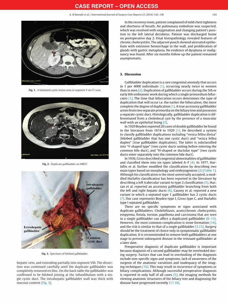
sea and vomiting. She had no history of jaundice or fever. Shehad been on iron supplement and oral contraceptive pill for irondeficiency anemia secondary to menorrhagia. Physical examina-tion revealed soft abdomen with no tenderness or palpable mass.Her blood investigations were normal including complete bloodcount, liver function test, bilirubin and tumor markers. Abdomi-nal ultrasound (US) showed a multi-septated echoic cystic lesionin the right liver adjacent to segment V and gallbladder. Abdomi-nal computed tomography (CT) showed a non-enhancing lobulatedcystic lesion in segment V with extension reaching the gallblad-der (Fig. 1). She was further investigated with magnetic resonancecholangiopancreatography (MRCP), which demonstrated a multi-locular cystic lesion communicating with biliary tree most likelyrepresenting duplicated gallbladder (Fig. 2). Patient was admittedfor elective laparoscopic cholecystectomy. Informed consent wasobtained after explaining the surgical procedure and possible com-plications. During surgery, while dissecting the gallbladder fromthe liver bed, a thick fibrous structure was found adherent to gall-bladder’s posterior surface at the infundibulum. Careful dissectionof this fibrous band revealed its communication with the intrahep-
atic cystic lesion that was anticipated and confirmed later to be theduplicate gallbladder.The dissection of the intrahepatic gallbladder was challengingbecause of its close proximity to the right portal vein and middle
Group Ltd. This is an open access article under the CC BY-NC-ND license (http://
CASE REPORT – OA. Al Rawahi et al. / International Journal of Su
Fig. 1. A lobulated cystic lesion seen in segment V on CT scan.
Fig. 2. Duplicate gallbladder on MRCP.
htccgm
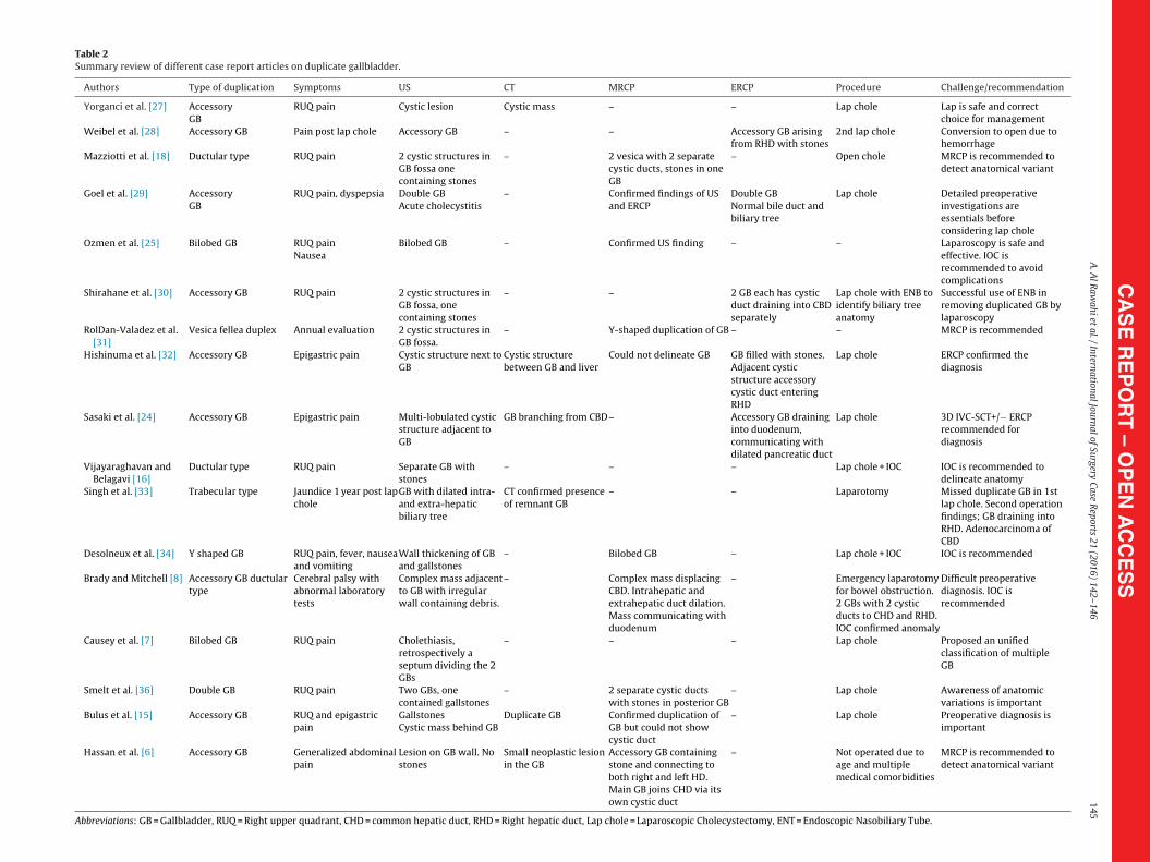
Fig. 3. Specimen of bilobed gallbladder.
epatic vein, and extending partially into segment VIII. The dissec-ion was continued carefully until the duplicate gallbladder was
ompletely removed en bloc. On the back table the gallbladder wasonfirmed to be bilobed joining at the infundibulum with a sin-le cystic duct. The intrahepatic gallbladder wall was thick withucous content (Fig. 3).PEN ACCESSrgery Case Reports 21 (2016) 142–146 143
In the recovery room, patient complained of mild chest tightnessand shortness of breath. Air pulmonary embolism was suspected,which was resolved with oxygenation and changing patient’s posi-tion to the left lateral decubitus. Patient was discharged homeon postoperative day 2. Final histopathology revealed features ofchronic cholecystitis. The adjacent pouch showed ulcerated epithe-lium with extensive hemorrhage in the wall, and proliferation ofglands with gastric metaplasia. No evidence of dysplasia or malig-nancy was found. After six months follow up the patient remainedasymptomatic.
3. Discussion
Gallbladder duplication is a rare congenital anomaly that occursin 1 per 4000 individuals [1], occurring nearly twice in womenthan in men [2]. Duplication of gallbladder occurs during the 5th orearly 6th embryonic week during which a single primordium bifur-cates [1]. The time that bifurcation occurs determines the type ofduplication that will occur i.e. the earlier the bifurcation; the morecomplete the degree of duplication [1]. A true accessory gallbladderarises from two separate primordia on the biliary tree and possessesa separate cystic duct. Histologically, gallbladder duplication is dif-ferentiated from a choledocal cyst by the presence of a muscularwall with an epithelial lining [3].
In 1929 Boyden reported 20 cases of double gallbladder he foundin the literature from 1674 to 1929 [1]. He described a systemto classify gallbladder duplications including “vesica fellea divisa”(bilobed gallbladder that has one cystic duct) and “vesica felleaduplex” (true gallbladder duplication). The latter is subclassifiedinto “Y-shaped type” (two cystic ducts uniting before entering thecommon bile duct), and “H-shaped or ductular type” (two cysticducts enter separately into the common bile duct).
In 1936, Gross described congenital abnormalities of gallbladderand classified them into six types labeled A–F [4]. In 1977, Har-laftis et al. further modified the classification by describing twomain types based on morphology and embryogenesis [2] (Table 1).Although his classification is the most universally accepted, a mod-ified Harlaftis classification has been reported in the literature bydescribing a left trabecular variant to type 2 classification [5]. Has-san et al. reported an accessory gallbladder branching from boththe left and right hepatic ducts [6]. Causey et al. reported a newvariant in which a septated type 1 gallbladder has 2 cystic ducts[7]. Our case represents Boyden type I, Gross type C, and Harlaftistype I septated gallbladder.
There are no specific symptoms or signs associated withduplicate gallbladders. Cholelithiasis, acute/chronic cholecystitis,empyema, fistula, torsion, papilloma and carcinoma that are seenin a single gallbladder can affect a duplicated gallbladder [8–13].However, the most common complication is stone formation [14],and the risk is similar to that of a single gallbladder [3,15]. Surgeryshould be the treatment of choice only in symptomatic gallbladderduplication. It is recommended to remove both gallbladders at onestage to prevent subsequent disease in the remnant gallbladder ata later date.
Preoperative diagnosis of duplicate gallbladder is importantbecause diagnosis of a second gallbladder may be overlooked dur-ing surgery. Factors that can lead to overlooking of the diagnosisinclude non-specific signs and symptoms, lack of awareness of thesurgeon of the anatomic variations and inadequacy of the imag-ing techniques [16]. This may result in recurrence of symptoms or
biliary complications. Although successful preoperative diagnosisis reported in only half of all cases [5], the imaging methods forviewing anatomic structures of the biliary tree and diagnosing thedisease have progressed recently [17,18].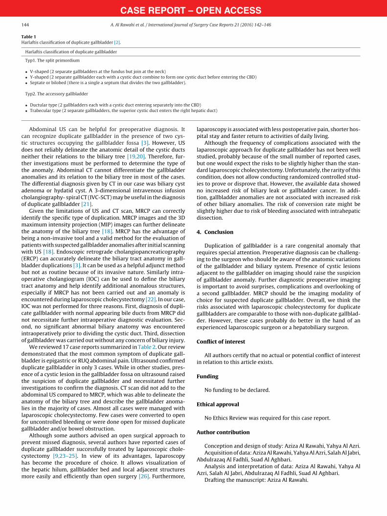
CASE REPORT – OPEN ACCESS144 A. Al Rawahi et al. / International Journal of Surgery Case Reports 21 (2016) 142–146
Table 1Harlaftis classification of duplicate gallbladder [2].
Harlaftis classification of duplicate gallbladder
Typ1. The split primordium
• V-shaped (2 separate gallbladders at the fundus but join at the neck)• Y-shaped (2 separate gallbladder each with a cystic duct combine to form one cystic duct before entering the CBD)• Septate or bilobed (there is a single a septum that divides the two gallbladder).
Typ2. The accessory gallbladder
he CBDht hep
ctdnttaTaco
imtbpw(bboteeIcnoio
dbdetiaallfg
pdchtm
• Ductular type (2 gallbladders each with a cystic duct entering separately into t• Trabecular type (2 separate gallbladders, the superior cystic duct enters the rig
Abdominal US can be helpful for preoperative diagnosis. Itan recognize duplicate gallbladder in the presence of two cys-ic structures occupying the gallbladder fossa [3]. However, USoes not reliably delineate the anatomic detail of the cystic ductseither their relations to the biliary tree [19,20]. Therefore, fur-her investigations must be performed to determine the type ofhe anomaly. Abdominal CT cannot differentiate the gallbladdernomalies and its relation to the biliary tree in most of the cases.he differential diagnosis given by CT in our case was biliary cystdenoma or hydatid cyst. A 3-dimensional intravenous infusionholangiography- spiral CT (IVC-SCT) may be useful in the diagnosisf duplicate gallbladder [21].
Given the limitations of US and CT scan, MRCP can correctlydentify the specific type of duplication. MRCP images and the 3D
aximum intensity projection (MIP) images can further delineatehe anatomy of the biliary tree [18]. MRCP has the advantage ofeing a non-invasive tool and a valid method for the evaluation ofatients with suspected gallbladder anomalies after initial scanningith US [18]. Endoscopic retrograde cholangiopancreaticography
ERCP) can accurately delineate the biliary tract anatomy in gall-ladder duplications [3]. It can be used as a helpful adjunct methodut not as routine because of its invasive nature. Similarly intra-perative cholangiogram (IOC) can be used to define the biliaryract anatomy and help identify additional anomalous structures,specially if MRCP has not been carried out and an anomaly isncountered during laparoscopic cholecystectomy [22]. In our case,OC was not performed for three reasons. First, diagnosis of dupli-ate gallbladder with normal appearing bile ducts from MRCP didot necessitate further intraoperative diagnostic evaluation. Sec-nd, no significant abnormal biliary anatomy was encounteredntraoperatively prior to dividing the cystic duct. Third, dissectionf gallbladder was carried out without any concern of biliary injury.
We reviewed 17 case reports summarized in Table 2. Our reviewemonstrated that the most common symptom of duplicate gall-ladder is epigastric or RUQ abdominal pain. Ultrasound confirmeduplicate gallbladder in only 3 cases. While in other studies, pres-nce of a cystic lesion in the gallbladder fossa on ultrasound raisedhe suspicion of duplicate gallbladder and necessitated furthernvestigations to confirm the diagnosis. CT scan did not add to thebdominal US compared to MRCP, which was able to delineate thenatomy of the biliary tree and describe the gallbladder anoma-ies in the majority of cases. Almost all cases were managed withaparoscopic cholecystectomy. Few cases were converted to openor uncontrolled bleeding or were done open for missed duplicateallbladder and/or bowel obstruction.
Although some authors advised an open surgical approach torevent missed diagnosis, several authors have reported cases ofuplicate gallbladder successfully treated by laparoscopic chole-
ystectomy [9,23–25]. In view of its advantages, laparoscopyas become the procedure of choice. It allows visualization ofhe hepatic hilum, gallbladder bed and local adjacent structuresore easily and efficiently than open surgery [26]. Furthermore,
)atic duct)
laparoscopy is associated with less postoperative pain, shorter hos-pital stay and faster return to activities of daily living.
Although the frequency of complications associated with thelaparoscopic approach for duplicate gallbladder has not been wellstudied, probably because of the small number of reported cases,but one would expect the risks to be slightly higher than the stan-dard laparoscopic cholecystectomy. Unfortunately, the rarity of thiscondition, does not allow conducting randomized controlled stud-ies to prove or disprove that. However, the available data showedno increased risk of biliary leak or gallbladder cancer. In addi-tion, gallbladder anomalies are not associated with increased riskof other biliary anomalies. The risk of conversion rate might beslightly higher due to risk of bleeding associated with intrahepaticdissection.
4. Conclusion
Duplication of gallbladder is a rare congenital anomaly thatrequires special attention. Preoperative diagnosis can be challeng-ing to the surgeon who should be aware of the anatomic variationsof the gallbladder and biliary system. Presence of cystic lesionsadjacent to the gallbladder on imaging should raise the suspicionof gallbladder anomaly. Further diagnostic preoperative imagingis important to avoid surprises, complications and overlooking ofa second gallbladder. MRCP should be the imaging modality ofchoice for suspected duplicate gallbladder. Overall, we think therisks associated with laparoscopic cholecystectomy for duplicategallbladders are comparable to those with non-duplicate gallblad-der. However, these cases probably do better in the hand of anexperienced laparoscopic surgeon or a hepatobiliary surgeon.
Conflict of interest
All authors certify that no actual or potential conflict of interestin relation to this article exists.
Funding
No funding to be declared.
Ethical approval
No Ethics Review was required for this case report.
Author contribution
Conception and design of study: Aziza Al Rawahi, Yahya Al Azri.Acquisition of data: Aziza Al Rawahi, Yahya Al Azri, Salah Al Jabri,
Abdulrazaq Al Fadhli, Suad Al Aghbari.Analysis and interpretation of data: Aziza Al Rawahi, Yahya Al
Azri, Salah Al Jabri, Abdulrazaq Al Fadhli, Suad Al Aghbari.Drafting the manuscript: Aziza Al Rawahi.
CA
SE
RE
PO
RT
– O
PE
N A
CC
ES
SA
. A
l R
awahi
et al.
/ International
Journal of
Surgery Case
Reports
21 (2016)
142–146
145Table 2Summary review of different case report articles on duplicate gallbladder.
Authors Type of duplication Symptoms US CT MRCP ERCP Procedure Challenge/recommendation
Yorganci et al. [27] AccessoryGB
RUQ pain Cystic lesion Cystic mass – – Lap chole Lap is safe and correctchoice for management
Weibel et al. [28] Accessory GB Pain post lap chole Accessory GB – – Accessory GB arisingfrom RHD with stones
2nd lap chole Conversion to open due tohemorrhage
Mazziotti et al. [18] Ductular type RUQ pain 2 cystic structures inGB fossa onecontaining stones
– 2 vesica with 2 separatecystic ducts, stones in oneGB
– Open chole MRCP is recommended todetect anatomical variant
Goel et al. [29] AccessoryGB
RUQ pain, dyspepsia Double GBAcute cholecystitis
– Confirmed findings of USand ERCP
Double GBNormal bile duct andbiliary tree
Lap chole Detailed preoperativeinvestigations areessentials beforeconsidering lap chole
Ozmen et al. [25] Bilobed GB RUQ painNausea
Bilobed GB – Confirmed US finding – – Laparoscopy is safe andeffective. IOC isrecommended to avoidcomplications
Shirahane et al. [30] Accessory GB RUQ pain 2 cystic structures inGB fossa, onecontaining stones
– – 2 GB each has cysticduct draining into CBDseparately
Lap chole with ENB toidentify biliary treeanatomy
Successful use of ENB inremoving duplicated GB bylaparoscopy
RolDan-Valadez et al.[31]
Vesica fellea duplex Annual evaluation 2 cystic structures inGB fossa.
– Y-shaped duplication of GB – – MRCP is recommended
Hishinuma et al. [32] Accessory GB Epigastric pain Cystic structure next toGB
Cystic structurebetween GB and liver
Could not delineate GB GB filled with stones.Adjacent cysticstructure accessorycystic duct enteringRHD
Lap chole ERCP confirmed thediagnosis
Sasaki et al. [24] Accessory GB Epigastric pain Multi-lobulated cysticstructure adjacent toGB
GB branching from CBD– Accessory GB draininginto duodenum,communicating withdilated pancreatic duct
Lap chole 3D IVC-SCT+/− ERCPrecommended fordiagnosis
Vijayaraghavan andBelagavi [16]
Ductular type RUQ pain Separate GB withstones
– – – Lap chole + IOC IOC is recommended todelineate anatomy
Singh et al. [33] Trabecular type Jaundice 1 year post lapchole
GB with dilated intra-and extra-hepaticbiliary tree
CT confirmed presenceof remnant GB
– – Laparotomy Missed duplicate GB in 1stlap chole. Second operationfindings; GB draining intoRHD. Adenocarcinoma ofCBD
Desolneux et al. [34] Y shaped GB RUQ pain, fever, nauseaand vomiting
Wall thickening of GBand gallstones
– Bilobed GB – Lap chole + IOC IOC is recommended
Brady and Mitchell [8] Accessory GB ductulartype
Cerebral palsy withabnormal laboratorytests
Complex mass adjacentto GB with irregularwall containing debris.
– Complex mass displacingCBD. Intrahepatic andextrahepatic duct dilation.Mass communicating withduodenum
– Emergency laparotomyfor bowel obstruction.2 GBs with 2 cysticducts to CHD and RHD.IOC confirmed anomaly
Difficult preoperativediagnosis. IOC isrecommended
Causey et al. [7] Bilobed GB RUQ pain Cholethiasis,retrospectively aseptum dividing the 2GBs
– – – Lap chole Proposed an unifiedclassification of multipleGB
Smelt et al. [36] Double GB RUQ pain Two GBs, onecontained gallstones
– 2 separate cystic ductswith stones in posterior GB
– Lap chole Awareness of anatomicvariations is important
Bulus et al. [15] Accessory GB RUQ and epigastricpain
GallstonesCystic mass behind GB
Duplicate GB Confirmed duplication ofGB but could not showcystic duct
– Lap chole Preoperative diagnosis isimportant
Hassan et al. [6] Accessory GB Generalized abdominalpain
Lesion on GB wall. Nostones
Small neoplastic lesionin the GB
Accessory GB containingstone and connecting toboth right and left HD.Main GB joins CHD via itsown cystic duct
– Not operated due toage and multiplemedical comorbidities
MRCP is recommended todetect anatomical variant
Abbreviations: GB = Gallbladder, RUQ = Right upper quadrant, CHD = common hepatic duct, RHD = Right hepatic duct, Lap chole = Laparoscopic Cholecystectomy, ENT = Endoscopic Nasobiliary Tube.
– O1 l of Su
t
A
G
R
[
[
[
[
[
[
[
[
[
[
[
[
[
[
[
[
[
[
[
[
[
[
[
[
[
[
OTpc
CASE REPORT46 A. Al Rawahi et al. / International Journa
Revising the manuscript critically for important intellectual con-ent: Aziza Al Rawahi, Yahya Al Azri.
Approval of the version of the manuscript to be published: Azizal Rawahi, Yahya Al Azri, Salah Al Jabri,
Abdulrazaq Al Fadhli, Suad Al Aghbari.
uarantor
Aziza Al Rawahi.Yahya Al Azri.
eferences
[1] E.A. Boyden, The accessory gallbladder—an embryological and comprarativestudy of aberrant biliary vesicles occurring in man and the domesticmammals, Am. J. Anat. 38 (1926) 177–231.
[2] N. Harlaftis, S.W. Gray, J.E. Skandalakis, Multiple gallbladders, Surg. Gynecol.Obstet. 145 (1977) 928–934.
[3] J. Gigot, B. Van Beers, L. Goncette, et al., Laparoscopic treatment of gallbladderduplication: a plea for removal of both gallbladders, Surg. Endosc. 11 (1997)479–482.
[4] R.E. Gross, Congenital anomalies of the gallbladder: a review of 148 caseswith a report of double gallbladder, Arch. Surg. 32 (1936) 131–159.
[5] R.D. Kim, I. Zendejas, C. Velopulos, et al., Duplicate gallbladder arising fromthe left hepatic duct: report of a case, Surg. Today 39 (2009) 536–553.
[6] S. Hassan, A.L. Young, M.F. Farooq, et al., Accessory gallbladder: a newanatomical variation arising from both left and right hepatic ducts, Ann. R.Coll. Surg. Engl. 94 (2012) e204–e205.
[7] M.W. Causey, S. Miller, A. Colby, et al., Gallbladder duplication: evaluation,treatment and classification, J. Pediatr. Surg. 45 (2010) 443–446.
[8] K. Brady, A. Mitchell, Cholecystitis of a duplicated gallbladder complicated bya cholecystoenteric fistula, Pediatr. Radiol. 39 (2009) 385–388.
[9] J.C. Garcia, A. Weber, F.S. Berry, et al., Double gallbladder treated successfullyby laparoscopy, J. Laparoendosc. Surg. 3 (1993) 153–155.
10] J.J. Cunningham, Empyema of duplicated gallbladder: echographic findings, J.Clin. Ultrasound 8 (1980) 511–512.
11] S.W. Raymond, C.B. Thrift, Carcinoma of a duplicated gallbladder, III Med. J.110 (1956) 239–240.
12] A.W.S. Ritchie, V. Crucioli, Double gallbladder with cholecystocolic fistula: acase report, Br. J. Surg. 67 (1980) 145–146.
13] W.J. Roeder, W.L. Mersheimer, K.K. Kazarian, Triplication of the gallbladderwith cholecystitis, cholethiasis and papillary adenocarcinoma, Am. J. Surg.
121 (1971) 746–748.14] R. Silvis, A.J. Van Wieringen, C.H. Van Der Werken, Re-operation forsymptomatic double gallbladder, Surg. Endosc. 10 (1996) 336–337.
15] K. Bulus, A. Koyuncu, A. Coskun, Preoperative diagnosis of double gallbladder:a case report, Turk. J. Gastroenterol. 23 (2) (2012) 172–174.
[
pen Accesshis article is published Open Access at sciencedirect.com. It is distribermits unrestricted non commercial use, distribution, and reproductredited.
PEN ACCESSrgery Case Reports 21 (2016) 142–146
16] R. Vijayaraghavan, C. Belagavi, Double gallbladder with different diseaseentities: a case report, J. Minim. Access Surg. 2 (1) (2006) 23–26.
17] D. Urbain, J. Jeanmort, P. Janne, Double gallbladder with transient cholestosis:preoperative demonstration by endoscopic retrpgrade cholangiography,Gastrointest. Endosc. 35 (1980) 346–348.
18] S. Mazziottie, F. Minutoli, A. Blandino, et al., Gallbladder duplication: MRcholangiography demonstration, Abdom. Imaging 26 (2001) 287–289.
19] O. Oyar, A. Yesildag, U. Gulsoy, et al., Bilobed gallbladder diagnosed by oralcholecysto-CT, Comput. Med. Imaging Gr. 27 (2003) 315–319.
20] R.A. Lichtenbaum, H.F. McMullen, R.M. Newman, Preoperative abdominalultrasound may be misleading in risk stratification for presence of commonbile duct abnormalities, Surg. Endosc. 14 (2000) 254–257.
21] A.H. Kwon, H. Inui, A. Inamura, et al., Preoperative assessment forlaparoscopic cholecystectomy: feasibility of using spiral computatedtomography, Ann. Surg. 227 (1998) 351–356.
22] R.D. Cummiskey, L.P. Champagne, Duplicate gallbladder during laparoscopiccholecystectomy, Surg. Laparosc. Endosc. 7 (3) (1997) 268–270.
23] N. Miyajima, T. Yamakava, A. Varma, et al., Experience with laparoscopicdouble gallbladder removal, Surg. Endosc. 9 (1995) 63–66.
24] A. Sasaki, T. Yoshida, K. Lalisako, et al., Laparoscopic cholecystectomy for adouble gallbladder of the duodenal type, Surg. Laparosc. Endosc. Percutan.Tech. 15 (6) (2005) 355–358.
25] V. Ozmen, E. Gorgun, E.S. Unal, et al., Laparoscopic treatment of a bilobedgallbladder: a case report and review of the literature, Surg. Laparosc. Endosc.Percutan. Tech. 13 (5) (2003) 345–347.
26] D.W. Crist, T.R. Gadacz, Laparoscopic anatomy of the biliary tree, Surg. Clin.North Am. 73 (1993) 785–798.
27] K. Yorganci, B. Kabay, O. Aran, Laparoscopic double cholecystectomy, Surg.Laparosc. Endosc. Percutan. Tech. 11 (2) (2001) 126–128.
28] D. Weibel, M. Kaufmann, H. Reidtmann-Klee, Accessory gallbladderoriginating from the right hepatic duct, Surg. Endosc. 15 (5) (2001) 519.
29] A. Goel, K.N. Strivastava, A.K. Rana, Double gallbladder—a laparoscopicmanagement, Surg. Laparosc. Endosc. Percutan. Tech. 13 (5) (2003) 348–349.
30] K. Shirahane, K. Yamaguchi, T. Ogawa, et al., Gallbladder duplicationsuccessfully removed laparoscopically using endoscopic nasobiliary tube,Surg. Endosc. 17 (2003) 1156–1157.
31] E. RolDan-Valadez, S. Osorio-Peralta, et al., Asymptomatic true gallbladderduplication: a case report and review of the literature, Acta Radiol. 45 (2004)810–814.
32] M. Hishinuma, Y. Isogai, Y. Matsuura, et al., Double gallbladder, J.Gastroenterol. Hepatol. 19 (2004) 232–241.
33] B. Singh, L. Ramsaroop, L. Allopi, et al., Duplicate gallbladder: an usual casereport, Surg. Radiol. Anat. 28 (2006) 654–657.
34] G. Desolneux, S. Mucci, J. Lebigot, et al., Duplication of the gallbladder: a casereport, Gastroenterol. Res. Pract. 3 (2009) 1–3.
35] J.J. Gagnier, G. Kienle, D.G. Altman, D. Moher, H. Sox, D. Riley, CARE Group. TheCARE guidelines: consensus-based clinical case report guidelinedevelopment, J. Clin. Epidemiol. 67 (1) (Jan 2014) 46–51.
36] J.L. Smelt, H. Wright, J. Sagar, D. Nehra, Laparoscopic removal of a doublegallbladder for cholelithiasis: a case report, Ann. R. Coll. Surg. Engl. 93(September (6)) (2011) e105–e106.
uted under the IJSCR Supplemental terms and conditions, whichion in any medium, provided the original authors and source are


![Gallbladder · Latin vesica fellea; vesica biliaris Gray's subject #250 1197 [1] System Digestive system (GI Tract) Artery Cystic artery Vein Cystic vein Nerve Celiac ganglia, vagus[2]](https://img.pdfslide.us/doc/110x75/5c8da86009d3f219388ce415/gallbladder-latin-vesica-fellea-vesica-biliaris-grays-subject-250-1197-1.jpg)


![[1] Gallia est omnis divisa [in partes tres], quarum...[1] Gallia est omnis divisa [in partes tres], quarum unam incolunt Belgae, aliam Aquitani, tertiam qui ipsorum [lingua] Celtae,](https://img.pdfslide.us/doc/110x75/5fb798a481c7ff0b6e21b14c/1-gallia-est-omnis-divisa-in-partes-tres-quarum-1-gallia-est-omnis-divisa.jpg)