Embed Size (px)
Citation preview
Ramteke, et al. Int J Pharm 2014; 4(4):297-308 ISSN 2249-1848
www.pharmascholars.com 297
Review Article CODEN: IJPNL6
BUCCAL PATCHES- A REVIEW
Ramteke K.H*1
, Dighe P.A*1
, Kharat A. R1, Patil S.V
2
1PES’s Modern College of Pharmacy (for ladies), Moshi, Pune, India
2Ashokrao Mane College of Pharmacy, Peth-Vadgaon, Kolhapur, India
*Corresponding author e-mail: [email protected], [email protected]
ABSTRACT
Transmucosal drug delivery is an alternative method of systemic delivery of the drug which offers different
advantages over existing methods by enhancing the bioavailability of drug due to rich in blood supply in mucosal
surface and prolongs residence time at the site of application to permit once or twice daily dosing. Buccal route is an
attractive and easy transmucosal route of administration for systemic drug delivery. Delivery of the drug via buccal
route leads direct access to the systemic circulation through the internal jugular vein bypasses drugs from hepatic
first pass metabolism leading to higher bioavailability. Buccal bioadhesive patches, releases topical drugs in the oral
cavity at a slow and predetermined rate and provides advantages over traditional dosage forms for treatment of many
diseases and disorders. The objective of this article is to review buccal patches by discussing their composition,
method of preparation and evaluation.
KEYWORDS: Transmucosal, bioavailability, mucosal surface, residence time, buccal patches.
INTRODUCTION
There are different routes of administration of dosage
form of specific drug; among that oral route is most
commonly preferred route. But there are several
disadvantages of this route like hepatic first pass
metabolism and enzymatic degradation in the GI
tract, that prohibit oral administration of number of
drugs especially peptides and proteins. These
problems are overcome by using transmucosal route.
This route involves the delivery of the drug through
the mucosa hence, also called as “Bioadhesive drug
delivery system”.
The term bioadhesion is generally used to describe
the adhesion between polymer (synthetic or natural)
to soft tissue of the body. In the mucus membrane the
goblet cells are present for the secretion of mucus,
which is composed of glycoprotein mucin.
Buccal delivery of drug provides an easy alternative
to the oral route of drug administration. As drug
action can be terminated in case of toxicity by
removing the dosage from the buccal cavity, buccal
delivery offers a safer method of drug delivery. It is
also possible to administer drugs to patients who
cannot be given drugs orally1-2
.
Buccal route provides the direct access to the
systemic circulation through the jugular vein
bypassing the first pass hepatic metabolism leading to
higher bioavailability. Other advantages such as
excellent accessibility, low enzymatic activity,
suitability for drugs or excipients that damages or
irritate the mucosa, administration without pain, easy
removal, facility to include permeation enhancer or
enzyme inhibitor or pH modifier in the formulation,
versatility in designing as multidirectional or
unidirectional release system for local or systemic
effect3.
Buccal patch is a non dissolving thin matrix
modified release dosage form composed of one or
more polymer films or layers containing the drug
and/or other excipients. The patch may contain a
mucoadhesive polymer layer which bonds to the oral
mucosa, gingiva, or teeth for controlled release of the
drug into the oral mucosa (unidirectional release),
oral cavity (unidirectional release), or both
(bidirectional release). The patch is removed from the
mouth and disposed of after a specified time4.
Buccal patches are highly flexible and thus
readily tolerated by the patient than tablets or
other dosage form. Patches are also ensure more
International Journal of Pharmacy Journal Homepage: http://www.pharmascholars.com
Ramteke, et al. Int J Pharm 2014; 4(4):297-308 ISSN 2249-1848
www.pharmascholars.com 298
accurate dosing of the drug as compared to gels and
ointments5.An ideal buccal patch should be flexible,
elastic, soft and strong to withstand breakage due to
stress because of mouth activities. Moreover, it must
also exhibit good mucoadhesive strength so that it
can be retained in the mouth for appropriate time. As
such, the mechanical, mucoadhesive, and swelling
properties of buccal patches are important and
essential to be evaluated.
Structure of buccal mucosa6-7
:-
The oral mucosa is composed of (Fig.1.)6
a) Stratified squamous epithelium
b) Lamina propria
c) Submucosa
a) Stratified squamous epithelium:-
The buccal epithelium contains of 40 to 50 layers of
non keratinized stratified squamous cells. It is about
500 to 800μm in thickness with varying degrees of
maturity. The uppermost superficial layer of cells is
made up of flattened compact differentiated cells
having 150μm thickness. Oral mucosae are leaky
epithelia intermediate between the epidermis and
intestinal mucosa. Permeability is 4- 4000 times
greater than that of skin.
b) Lamina propria:-
The lamina propria is also called as basement
membrane it is a continuous layer of extracellular
materials and forms a boundary between the basal
layer of epithelium and the connective tissues. The
lamina propria comprises collagen fibrils, which is a
supporting layer of connective tissue, blood vessels,
and smooth muscle. The structure of the lamina
propria is not much dense and it is not a barrier to
drug permeation.
c) Submucosa:-
The submucosa is a dense connective tissue that
contains a few accessory salivary glands, called as
mucus acinus. Mucus acini are surrounded by
myoepithelial cells that help in the secretion of saliva.
The oral cavity is marked by the presence of saliva
produced by the salivary glands. The mucus is
secreted by the major and minor salivary glands as
part of saliva.
Mechanism of buccal absorption8:-
The absorption of the drug through buccal mucosa
follows three steps mechanism.
Step 1:- Wetting and swelling of polymer to permit
intimate contact with biological tissue (Fig.2.)9.
Step 2:- Inter-penetration of bioadhesive polymer
(BP) chains and entanglement of polymer and mucin
chains (Fig.3.)9.
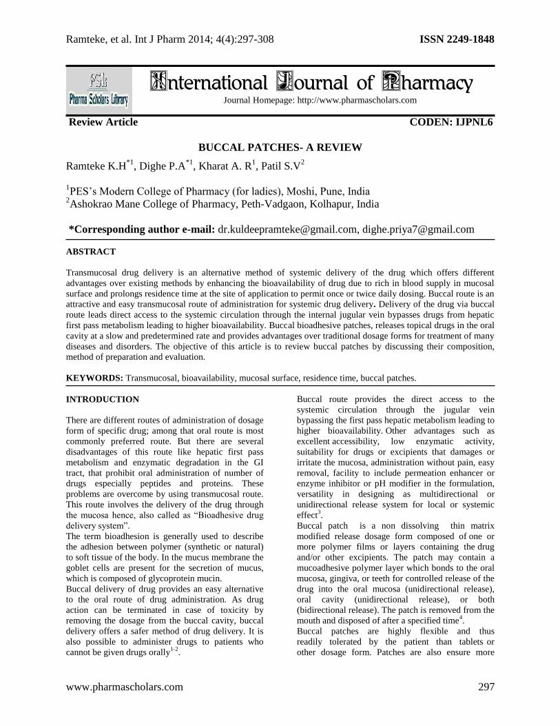
Step 3:- Formation of chemical bonds between the
entangled chains (Fig.4.)9.
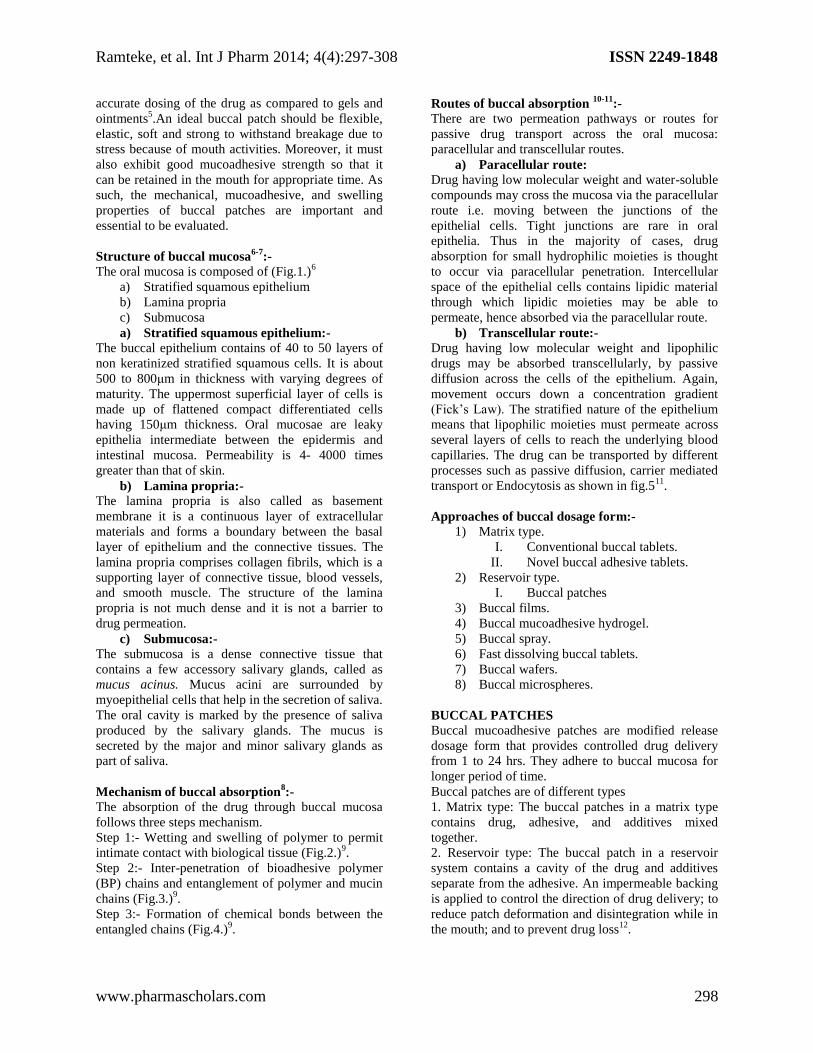
Routes of buccal absorption 10-11
:-
There are two permeation pathways or routes for
passive drug transport across the oral mucosa:
paracellular and transcellular routes.
a) Paracellular route:
Drug having low molecular weight and water-soluble
compounds may cross the mucosa via the paracellular
route i.e. moving between the junctions of the
epithelial cells. Tight junctions are rare in oral
epithelia. Thus in the majority of cases, drug
absorption for small hydrophilic moieties is thought
to occur via paracellular penetration. Intercellular
space of the epithelial cells contains lipidic material
through which lipidic moieties may be able to
permeate, hence absorbed via the paracellular route.
b) Transcellular route:-
Drug having low molecular weight and lipophilic
drugs may be absorbed transcellularly, by passive
diffusion across the cells of the epithelium. Again,
movement occurs down a concentration gradient
(Fick’s Law). The stratified nature of the epithelium
means that lipophilic moieties must permeate across
several layers of cells to reach the underlying blood
capillaries. The drug can be transported by different
processes such as passive diffusion, carrier mediated
transport or Endocytosis as shown in fig.511
.
Approaches of buccal dosage form:-
1) Matrix type.
I. Conventional buccal tablets.
II. Novel buccal adhesive tablets.
2) Reservoir type.
I. Buccal patches
3) Buccal films.
4) Buccal mucoadhesive hydrogel.
5) Buccal spray.
6) Fast dissolving buccal tablets.
7) Buccal wafers.
8) Buccal microspheres.
BUCCAL PATCHES
Buccal mucoadhesive patches are modified release
dosage form that provides controlled drug delivery
from 1 to 24 hrs. They adhere to buccal mucosa for
longer period of time.
Buccal patches are of different types
1. Matrix type: The buccal patches in a matrix type
contains drug, adhesive, and additives mixed
together.
2. Reservoir type: The buccal patch in a reservoir
system contains a cavity of the drug and additives
separate from the adhesive. An impermeable backing
is applied to control the direction of drug delivery; to
reduce patch deformation and disintegration while in
the mouth; and to prevent drug loss12
.

Ramteke, et al. Int J Pharm 2014; 4(4):297-308 ISSN 2249-1848
www.pharmascholars.com 299
They consist of solid matrix (non-dissolvable or
slowly dissolvable). They may be
Unidirectionally.
Bidirectionally.
Multidirectionally.
Adhesive polymer itself acts as drug carrier or
adhesive layer link between drugs loaded layer and
mucosa. Size generally 1-16 cm2 but 1-3 cm
2 used
.Large sized patches are placed at central position of
buccal mucosa.
Three basic types of buccal patches to achieve
targeted drug release.
a. Monolithic matrix( for multidirection
release)
b. Multilayer matrix ( having semi permeable
backing layer)
c. Multilayer matrix (having impermeable
layer over back and side of device)
COMPOSITION OF BUCCAL PATCHES:
1. Active ingredient
2. Polymers (adhesive layer)
3. Diluents
5. Sweetening agents
6. Flavoring agents
7. Backing layer
8. Penetration enhancer
9. Plasticizers
1. Active ingredients13-14
:-
Active drug (s) used in the formulation of buccal
patches, should have following characteristics
1. The conventional single dose of the drug should be
small.
The drugs having biological half-life between 2-8
hours are good candidates for controlled drug
delivery.
2. Tmax of the drug shows wider-fluctuations or higher
values when given orally.
3. The drug absorption should be passive when given
orally.
2. Polymers(adhesive layer)15
:-
The first step in the development of buccoadhesive
dosage forms is the selection and characterization of
bioadhesive polymers in the formulation.
Bioadhesive polymers are most important in
buccoadhesive drug delivery systems of drugs.
Polymers are also used in matrix devices in which the
drug is mixed in the polymer matrix, which controls
the time of release of drugs. An ideal polymer for
buccoadhesive drug delivery systems should have
following characteristics.
1. It should be inert and compatible with the
environment
2. The polymer and its degradation products should
be non-toxic absorbable from the mucous layer.
3. It should adhere quickly to moist tissue surface and
should possess some site Specificity.
4. The polymer must not decompose on storage or
during the shelf life of the dosage form.
5. The polymer should be easily available in the
market and economical.
Criteria followed in polymer selection
Non-toxic, non-irritant and free from leachable
impurities.
Should adhere quickly to buccal mucosa and
should possess sufficient mechanical strength
Should possess peel, tensile and shear strengths
at the bioadhesive range.
Good spreadability, wetting, swelling and
solubility and biodegradability properties.
It should form a strong non covalent bond with
the mucin or epithelial surface
It must have high molecular weight and narrow
distribution.
Should show bioadhesive properties in both dry
and liquid state.
It should be compatible with the biological
membrane.
The polymers that are commonly used as bioadhesive
in pharmaceutical applications are16
:
1. Natural polymers
i. Tragacanth
ii. Sodium alginate
iii. Guar gum
iv. Xanthan gum
v. Soluble starch
vi. Gelatin
vii. Chitosan
2. Synthetic and semisynthetic polymers
i. Cellulose derivatives (Methylcellulose, Ethyl
cellulose, HEC, HPC, HPMC, Sod.CMC).
ii. Poly (Acrylic acid) polymers (Carbomers,
Polycarbophil).
iii. Poly ethylene oxide.
iv. Poly vinyl alcohol.
v. Poly hydroxyl ethyl methylacrylate.
vi. Poly vinyl pyrrolidone.
3. Diluents, Sweeteners and Flavors:-
These are the pharmaceutical excipients used to
enhance the morphological properties of the dosage
form, such as size, taste, and odor.
As the patches are of buccal use taste and odor are
also taken into considerations for that purpose
flovours and sweeteners are used. Diluents are used
as fillers for the low dose of the drug.
Diluents- eg. Lactose, Microcrystaline starch, starch.

Ramteke, et al. Int J Pharm 2014; 4(4):297-308 ISSN 2249-1848
www.pharmascholars.com 300
Sweetening agents- eg. Sucralose, aspartame,
mannitol
Flavoring agents- eg. Menthol, vanillin, clove oil
4. Backing layer17
:-
Backing membrane plays important role in the
attachment of bioadhesive devices to the mucus
membrane. The materials used as backing membrane
should be inert, and impermeable to the drug and
penetration enhancer to prevent release of it from the
patch.
The commonly used materials include carbopol,
magnesium separate, HPMC, HPC, CMC, sodium
CMC, polycarbophil etc.
5. Penetration enhancer17-18
:-
Membrane permeation is the major limiting factor for
many drugs in the development of buccal adhesive
patches. The epithelium which lines the buccal
mucosa is a very effective barrier to the absorption of
drugs. Materials that facilitate the permeation of drug
through buccal mucosa are called as permeation
enhancers.
Penetration enhancers are enhances the release of the
drug. They are also helps in the systemic drug
delivery by allowing the drug to penetrate more
readily into the viable tissues.
Mechanism of penetration enhancers are as follows19
Changing mucus rheology
Increasing the fluidity of lipid bilayer membrane
Acting on the components at tight junctions
By overcoming the enzymatic barrier
Increasing the thermodynamic activity of drugs
Eg.
Chelators: EDTA, citric acid, sodium salicylate,
methoxy salicylates.
Surfactants: sodium lauryl sulphate,
polyoxyethylene,, Benzalkonium chloride,
cetylpyridinium chloride, cetyltrimethyl
ammonium bromide.
Bile salts: sodium glycocholate, sodium
deoxycholate, sodium taurocholate, sodium
glycodeoxycholate, sodium taurodeoxycholate.
Fatty acids: oleic acid, capric acid, lauric acid,
lauric acid/ propylene glycol, methyloleate,
phosphatidylcholine.
Non-surfactants: unsaturated cyclic ureas.
Inclusion complexes: cyclodextrins.
6. Plasticizers20
:-
Plasticizers are important factors that affect
mechanical properties of films. The mechanical
properties like tensile strength and elongation to the
films. Variation in the concentration of plasticizers
may affect these properties. The commonly used
plasticizers are glycerol, di-butylpthallate, and
polyethylene glycol etc.
Method of preparation21
:-
There are six methods of preparation of buccal
patches.
1. Solvent casting method
2. Semisolid casting method
3. Solid dispersion extrusion method
4. Rolling Method
5. Direct milling method
6. Hot melt extrusion method
1. Solvent casting method:-
In this method, all patch excipients including the drug
co-dispersed in an organic solvent and coated onto a
sheet of release liner. After solvent evaporation a thin
layer of the protective backing material is laminated
onto the sheet of coated release liner to form a
laminate that is die- cut to form parches of the
desired size and geometry.
2. Semisolid casting method:-
In semisolid casting method firstly a solution of
water soluble film forming polymer is prepared.
The resulting solution is added to a solution of acid
insoluble polymer (e.g. cellulose acetate phthalate,
cellulose acetate butyrate), which was prepared in
ammonium or sodium hydroxide. Then appropriate
amount of plasticizer is added so that a gel mass
is obtained. Finally the gel mass is casted in to the
films or ribbons using heat controlled drums. The
thickness of the film is about 0.015-0.05 inches. The
ratio of the acid insoluble polymer to film forming
polymer should be 1:4.
3. Solid dispersion extrusion method:-
In this method immiscible components are extrude
with drug and then solid dispersions are prepared.
Finally the solid dispersions are shaped in to films by
means of dies.
4. Rolling Method:-
In rolling method a solution or suspension
containing drug is rolled on a carrier. The solvent
is mainly water and mixture of water and alcohol.
The film is dried on the rollers and cut in to desired
shapes and sizes. (Fig.7)22
5. Direct milling method21-22
:-
In this, patches are manufactured without the use of
solvents. Drug and excipients are mechanically
mixed by direct milling or by kneading, usually
without the presence of any liquids. After the mixing
process, the resultant material is rolled on a release
liner until the desired thickness is achieved. The
backing material is then laminated as previously
described. While there are only minor or even no
differences in patch performance between patches
fabricated by the two processes, the solvent-free
process is preferred because there is no possibility of

Ramteke, et al. Int J Pharm 2014; 4(4):297-308 ISSN 2249-1848
www.pharmascholars.com 301
residual solvents and no associated solvent-related
health issues.
6. Hot melt extrusion method:-
In hot melt extrusion blend of pharmaceutical
ingredients is molten and then forced through an
orifice to yield a more homogeneous material in
shape of films. Hot melt extrusion has been used
for the manufacture of controlled release matrix
tablets, pellets and granules, as well as oral
disintegrating films.
The normal buccal patch is as given in Fig.822
.
EVALUATION
1. Thickness23-25
The thickness of each patch measured by using
thickness tester or standard screw gauge or
Electronic digital micrometer at different positions
of the patch and calculate the average.
2. Weight uniformity23
Cut the patch size of 1 x 1 cm2 or 10 mm. Take the
weight of each patch and calculate the weight
variation.
3. Surface pH study26-28
Buccal patches are swell within 1 hr on the
surface of the agar plate, prepared by dissolving
2% (w/v) agar in warmed isotonic phosphate buffer
of pH 6.8 under stirring and then poured the solution
into the petridish allowed to stand till gelling at room
temperature. Measure the surface pH by pH paper
placed on the surface of the swollen patch. Calculate
the mean of three readings.
4. Morphological characterization
Morphological characters are studied by using
scanning electron microscope (SEM).
5. Content uniformity29-37
Drug content uniformity was determined by
dissolving the buccal patch (10 mm in diameter) from
each batch by homogenization in 100 ml of an
isotonic phosphate buffer (pH 6.8) for 6 hrs under
occasional shaking. The 5 ml of resulting solution
was diluted to 20ml with buffer and filtered through a
whattman filter paper. The drug content was then
determined after proper dilution and measured the
absorbance by using a UV-visible
spectrophotometer.
6. Folding endurance38-39
Folding endurance of the patches determined by
repeatedly folding one patch at the same place
till it broke or folded upto 200 or 300 times
manually, which was considered satisfactory to
reveal good patch properties. The number of times of
film could be folded at the same place without
breaking gave the value of the folding endurance.
7. Swelling % study40-41
Buccal patches are weighed individually (W1), and
placed separately in 2% agar gel plates, incubated at
37°C ± 1°C. Plates examined for any physical
changes at regular 1 hour time intervals until 3 hours,
patches are removed from the gel plates and excess
surface water is removed carefully using the filter
paper. The swollen patches are then reweighed (W2)
and the swelling index (SI) is calculated using the
following formula.
8. Water absorption capacity test42
Circular Patches, with a surface area of 2.3 cm2 are
allowed to swell on the surface of agar plates
prepared in simulated saliva (2.38 g Na2HPO4, 0.19
gKH2 PO4, and 8 g NaCl per lit. of distilled water
adjusted with phosphoric acid to pH 6.7), and kept in
an incubator maintained at 37°C ± 0.5°C. At various
time intervals (0.25, 0.5, 1, 2, 3, and 4 hours),
samples are weighed (wet weight) and then left to dry
for 7 days in a desicator over anhydrous calcium
chloride at room temperature then the final constant
weights are recorded. Water uptake (%) is calculated
using the following equation,
Where, Ww is the wet weight, Wi is the initial weight
and Wf is the final weight. The swelling of each film
is measured.
9. Thermal analysis study
Thermal analysis study is performed using
differential scanning calorimeter (DSC).
10. Ex vivo Bioadhesion test43
The fresh sheep mouth separated and washed with
phosphate buffer (pH 6.8). A piece of gingival
mucosa is tied in the open mouth of a glass vial,
filled with phosphate buffer (pH 6.8). This glass vial
is tightly fitted into a glass beaker filled with
phosphate buffer (pH 6.8, 37°C ± 1°C) so it just
touched the mucosal surface. The patch is stuck to
the lower side of a rubber stopper with cyano acrylate
adhesive. Two pans of the balance are balanced with
a 5g weight. The 5g weight is removed from the left
hand side pan, which loaded the pan attached with
the patch over the mucosa. The balance is kept in this
position for 5 minutes of contact time. The water is

Ramteke, et al. Int J Pharm 2014; 4(4):297-308 ISSN 2249-1848
www.pharmascholars.com 302
added slowly at 100 drops/min to the right-hand side
pan until the patch detached from the mucosal
surface (Fig.9.)43
. The weight, in grams, required to
detach the patch from the mucosal surface provided
the measure of mucoadhesive strength.
11. In vitro drug release44
The United States Pharmacopeia (USP) XXIII-B
rotating paddle apparatus or type I apparatus is used
to study the drug release from the patches.
Dissolution medium- phosphate buffer pH 6.8
Temperature- 37°C ± 0.5°C
Rotations- 50 rpm.
The backing layer of buccal patch is attached to the
glass disk with instant adhesive material. The disk is
allocated to the bottom of the dissolution vessel.
Samples (5 ml) are withdrawn at predetermined time
intervals and replaced with fresh medium. The
samples filtered through whattman filter paper and
analyzed for drug content after appropriate dilution.
12. In vitro permeation study of buccal patch45-46
The in- vitro buccal permeation through the buccal
mucosa (sheep and rabbit) is performed using
Keshary-Chien/Franz type glass diffusion cell at
37°C± 0.2°C (Fig.10.). Fresh buccal mucosa
is mounted between the donor and receptor
compartments. The buccal patch is placed with the
core facing the mucosa and the compartments
clamped together. The receptor compartment is filled
with phosphate buffer pH 6.8, and the hydrodynamics
in the receptor compartment is maintained by stirring
with a magnetic bead at 50 rpm. Samples are
withdrawn at predetermined time intervals and
analyzed for drug content.
13. Ex vivo mucoadhesion time47
The fresh buccal mucosa is tied on the glass slide,
and a mucoadhesive patch is wetted with 1 drop of
phosphate buffer pH 6.8 and pasted to the buccal
mucosa by applying a light force with a fingertip for
30 seconds. The glass slide is then put in the beaker,
which is filled with 200 ml of the phosphate buffer
pH 6.8, is kept at 37°C ± 1°C. After 2 minutes, a 50-
rpm stirring rate is applied to simulate the buccal
cavity environment, and patch adhesion is monitored
for 12 hours. The time for changes in colour, shape,
collapsing of the patch and drug content is noted.
14. In vivo residence time48
The experiment was performed in eight healthy adult
male volunteers, aged between 22 and 28 years. The
volunteers were asked to record the residence time of
the patch on buccal mucosa in the oral cavity, which
was taken as the time for the patch to dislodge
completely from the buccal mucosa by continual
sensation of the patch as well as the backing
membrane. In vivo residence time was recorded in
each case.
15. Tensile strength49-50
Tensile strength of the films (patches) includes
evaluated using a tensile tester. Film strip with the
dimensions of 60 x 10 mm cut and positioned
between two clamps separated by a distance of 3 cm.
The lower clamp held stationary and the strips are
pulled apart by the upper clamp moving at a rate of 2
mm/sec until the strip break. The force and
elongation of the film at the point when the trip break
is recorded. The tensile strength and elongation at
break values are calculated using the formula,
Where,
M-Mass in gm, g – Acceleration due to gravity (980
cm/sec2), B- Breadth of the specimen in cm
T- Thickness of specimen in cm
16. Stability study in human saliva51
The human saliva is collected from humans (age 18-
50years). Buccal patches are placed in separate
petridishes containing 5ml of human saliva at temp.
37°C ± 0.2°C for 6 hours. At regular time intervals
(0, 1, 2, 3, and 6 hours), the sample is collected and
absorbance is taken to check stability.
17. Vapor transmission test (VTR)49,52
Vapor transmission method was employed for the
determination of vapor transmission from the patch.
Glass-bottle (length= 5 cm, narrow mouth with
internal diameter =0.8 cm) filled with 2 g anhydrous
calcium chloride and an adhesive spread across its
rim, was used in the study. The patch was fixed over
the adhesive and the assembly was placed in a
constant humidity chamber, prepared using saturated
solution of ammonium chloride and maintained at
37±2 °C. The difference in weight after 24 h, 3rd
day
and 1 week was calculated. The experiments were
carried out in triplicate and vapor transmission rate
was obtained as follow:
VTR = (Amount of moisture transmitted) / (Area x
Time)
ADVANTAGES53-58
:-
i. Avoid first pass effect
ii. Painless, easy and comfortable application.
iii. Larger buccal area to allow drug delivery to be
placed at different occasion (i.e. left or right cheek).
iv. Drug is protected from degradation due to pH and
digestive enzymes of the middle gastrointestinal
tract.
v. Easy termination in case of overdose.

Ramteke, et al. Int J Pharm 2014; 4(4):297-308 ISSN 2249-1848
www.pharmascholars.com 303
vi. Enhances the stability of drug.
vii. Rapid onset of action.
viii. Easy for unconscious and uncapaciated patients.
ix. Improved patient compliance due to the elimination
of associated pain with injections.
x. Sustained drug delivery.
xi. In comparison to TDDS, mucosal surfaces do not
have a stratum corneum. Thus, the major barrier
layer to transdermal drug delivery is not a factor in
transmucosal routes of administration. Hence
transmucosal systems exhibit a faster initiation and
decline of delivery than do transdermal patches.
xii. Though less permeable than the sublingual area, the
buccal mucosa is well vascularized, and drugs can
be rapidly absorbed into the venous system
underneath the oral mucosa.
xiii. Transmucosal delivery occurs fewer variables
between patients, resulting in lower intersubject
variability as compared to transdermal patches.
LIMITATIONS59-62
:-
i. Saliva is continuously secreted into the oral
cavity diluting drugs at the site of absorption
resulting in low drug concentrations at the
surface of the absorbing membrane. Involuntary
swallowing of saliva results in a major part of
dissolved or suspended released drug being
removed from the site of absorption.
Furthermore, there is risk that the delivery system
itself would be swallowed.
ii. Drug characteristics may limit the use of the
oral cavity as a site for drug delivery. Taste,
irritancy, allergy and adverse properties such as
discoloration or erosion of the teeth may limit the
drug candidate list for this route. Conventional type
of buccal drug delivery systems did not allow the
patient to concurrently eat, drink or in some cases,
talk.
iii. The area of absorptive membrane is relatively
smaller. If the effective area for absorption is
dictated by the dimensions of a delivery system,
this area then becomes even smaller.
iv. Drugs which are unstable at buccal pH cannot be
administered.
v. Drug required with small dose can only be
administered.
vi. Those drugs which are absorbed by passive
diffusion can only be administered by this route.
Patented and Marketed Preparations63
Patented and marketed preparations are given in table
no. 1 and 2 respectively.
CONCLUSION
Buccal drug delivery is useful for the drugs that
undergo first pass metabolism and GI degradation. In
this drug delivery system the formulation keeps in
contact with the mucosal surface resulting in better
absorption and prolonged resident time. Buccal
patches are shows better patient compliance because
of decrease in frequency of administration, hence
increases bioavailability of the drug. Hence, buccal
drug delivery is more advantageous over the other
oral dosage forms.
Table 1: Patented preparations
Patent no. Inventors Work
2011003541 Myers, Garry L, Hilbert,Samuel
D., Boone, Bill J.,Bogue, Sanghvi.
Used the polymers of cellulose for the preparation of buccal
patch of buprenorphine.
4900552 Sanvordeker, Dilip R., Leung, Sau-
hung S
Development of trilaminate patch showing sustained
release of active ingredient in buccal cavity.
594294
Repka, Staci L., McGinity, James
W.Michael A.
Prepared a patches using water soluble & swellable
polymers like HPC or polyethylene oxide for controlled
release of drug depending on size & shape of films.
20070172515 Fuisz, Richard C.
Development of multicomponent system comprises of one
or more mucoadhesive films that adhere to mucosal
surface.
20100266669
Meyer, Stephan, Slominski, Greg,
Fankhauser, Christopher, Edward
ouis, Nicole
Development of single layer oral disintgrating films having
two different zones which consist of nicotine for buccal
absorption.
20100063110 Meyer, Stephan, Slominski, Greg,
Fankhauser, Christopher, Edward
Development of mucoadhesive oral disintegrating film that
completely disintgrate to mouth within 1-10 min.
200715577 Moormann, Joachim opitz, Klaus,
Hoffmann, Hans rainer
Development of films of Deoxypeganin and its derivatives
for transmucosal administration.

Ramteke, et al. Int J Pharm 2014; 4(4):297-308 ISSN 2249-1848
www.pharmascholars.com 304
Table 2: Marketed preparations.
Trade name Drug Company Indication
DentiPatch® Lidocaine Noven Local analgesia
Cydot® Melatonin -- Normalizing circadian
rhythms.
Onsolis® Rozatriptan
Fentanyl Merck --
Figure1: Structure of buccal mucosa.
Figure 2: Wetting and swelling of polymer.
Figure 3: Inter-penetration of two polymer chains.
Ramteke, et al. Int J Pharm 2014; 4(4):297-308 ISSN 2249-1848
www.pharmascholars.com 305
Figure 4: Chemical bond formation.
Figure 5: Routes of buccal absorption.
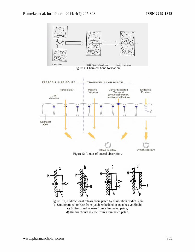
Figure 6: a) Bidirectional release from patch by dissolution or diffusion;
b) Unidirectional release from patch embedded in an adhesive Shield
c) Bidirectional release from a laminated patch;
d) Unidirectional release from a laminated patch.

Ramteke, et al. Int J Pharm 2014; 4(4):297-308 ISSN 2249-1848
www.pharmascholars.com 306
Figure 7: Rolling method.
Figure 8: Normal Buccal Patch.
Figure 9: Ex vivo bioadhesion test.

Ramteke, et al. Int J Pharm 2014; 4(4):297-308 ISSN 2249-1848
www.pharmascholars.com 307
Figure 10: Franz diffusion cell for buccal patch.
REFERENCES
1. Shojaei AH, Chang RK, Guo X, Burnside BA, Couch RA. Pharm. Technol, 2001; 6(25): 70.
2. Jasti B, Xiaoling Li X, Cleary G. Bus. Brief. Pharmatech., 2003;194-96.
3. Saxena S, Sharma P, Nimisha. Pharma Tutor, Pharmcy infopedia, 1207.
4. Sahni JS, Ahmad FJ, Khar RK. Ind. J. Pharm. Sci., 2008; 70(1): 61-5.
5. Rajesh M, Dhamande K, Wankhede UR, Angure S. Int. J. Pharm, 2011; 2(3).
6. Michael J, Rathbone, Jonathan H, Michael S, Roberts. Modified-Release Drug Delivery Technology,
Marcel Dekker, Inc. New York Basel.
7. Javier O, Jason T. Eur. J. pharm. Biopharm, 2010; 11(23): 1-13.
8. Serra L, Domenech J, Nicholas AP. Eur. J. Pharm, Biopharm, 2009; 71: 519-28.
9. Alagusundaram M, Madhusudhan Chetty C, Dhachinamurthy D, Int. J.Rev. Life. Sci., 2011; 1(2): 35-44.
10. Bhaskara Jasti, Xiaoling Li, Gary Cleary. Bussiness Briefing: Pharm tech, 2004; 194-96.
11. Marcos Luciano Bruschi and Osvaldo de Freitas. Oral Bioadhesive Drug Delivery Systems, Drug
Development and Industrial Pharmacy, 2005; 31(3): 293-310.
12. Mitra AK, Alur HH, Johnston. Peptides and Protein- Buccal Absorption. Encyclopedia of Pharmaceutical
technology. Marcel Dekker Inc., Edi., 2002; 2081-93.
13. Shojaei AH and Li X. In vitro permeation of acyclovir through porcine buccal mucosa, Proceedings of
International Symposium on Controlled Release of Bioactive Materials, 1996; 23: 507-8.
14. Siegel IA, Gordon HP. Arch. Oral Biol., 1985; 30: 43-7.
15. Manganaro AM, Wertz PW. The effects of permeabilizers on the in vitro penetration of propranolol
through porcine buccal epithelium. Mil. Med., 1996; 161: 669-72.
16. Patil SB, Murthy RSR, Mahajan HS, Wagh RD, Gattani SG. Pharm. Times, 2006; 38 (4): 25-28.
17. Salamat-Miller N, Chittchang M, Johnston TP. Advance Drug Delivery Review, Nov 2005; 57(11): 1666-
91.
18. Smart JD. Expert Opinion Drug Delivery, 2005; 2(3): 507.
19. Chinna Reddy P, Chaitanya KSC, Madhusudan Rao Y. DARU, 2011; 19(6): 385-403.
20. Narasimha Rao R, Kandhadi SR, Swapna U, Konasree S, Enugala S. Int. J. Pharm. Bio. Sci., 2011; 1: 145-
59.
21. Kumar M. Int. J. pharm. Pharm. Sci., 2010; 2(3): 86-90.
22. Semalty M. Int. J. Pharma. Sci. nanotech., 2008; 1(2).
23. Sathish D, Shayeda. Int. J. Pharm. Sci. and Nanotechnology, 2010; 3(1): 860-6.
24. Goudanavar PS. Der pharmacialettre, 2010; 2 (1): 382-7.
25. Rao Y, Balarameshachary MR, Vani G. Drug Dev. Ind. Pharm, 1999; 25: 685.
26. Zhang J. Int. J. Pharm., 1994; 101:15–22.
27. Noha AN, Nabila AB, Fatma AI,Lobna MM. Acta Pharm, 2003; 53: 199-212.
28. Supriya SS, Nilesh SS, Sagar S, Vilasrao K. American Association of Pharmaceutical Scientists Pharm Sci.
Tech., 2008; 9(3): 127-32.
29. Subhash VD, Madhuri A, Channawar, Anil VC, Umesh MJ, Kailash RB. Int. J. of Pharmacy and Pharma.
Sci., 2009; 1(1): 216-29.
30. Nafee NA, Ismail FA, Boraie NA, Mortada LM. Acta Pharm., 2003; 53: 199.

Ramteke, et al. Int J Pharm 2014; 4(4):297-308 ISSN 2249-1848
www.pharmascholars.com 308
31. Shemalty A, Bhojwani M, Bhatt GK, Gupta GD, Srivastava AK. Indian J. Pharm. Sci., 2005; 67: 548.
32. Challa R, Ahuja A, Ali J, Khar RK. AAPS Pharm. Sci. Tech., 2005; 6: 327.
33. Paradkar A, Maheshwari M, Tyagi AK, Chouhan B, Kadam SS. APS Pharm Sci Tech., 2003; 4: 65.
34. Pavankumar GV, Ramkrishna V, William GJ, Konde A. Indian J. Pharm. Sci., 2005; 67: 160-4.
35. Cirri M, Rangoni C, Maestrelli F, Corti G, Mura P. Drug Dev. Ind. Pharm., 2005; 31: 697-707.
36. Panigrahi L, Snigdha P, Ghosal SK. Indian J. Pharm. Sci., 2005; 67: 319.
37. Mukne AP, Nagarsenker MS. AAPS Pharm. Sci. Tech., 2004; 5: E19.
38. Kamel AH. AAPS pharm. Sci. tech., 2007; 8 (3): 75.
39. Biswajit B, Kevin G, Thimmasetty J. Int. J. Pharm. Sci. and Nanotechnology, 2010; 2(4): 739-48.
40. Choudhary A. Int. J. Res. Pharm. Sci., 2010; 1(4): 396-401.
41. Thimmasetty J, Pandey GS, Satheshbabu PR. Design Pak. J. Pharm. Sci., 2008; 21(3): 241-8.
42. Giradkar KP. Int. J. Pharma. res. & development, 2010; 2.
43. Leung SS, JR Robinson. J. Contr. Rel., 1990; 12: 187–94.
44. Patel R, Poddar SS. Current drug delivery, 2009; 6: 140-4.
45. Gandhi RE, Robinson JR. Indian J. Pharm. Sci., 1988; 50: 145–15.
46. Khanna R, Agarwal SP, Ahuja A. Indian J. Pharm. Sci., 1997; 59: 299.
47. Park K, Robinson JR. Int. J. Pharm., 1984; 19: 107–27.
48. Chinna Reddy P, Madhusudan Rao Y. Buccal Drug Delivery Systems. In: Madhusudan Rao Y, Jithan AV
ed. Advances in drug delivery: 2010, pp. 139-210.
49. Singh S, Jain S, Muthu MS, Tiwari S, Tilak R. AAPS Pharm. Sci. Tech., 2008; 9(2): 660-67.
50. Betz G, Burgin PJ, Leuenberger H. Int. J. Pharm, 2003; 252: 11-25.
51. Vishnu MP, Bhupendra Prajapati G, Madhabhai MP. Acta Pharm, 2005; 57: 61-72.
52. Kanig JL, Goodman HJ. Pharm. Sci., 1962; 51: 77–83.
53. Wani MS, Parakh SR, Dehghan MH, Polshettiwar SA, Chopade VV, Pande VV. Pharmainfo.net., 2007;
5(2).
54. Aungst BJ, Rogers NJ. Int. J. Pharm., 1989; 53: 227-35.
55. Siegel IA, Gordon HP. Arch. Oral Biol., 1985; 30: 43-47.
56. Kandjia J, Anderson MJD, & Muller Ruchholtz W. J. Cancer. Res. Clin. Oncol., 1981; 101-65.
57. Salamat-Miller N, Chittchang M, Johnston TP. The use of mucoadhesive polymers in buccal drug delivery,
2005; 57(11):1666-91
58. NK Jain. Controlled and novel drug delivery, 65-75,371-377.
59. Gandhi SD, Pandya PR, Umbarkar R, Tambawala T, and Shah MA. Int. J. Pharm. Sci., 2011; 851-72.
60. Sevda Senel, Mary Kremer, Katalin Nagy and Christopher Squier. Current Pharmaceutical Biotechnology,
2001; 2: 175-86.
61. Amir H, Shojaei. J. of Pharm. Pharm. Sci., 1998; 1(1): 15-30.
62. Ind. J. Pharma. Sci., 2004; 66(4): 371-536, 556-562.
63. Ravi S, Rishabha M, P Sharma. Trends in Buccal Film: Formulation characteristics, recent studies and
patents. Eur. J. App. Sci. 2011; 3(3): 93-101.