Embed Size (px)
Citation preview
International Journal of Pharmaceutics 472 (2014) 82–87
Microfluidics-assisted engineering of polymeric microcapsules withhigh encapsulation efficiency for protein drug delivery
Jenni Pessi a,b,*, Hélder A. Santos a, Inna Miroshnyk b,c, JoukoYliruusi a, David A. Weitz b,Sabiruddin Mirza b
aDivision of Pharmaceutical Chemistry and Technology, Faculty of Pharmacy, University of Helsinki, FI-00014 Helsinki, Finlandb School of Engineering and Applied Science, Department of Physics, Harvard University, Cambridge, MA 02138, USAc LECOM School of Pharmacy, Erie, PA 16509, USA
A R T I C L E I N F O
Article history:Received 14 April 2014Received in revised form 6 June 2014Accepted 8 June 2014Available online 10 June 2014
PubChem Classifications:Ethyl acetate (PubChem CID 8857)Polycaprolactone (PubChem CID 10401)Polyethylene glycol (PubChem CID 174)Poly(vinyl alcohol) (PubChem CID 11199)
Keywords:MicrocapsulesPolycaprolactoneMicrofluidicsTherapeutic proteinsDrug delivery
A B S T R A C T
In this study, microfluidic technology was employed to develop protein formulations. The microcapsuleswere produced with a biphasic flow to create water–oil–water (W/O/W) double emulsion droplets withultrathin shells. Optimized microcapsule formulations containing 1% (w/w) bovine serum albumin (BSA)in the inner phase were prepared with poly(vinyl alcohol), polycaprolactone and polyethylene glycol. Allthe particles were found to be intact and with a particle size of 23–47 mm. Furthermore, the particleswere monodisperse, non-porous and stable up to 4 weeks. The encapsulation efficiency of BSA in themicrocapsules was 84%. The microcapsules released 30% of their content within 168 h. This studydemonstrates that microfluidics is a powerful technique for engineering formulations for therapeuticproteins.
ã 2014 Elsevier B.V. All rights reserved.
Contents lists available at ScienceDirect
International Journal of Pharmaceutics
journa l home page : www.e l sev ier .com/ loca te / i jpharm
1. Introduction
Protein and peptide therapeutics often have poor oralbioavailability. Increasing the bioavailability is challenging, sincethe gastrointestinal tract has various barriers for the protein drugsto overcome before reaching the bloodstream (Zhou, 1994).Proteins and peptides are very sensitive to enzymatic degradation,aggregation, adsorption, and denaturation (Fix, 1996; Saffran et al.,1986). Other physical barriers for proteins and peptides absorptionare the size, charge and solubility constraints (Cox et al., 2002).Crossing the epithelial intestinal cell layer is possible via diffusionthrough the hydrophobic tight junctions of the cells by passivetransport, via facilitated transcellular diffusion through thelipophilic absorptive cells, or via active carrier-mediated transportsystems or transcytosis (Ingemann et al., 2000). Thus, drug carrier
* Corresponding author at: Division of Pharmaceutical Chemistry and Technolo-gy, Faculty of Pharmacy, University of Helsinki, FI-00014 Helsinki, Finland.Tel.: +358 50 381 0225.
E-mail address: [email protected] (J. Pessi).
http://dx.doi.org/10.1016/j.ijpharm.2014.06.0120378-5173/ã 2014 Elsevier B.V. All rights reserved.
systems for oral delivery of proteins have an important role in thedevelopment of protein-based formulations (Langer, 1998).
Polymeric microcapsules hold great potential as deliverysystems for oral protein delivery (Freiberg and Zhu, 2004).Polymeric microcapsules can be widely applied to many situationswhere continuous and controlled drug administration is essential,and the use of microcapsules for drug delivery is not limited to anyspecific illness. For example, polycaprolactone (PCL), a semi-crystalline, hydrophobic, (Chandra and Rustgi, 1998) biocompati-ble and biodegradable polymer (Pitt, 1990; Chen et al., 2000) hasbeen widely used for the preparation of microcapsules for drugdelivery applications (Jeong et al., 2003; Natarajan et al., 2011;Scala-Bertola et al., 2012; Somavarapu et al., 2005).
Microfluidic technology has various advantages for the prepara-tion of polymeric microcapsules, (Utada et al., 2005) because itallows precise control over the fabrication process (Umbanhowaret al., 2000). With microfluidic devices it is possible to miximmiscible liquids in a tunable manner by using three-dimensionalflows (Squiresand Quake, 2005). Thismakesthe precisemanufactur-ing process possible and enables choosing the chemical composi-tions and structures of the prepared particles independently
J. Pessi et al. / International Journal of Pharmaceutics 472 (2014) 82–87 83
(Duncanson et al., 2012b). More importantly, double emulsiondroplets with ultra-thin shells for preparing microcapsules can becreated by using a biphasic flow in a glass capillary device thatcombines co-flow and flow-focusing without additional energyinput that affects protein structures (Kim et al., 2011, 2013).
In this study, the microfluidic technique was employed as a toolfor templating and fabricating biocompatible polymeric micro-capsules for protein drug delivery. The microcapsules were loadedwith bovine serum albumin (BSA) and the properties of the formedmicrocapsules were evaluated.
2. Methods
2.1. Chemicals
The model protein used in this study, BSA, was purchased fromSigma–Aldrich, Germany. The polymers used were poly(vinylalcohol) (PVA, 87–89% hydrolyzed, Mw 13,000–23,000;Sigma–Aldrich, USA), polyethylene glycol 6000 (PEG6000, Mw5000–7000; Fluka Analytical, Germany) and polycaprolactone(PCL, Mw 70,000–90,0000; Sigma–Aldrich, USA). Phosphatebuffers (100 mM) at pH 7.2 used in the dissolution tests wereprepared according to the European Pharmacopoeia (Ph. Eur. 7thedition). All the other reagents were used as received and were ofanalytical grade.
2.2. Fabrication of the microfluidic devices
All microcapsule formulations were fabricated using glassmicrocapillary devices (Chu et al., 2007; Duncanson et al., 2012a;Kim and Weitz, 2011; Shum et al., 2011). Combining co-flow andflow-focusing within the glass capillary device enabled thepreparation of microcapsules from more complex and viscousmaterials (Utada et al., 2005) Cylindrical capillaries were pulledwith a Flaming/Brown micropipette puller (Model P-97, SutterInstrument Co., USA) to obtain tapered tips and to form tips withdiameter of 100 and 150 mm. Cylindrical capillaries were coatedwith hydrophilic or hydrophobic coating, corresponding towhether they contained the water or the oil phase. Hydrophobicand hydrophilic coating agents were trimethoxy(octadecyl) silane(Sigma–Aldrich, USA) and 2-[methoxy(polymethyleneoxy) pro-pyl]-9-12 trimethoxysilane (Gelest Inc., Netherlands).
2.3. Microfluidic encapsulation of BSA
The droplet formation in microfluidic devices is based on jettingto dripping transition and taking advantage of the hydrodynamicinstability (Powers et al., 1998). With the jetting to drippingtransition the drop formation involves a balance between theviscous drag of the coaxial fluid that pulls on the drop and thesurface tension forces (Umbanhowar et al., 2000). The surfaceenergy is decreased as the jet breaks into drops, and thus, the drop
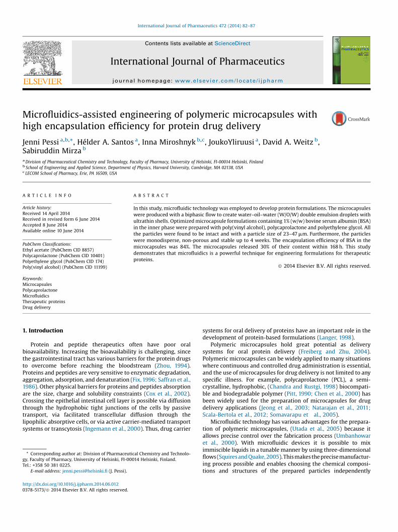
Fig. 1. The inner and middle phases flow in the cylindrical capillary on the left. Stretchedit, forming large droplets of water phase into the oil phase. This forms droplets with ultrdouble emulsion with the outer phase flowing from the square capillary.
formation mechanism can be explained via the Rayleigh–Plateauinstability (Squires and Quake 2005; Utada et al., 2007) (Fig. 1).
Because the physics of the process is well understood,microfluidic devices make is possible to mix immiscible liquidswith precise control (Squires and Quake, 2005). The microfluidictechnology has various advantages, particularly the ability tocreate actually three-dimensional flows (Utada et al., 2005). Thismakes the precise manufacturing process possible and enablesgaining the control over the immiscible fluids and theirdimensions. More specifically, the droplets of desired structureand size can be created by simply adjusting the process andformulation variables.
The microfluidic device employed a biphasic flow to producemicrocapsules from double emulsion droplets with ultrathin shells(Fig. 2) (Kim et al., 2011). Emulsion phases of the water–oil–water(W/O/W) emulsion were pumped into the glass capillary deviceswith syringes using Harvard pumps (Harvard Apparatus Hollston,USA). Syringes were attached to the inlets of the glass capillarydevice with plastic tubing (PE5 0.86 � 1.32 mm, Scientific Com-modities Inc., USA). Microfluidic technology requires specificformulation to successfully produce double emulsion droplets.Various formulations with different flow rates were examinedduring the formulation optimization process (Table S1). Theformulations investigated in the screening study were chosenbased on the viscosity and compatibility of the components. Theoptimized formulation contained 5% (w/w) of PVA in water as theouter phase, 3% (w/w) of PCL in ethyl acetate as the middle phase,and 20% (w/w) of PEG6000 and PVA (1:4) and 1% (w/w) BSA inwater as the inner phase. The water-solubility of BSA and increasein viscosity limit the maximum amount of BSA used in thisformulation to 1% (w/w). The inner and middle phases flowed atthe rate of 1000 mL/h and the flow rate of the outer phase was3000 mL/h, respectively.
2.4. Particle characterization: particle morphology, particle size,stability and core shell structure
The morphology and surface properties of the microcapsuleswere examined by electron scanning microscopy (SEM). SEMimages were taken with an environmental SEM microscope (CarlZeiss AG, EVO 55, Germany) with wet stage at chamber pressure of682 Pa and 26 kV. Samples were placed on wet paper and furtherpreparation was not required. The chamber was cooled down withliquid nitrogen to water vapor state and the electric beam was runthrough upper and lower aperture of 100 and 500 mm.
The particle size was determined by optical microscopy anddiameter measurements from 5 batches (n = 100). Diametermeasurements were conducted with software for scientific imageanalysis (ImageJ freeware, National Institutes of Health, USA) andmeasured according to 1 mm scale for the optical microscope.Short time stability was examined by monitoring the collapse rateof the particles with optical microscopy. 5 batches of particles were
capillary is inserted into this cylindrical capillary and the inner phase flows throughathin shells as the phases move to the collection capillary (on the right) and form a
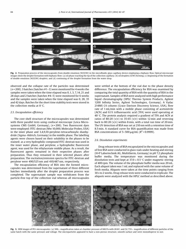
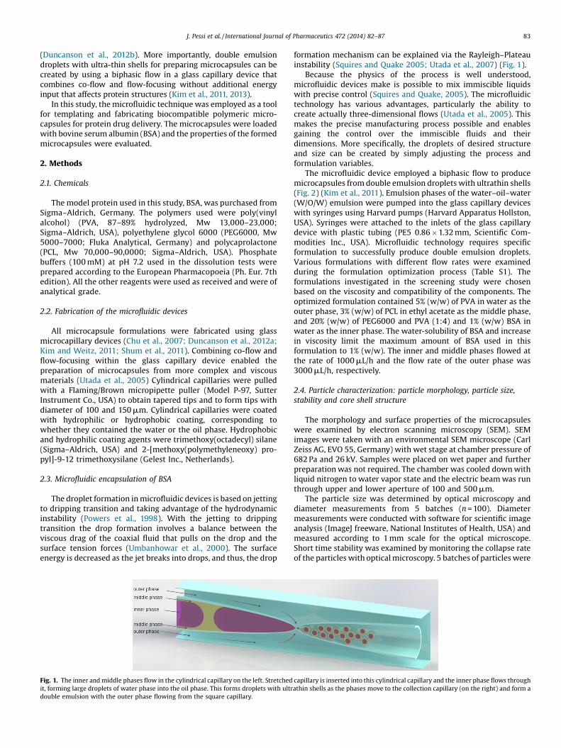
Fig. 2. Preparation process of the microcapsules from double emulsion (W/O/W) in the microfluidic glass capillary device employing a biphasic flow. Optical microscopeimages show the droplet formation with biphasic flow: (a) oil phase reaching the tip of the collection capillary; (b) oil droplets (O/W) forming; (c) beginning of the formationof double emulsion (W/O/W) droplets; and (d) continuing of the droplet formation from the water drop.
84 J. Pessi et al. / International Journal of Pharmaceutics 472 (2014) 82–87
observed and the collapse rate of the particles was determined(n = 200). 3 batches (batches #1–3) were monitored for 4 weeks thesamples were taken when the time elapsed was 0, 1, 3, 7, 14, 21 and28 days and 2 batches (batches #4–5) were monitored for 6 weeksand the samples were taken when the time elapsed was 0, 28, 35and 42 days. Batches for the short time stability tests were stored inthe collection media at 8 �C.
2.5. Encapsulation efficiency
The core shell structure of the microcapsules was determinedwith three parallel tests using confocal microscope (Leica Micro-systems CMS GmbH, Germany), (n = 200). Two fluorescent dyeswere employed: FITC-dextran (Mw 10,000, Molecular Probes, USA)in the inner phase and 3,4,9,10-perylene-tetracarboxylic dianhy-dride (Sigma–Aldrich, Germany) in the middle phase. The labellingagents were chosen based on their solubility in the phases to bestained; as a very hydrophilic compound FITC-dextran was used inthe inner water phase, and perylene, a hydrophobic fluorescentagent, was used for the ethylacetate middle phase. As a result, thefluorescent agents remained in their respective phases afterpreparation. Thus they remained in their selected phases afterpreparation. The excitation/emission spectra for FITC-dextran andperylene were 490/525 nm and 410/487 nm, respectively.
The encapsulation efficiency of BSA into the formed micro-capsules was determined from the supernatant of three differentbatches immediately after the droplet preparation process wascompleted. The supernatant sample was withdrawn from theparticle-free top of the collection vial whereas all microcapsules
Fig. 3. SEM images of PCL microcapsules: (a) 506� magnification taken at chamber presame batch with the same pressure and voltage. The microparticles appeared to have
were settled at the bottom of the vial due to the phase densitydifference. The encapsulation efficiency for BSA was examined bycomparing the total quantity of BSA with the quantity of BSA in thesupernatant. Samples of BSA were analyzed with high performanceliquid chromatography (HPLC Thermo System Products, Agilent1200 Infinity Series, Agilent Technologies, Germany). A Vydac214MS C4 column (Grace Davison Discovery Science, USA), flowrate of 1 mL/min with a mobile phase consisting of acetonitrile(ACN) and 0.1% trifluoroacetic acid (TFA) were used operating at40 �C. The protein analysis required a gradient of TFA and ACN atratios of 80:20 (v/v) to 35:65 (v/v) within 12 min and reversingback to 80:20 (v/v) within 8 min, with a total run time of 20 min.The UV detection of BSA was set at 210 nm with a retention time of8.5 min. A standard curve for BSA quantification was made fromBSA concentrations of 5–500 mg/mL (R2 = 0.9999).
2.6. Dissolution experiments
Drug release tests of BSA encapsulated in the microcapsules andof free BSA were conducted in glass vials under heating and stirring(H+P Labortechnik AG, Multitherm, Germany) in pH 7.2 phosphatebuffer media. The temperature was monitored during thedissolution tests and kept at 37.0 � 0.5 �C under magnetic stirringof 400 rpm. The volume of the phosphate buffer media was 20 mL.Each aliquot taken was 1 mL and replaced with the same volume offresh media. Aliquots were taken at the time points ranging from30 s to 2 weeks. Drug release tests were conducted in triplicate. Thealiquots were analyzed with the HPLC method as described above.
ssure of 682 Pa with 26 kV; and (b) 735� magnification of different particles of thea non-porous structure, smooth surface and were monodisperse in size.
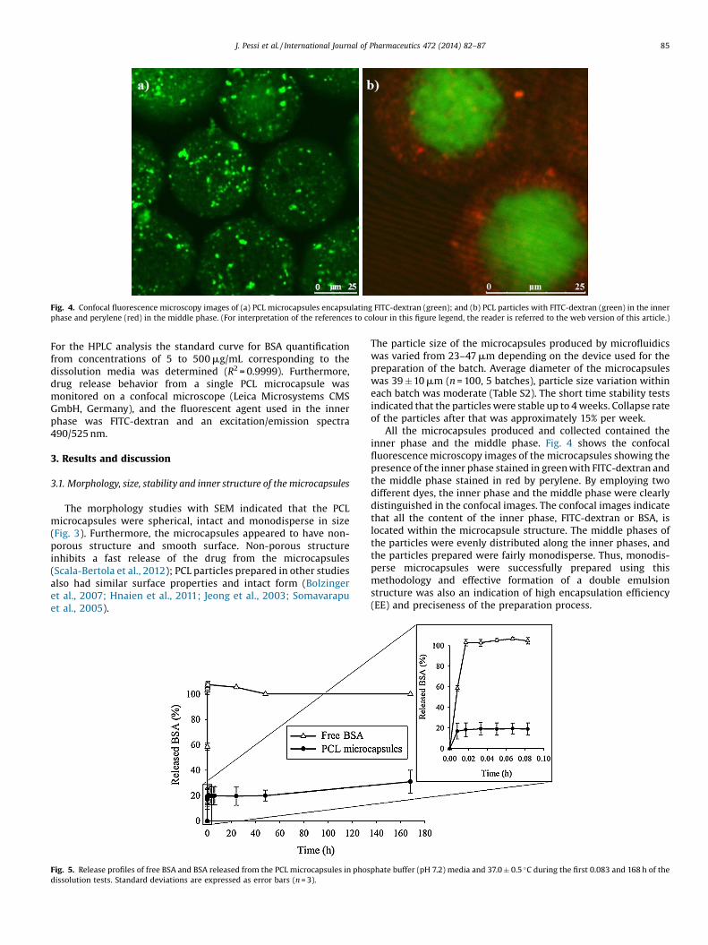
Fig. 4. Confocal fluorescence microscopy images of (a) PCL microcapsules encapsulating FITC-dextran (green); and (b) PCL particles with FITC-dextran (green) in the innerphase and perylene (red) in the middle phase. (For interpretation of the references to colour in this figure legend, the reader is referred to the web version of this article.)
J. Pessi et al. / International Journal of Pharmaceutics 472 (2014) 82–87 85
For the HPLC analysis the standard curve for BSA quantificationfrom concentrations of 5 to 500 mg/mL corresponding to thedissolution media was determined (R2 = 0.9999). Furthermore,drug release behavior from a single PCL microcapsule wasmonitored on a confocal microscope (Leica Microsystems CMSGmbH, Germany), and the fluorescent agent used in the innerphase was FITC-dextran and an excitation/emission spectra490/525 nm.
3. Results and discussion
3.1. Morphology, size, stability and inner structure of the microcapsules
The morphology studies with SEM indicated that the PCLmicrocapsules were spherical, intact and monodisperse in size(Fig. 3). Furthermore, the microcapsules appeared to have non-porous structure and smooth surface. Non-porous structureinhibits a fast release of the drug from the microcapsules(Scala-Bertola et al., 2012); PCL particles prepared in other studiesalso had similar surface properties and intact form (Bolzingeret al., 2007; Hnaien et al., 2011; Jeong et al., 2003; Somavarapuet al., 2005).
Fig. 5. Release profiles of free BSA and BSA released from the PCL microcapsules in phosdissolution tests. Standard deviations are expressed as error bars (n = 3).
The particle size of the microcapsules produced by microfluidicswas varied from 23–47 mm depending on the device used for thepreparation of the batch. Average diameter of the microcapsuleswas 39 � 10 mm (n = 100, 5 batches), particle size variation withineach batch was moderate (Table S2). The short time stability testsindicated that the particles were stable up to 4 weeks. Collapse rateof the particles after that was approximately 15% per week.
All the microcapsules produced and collected contained theinner phase and the middle phase. Fig. 4 shows the confocalfluorescence microscopy images of the microcapsules showing thepresence of the inner phase stained in green with FITC-dextran andthe middle phase stained in red by perylene. By employing twodifferent dyes, the inner phase and the middle phase were clearlydistinguished in the confocal images. The confocal images indicatethat all the content of the inner phase, FITC-dextran or BSA, islocated within the microcapsule structure. The middle phases ofthe particles were evenly distributed along the inner phases, andthe particles prepared were fairly monodisperse. Thus, monodis-perse microcapsules were successfully prepared using thismethodology and effective formation of a double emulsionstructure was also an indication of high encapsulation efficiency(EE) and preciseness of the preparation process.
phate buffer (pH 7.2) media and 37.0 � 0.5 �C during the first 0.083 and 168 h of the
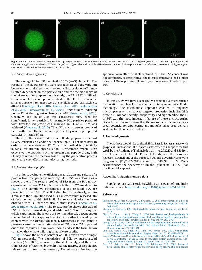
Fig. 6. Confocal fluorescence microscope follow-up images of one PCL microcapsule, showing the release of the FITC-dextran (green) content: (a) the shell rupturing from thethinnest spot; (b) particle releasing FITC-dextran; (c) and (d) particles with no visible FITC-dextran content. (For interpretation of the references to colour in this figure legend,the reader is referred to the web version of this article.)
86 J. Pessi et al. / International Journal of Pharmaceutics 472 (2014) 82–87
3.2. Encapsulation efficiency
The average EE for BSA was 84.0 � 10.5% (n = 3) (Table S3). Theresults of the EE experiment were reproducible and the variationbetween the parallel tests was moderate. Encapsulation efficiencyis often dependent on the particle size and for the size range ofthe microcapsules prepared in this study, the EE of 84% is difficultto achieve. In several previous studies the EE for similar orsmaller particle size ranges were at the highest approximately ca.40–60% (Bolzinger et al., 2007; Hnaien et al., 2011; Scala-Bertolaet al., 2012; Somavarapu et al., 2005). Other studies indicatedpoorer EE at the highest of barely ca. 40% (Hnaien et al., 2011).Generally, the EE of 70% was considered high, even forsignificantly larger particles. For example, PCL particles preparedwith flow-focused jetting cell achieved an EE of 42–79% wasachieved (Cheng et al., 2010). Thus, PCL microcapsules producedhere with microfluidics were superior to previously reportedparticles in terms of EE.
These results indicate that the microfluidic preparation methodis very efficient and additional energy input is not necessary inorder to achieve excellent EE. Thus, this method is potentiallysuitable for protein encapsulation. Furthermore, when usingexpensive therapeutics, such as protein and peptide drugs, highEE may minimize the material loss during the preparation processand create cost-effective manufacturing methods.
3.3. Protein release profile
In order to evaluate the efficient encapsulation and release of aprotein from the prepared microcapsules, BSA was chosen as amodel protein. The release profiles of BSA from the PCL micro-capsules and of free BSA in phosphate buffer pH 7.2 are shown inFig. 5. The cumulative percentages of the released BSA arepresented up to 168 h. Free BSA dissolved immediately, within1 min, into the dissolution media. PCL microcapsules released 30%of their content within 168 h. Similar release kinetics has beenobserved with PCL particles also in other studies (Coccoli et al.,2008; Hnaien et al., 2011). The release profile shows that 20% ofBSA is released immediately and additional 10% of it during thewhole experiment. The release of BSA is not directly dependent onthe number of microcapsules breaking; it is rather initiated by thecontact with the dissolution media. Differences in the osmoticpressure cause the immediate release of BSA, since BSA is pushedout of the capsules. Future work should address the formulationvariables that enable tailoring drug release profile.
Fig. 6 shows the release behavior of FITC-dextran from a singlePCL microcapsule. The degradation of PCL by autocatalyzedreaction (Pitt, 2009), occurred in the shell evenly, and thus, thethinnest part of the shell broke first. All the microcapsules did notrelease their content simultaneously. The microcapsules kept the
spherical form after the shell ruptured, thus the BSA content wasnot completely release from all the microcapsules and led to initialrelease of 20% of protein, followed by a slow release of protein up to30%.
4. Conclusions
In this study, we have successfully developed a microcapsuleformulation template for therapeutic proteins using microfluidictechnology. The microfluidic approach enabled to engineermicrocapsules with enhanced targeted properties, including highprotein EE, monodispersity, low porosity, and high stability. The EEof 84% was the most important feature of these microcapsules.Overall, this research shows that the microfluidic technique has agreat potential for engineering and manufacturing drug deliverysystems for therapeutic proteins.
Acknowledgements
The authors would like to thank Ilkka Lassila for assistance withgraphical illustrations. H.A. Santos acknowledges support for thiswork by the Academy of Finland (decision no. 252215 and 256394),the University of Helsinki Research Funds, and the EuropeanResearch Council under the European Union's Seventh FrameworkProgramme (FP/2007–2013) grant no. 310892. Dr. S. Mirzaacknowledges the Academy of Finland (grants no. 1132726) forthe financial support.
Appendix A. Supplementary data
Supplementarydataassociatedwiththisarticlecanbefound,intheonline version, at http://dx.doi.org/10.1016/j.ijpharm.2014.06.012.
References
Bolzinger, M., Bordes, C., Gauvrit, J., Briançon, S., 2007. Improvement of a bovineserum albumin microencapsulation process by screening design. Int. J. Pharm.344, 16–25.
Chandra, R., Rustgi, R., 1998. Biodegradable polymers. Prog. Polym. Sci. 23, 1273–1335.
Chen, D., Chen, H., Bei, J., Wang, S., 2000. Morphology and biodegradation ofmicrospheres of polyester–polyether block copolymer based on polycaprolac-tone/polylactide/poly (ethylene oxide). Polym. Int. 49, 269–276.
Cheng, X., Liu, R., He, Y., 2010. A simple method for the preparation of monodisperseprotein-loaded microspheres with high encapsulation efficiencies. Eur. J.Pharm. Biopharm. 76, 336–341.
Chu, L.Y., Utada, A.S., Shah, R.K., Kim, J.W., Weitz, D.A., 2007. Controllablemonodisperse multiple emulsions. Angew. Chem. Int. Ed. 46, 8970–8974.
Coccoli, V., Luciana, A., Orsi, S., Guarino, V., Causa, F., Netti, P.A., 2008. Engineering ofpoly(e-caprolactone) micro-carriers to modulate protein encapsulation capa-bility and release kinetic. J. Mater. Sci. Mater. Med. 19, 1703–1711.
Cox, D.S., Raje, S., Gao, H., Salami, N.N., Eddington, N.D., 2002. Enhancedpermeability of molecular weight markers and poorly bioavailable compounds
J. Pessi et al. / International Journal of Pharmaceutics 472 (2014) 82–87 87
across Caco-2cell monolayers using the absorption enhancer, zonula occludenstoxin. Pharm. Res. 19, 1680–1688.
Duncanson, W.J., Abbaspourrad, A., Shum, H.C., Kim, S., Adams, L.L., Weitz, D.A.,2012a. Monodisperse gas-filled microparticles from reactions in doubleemulsions. Langmuir 28, 6742–6745.
Duncanson, W.J., Lin, T., Abate, A.R., Seiffert, S., Shah, R.K., Weitz, D.A., 2012b.Microfluidic synthesis of advanced microparticles for encapsulation andcontrolled release. Lab Chip 12, 2135–2145.
Fix, J.A., 1996. Oral controlled release technology for peptides: status and futureprospects. Pharm. Res. 13, 1760–1764.
Freiberg, S., Zhu, X., 2004. Polymer microspheres for controlled drug release. Int. J.Pharm. 282, 1–18.
Hnaien, M., Ruffin, E., Bordes, C., Marcillat, O., Lagarde, F., Jaffrezic-Renault, N.,Briançon, S., 2011. Integrity characterization of myoglobin released from poly (ç-caprolactone) microspheres using two analytical methods: UV–vis spectrome-try and conductometric bi-enzymatic biosensor. Eur. J. Pharm. Biopharm. 78,298–305.
Ingemann, M., Frokjaer, S., Hovgaard, L., Bronsted, H., 2000. Peptide and proteindrug delivery systems for non-parenteral routes of administration. In: Frokjaer,S., Hovgaard, L. (Eds.), Pharmaceutical Formulation Development of Peptidesand Proteins. Taylor & Francis, London, pp. 189–205.
Jeong, J.C., Lee, J., Cho, K., 2003. Effects of crystalline microstructure on drugrelease behavior of poly(q-caprolactone) microspheres. J. Control. Release 92,249–258.
Kim, S.H., Kim, J.W., Cho, J., Weitz DA:, 2011. Double-emulsion drops with ultra-thin shells for capsule templates. Lab Chip 11, 3162–3166.
Kim, S.H., Kim, J.W., Kim, D., Han, S., Weitz, D.A., 2013. Polymersomes containing ahydrogel network for high stability and controlled release. Small 9, 124–131.
Kim, S.H., Weitz, D.A., 2011. One-step emulsification of multiple concentric shellswith capillary microfluidic devices. Angew. Chem. 123, 8890–8893.
Langer, R., 1998. Drug delivery and targeting. Nature 392, 5–10.
Natarajan, V., Krithica, N., Madhan, B., Sehgal, P.K., 2011. Formulation and evaluationof quercetin polycaprolactone microspheres for the treatment of rheumatoidarthritis. J. Pharm. Sci. 100, 195–205.
Pitt, C.G., 1990. Poly-e-caprolactone and its copolymers. In: Chasin, M., Langer, R.(Eds.), Biodegradable Polymers as Drug Delivery Systems. Marcel Dekker, NewYork, pp. 71–120.
Powers, T.R., Zhang, D., Goldstein, R.E., Stone, H.A.,1998. Propagation of a topologicaltransition: the Rayleigh instability. Phys. Fluids 10, 1052.
Scala-Bertola, J., Javot, L., Camargo, J., Bonneaux, F., Lecompte, T., Maincent, P., Sapin,A., 2012. Evaluation of subcutaneous forms in the improvement of pharmaco-kinetic profile of warfarin. Int. J. Pharm. 431, 33–38.
Saffran, M., Kumar, G.S., Savariar, C., Burnham, J.C., Williams, F., Neckers, D.C.,1986. Anew approach to the oral administration of insulin and other peptide drugs.Science 233, 1081–1084.
Shum, H.C., Zhao, Y., Kim, S.H., Weitz, D.A., 2011. Multicompartment polymersomesfrom double emulsions. Angew. Chem. 123, 1686–1689.
Somavarapu, S., Pandit, S., Gradassi, G., Bandera, M., Ravichandran, E., Alpar, O.H.,2005. Effect of vitamin E TPGS on immune response to nasally delivereddiphtheria toxoid loaded poly (caprolactone) microparticles. Int. J. Pharm. 298,344–347.
Squires, T.M., Quake, S.R., 2005. Microfluidics: fluid physics at the nanoliter scale.Rev. Mod. Phys. 77, 977.
Umbanhowar, P., Prasad, V., Weitz, D.A., 2000. Monodisperse emulsion generationvia drop break off in a coflowing stream. Langmuir 16, 347–351.
Utada, A., Chu, L.Y., Fernandez-Nieves, A., Link, D., Holtze, C., Weitz, D.A., 2007.Dripping, jetting, drops, and wet-ting: the magic of microfluidics. MRS Bull. 32,702–708.
Utada, A., Lorenceau, E., Link, D., Kaplan, P., Stone, H., Weitz, D.A., 2005.Monodisperse double emulsions generated from a microcapillary device.Science 308, 537–541.
Zhou, X.H., 1994. Overcoming enzymatic and absorption barriers to non-parenterally administered protein and peptide drugs. J. Control. Release 29,239–252.