Embed Size (px)
Citation preview

International Journal of Mass Spectrometry 377 (2015) 228–234
Contents lists available at ScienceDirect
International Journal of Mass Spectrometry
journa l homepage: www.e lsev ier .com/ locate / i jms
Soft-landing electrospray ion beam deposition of sensitive oligoyneson surfaces in vacuum
Gordon Rinkea, Stephan Rauschenbacha,∗, Stephen Schrettlb, Tobias N. Hoheiselb,Jonathan Blohma,1, Rico Gutzlera, Federico Rosei c, Holger Frauenrathb, Klaus Kerna,d
a Max-Planck-Institute for Solid State Research, Nanoscale Science Department, Stuttgart, Germanyb Ecole Polytechnique Fédérale de Lausanne, Institute of Materials, Laboratory of Macromolecular and Organic Materials, Lausanne, Switzerlandc Centre for Energy, Materials and Telecommunications, Institut National de la Recherche Scientifique, Varennes, QC, Canadad Institut de Physique de la Matière Condensée, Ecole Polytechnique Fédérale de Lausanne, Switzerland
a r t i c l e i n f o
Article history:Received 19 March 2014Received in revised form 6 June 2014Accepted 27 June 2014Available online 4 July 2014
a b s t r a c t
Advances in synthetic chemistry permit the synthesis of large, highly functional, organic molecules.Characterizing the complex structure of such molecules with highly resolving, vacuum-based methodslike scanning probe microscopy requires their transfer into the gas phase and further onto an atomicallyclean surface in ultrahigh vacuum without causing additional contamination. Conventionally this is donevia sublimation in vacuum. However, similar to biological molecules, large synthetic compounds canbe non-volatile and decompose upon heating. Soft-landing ion beam deposition using soft ionizationmethods represents an alternative approach to vacuum deposition. Using different oligoyne derivativesof the form of R1 (C C)n R2, here we demonstrate that even sensitive molecules can be handled bysoft-landing electrospray ion beam deposition. We generate intact molecular ions as well as fragmentions with intact hexayne parts and deposit them on clean metal surfaces. Scanning tunneling microscopyshows that the reactive hexayne segments of the molecules of six conjugated triple bonds are intact. Themolecules agglomerate into ribbon-like islands, whose internal structure can be steered by the choice ofthe substituents. Our results suggest the use of ion beam deposition to arrange reactive precursors forsubsequent polymerization reactions.
© 2014 The Authors. Published by Elsevier B.V. This is an open access article under the CC BY-NC-NDlicense (http://creativecommons.org/licenses/by-nc-nd/3.0/).
1. Introduction
Soft ionization methods such as electrospray ionization (ESI)[1] or matrix assisted laser desorption ionization (MALDI) [2] areimportant methods in mass spectrometry as they allow researchersto study a broad variety of molecules with high precision. These ion-ization methods typically generate intact molecular gas phase ions,even of fragile biological molecules like proteins or peptides thatwould decompose in conventional gas phase ion sources based onelectron impact or chemical ionization [3].
A less common application of soft ionization is its use as agas phase particle source for vacuum deposition. The prepara-tion of well-defined molecular adsorbates on atomically clean
∗ Corresponding author. Tel.: +49 0711 689 1433; fax: +49 (0) 711 689 1662.E-mail addresses: [email protected] (G. Rinke), [email protected]
(S. Rauschenbach).1 Present address: Department of Chemistry, University of Warwick, Coventry,
United Kingdom.
substrates for use in surface science experiments requires a well-controlled environment, such as ultrahigh vacuum (UHV, p ≈ 10−10
mbar). This environment enables the investigation of moleculeswith methods of highest precision, like spatial mapping of sur-faces and adsorbates at atomic resolution with scanning tunnelingmicroscopy (STM). For this purpose, however, the molecules haveto be transferred to the surface as gas phase molecules, either byleaking them into vacuum or by sublimation within the UHV sys-tem. Thus the application is limited to volatile molecules, whichcan be evaporated without decomposition. Soft ionization sourceslike ESI or MALDI generate intact gas phase ions of molecules thatare otherwise not accessible by conventional evaporation meth-ods due to the fragility or high reactivity of the correspondingorganic species. For ion beam deposition, the intact molecularions are transferred from the ion source, which can be at ambi-ent pressure, to vacuum through differentially pumped ion optics[4–7]. Although ion beam deposition is experimentally challenging,the methodology of intact deposition of molecules onto surfaces,also called ion soft-landing, can be considered as well established[8–10]. Also the deposition of molecular ions that undergo chemical
http://dx.doi.org/10.1016/j.ijms.2014.06.0261387-3806/© 2014 The Authors. Published by Elsevier B.V. This is an open access article under the CC BY-NC-ND license (http://creativecommons.org/licenses/by-nc-nd/3.0/).
G. Rinke et al. / International Journal of Mass Spectrometry 377 (2015) 228–234 229
reactions upon deposition, called reactive landing, is well studiedmainly by chemical characterization of the surface [11–16].
In situ scanning probe microscopy investigations of surfacesafter the soft-landing deposition of intact molecular ions enablesthe imaging of individual non-volatile molecules with submolecu-lar resolution [17–21]. In a few examples, ordered two-dimensional[22,23] as well as three-dimensional thin film growth [23] has beenobserved after soft landing of intrinsically stable molecules.
Here, we extend the approach of soft-landing electrospray ionbeam deposition (ES-IBD) combined with high resolution scanningtunneling microscopy to reactive molecules. We demonstrate thatpotentially reactive species can be brought to the surface withoutimmediate covalent reaction. By adapting the molecular structure,we tune weak physisorption interactions responsible for the self-assembly of the molecules into ordered precursor structures, whichmay be covalently cross-linked in a second step by light or temper-ature treatment [24].
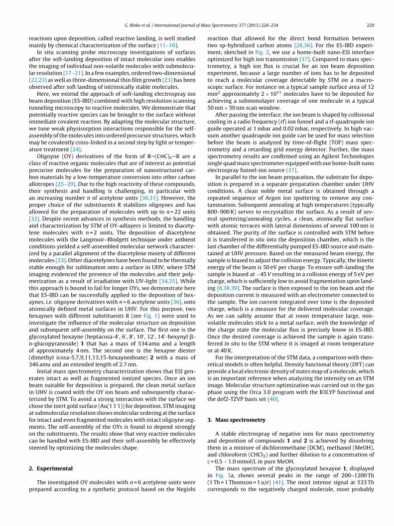
Oligoyne (OY) derivatives of the form of R (C C)n R are aclass of reactive organic molecules that are of interest as potentialprecursor molecules for the preparation of nanostructured car-bon materials by a low-temperature conversion into other carbonallotropes [25–29]. Due to the high reactivity of these compounds,their synthesis and handling is challenging, in particular withan increasing number n of acetylene units [30,31]. However, theproper choice of the substituents R stabilizes oligoynes and hasallowed for the preparation of molecules with up to n = 22 units[32]. Despite recent advances in synthesis methods, the handlingand characterization by STM of OY-adlayers is limited to diacety-lene molecules with n = 2 units. The deposition of diacetylenemolecules with the Langmuir–Blodgett technique under ambientconditions yielded a self-assembled molecular network character-ized by a parallel alignment of the diacetylene moiety of differentmolecules [33]. Other diacetelynes have been found to be thermallystable enough for sublimation onto a surface in UHV, where STMimaging evidenced the presence of the molecules and their poly-merization as a result of irradiation with UV-light [34,35]. Whilethis approach is bound to fail for longer OYs, we demonstrate herethat ES-IBD can be successfully applied to the deposition of hex-aynes, i.e. oligoyne derivatives with n = 6 acetylene units [36], ontoatomically defined metal surfaces in UHV. For this purpose, twohexaynes with different substituents R (see Fig. 1) were used toinvestigate the influence of the molecular structure on depositionand subsequent self-assembly on the surface. The first one is theglycosylated hexayne (heptacosa-4′, 6′, 8′, 10′, 12′, 14′-hexynyl �-d-glucopyranoside) 1 that has a mass of 534 amu and a lengthof approximately 4 nm. The second one is the hexayne diester(dimethyl icosa-5,7,9,11,13,15-hexaynedioate) 2 with a mass of346 amu and an extended length of 2.7 nm.
Initial mass spectrometry characterization shows that ESI gen-erates intact as well as fragmented ionized species. Once an ionbeam suitable for deposition is prepared, the clean metal surfacein UHV is coated with the OY ion beam and subsequently charac-terized by STM. To avoid a strong interaction with the surface wechose the inert gold surface (Au(1 1 1)) for deposition. STM imagingat submolecular resolution shows molecular ordering at the surfacefor intact and even fragmented molecules with intact oligoyne seg-ments. The self-assembly of the OYs is found to depend stronglyon the substituents. The results show that very reactive moleculescan be handled with ES-IBD and their self-assembly be effectivelysteered by optimizing the molecules shape.
2. Experimental
The investigated OY molecules with n = 6 acetylene units wereprepared according to a synthetic protocol based on the Negishi
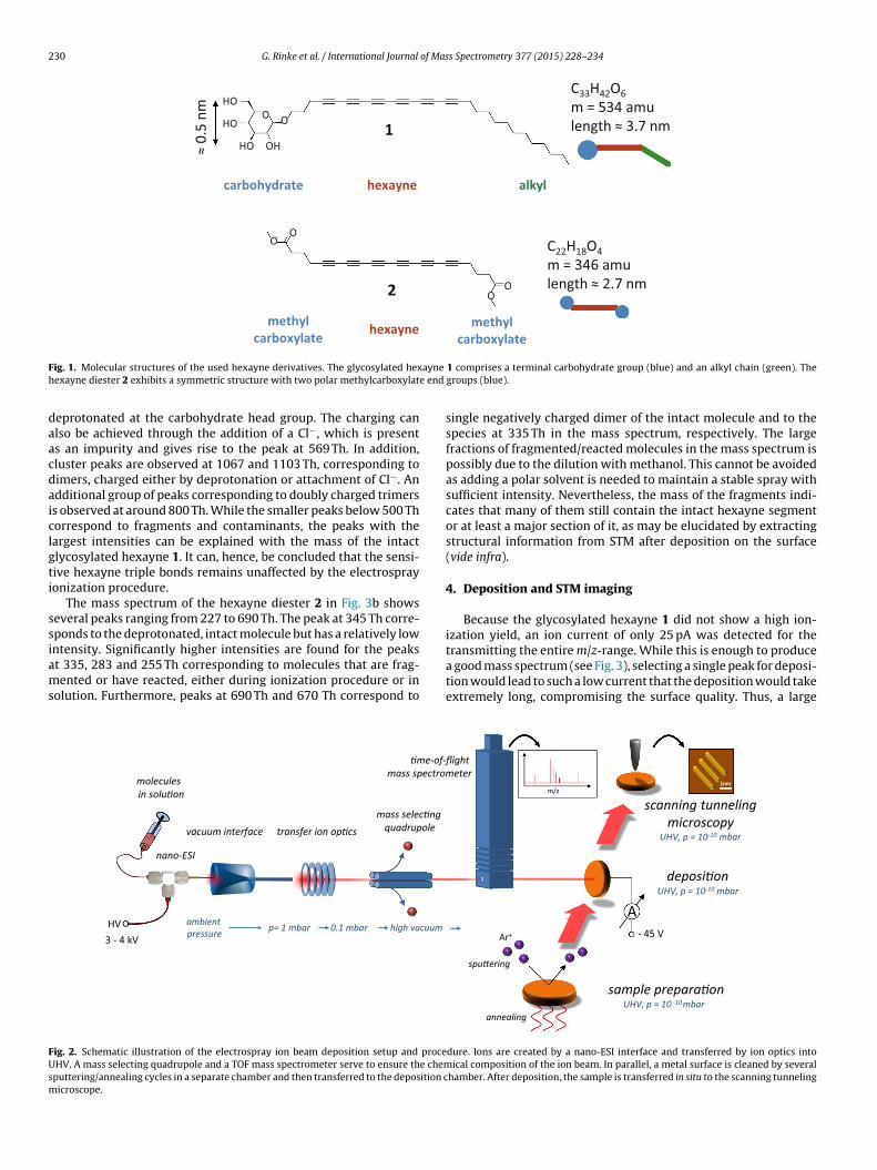
reaction that allowed for the direct bond formation betweentwo sp-hybridized carbon atoms [28,36]. For the ES-IBD experi-ment, sketched in Fig. 2, we use a home-built nano-ESI interfaceoptimized for high ion transmission [37]. Compared to mass spec-trometry, a high ion flux is crucial for an ion beam depositionexperiment, because a large number of ions has to be depositedto reach a molecular coverage detectable by STM on a macro-scopic surface. For instance on a typical sample surface area of 12mm2 approximately 2 × 10¹¹ molecules have to be deposited forachieving a submonolayer coverage of one molecule in a typical50 nm × 50 nm scan window.
After passing the interface, the ion beam is shaped by collisionalcooling in a radio frequency (rf) ion funnel and a rf-quadrupole ionguide operated at 1 mbar and 0.02 mbar, respectively. In high vac-uum another quadrupole ion guide can be used for mass selectionbefore the beam is analyzed by time-of-flight (TOF) mass spec-trometry and a retarding grid energy detector. Further, the massspectrometry results are confirmed using an Agilent Technologiessingle quad mass spectrometer equipped with our home-built nanoelectrospray funnel-ion source [37].
In parallel to the ion beam preparation, the substrate for depo-sition is prepared in a separate preparation chamber under UHVconditions. A clean noble metal surface is obtained through arepeated sequence of Argon ion sputtering to remove any con-tamination. Subsequent annealing at high temperatures (typically800–900 K) serves to recrystallize the surface. As a result of sev-eral sputtering/annealing cycles, a clean, atomically flat surfacewith atomic terraces with lateral dimensions of several 100 nm isobtained. The purity of the surface is controlled with STM beforeit is transferred in situ into the deposition chamber, which is thelast chamber of the differentially pumped ES-IBD source and main-tained at UHV pressure. Based on the measured beam energy, thesample is biased to adjust the collision energy. Typically, the kineticenergy of the beam is 50 eV per charge. To ensure soft-landing thesample is biased at −45 V resulting in a collision energy of 5 eV percharge, which is sufficiently low to avoid fragmentation upon land-ing [8,38,39]. The surface is then exposed to the ion beam and thedeposition current is measured with an electrometer connected tothe sample. The ion current integrated over time is the depositedcharge, which is a measure for the delivered molecular coverage.As we can safely assume that at room temperature large, non-volatile molecules stick to a metal surface, with the knowledge ofthe charge state the molecular flux is precisely know in ES-IBD.Once the desired coverage is achieved the sample is again trans-ferred in situ to the STM where it is imaged at room temperatureor at 40 K.
For the interpretation of the STM data, a comparison with theo-retical models is often helpful. Density functional theory (DFT) canprovide a local electronic density of states map of a molecule, whichis an important reference when analyzing the intensity on an STMimage. Molecular structure optimization was carried out in the gasphase using the Orca 3.0 program with the B3LYP functional andthe def2-TZVP basis set [40].
3. Mass spectrometry
A stable electrospray of negative ions for mass spectrometryand deposition of compounds 1 and 2 is achieved by dissolvingthem in a mixture of dichloromethane (DCM), methanol (MeOH),and chloroform (CHCl3) and further dilution to a concentration ofc = 0.5 − 1.0 mmol/L in pure MeOH.
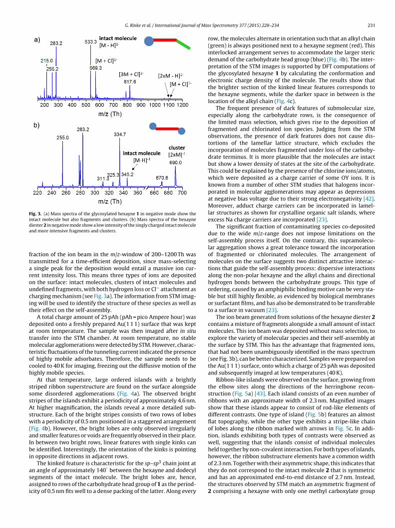
The mass spectrum of the glycosylated hexayne 1, displayedin Fig. 3a, shows several peaks in the range of 200–1200 Th(1 Th = 1 Thomson = 1 u/e) [41]. The most intense signal at 533 Thcorresponds to the negatively charged molecule, most probably
230 G. Rinke et al. / International Journal of Mass Spectrometry 377 (2015) 228–234
1
C33HHO
HO
HO OH
O
OO
OO
O
42O6m = 534 amulength ≈ 3.7 nm
carbohydrate hexayne alkyl≈
0.5
nm
2
C22H18O4m = 346 amulength ≈ 2.7 nm
hexaynemethylcarboxylate
methylcarboxylate
Fig. 1. Molecular structures of the used hexayne derivatives. The glycosylated hexayne 1 comprises a terminal carbohydrate group (blue) and an alkyl chain (green). Thehexayne diester 2 exhibits a symmetric structure with two polar methylcarboxylate end groups (blue).
deprotonated at the carbohydrate head group. The charging canalso be achieved through the addition of a Cl−, which is presentas an impurity and gives rise to the peak at 569 Th. In addition,cluster peaks are observed at 1067 and 1103 Th, corresponding todimers, charged either by deprotonation or attachment of Cl−. Anadditional group of peaks corresponding to doubly charged trimersis observed at around 800 Th. While the smaller peaks below 500 Thcorrespond to fragments and contaminants, the peaks with thelargest intensities can be explained with the mass of the intactglycosylated hexayne 1. It can, hence, be concluded that the sensi-tive hexayne triple bonds remains unaffected by the electrosprayionization procedure.
The mass spectrum of the hexayne diester 2 in Fig. 3b showsseveral peaks ranging from 227 to 690 Th. The peak at 345 Th corre-sponds to the deprotonated, intact molecule but has a relatively lowintensity. Significantly higher intensities are found for the peaksat 335, 283 and 255 Th corresponding to molecules that are frag-mented or have reacted, either during ionization procedure or insolution. Furthermore, peaks at 690 Th and 670 Th correspond to
single negatively charged dimer of the intact molecule and to thespecies at 335 Th in the mass spectrum, respectively. The largefractions of fragmented/reacted molecules in the mass spectrum ispossibly due to the dilution with methanol. This cannot be avoidedas adding a polar solvent is needed to maintain a stable spray withsufficient intensity. Nevertheless, the mass of the fragments indi-cates that many of them still contain the intact hexayne segmentor at least a major section of it, as may be elucidated by extractingstructural information from STM after deposition on the surface(vide infra).
4. Deposition and STM imaging
Because the glycosylated hexayne 1 did not show a high ion-ization yield, an ion current of only 25 pA was detected for thetransmitting the entire m/z-range. While this is enough to producea good mass spectrum (see Fig. 3), selecting a single peak for deposi-tion would lead to such a low current that the deposition would takeextremely long, compromising the surface quality. Thus, a large
A
sample prepara�on
deposi�on
scanning tunneling microscopy
transfer ion op�csmass selec�ng
quadrupole
�me-of-flight mass spectrometer
Ar+
spu�ering
annealing
nano-ESI
HV
UHV, p = 10 -10mbar
UHV, p = 10-10 mbar
UHV, p = 10-10 mbarvacuum interface
m/zmolecules in solu�on
ambient pressure p= 1 mbar 0.1 mbar high vacuum
1nm
3 - 4 kV - 45 V
Fig. 2. Schematic illustration of the electrospray ion beam deposition setup and procedure. Ions are created by a nano-ESI interface and transferred by ion optics intoUHV. A mass selecting quadrupole and a TOF mass spectrometer serve to ensure the chemical composition of the ion beam. In parallel, a metal surface is cleaned by severalsputtering/annealing cycles in a separate chamber and then transferred to the deposition chamber. After deposition, the sample is transferred in situ to the scanning tunnelingmicroscope.
G. Rinke et al. / International Journal of Mass Spectrometry 377 (2015) 228–234 231
Fig. 3. (a) Mass spectra of the glycosylated hexayne 1 in negative mode show theintact molecule but also fragments and clusters. (b) Mass spectra of the hexaynediester 2 in negative mode show a low intensity of the singly charged intact moleculeand more intensive fragments and clusters.
fraction of the ion beam in the m/z-window of 200–1200 Th wastransmitted for a time-efficient deposition, since mass-selectinga single peak for the deposition would entail a massive ion cur-rent intensity loss. This means three types of ions are depositedon the surface: intact molecules, clusters of intact molecules andundefined fragments, with both hydrogen loss or Cl− attachment ascharging mechanism (see Fig. 3a). The information from STM imag-ing will be used to identify the structure of these species as well astheir effect on the self-assembly.
A total charge amount of 25 pAh (pAh = pico Ampere hour) wasdeposited onto a freshly prepared Au(1 1 1) surface that was keptat room temperature. The sample was then imaged after in situtransfer into the STM chamber. At room temperature, no stablemolecular agglomerations were detected by STM. However, charac-teristic fluctuations of the tunneling current indicated the presenceof highly mobile adsorbates. Therefore, the sample needs to becooled to 40 K for imaging, freezing out the diffusive motion of thehighly mobile species.
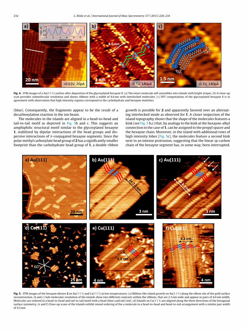
At that temperature, large ordered islands with a brightlystriped ribbon superstructure are found on the surface alongsidesome disordered agglomerations (Fig. 4a). The observed brightstripes of the islands exhibit a periodicity of approximately 4.6 nm.At higher magnification, the islands reveal a more detailed sub-structure. Each of the bright stripes consists of two rows of lobeswith a periodicity of 0.5 nm positioned in a staggered arrangement(Fig. 4b). However, the bright lobes are only observed irregularlyand smaller features or voids are frequently observed in their place.In between two bright rows, linear features with single kinks canbe identified. Interestingly, the orientation of the kinks is pointingin opposite directions in adjacent rows.
The kinked feature is characteristic for the sp–sp3 chain joint atan angle of approximately 140
◦between the hexayne and dodecyl
segments of the intact molecule. The bright lobes are, hence,assigned to rows of the carbohydrate head group of 1 as the period-icity of 0.5 nm fits well to a dense packing of the latter. Along every
row, the molecules alternate in orientation such that an alkyl chain(green) is always positioned next to a hexayne segment (red). Thisinterlocked arrangement serves to accommodate the larger stericdemand of the carbohydrate head group (blue) (Fig. 4b). The inter-pretation of the STM images is supported by DFT computations ofthe glycosylated hexayne 1 by calculating the conformation andelectronic charge density of the molecule. The results show thatthe brighter section of the kinked linear features corresponds tothe hexayne segments, while the darker space in between is thelocation of the alkyl chain (Fig. 4c).
The frequent presence of dark features of submolecular size,especially along the carbohydrate rows, is the consequence ofthe limited mass selection, which gives rise to the deposition offragmented and chlorinated ion species. Judging from the STMobservations, the presence of dark features does not cause dis-tortions of the lamellar lattice structure, which excludes theincorporation of molecules fragmented under loss of the carbohy-drate terminus. It is more plausible that the molecules are intactbut show a lower density of states at the site of the carbohydrate.This could be explained by the presence of the chlorine ions/atoms,which were deposited as a charge carrier of some OY ions. It isknown from a number of other STM studies that halogens incor-porated in molecular agglomerations may appear as depressionsat negative bias voltage due to their strong electronegativity [42].Moreover, adduct charge carriers can be incorporated in lamel-lar structures as shown for crystalline organic salt islands, whereexcess Na charge carriers are incorporated [23].
The significant fraction of contaminating species co-depositeddue to the wide m/z-range does not impose limitations on theself-assembly process itself. On the contrary, this supramolecu-lar aggregation shows a great tolerance toward the incorporationof fragmented or chlorinated molecules. The arrangement ofmolecules on the surface suggests two distinct attractive interac-tions that guide the self-assembly process: dispersive interactionsalong the non-polar hexayne and the alkyl chains and directionalhydrogen bonds between the carbohydrate groups. This type ofordering, caused by an amphiphilic binding motive can be very sta-ble but still highly flexible, as evidenced by biological membranesor surfactant films, and has also be demonstrated to be transferableto a surface in vacuum [23].
The ion beam generated from solutions of the hexayne diester 2contains a mixture of fragments alongside a small amount of intactmolecules. This ion beam was deposited without mass selection, toexplore the variety of molecular species and their self-assembly atthe surface by STM. This has the advantage that fragmented ions,that had not been unambiguously identified in the mass spectrum(see Fig. 3b), can be better characterized. Samples were prepared onthe Au(1 1 1) surface, onto which a charge of 25 pAh was depositedand subsequently imaged at low temperatures (40 K).
Ribbon-like islands were observed on the surface, growing fromthe elbow sites along the directions of the herringbone recon-struction (Fig. 5a) [43]. Each island consists of an even number ofribbons with an approximate width of 2.3 nm. Magnified imagesshow that these islands appear to consist of rod-like elements ofdifferent contrasts. One type of island (Fig. 5b) features an almostflat topography, while the other type exhibits a stripe-like chainof lobes along the ribbon marked with arrows in Fig. 5c. In addi-tion, islands exhibiting both types of contrasts were observed aswell, suggesting that the islands consist of individual moleculesheld together by non-covalent interaction. For both types of islands,however, the ribbon substructure elements have a common widthof 2.3 nm. Together with their asymmetric shape, this indicates thatthey do not correspond to the intact molecule 2 that is symmetricand has an approximated end-to-end distance of 2.7 nm. Instead,the structures observed by STM match an asymmetric fragment of2 comprising a hexayne with only one methyl carboxylate group
232 G. Rinke et al. / International Journal of Mass Spectrometry 377 (2015) 228–234
Fig. 4. STM images of a Au(1 1 1) surface after deposition of the glycosylated hexayne 1. (a) The intact molecule self-assembles into islands with bright stripes. (b) A close-upscan provides submolecular resolution and shows ribbons with a width of 4.6 nm with interlocked molecules. (c) DFT computations of the glycosylated hexayne 1 is inagreement with observation that high intensity regions correspond to the carbohydrate and hexayne moieities.
(blue). Consequently, the fragments appear to be the result of adecarboxylation reaction in the ion beam.
The molecules in the islands are aligned in a head-to-head andtail-to-tail motif as depicted in Fig. 5b and c. This suggests anamphiphilic structural motif similar to the glycosylated hexayne1, stabilized by dipolar interactions of the head groups and dis-persive interactions of �-conjugated hexayne segments. Since thepolar methyl carboxylate head group of 2 has a significantly smallerfootprint than the carbohydrate head group of 1, a double ribbon
growth is possible for 2 and apparently favored over an alternat-ing interlocked mode as observed for 1. A closer inspection of theisland topography shows that the shape of the molecules features akink (see Fig. 5 b,c) that, by analogy to the kink at the hexayne-alkylconnection in the case of 1, can be assigned to the propyl spacer andthe hexayne chain. Moreover, in the island with additional rows ofhigh intensity lobes (Fig. 5c), the molecules feature a second kinknext to an intense protrusion, suggesting that the linear sp-carbonchain of the hexayne segment has, in some way, been interrupted.
Fig. 5. STM images of the hexayne diester 2 on Au(1 1 1) and Cu(1 1 1) at low temperatures. (a) Ribbon-like island growth on Au(1 1 1) along the elbow site of the gold surfacereconstruction. (b and c) Sub-molecular resolution of the islands show two different contrasts within the ribbons, that are 2.3 nm wide and appear in pairs of 4.6 nm width.Molecules are ordered in a head-to-head and tail-to-tail motif with a head (blue) and tail (red). (d) Islands on Cu(1 1 1) are aligned along the three directions of the hexagonalsurface symmetry. (e and f) Close-up scans of the islands exhibit mixed ordering of the a molecule in a head-to-head and head-to-tail arrangement with a similar pair widthof 4.3 nm.
G. Rinke et al. / International Journal of Mass Spectrometry 377 (2015) 228–234 233
Hence, the islands on Au(1 1 1) are clearly formed from frag-ments of the hexayne diester 2. Their shape and size appears tobe uniform, however, which seems to contradict the presence ofvarious fragments in the corresponding mass spectrum. Streaksfrequently observed in the STM images indicate that, despite thelow temperature imaging, mobile molecules are present at the sur-face. In order to be able to immobilize these molecules as well,the Cu(1 1 1) surface was chosen for further investigations, since itinteracts stronger with adsorbates and may inhibit molecular dif-fusion more efficiently. Both the intact hexayne diester 2 itself aswell as very small fragments are excluded from the deposition thistime by mass selection of the m/z range of 250–300 Th.
Since STM imaging on the Cu(1 1 1) surface at room tempera-ture did not show any immobilized structures, it was carried outat a temperature of 40 K. At this temperature STM images of thesurface showed similar ribbon-like islands with bright stripes asobserved on Au(1 1 1), aligned along three directions at an angleof 60
◦, in accordance with the surface symmetry (Fig. 5d–f). In
addition, disordered agglomerations surrounding the islands arealso observed. The islands on Cu(1 1 1) are, again, constitutedfrom ribbons composed of asymmetrical building blocks compris-ing a single kink. The width of a double ribbon on Cu(1 1 1) of4.3 nm is almost equal to the width observed on the gold surface(4.6 nm). In addition to the head-to-head/tail-to-tail configurationof the ribbons on Au(1 1 1), a head-to-tail arrangement is some-times observed on Cu(1 1 1), which means that paired as wellas unpaired ribbons are present. This becomes obvious since thedominant bright stripes along the ribbons are only produced bythe head-to-head configuration, while the coupling line betweentwo ribbons is less pronounced in the head-to-tail configuration.The shape identified for the single molecule matches the shapefound for the Au(1 1 1) islands closely, suggesting that the samespecies are observed on Au(1 1 1) and Cu(1 1 1). The disorderedsurroundings of the ordered islands, however, indicates that onlycertain hexayne diester fragments can form ribbons on Cu(1 1 1),while the variety of other fragments is frozen out into unorderedpatches.
5. Summary and conclusion
In summary, the successful deposition of sensitive oligoynederivatives and their fragments with n = 6 conjugated triple bondswas demonstrated by means of ES-IBD. The soft ionization by ESIgenerates intact molecular ions or fragments with a still intact hex-ayne segment.
The molecules or fragments are detected on the surface by highresolution STM imaging, which allowed us to identify the molec-ular shape and the structure of agglomerations. The asymmetricglycosylated hexayne 1 has turned out to be more stable underthe ionization and deposition conditions, and arranges into aninterlocked ribbon structure of intact molecules. By contrast, thesymmetric hexayne diester 2 largely underwent fragmentation,supposedly, because the chosen methyl carboxylate head groupscan be eliminated following ionization-induced decarboxylationpathways. Nevertheless, certain fragments of 2 were observed togive rise to ordered surface structures, in which they were arrangedinto double ribbons by parallel alignment of the still intact hexaynesegments.
Hence, the choice of substituents can be used to influence theionization yield, the creation of fragments, as well as the molecu-lar arrangement on the surface. We find that the substituents donot need to provide specific binding sites. Instead, the amphiphiliccharacter of the hexayne molecules is a straightforward structuralmotif that is sufficient to achieve long range order and promotesanisotropic growth.
With reactive, carbon-rich molecular precursors like the twohexayne 1 and 2 investigated here, the rational fabrication ofnovel types of carbon materials on surfaces, for instance graphenenanoribbons (GNR), can be envisioned [44–47]. Such a fabricationroute may intrinsically result in GNRs with well-defined edges andchemical functionalities, which would allow to adjust their elec-tronic properties [48–52]. However, since the molecules are verymobile on the surface, thermal activation does not seem to be agood choice for initiating a carbonization reaction at low coverage[53,54]. Instead, UV-light or electron irradiation [33,34], or usingcatalytic surfaces [55,56] appear to be more feasible routes. Indeedsuch mild activation conditions are available when using reactiveprecursors like OY derivatives, for which we have shown the depo-sition and assembly here. While metal surfaces are required for STMcharacterization of the deposits, the use of an insulating surfacegeared toward materials preparation for device fabrication is wellwithin the scope of the electrospray deposition technique [23,38].
Acknowledgment
F.R. is grateful to the Canada Research Chairs program for par-tial salary support and acknowledges the Alexander von HumboldtFoundation for a F.W. Bessel Award. H.F., S.S., and T.N.H. grate-fully acknowledge funding from the European Research Council(ERC Grant 239831, ‘OrgElNanoCarbMater’) and ETH Zurich (Pro-jekt ETH-05 08-2). S.R. and G.R. are grateful for support from AgilentTechnologies.
References
[1] J.B. Fenn, M. Mann, C.K. Meng, S.F. Wong, C.M. Whitehouse, Science 246 (1989)64–71.
[2] M. Karas, D. Bachmann, U. Bahr, F. Hillenkamp, Int. J. Mass Spectrom. Ion Pro-cess. 78 (1987) 53–68.
[3] E. de Hoffmann, V. Stroobant, Mass Spectrometry Principles and Applications,John Wiley & Sons Ltd., Chichester, West Sussex, England, 2007.
[4] V. Franchetti, B.H. Solka, W.E. Baitinger, J.W. Amy, R.G. Cooks, Int. J. Mass Spec-trom. Ion Process. 23 (1977) 29–35.
[5] S. Rauschenbach, F.L. Stadler, E. Lunedei, N. Malinowski, S. Koltsov, G. Costan-tini, K. Kern, Small 2 (2006) 540–547.
[6] O. Hadjar, P. Wang, J.H. Futrell, Y. Dessiaterik, Z. Zhu, J.P. Cowin, M.J. Iedema, J.Laskin, Anal. Chem. 79 (2007) 6566–6574.
[7] C. Hamann, R. Woltmann, I.-P. Hong, N. Hauptmann, S. Karan, R. Berndt, Rev.Sci. Instrum. 82 (2011) 033903.
[8] B. Gologan, J.R. Green, J. Alvarez, J. Laskin, R.G. Cooks, Phys. Chem. Chem. Phys.7 (2005) 1490–1500.
[9] G.E. Johnson, Q. Hu, J. Laskin, Annu. Rev. Anal. Chem. 4 (2011) 83–104.[10] G. Verbeck, W. Hoffmann, B. Walton, Analyst 137 (2012) 4393–4407.[11] E.T. Ada, O. Kornienko, L. Hanley, J. Phys. Chem. B 102 (1998) 3959–3966.[12] N. Wade, C. Evans, S.-C. Jo, R.G. Cooks, J. Mass Spectrom. 37 (2002) 591–602.[13] P. Wang, O. Hadjar, P.L. Gassman, J. Laskin, Phys. Chem. Chem. Phys. 10 (2008)
1512–1522.[14] F. Mazzei, G. Favero, M. Frasconi, A. Tata, F. Pepi, Chem. – Eur. J. 15 (2009)
7359–7367.[15] G.E. Johnson, J. Laskin, Chem. – Eur. J. 16 (2010) 14433–14438.[16] S. Nagaoka, K. Ikemoto, K. Horiuchi, A. Nakajima, J. Am. Chem. Soc. 133 (2011)
18719–18727.[17] N. Thontasen, G. Levita, N. Malinowski, Z. Deng, S. Rauschenbach, K. Kern, J.
Phys. Chem. C 114 (2010) 17768–17772.[18] Z. Deng, N. Thontasen, N. Malinowski, G. Rinke, L. Harnau, S. Rauschenbach, K.
Kern, Nano Lett. 12 (2012) 2452–2458.[19] S. Kahle, Z. Deng, N. Malinowski, C. Tonnoir, A. Forment-Aliaga, N. Thontasen,
G. Rinke, D. Le, V. Turkowski, T.S. Rahman, S. Rauschenbach, M. Ternes, K. Kern,Nano Lett. 12 (2012) 518–521.
[20] N. Hauptmann, C. Hamann, H. Tang, R. Berndt, J. Phys. Chem. C 117 (2013)9734–9738.
[21] C.S. Kley, C. Dette, G. Rinke, C.E. Patrick, J. Cechal, S.J. Jung, M. Baur, M. Durr, S.Rauschenbach, F. Giustino, S. Stepanow, K. Kern, Nano Lett. 14 (2014) 563–569.
[22] H.J. Rader, A. Rouhanipour, A.M. Talarico, V. Palermo, P. Samori, K. Mullen, Nat.Mater. 5 (2006) 276–280, 31.
[23] S. Rauschenbach, G. Rinke, N. Malinowski, R.T. Weitz, R. Dinnebier, N. Thon-tasen, Z. Deng, T. Lutz, P.M. de Almeida Rollo, G. Costantini, L. Harnau, K. Kern,Adv. Mater. 24 (2012) 2761–2767.
[24] L. Lafferentz, V. Eberhardt, C. Dri, C. Africh, G. Comelli, F. Esch, S. Hecht, L. Grill,Nat. Chem. 4 (2012) 215–220.
[25] T.N. Hoheisel, S. Schrettl, R. Szilluweit, H. Frauenrath, Angew. Chem. Int. Ed. 49(2010) 6496–6515.

234 G. Rinke et al. / International Journal of Mass Spectrometry 377 (2015) 228–234
[26] E.T. Chernick, R.R. Tykwinski, J. Phys. Org. Chem. 26 (2013) 742–749.[27] J.-F. Morin, Synlett 24 (16) (2013) 2032–2044.[28] S. Schrettl, C. Stefaniu, C. Schwieger, G. Pasche, E. Oveisi, Y. Fontana, A. Fontcu-
berta i Morral, J. Reguera, R. Petraglia, C. Corminboeuf, G. Brezesinski, H.Frauenrath, Nat. Chem. 6 (2014) 468–476.
[29] R. Szilluweit, T.N. Hoheisel, M. Fritzsche, B. Ketterer, A. Fontcuberta i Morral, D.Demurtas, V. Laporte, R. Verel, S. Bolisetty, R. Mezzenga, Nano Lett. 12 (2012)2573–2578.
[30] S. Szafert, J.A. Gladysz, Chem. Rev. 106 (2006) PR1–PR33.[31] R.R. Tykwinski, W.A. Chalifoux, S. Eisler, A. Lucotti, M. Tommasini, D. Fazzi, M.
Del Zoppo, G. Zerbi, Pure Appl. Chem. 82 (2010) 891–904.[32] W.A. Chalifoux, R.R. Tykwinski, Nat. Chem. 2 (2010) 967–971.[33] A. Miura, S. De Feyter, M.M.S. Abdel-Mottaleb, A. Gesquiere, P.C.M. Grim, G.
Moessner, M. Sieffert, M. Klapper, K. Mullen, F.C. De Schryver, Langmuir 19(2003) 6474–6482.
[34] T. Takami, H. Ozaki, M. Kasuga, T. Tsuchiya, A. Ogawa, Y. Mazaki, D. Fukushi, M.Uda, M. Aono, Angew. Chem. Int. Ed. 36 (1997) 2755–2757.
[35] A. Deshpande, C.-H. Sham, J.M.P. Alaboson, J.M. Mullin, G.C. Schatz, M.C. Her-sam, J. Am. Chem. Soc. 134 (2012) 16759–16764.
[36] T.N. Hoheisel, H. Frauenrath, Org. Lett. 10 (2008) 4525–4528.[37] M. Pauly, M. Sroka, J. Reiss, G. Rinke, A. Albarghash, R. Vogelgesang, H. Hahne, B.
Kuster, J. Sesterhenn, K. Kern, S. Rauschenbach, Analyst 139 (2014) 1856–1867.[38] J. Laskin, P. Wang, O. Hadjar, Phys. Chem. Chem. Phys. 10 (2008) 1079–1090.[39] S. Rauschenbach, R. Vogelgesang, N. Malinowski, J.W. Gerlach, M. Benyoucef,
G. Costantini, Z. Deng, N. Thontasen, K. Kern, ACS Nano 3 (2009) 2901–2910.[40] F. Neese, WIREs Comput. Mol. Sci. 2 (2012) 73–78.[41] R.G. Cooks, A.L. Rockwood, Rapid Commun. Mass Spectrom. 5 (1991) 93.
[42] M.M. Blake, S.U. Nanayakkara, S.A. Claridge, L.C. Fernandez-Torres, E.C.H. Sykes,P.S. Weiss, J. Phys. Chem. A 113 (2009) 13167–13172.
[43] J.V. Barth, H. Brune, G. Ertl, R.J. Behm, Phys. Rev. B 42 (1990) 9307–9318.[44] J. Cai, P. Ruffieux, R. Jaafar, M. Bieri, T. Braun, S. Blankenburg, M. Muoth, A.P.
Seitsonen, M. Saleh, X. Feng, K. Mullen, R. Fasel, Nature 466 (2010) 470–473.[45] K. Amsharov, N. Abdurakhmanova, S. Stepanow, S. Rauschenbach, M. Jansen, K.
Kern, Angew. Chem. 122 (2010) 9582–9586.[46] L. Chen, Y. Hernandez, X. Feng, K. Mullen, Angew. Chem. Int. Ed. 51 (2012)
7640–7654.[47] D.G. de Oteyza, P. Gorman, Y.-C. Chen, S. Wickenburg, A. Riss, D.J. Mowbray,
G. Etkin, Z. Pedramrazi, H.-Z. Tsai, A. Rubio, M.F. Crommie, F.R. Fischer, Science340 (2013) 1434–1437.
[48] M. Fujita, K. Wakabayashi, K. Nakada, K. Kusakabe, J. Phys. Soc. Jpn. 65 (1996)1920–1923.
[49] K. Nakada, M. Fujita, G. Dresselhaus, M.S. Dresselhaus, Phys. Rev. B 54 (1996)17954–17961.
[50] K. Wakabayashi, M. Fujita, H. Ajiki, M. Sigrist, Phys. Rev. B 59 (1999) 8271–8282.[51] V. Barone, O. Hod, G.E. Scuseria, Nano Lett. 6 (2006) 2748–2754.[52] Y.-W. Son, M.L. Cohen, S.G. Louie, Nature 444 (2006) 347–349.[53] L. Grill, M. Dyer, L. Lafferentz, M. Persson, M.V. Peters, S. Hecht, Nat. Nanotech.
2 (2007) 687–691.[54] R. Gutzler, L. Cardenas, J. Lipton-Duffin, M. El Garah, L.E. Dinca, C.E. Szakacs,
C. Fu, M. Gallagher, M. Vondrácek, M. Rybachuk, D.F. Perepichka, F. Rosei,Nanoscale 6 (2014) 2660–2668.
[55] F. Monnier, M. Taillefer, Angew. Chem. Int. Ed. 47 (2008) 3096–3099.[56] J.A. Lipton-Duffin, J.A. Miwa, M. Kondratenko, F. Cicoira, B.G. Sumpter, V. Meu-
nier, D.F. Perepichka, F. Rosei, Proc. Natl. Acad. Sci. 107 (2010) 11200–11204.


![Casadei, Marco; Ren, Xinguo; Rinke, Patrick; Rubio, Angel ...CASADEI, REN, RINKE, RUBIO, AND SCHEFFLER PHYSICAL REVIEW B 93, 075153 (2016) FIG. 1. Experimental phase diagram [6,24]](https://img.pdfslide.us/doc/110x75/6108a49b4ebb3b55d6498acc/casadei-marco-ren-xinguo-rinke-patrick-rubio-angel-casadei-ren-rinke.jpg)


![International Journal of Mass Spectrometry Journal of Mass Spectrometry 287 ... [12,13], laser ablation/vaporization [2,14,15], ... ESI is a gentle ionization](https://img.pdfslide.us/doc/110x75/5b077db27f8b9a79538dfe58/international-journal-of-mass-spectrometry-journal-of-mass-spectrometry-287-.jpg)


![International Journal of Mass Spectrometry - - [email protected]](https://img.pdfslide.us/doc/110x75/61fb44082e268c58cd5c211e/international-journal-of-mass-spectrometry-emailprotected.jpg)