Embed Size (px)
Citation preview
Ts
CDa
b
c
d
a
ARRAA
KMMiA
1
vbusio(ocatn
b
M
1d
International Journal of Mass Spectrometry 303 (2011) 164–172
Contents lists available at ScienceDirect
International Journal of Mass Spectrometry
journa l homepage: www.e lsev ier .com/ locate / i jms
hermal desorption metastable atom bombardment ionization aerosol masspectrometer
arly B. Robinsona,b, Joel R. Kimmelb,c,d,∗, Donald E. Davida,b, John T. Jaynec, Achim Trimbornc,ouglas R. Worsnopc, Jose L. Jimeneza,b
Department of Chemistry and Biochemistry, University of Colorado, Boulder, CO, United StatesCooperative Institute for Research in Environmental Sciences (CIRES), University of Colorado, Boulder, CO, United StatesAerodyne Research Inc., Billerica, MA, United StatesTofwerk AG, Thun, Switzerland
r t i c l e i n f o
rticle history:eceived 13 December 2010eceived in revised form 25 January 2011ccepted 29 January 2011vailable online 15 February 2011
eywords:etastable atom beam source
a b s t r a c t
A metastable atom bombardment (MAB) ionization source has been coupled to an existing thermal des-orption aerosol mass spectrometer. The design allows real-time alternation between MAB and electronionization (EI). A jet of metastable species produced in a DC discharge is directed at the ionization volumeof the mass spectrometer, where Penning ionization is thought to be the dominant mechanism. Perfor-mance is characterized in experiments with oleic acid particles. By changing discharge gases between N2,Kr, and Ar, the excited state energy of the metastable species can be adjusted in the range 8.5–11.7 eV.For vaporization at 180 ◦C, all gases yield significantly less fragmentation than EI, which could improve
etastable atom bombardment (MAB)onizationerosol mass spectrometry
results of factor analysis. Fragmentation increases with vaporization temperature, but generated frag-ments have higher average mass than those produced by EI. Analyte signal levels are 0.1% and 0.006% ofequivalent analysis with EI when using Ar* and Kr*, respectively. These sensitivities are not practical forambient studies, but are sufficient for source measurements, as demonstrated with direct measurementsof biomass burning emissions. The measured Ar* flux of 3.6 × 10¹³ sr−1 s−1 is ∼30 times lower than thebest literature values for similar metastable beam sources, suggesting that sensitivity can be increased
men
by source design improve. Introduction
Aerosols have important effects on regional and global climate,isibility, and human health. At present aerosols are consideredy the Intergovernmental Panel on Climate Change as the mostncertain component in the radiative forcing of climate [1]. Atmo-pheric aerosols are mixtures of organic and inorganic matter. Thenorganic fraction is better understood, due to the smaller numberf species, fewer sources, and simpler chemistry. Organic aerosolsOA), on the other hand, are complex mixtures of a wide varietyf species with both natural and anthropogenic sources. Thoroughhemical characterization of OA remains a significant challenge,nd the sources and processing of OA are poorly known. This leads
o inaccuracies in predictions of future climate forcing and requiresew approaches for the analysis of OA [2].A number of techniques can quantify and characterize OA,ut inevitably each technique has limitations. Thermal–optical
∗ Corresponding author at: Aerodyne Research Inc., 45 Manning Road, Billerica,A 01821-3976, United States.
E-mail address: [email protected] (J.R. Kimmel).
387-3806/$ – see front matter © 2011 Elsevier B.V. All rights reserved.oi:10.1016/j.ijms.2011.01.027
ts.© 2011 Elsevier B.V. All rights reserved.
instruments can quantify total organic carbon [3], while totalwater-soluble organic carbon can be quantified via capture into aliquid and online analysis [4]. However both techniques are limitedby their lack of chemical resolution, as they cannot identify sub-types of OA. Ideally OA could be characterized at a molecular level,but due to the extreme range of physical and chemical propertiesof OA species, only a small fraction of the mass of ambient OA hasbeen compositionally resolved [2,5]. Several approaches attemptto characterize the composition of the total bulk OA, althoughat the expense of molecular information. These include FTIR [6],NMR [7], and online aerosol mass spectrometers, such as the Aero-dyne aerosol mass spectrometer (AMS) used in this work [8]. TheAMS flash vaporizes particles and ionizes the gaseous plume withelectron ionization (EI). Recorded MS signals are quantitativelyapportioned to total OA and non-refractory inorganic species. Fac-tor analysis of AMS data allows the identification of several OAcomponents which provide useful information about OA sources
and processing [8]. But, the high degree of molecular fragmenta-tion generated by EI limits the information that can be extractedabout molecular composition as well as source identification.Better resolution of different sources of OA and classes ofcompounds comprising OA is highly desirable. Softer ionization
al of M
tiV[afeAbOdtppcsa
ttmtgieittam
A
it
B
ccfI8f[
poiiiTtvPamlanpiacsfi
C.B. Robinson et al. / International Journ
echniques reduce or eliminate molecular fragmentation bymparting much less energy than EI during the ionization process.acuum ultraviolet (VUV) photoionization [9], chemical ionization
10–13], and low energy electron capture ionization [14] havell been recently applied to OA analysis in the laboratory. Evenor a softer ionization source the mass spectra of ambient OA arextremely complex. Thus, for a direct analysis instrument like theMS the main advantage of the more distinct mass spectra woulde identification of additional chemical classes of atmosphericA via factor analysis methods [15]. The AMS represents a well-eveloped platform for atmospheric aerosol analysis [8], and thushe implementation of a soft-ionization source is simplified. Arevious effort coupled a VUV lamp to the thermal desorption AMSlatform [16]. The VUV–AMS showed much reduced fragmentationompared to EI, but the sensitivity was 0.02% of EI. Currently aignificantly more intense VUV lamp is not available that wouldllow for increased sensitivity.
This work develops and characterizes hardware for analysis ofhe AMS-vaporized aerosols by Penning ionization and mass spec-rometry. The mechanism of Penning ionization imparts analyte
olecules with energies near those imparted by VUV photoioniza-ion, and, to a first approximation, one could expect mass spectraenerated by the two methods to be qualitatively similar. Penningonization involves reaction of an analyte molecule (BC) with anxcited-state, metastable molecule or atom (A*) [17,18]. If the ion-zation potential of the analyte is lower than the internal energy ofhe metastable species, an electron from the analyte molecule mayransfer to a low-lying state of the metastable species forming annalyte cation and causing the loss of the excited electron from theetastable species.
∗ + BC → A + BC+ + e− (1)
For cases where the internal energy of the metastable speciess significantly greater than the ionization potential of the analyte,he generated cation may fragment:
C+ → B+ + C (2)
Metastable species are commonly formed via controlled gas dis-harges. For cases where the discharge medium is a pure gas, onean theoretically tailor conditions to yield ionization with minimalragmentation [19] and/or to ionize only select classes of molecules.n this work we use gases with excited state energies between.45 eV (N2
*) [20,21] and 11.72 eV (Ar*) [22], which are well suitedor the typical ionization energies of organic molecules (9–11 eV)23].
Current literature includes reports of many new atmosphericressure ionization schemes that rely on the controlled productionf metastable species. In some cases, analyte molecules are ion-zed directly by Penning ionization [24,25] while other mechanismsnvolve the Penning ionization of a reagent gas and subsequent ion-zation of the analyte by chemical ionization mechanisms [26,27].his work targets the analysis of aerosols vaporized in the ioniza-ion volume of the AMS, which must be maintained under highacuum. In 1993, Faubert et al. [22] demonstrated a new concept forenning ionization at low pressure, which they termed metastabletom bombardment (MAB) ionization. The design was based on theetastable beam sources of Fahey [28] and Searcy [29], where a
ow voltage (300–1000 V) corona discharge is maintained betweensharpened needle and a skimmer electrode separated by a sonicozzle. The sharpened electrode resides in a chamber containing aure gas at 10–100 mbar and the skimmer electrode resides in an
ndependently pumped stage. Expansion through the nozzle cre-tes a jet containing metastable, neutral, and charged species. Theore of the jet passes through the skimmer, and into the low pres-ure ionization volume of the mass spectrometer. By using electricelds [30] to remove charged species, the MAB ionization source
ass Spectrometry 303 (2011) 164–172 165
created an ionization environment that was less complex than stan-dard glow discharge methods in which the analyte is introducedinto the discharge region. In this first application of the metastablebeam source to mass spectrometry, Faubert et al. demonstrated theability to tune ionization energy and the degree of fragmentationby changing the discharge gas (He, Ne, Ar, and Kr) and they reportedsensitivities for organic molecules that were approximately 20% ofEI. Little has been published on the use of MAB ionization since thattime. Most recently Le Vot et al. have coupled a MAB source to anFTICR mass spectrometer and achieved reported sensitivities com-parable to EI [31], and a metastable beam source has recently beenused for Ar* induced fragmentation of peptide ions in a linear iontrap [32].
In this work we have coupled a metastable beam source withan AMS to provide an instrument, termed the MAB–AMS, whichmeasures the mass spectra of aerosol organic species with muchreduced molecular fragmentation. The design of the implementedsource is presented, key operating parameters are characterized,and the sensitivity and degree of fragmentation of the MAB–AMSare compared with those of the EI–AMS and of a previously pub-lished VUV–AMS coupling [16].
2. Experimental methods
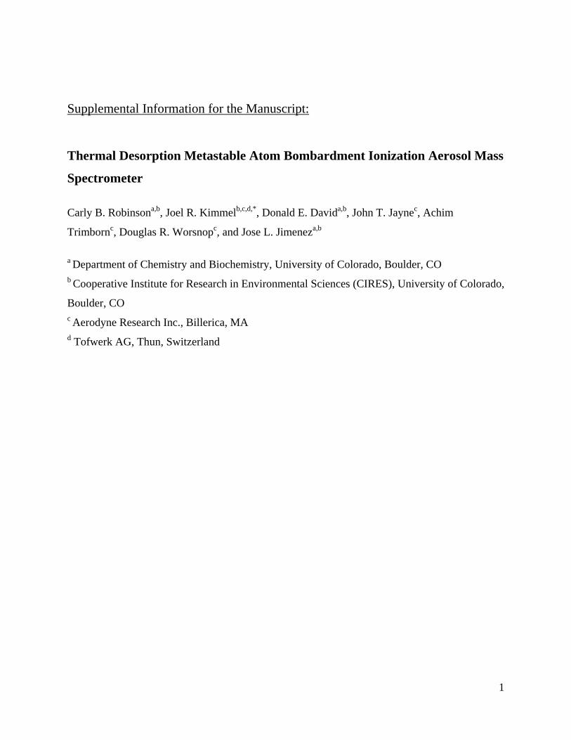
The AMS hardware and its application were detailed in arecent review [8], and the time-of-flight AMS is shown inFig. S1 (supp. Info.). Briefly, ambient particles are sampled directlyfrom atmospheric pressure into the vacuum system of the AMSvia an aerodynamic lens [33], which focuses particles into a tightbeam. The beam traverses a high vacuum particle flight chamber.A rotating mechanical chopper at the beginning of the particletime-of-flight region can modulate the beam for size-resolvedmeasurements (PToF mode). Alternatively, this chopper is alter-nated between discrete beam transmitting and non-transmittingpositions for background-subtracted ensemble measurements (MSmode). At the end of the particle drift region, particles impacta heated surface (typically 600 ◦C) which leads to vaporizationof non-refractory species. In the standard implementation [8],the resultant plume of vapor is analyzed by electron ionization(EI) mass spectrometry. Typically the filament used to create theelectrons for EI has an emission current of 2.0 mA. The AMS(Aerodyne Research, Billerica, MA) is available with three differ-ent mass spectrometers: a quadrupole mass spectrometer [34](QMG 422, Balzers, Furstentum, Liechtenstein), a compact, high-sensitivity TOF mass spectrometer [35] (CTOF, Tofwerk AG, Thun,Switzerland), or a high-resolution TOFMS [36] (HTOF, Tofwerk).This work used the HTOF-based instrument, called the HR-ToF-AMS[36], but the MAB source design is not specific to this plat-form.
A schematic representation of the metastable beam source andthe ionization region of the AMS is shown in Fig. 1. The standardAMS has two EI filaments mounted on opposite sides of the ion-ization chamber. Here, one filament has been removed, and themetastable beam enters the ionization chamber through a 5 mmdiameter hole in the ionization chamber at the position of theremoved filament. The self-contained beam source is housed in astainless-steel vacuum chamber (mechanical drawing available inFig. S2), which attaches to the AMS vacuum system at an ISO-63 portimmediately adjacent to the EI-vaporizer assembly. The beam-exitend of the source chamber extends into the AMS ionization cham-ber on an axis exactly opposite the remaining EI filament, to the
point where the exit aperture of the source is in near contact withthe ionization chamber assembly. Metastable species enter the ion-ization volume a few millimeters from the vaporizer surface. EI iskept inactive during MAB operation. The sensitivity of EI analysisis not significantly affected by the installation of the MAB source.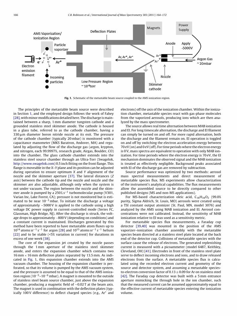
166 C.B. Robinson et al. / International Journal of Mass Spectrometry 303 (2011) 164–172
sour
i[tgi3oclaishfldnasnmVmovGaam1[e
ta1cvfatocTi
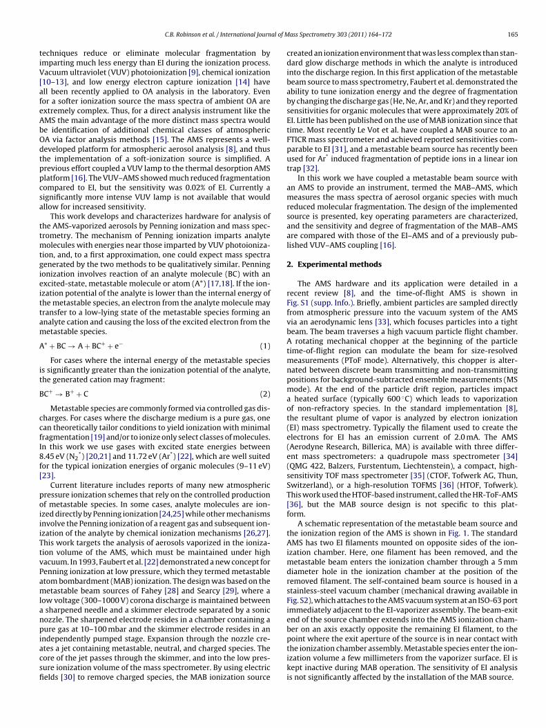
Fig. 1. Schematic of the metastable beam
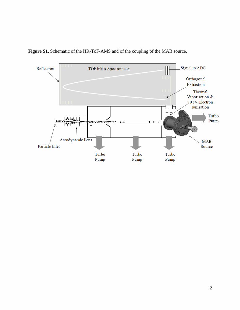
The principles of the metastable beam source were describedn Section 1, and the employed design follows the work of Fahey28], with minor modifications detailed here. The discharge is main-ained between a sharp, 1 mm diameter tungsten cathode and arounded stainless steel skimmer anode. The cathode is housedn a glass tube, referred to as the cathode chamber, having a30 �m diameter boron nitride nozzle at its exit. The pressuref the cathode chamber (typically 20 mbar) is monitored with aapacitance manometer (MKS Baratron, Andover, MA) and regu-ated by adjusting the flow of the discharge gas (argon, krypton,nd nitrogen, each 99.9997%, research grade, Airgas, Boulder, CO)nto the chamber. The glass cathode chamber extends into thetainless steel source chamber through an Ultra-Torr (Swagelok,ttp://www.swagelok.com) 0.5 inch fitting on the front flange. Thisange is moveable in the X–Y plane and its position can be adjusteduring operation to ensure optimum X and Y alignment of theozzle and the skimmer aperture [37]. The lateral distances (Zxis) between the cathode tip and the nozzle and nozzle and thekimmer are also adjustable, although only when the system isot under vacuum. The region between the nozzle and the skim-er anode is pumped by a 250 L s−1 turbomolecular pump (V301,arian Inc, Lake Forest, CA); pressure is not monitored but is esti-ated to be near 10−3 mbar. To initiate the discharge a voltage
f approximately −5000 V is applied to the cathode using a higholtage DC power supply in current-controlled mode (Series FC,lassman, High Bridge, NJ). After the discharge is struck, the volt-ge drops to approximately −800 V (depending on conditions) andconstant current is maintained. Discharges generated by thisethod have been reported to have metastable atom fluxes up to
014 atoms sr−1 s−1 for argon [28] and 1015 atoms sr−1 s−1 helium22] and to be stable (<5% variation in current) for durations inxcess of one week [38].
The core of the expansion jet created by the nozzle passeshrough the 1 mm aperture of the stainless steel skimmernode, and enters the expansion chamber which contains two6 mm × 16 mm deflection plates separated by 13.5 mm. As indi-ated in Fig. 1, this expansion chamber extends into the AMSacuum chamber. The housing of the expansion chamber is per-orated, so that its volume is pumped by the AMS vacuum system,nd the pressure is assumed to be equal to that of the AMS ioniza-
−5 −6
ion region (10 –10 mbar). A magnet is mounted to the outsidef stainless steel beam source chamber, just above the expansionhamber, producing a magnetic field of ∼0.02 T at the beam axis.he magnet is used in combination with the deflection plates (typ-cally 100 V difference) to deflect charged species (e.g., Ar+ andce coupled to the AMS ionization region.
electrons) off the axis of the ionization chamber. Within the ioniza-tion chamber, metastable species react with gas-phase moleculesfrom the vaporized aerosols, producing ions which are then ana-lyzed by the mass spectrometer.
The source allows real time alternation between MAB ionizationand EI. For long timescale alternation, the discharge and EI filamentcan simply be turned on and off. For more rapid alternation, boththe discharge and the filament remain on. EI operation is toggledon and off by switching the electron acceleration energy between70 eV (on) and 0 eV (off). For time periods where the electron energyis 0 V, mass spectra are equivalent to operation with only MAB ion-ization. For time periods where the electron energy is 70 eV, the EImechanism dominates the observed signal and the MAB ionizationis treated as effectively negligible. Background peaks associatedwith EI of the discharge gas are removed by subtraction.
Source performance was optimized by two methods: aerosolmass spectral measurements and direct measurement ofmetastable species flux. MS experiments allow characterizationof the instrument’s analytical capabilities. The flux measurementsallow the assembled source to be directly compared to otherpublished designs (MS and non-MS applications).
For MS-based characterization oleic acid, C18H34O2 (99.0%purity, Sigma Aldrich, St. Louis, MO) aerosols were created usinga TSI constant output atomizer (St. Paul, MN, model 3076) andanalyzed by the AMS using MAB ionization and EI. Aerosol con-centrations were not calibrated. Instead, the sensitivity of MABionization relative to EI was used as a sensitivity metric.
For metastable species flux measurements, a Faraday cupdetector [39,40] was mounted in the position of the AMSvaporizer–ionization chamber assembly with the metastablespecies beam directed at a stainless steel plate located at the backend of the detector cup. Collisions of metastable species with thesurface cause the release of electrons. The generated replenishingcurrent is measured with a picoammeter (model 6487, Keithley,Cleveland, OH) [41]. Electrodes in front of the stainless steel plateserve to deflect incoming electrons and ions, and to draw releasedelectrons from the surface. A metastable species flux is calcu-lated using the recorded electron current and geometry of thesource and detector system, and assuming a metastable species-to-electron conversion factor of 0.13 ± 0.09 for Ar on stainless steel
[42]. The Faraday cup detector was built with a 5 mm entranceaperture mimicking the through hole in the ion chamber, suchthat the measured current can be assumed approximately equal tothe effective current of metastable species entering the ionizationvolume.
al of M
aoaLtawsd
3
leidvridd
3
olicemria[tsbu
FaEMeo
C.B. Robinson et al. / International Journ
Field data are presented from the FLAME-3 experiment (Fire Labt Missoula Experiment, Phase 3), which focused on quantificationf emissions from controlled biomass burns simulating wildfires,nd was conducted at the United States Forest Service’s Fire Scienceaboratory in Missoula, MT [43]. An HR-ToF-AMS equipped withhe MAB source was positioned on the second story of the Fire Lab,nd sampled directly (using a 20 m-long 3/8 inch OD copper inletith a flow of 10 L min−1) from a large smoke stack through which
moke was directed. Data are presented from an experiment whereifferent materials were burned for ∼5 min each.
. Results and discussion
In the early stages of this work, features from multiple pub-ished metastable source designs [22,28,29,37,38,40,44–47] werexplored to identify a design having high metastable flux, stabil-ty sufficient for field experiments (minimum of 12 h of continuousata acquisition), and repeatable performance. Factors that werearied included: cathode shape, electrode geometry, cathode mate-ial, and nozzle material. Those results are not detailed here, butnterested readers are pointed to the referenced works, whichiscuss these designs. The presented results refer to the sourceescribed in Section 2.
.1. Metastable source optimization
The gas jet exiting the discharge nozzle contains a mixturef neutral species, metastable species, ions, and electrons. Ana-yte molecules can be ionized by numerous mechanisms involvingnteractions with the charged species, most notably EI, which typi-ally yields strong fragmentation. In order to simplify the ionizationnvironment and allow Penning ionization to be the dominantechanism, a combination of electric and magnetic fields is used to
emove charged species from the beam directed into the AMS ion-zation volume. The first descriptions of MAB ionization and recentpplications of beam source for metastable-induced fragmentation
32] used only electrostatic deflection. In this work, strong deflec-ion fields alone proved insufficient. Fig. 2 shows a difference masspectrum (particle beam transmitted data minus particle beamlocked data) of laboratory-generated oleic acid particles, collectedsing EI (Fig. 2a) and Ar* MAB (M(Ar)B) ionization with and with-1.5x10⁵
1.0
0.5
0.0
300250200150100500
m/z
1000
500
0
3000
1500
0
1000
500
0
1.5x10⁶
1.0
0.5
0.0
MAB - Deflection Plates and Magnet Fp = 0.080
MAB - Deflection Plates Only Fp = 0.041
MAB - Magnets Only Fp = 0.075
MAB - Neither Deflection Plates nor Magnet Fp = 0.020
To
tal S
ign
al (io
ns s
-1)
EI Fp = 0.016
b
a
c
d
e
ig. 2. Comparison of the background subtracted mass spectra of pure oleic aciderosol standards acquired with a vaporizer temperature of 200 ◦C ionized by (a)I (b) MAB without deflection or a magnet (c) MAB with only deflection plates, (d)AB with only a magnet, and (e) MAB with a magnet and deflection plates in the
xperimental setup. Fp is the ratio of the signal from the parent plus dehydratedleic acid ions to the total signal.
ass Spectrometry 303 (2011) 164–172 167
out the applied magnetic and electric deflection fields (Fig. 2b–e).When neither deflection plates nor a magnet are used (Fig. 2b) theMAB–AMS spectrum has a similar degree of fragmentation com-pared to the EI–AMS spectrum with the majority of the total signalbelow m/z 100, suggesting that ionization is dominated by dis-charge electrons. When only the deflection plate voltage is applied(Fig. 2c) the fraction of the signal from the parent plus dehydratedmolecular ion (Fp) doubles, and there is an increase in the relativeintensity of fragments above m/z 100. Simultaneously, deflection ofcharged species (Ar+ and/or e−) leads to a 23-fold reduction in totalrecorded ion current. Calculations suggest that the applied deflec-tion voltages should be more than sufficient to deflect all of theAr+ and e−, and operation at substantially higher voltages (up to10×) results in little change in the spectrum. But, when a magneticfield is applied alone (Fig. 2d) or concurrent to the deflection plates(Fig. 2e), fragments below m/z 50 are nearly eliminated. It is clearthat the magnets are more effectively removing electrons from theaxis of the ionization volume, but the exact mechanism of removalis unknown. For instance, the magnets could be changing the natureof the discharge between the nozzle and skimmer, or the magnetscould be removing electrons generated in processes downstream ofthe deflection plates (which could also explain the lack of efficiencyof the deflection plates). Unless otherwise noted, both magnets anddeflection plates were used for all data presented in the remainderof this paper. In future designs the deflection plates will not beincorporated into the design and only the magnets will be used.
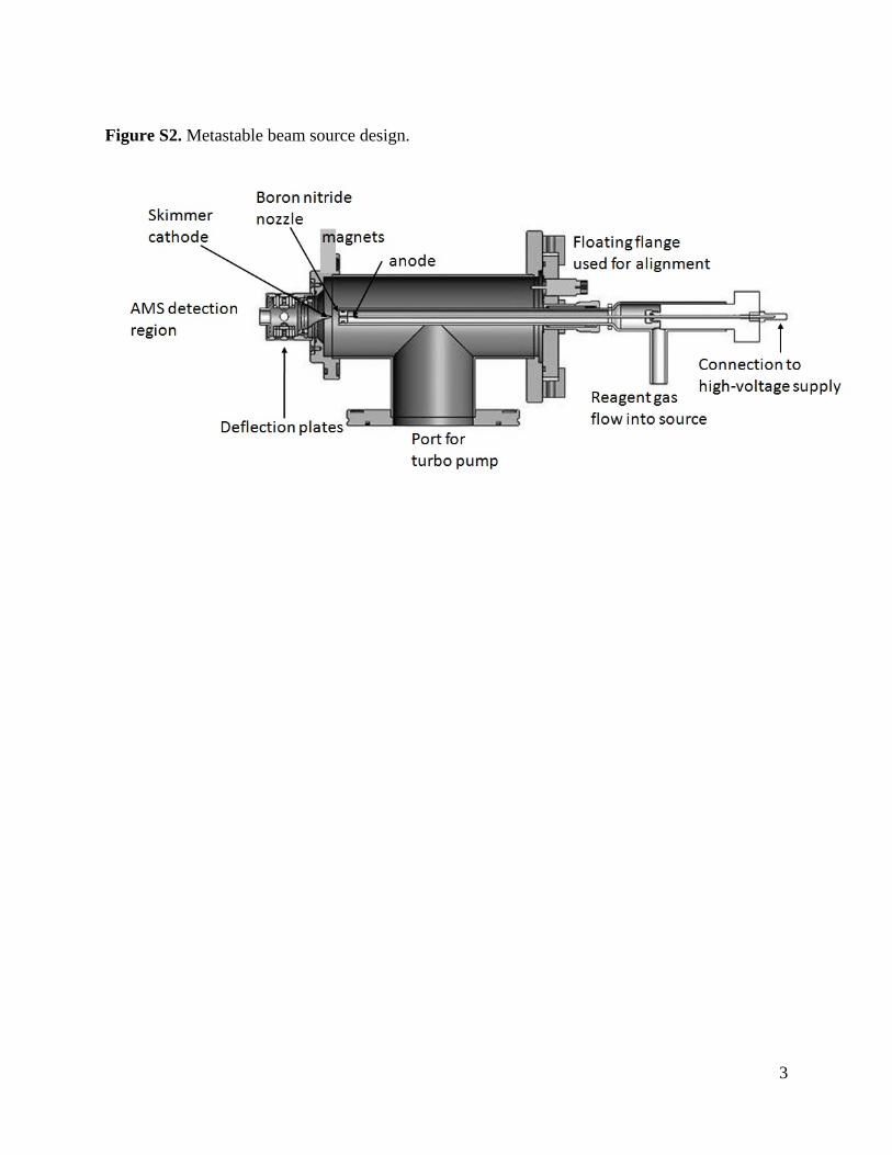
The primary tunable parameters of the MAB ionization sourceare discharge current and discharge gas pressure. Metastable beamsources of similar design are typically run near 10 mA [22,37]. Forour source, a current greater than 6 mA is required to maintain astable discharge. Above a current of 16 mA oxidized material startsto form on the tip of the tungsten cathode, leading to rapid degra-dation and discharge failure. The metastable flux is approximately30% greater at 16 mA than at 8 mA, and the observed MS ion currentis directly proportional to this flux (Fig. S3). Typical experiments arerun at 12 mA to ensure longer term stability.
Cathode chamber pressure varies greatly for Ar* beam sources(40 [22] to 400 mbar [45]), depending on design geometry andpumping. For our source, a discharge cannot be maintained below13 mbar and above 65 mbar the gas load becomes too high for the
vacuum system. The effect of cathode chamber pressure in thisrange was characterized using an argon discharge for the ioniza-tion of oleic acid particles. Fig. 3 shows the total signal at m/z40, 43, 264, and 282 as a function of cathode chamber pressure.These peaks correspond to Ar+, an oleic acid fragment indicative1000
800
600
400
200
0
Tota
l S
ign
al (io
ns s
-1)
m/z
28
2 a
nd
26
4
5045403530252015
Pressure (mbar)
30x103
20
10
0
To
tal S
ign
al (io
ns s
-1)m
/z 4
0
80
60
40
20
0
To
tal S
ign
al (io
ns s
-1)m
/z 4
3
m/z 282m/z 264m/z 43m/z 40
Fig. 3. Effect of the discharge gas pressure on AMS signals while using a constantconcentration of pure oleic acid aerosol standards. m/z 40 corresponds to Ar+. m/z 43is a marker for highly fragmented oleic acid molecules. m/z 264 and 282 correspondto dehydrated oleic acid and the oleic acid parent ion, respectively.
168 C.B. Robinson et al. / International Journal of Mass Spectrometry 303 (2011) 164–172
2x10⁶
1
0
To
tal S
ign
al (io
n s
-1)
300250200150100500m/z
2x10⁶
1
0
1000
500
0
300250200150100500
1000
500
0
To
tal S
ign
al (io
n s
-1)
1000
500
0
2x10⁶
1
0
1.5x10⁴
1.2
0.9
0.6
0.3
0.0To
tal S
ign
al (io
n s
-1)
600500400300200
Vaporizer Temperature (°C)
0.10
0.08
0.06
0.04
0.02
0.00F
p
Vaporizer Temperature 180°C
Vaporizer Temperature 600°C
EI M(Ar)B
a
b
d
c
e
EI M(Ar)B
M(Ar)BEI Vaporizer Temperature 305°C
M(Ar)B
f g
Total Signal Fp
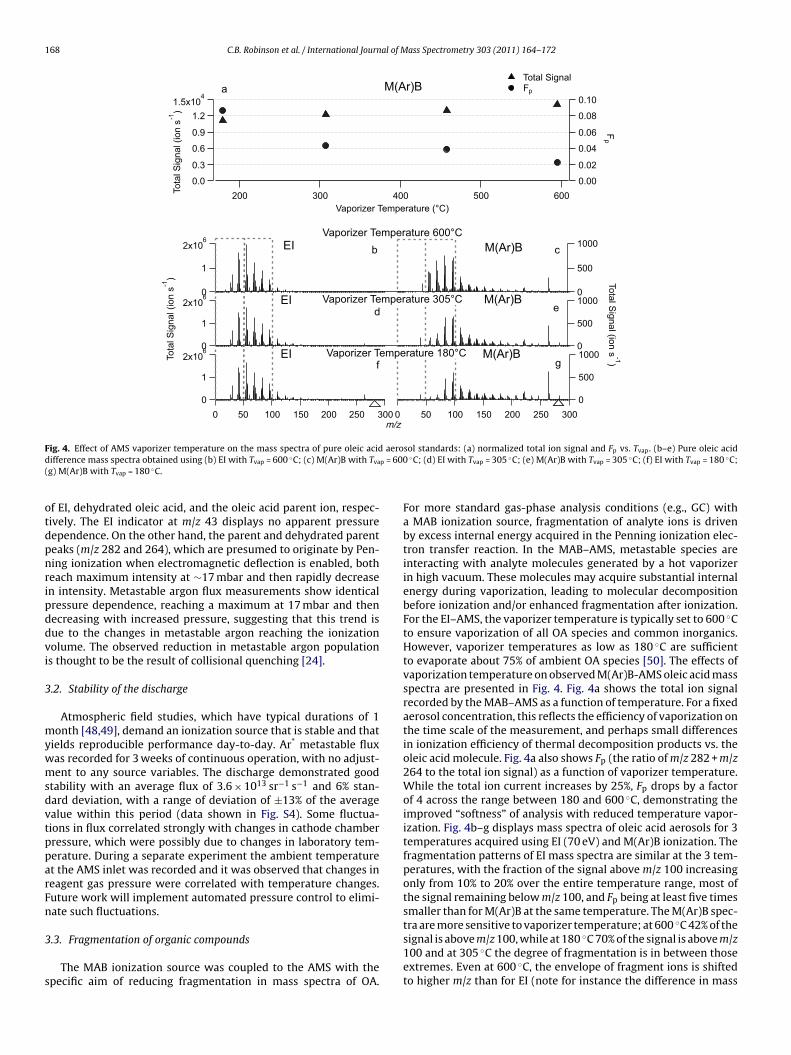
F aerod p = 600(
otdpnripddvi
3
mywmsdvtpparFn
3
s
ig. 4. Effect of AMS vaporizer temperature on the mass spectra of pure oleic acidifference mass spectra obtained using (b) EI with Tvap = 600 ◦C; (c) M(Ar)B with Tva
g) M(Ar)B with Tvap = 180 ◦C.
f EI, dehydrated oleic acid, and the oleic acid parent ion, respec-ively. The EI indicator at m/z 43 displays no apparent pressureependence. On the other hand, the parent and dehydrated parenteaks (m/z 282 and 264), which are presumed to originate by Pen-ing ionization when electromagnetic deflection is enabled, botheach maximum intensity at ∼17 mbar and then rapidly decreasen intensity. Metastable argon flux measurements show identicalressure dependence, reaching a maximum at 17 mbar and thenecreasing with increased pressure, suggesting that this trend isue to the changes in metastable argon reaching the ionizationolume. The observed reduction in metastable argon populations thought to be the result of collisional quenching [24].
.2. Stability of the discharge
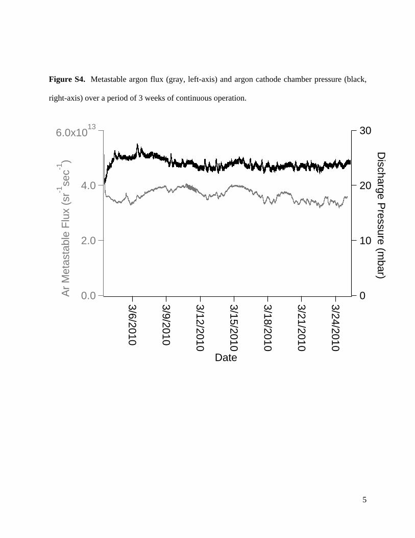
Atmospheric field studies, which have typical durations of 1onth [48,49], demand an ionization source that is stable and that
ields reproducible performance day-to-day. Ar* metastable fluxas recorded for 3 weeks of continuous operation, with no adjust-ent to any source variables. The discharge demonstrated good
tability with an average flux of 3.6 × 10¹³ sr−1 s−1 and 6% stan-ard deviation, with a range of deviation of ±13% of the averagealue within this period (data shown in Fig. S4). Some fluctua-ions in flux correlated strongly with changes in cathode chamberressure, which were possibly due to changes in laboratory tem-erature. During a separate experiment the ambient temperaturet the AMS inlet was recorded and it was observed that changes ineagent gas pressure were correlated with temperature changes.uture work will implement automated pressure control to elimi-ate such fluctuations.
.3. Fragmentation of organic compounds
The MAB ionization source was coupled to the AMS with thepecific aim of reducing fragmentation in mass spectra of OA.
sol standards: (a) normalized total ion signal and Fp vs. Tvap. (b–e) Pure oleic acid◦C; (d) EI with Tvap = 305 ◦C; (e) M(Ar)B with Tvap = 305 ◦C; (f) EI with Tvap = 180 ◦C;
For more standard gas-phase analysis conditions (e.g., GC) witha MAB ionization source, fragmentation of analyte ions is drivenby excess internal energy acquired in the Penning ionization elec-tron transfer reaction. In the MAB–AMS, metastable species areinteracting with analyte molecules generated by a hot vaporizerin high vacuum. These molecules may acquire substantial internalenergy during vaporization, leading to molecular decompositionbefore ionization and/or enhanced fragmentation after ionization.For the EI–AMS, the vaporizer temperature is typically set to 600 ◦Cto ensure vaporization of all OA species and common inorganics.However, vaporizer temperatures as low as 180 ◦C are sufficientto evaporate about 75% of ambient OA species [50]. The effects ofvaporization temperature on observed M(Ar)B-AMS oleic acid massspectra are presented in Fig. 4. Fig. 4a shows the total ion signalrecorded by the MAB–AMS as a function of temperature. For a fixedaerosol concentration, this reflects the efficiency of vaporization onthe time scale of the measurement, and perhaps small differencesin ionization efficiency of thermal decomposition products vs. theoleic acid molecule. Fig. 4a also shows Fp (the ratio of m/z 282 + m/z264 to the total ion signal) as a function of vaporizer temperature.While the total ion current increases by 25%, Fp drops by a factorof 4 across the range between 180 and 600 ◦C, demonstrating theimproved “softness” of analysis with reduced temperature vapor-ization. Fig. 4b–g displays mass spectra of oleic acid aerosols for 3temperatures acquired using EI (70 eV) and M(Ar)B ionization. Thefragmentation patterns of EI mass spectra are similar at the 3 tem-peratures, with the fraction of the signal above m/z 100 increasingonly from 10% to 20% over the entire temperature range, most ofthe signal remaining below m/z 100, and Fp being at least five timessmaller than for M(Ar)B at the same temperature. The M(Ar)B spec-
tra are more sensitive to vaporizer temperature; at 600 ◦C 42% of thesignal is above m/z 100, while at 180 ◦C 70% of the signal is above m/z100 and at 305 ◦C the degree of fragmentation is in between thoseextremes. Even at 600 ◦C, the envelope of fragment ions is shiftedto higher m/z than for EI (note for instance the difference in mass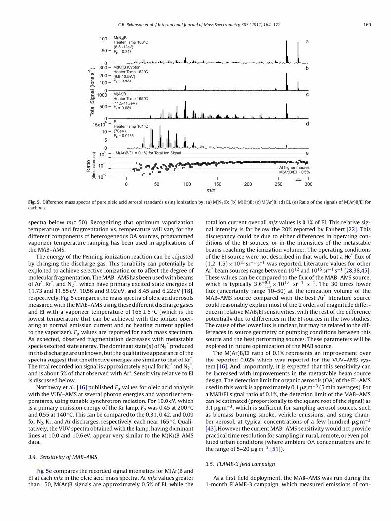
C.B. Robinson et al. / International Journal of Mass Spectrometry 303 (2011) 164–172 169
15x105
10
5
0
300250200150100500
100
50
0
Tota
lS
ignal(ions
s-1
)1000
500
0
300
200
100
0
10-6
10-3
100
Ra
tio
(dim
ensio
nle
ss)
EIHeater Temp 161°C(70eV)Fp = 0.0165
M(N2)B
Heater Temp 163°C(8.5 -12eV)Fp = 0.313
M(Ar)BHeater Temp 165°C(11.5-11.7eV)Fp = 0.089
M(Kr)B KryptonHeater Temp 162°C(9.9-10.5eV)Fp = 0.428
M(Ar)B/EI = 0.1% for Total Ion Signal
At higher massesM(Ar)B/EI ~ 0.5%
a
b
c
d
e
F n by:e
stdvt
bemo1rmalatAsisTai
wpiaftld
3
Et
ig. 5. Difference mass spectra of pure oleic acid aerosol standards using ionizatioach m/z.
pectra below m/z 50). Recognizing that optimum vaporizationemperature and fragmentation vs. temperature will vary for theifferent components of heterogeneous OA sources, programmedaporizer temperature ramping has been used in applications ofhe MAB–AMS.
The energy of the Penning ionization reaction can be adjustedy changing the discharge gas. This tunability can potentially bexploited to achieve selective ionization or to affect the degree ofolecular fragmentation. The MAB–AMS has been used with beams
f Ar*, Kr*, and N2*, which have primary excited state energies of
1.73 and 11.55 eV, 10.56 and 9.92 eV, and 8.45 and 6.22 eV [18],espectively. Fig. 5 compares the mass spectra of oleic acid aerosolseasured with the MAB–AMS using these different discharge gases
nd EI with a vaporizer temperature of 165 ± 5 ◦C (which is theowest temperature that can be achieved with the ionizer oper-ting at normal emission current and no heating current appliedo the vaporizer). Fp values are reported for each mass spectrum.s expected, observed fragmentation decreases with metastablepecies excited state energy. The dominant state(s) of N2
* producedn this discharge are unknown, but the qualitative appearance of thepectra suggest that the effective energies are similar to that of Kr*.he total recorded ion signal is approximately equal for Kr* and N2
*,nd is about 5% of that observed with Ar*. Sensitivity relative to EIs discussed below.
Northway et al. [16] published Fp values for oleic acid analysisith the VUV–AMS at several photon energies and vaporizer tem-eratures, using tunable synchrotron radiation. For 10.0 eV, which
s a primary emission energy of the Kr lamp, Fp was 0.45 at 200 ◦Cnd 0.55 at 140 ◦C. This can be compared to the 0.31, 0.42, and 0.09or N2, Kr, and Ar discharges, respectively, each near 165 ◦C. Quali-atively, the VUV spectra obtained with the lamp, having dominantines at 10.0 and 10.6 eV, appear very similar to the M(Kr)B-AMSata.
.4. Sensitivity of MAB–AMS
Fig. 5e compares the recorded signal intensities for M(Ar)B andI at each m/z in the oleic acid mass spectra. At m/z values greaterhan 150, M(Ar)B signals are approximately 0.5% of EI, while the
m/z
(a) M(N2)B; (b) M(Kr)B; (c) M(Ar)B; (d) EI. (e) Ratio of the signals of M(Ar)B/EI for
total ion current over all m/z values is 0.1% of EI. This relative sig-nal intensity is far below the 20% reported by Faubert [22]. Thisdiscrepancy could be due to either differences in operating con-ditions of the EI sources, or in the intensities of the metastablebeams reaching the ionization volumes. The operating conditionsof the EI source were not described in that work, but a He* flux of(1.2–1.5) × 1015 sr−1 s−1 was reported. Literature values for otherAr* beam sources range between 1012 and 1015 sr−1 s−1 [28,38,45].These values can be compared to the flux of the MAB–AMS source,which is typically 3.6+8.1
−1.5 × 10¹³ sr−1 s−1. The 30 times lowerflux (uncertainty range 10–50) at the ionization volume of theMAB–AMS source compared with the best Ar* literature sourcecould reasonably explain most of the 2 orders of magnitude differ-ence in relative MAB/EI sensitivities, with the rest of the differencepotentially due to differences in the EI sources in the two studies.The cause of the lower flux is unclear, but may be related to the dif-ferences in source geometry or pumping conditions between thissource and the best performing sources. These parameters will beexplored in future optimization of the MAB source.
The M(Ar)B/EI ratio of 0.1% represents an improvement overthe reported 0.02% which was reported for the VUV–AMS sys-tem [16]. And, importantly, it is expected that this sensitivity canbe increased with improvements in the metastable beam sourcedesign. The detection limit for organic aerosols (OA) of the EI–AMSused in this work is approximately 0.1 �g m−3 (5 min averages). Fora MAB/EI signal ratio of 0.1%, the detection limit of the MAB–AMScan be estimated (proportionally to the square root of the signal) as3.1 �g m−3, which is sufficient for sampling aerosol sources, suchas biomass burning smoke, vehicle emissions, and smog cham-ber aerosol, at typical concentrations of a few hundred �g m−3
[43]. However the current MAB–AMS sensitivity would not providepractical time resolution for sampling in rural, remote, or even pol-luted urban conditions (where ambient OA concentrations are inthe range of 5–20 �g m−3 [51]).
3.5. FLAME-3 field campaign
As a first field deployment, the MAB–AMS was run during the1-month FLAME-3 campaign, which measured emissions of con-
170 C.B. Robinson et al. / International Journal of Mass Spectrometry 303 (2011) 164–172
1000
800
600
400
200
0
Tota
l S
ignal (A
.U.)
3:20 PM
9/16/09
3:30 PM 3:40 PM 3:50 PM
Date and Time
0.1
2
4
6
81
2
4
6
810
M(A
r)B
No
rma
lize
dS
ign
al
0.12 3 4 5 6
12 3 4 5 6
10
EI Normalized Signal
46
78
10
1112
13
14
M(Ar)B EI
Burn 7Manzanita
Burn 6Ceanothus
1:12:1
1:2
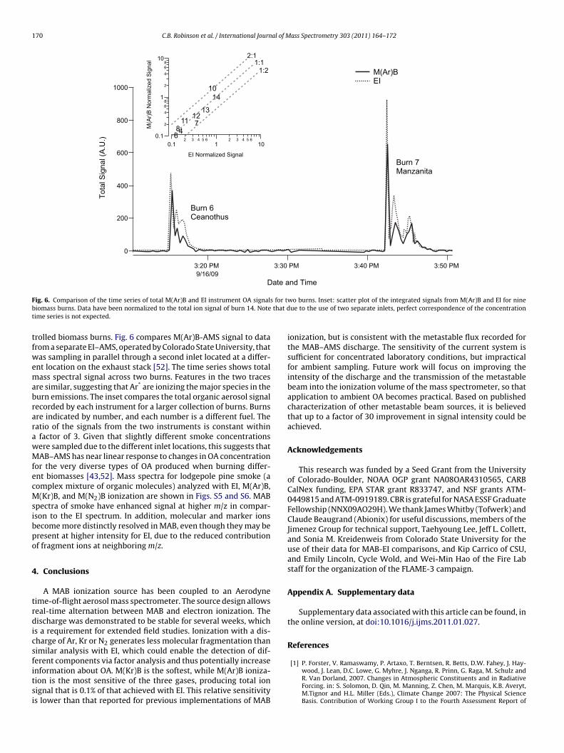
F for tb that dt
tfwemabrarawMfecMsibpo
4
trdicsfitsi
ig. 6. Comparison of the time series of total M(Ar)B and EI instrument OA signalsiomass burns. Data have been normalized to the total ion signal of burn 14. Noteime series is not expected.
rolled biomass burns. Fig. 6 compares M(Ar)B-AMS signal to datarom a separate EI–AMS, operated by Colorado State University, thatas sampling in parallel through a second inlet located at a differ-
nt location on the exhaust stack [52]. The time series shows totalass spectral signal across two burns. Features in the two traces
re similar, suggesting that Ar* are ionizing the major species in theurn emissions. The inset compares the total organic aerosol signalecorded by each instrument for a larger collection of burns. Burnsre indicated by number, and each number is a different fuel. Theatio of the signals from the two instruments is constant withinfactor of 3. Given that slightly different smoke concentrationsere sampled due to the different inlet locations, this suggests thatAB–AMS has near linear response to changes in OA concentration
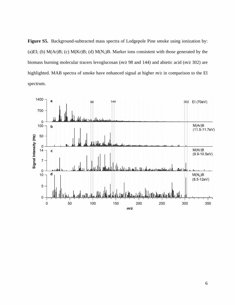
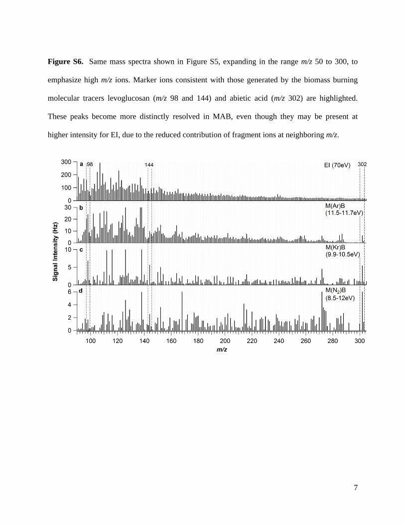
or the very diverse types of OA produced when burning differ-nt biomasses [43,52]. Mass spectra for lodgepole pine smoke (aomplex mixture of organic molecules) analyzed with EI, M(Ar)B,(Kr)B, and M(N2)B ionization are shown in Figs. S5 and S6. MAB
pectra of smoke have enhanced signal at higher m/z in compar-son to the EI spectrum. In addition, molecular and marker ionsecome more distinctly resolved in MAB, even though they may beresent at higher intensity for EI, due to the reduced contributionf fragment ions at neighboring m/z.
. Conclusions
A MAB ionization source has been coupled to an Aerodyneime-of-flight aerosol mass spectrometer. The source design allowseal-time alternation between MAB and electron ionization. Theischarge was demonstrated to be stable for several weeks, which
s a requirement for extended field studies. Ionization with a dis-harge of Ar, Kr or N2 generates less molecular fragmentation thanimilar analysis with EI, which could enable the detection of dif-
erent components via factor analysis and thus potentially increasenformation about OA. M(Kr)B is the softest, while M(Ar)B ioniza-ion is the most sensitive of the three gases, producing total ionignal that is 0.1% of that achieved with EI. This relative sensitivitys lower than that reported for previous implementations of MABwo burns. Inset: scatter plot of the integrated signals from M(Ar)B and EI for nineue to the use of two separate inlets, perfect correspondence of the concentration
ionization, but is consistent with the metastable flux recorded forthe MAB–AMS discharge. The sensitivity of the current system issufficient for concentrated laboratory conditions, but impracticalfor ambient sampling. Future work will focus on improving theintensity of the discharge and the transmission of the metastablebeam into the ionization volume of the mass spectrometer, so thatapplication to ambient OA becomes practical. Based on publishedcharacterization of other metastable beam sources, it is believedthat up to a factor of 30 improvement in signal intensity could beachieved.
Acknowledgements
This research was funded by a Seed Grant from the Universityof Colorado-Boulder, NOAA OGP grant NA08OAR4310565, CARBCalNex funding, EPA STAR grant R833747, and NSF grants ATM-0449815 and ATM-0919189. CBR is grateful for NASA ESSF GraduateFellowship (NNX09AO29H). We thank James Whitby (Tofwerk) andClaude Beaugrand (Abionix) for useful discussions, members of theJimenez Group for technical support, Taehyoung Lee, Jeff L. Collett,and Sonia M. Kreidenweis from Colorado State University for theuse of their data for MAB-EI comparisons, and Kip Carrico of CSU,and Emily Lincoln, Cycle Wold, and Wei-Min Hao of the Fire Labstaff for the organization of the FLAME-3 campaign.
Appendix A. Supplementary data
Supplementary data associated with this article can be found, inthe online version, at doi:10.1016/j.ijms.2011.01.027.
References
[1] P. Forster, V. Ramaswamy, P. Artaxo, T. Berntsen, R. Betts, D.W. Fahey, J. Hay-wood, J. Lean, D.C. Lowe, G. Myhre, J. Nganga, R. Prinn, G. Raga, M. Schulz andR. Van Dorland, 2007. Changes in Atmospheric Constituents and in RadiativeForcing. in: S. Solomon, D. Qin, M. Manning, Z. Chen, M. Marquis, K.B. Averyt,M.Tignor and H.L. Miller (Eds.), Climate Change 2007: The Physical ScienceBasis. Contribution of Working Group I to the Fourth Assessment Report of
al of M
[
[
[
[
[
[
[
[
[
[
[
[
[
[
[
[
[
[
[
[
[[
[
[
[
[
[
[
[
[
[
[
[
[
[
[
[
[
[
C.B. Robinson et al. / International Journ
the Intergovernmental Panel on Climate Change. Cambridge University Press,Cambridge, United Kingdom and New York, NY, USA, 2007, pp. 129–234.
[2] M. Hallquist, J.C. Wenger, U. Baltensperger, Y. Rudich, D. Simpson, M. Claeys, J.Dommen, N.M. Donahue, C. George, A.H. Goldstein, J.F. Hamilton, H. Herrmann,T. Hoffmann, Y. Iinuma, M. Jang, M.E. Jenkin, J.L. Jimenez, A. Kiendler-Scharr, W.Maenhaut, G. McFiggans, T.F. Mentel, A. Monod, A.S.H. Prevot, J.H. Seinfeld, J.D.Surratt, R. Szmigielski, J. Wildt, The Formation, properties and impact of sec-ondary organic aerosol: current and emerging issues, Atmospheric Chemistryand Physics 9 (2009) 5155–5236.
[3] J.C. Chow, J.G. Watson, L.C. Pritchett, W.R. Pierson, C.A. Frazier, R.G. Purcell,The DRI thermal optical reflectance carbon analysis system – description,evaluation and applications in United-States air-quality studies, AtmosphericEnvironment 27 (1993) 1185–1201.
[4] A.P. Sullivan, R.J. Weber, A.L. Clements, J.R. Turner, M.S. Bae, J.J. Schauer, Amethod for on-line measurement of water-soluble organic carbon in ambientaerosol particles: results from an urban site, Geophysical Research Letters 31(2004).
[5] B.J. Williams, A.H. Goldstein, D.B. Millet, R. Holzinger, N.M. Kreisberg, S.V. Her-ing, A.B. White, D.R. Worsnop, J.D. Allan, J.L. Jimenez, Chemical speciation oforganic aerosol during the international consortium for atmospheric researchon transport and transformation 2004: results from in situ measurements,Journal of Geophysical Research: Atmospheres 112 (2007).
[6] S. Gilardoni, S. Liu, S. Takahama, L.M. Russell, J.D. Allan, R. Steinbrecher, J.L.Jimenez, P.F. De Carlo, E.J. Dunlea, D. Baumgardner, Characterization of organicambient aerosol during MIRAGE 2006 on three platforms, Atmospheric Chem-istry and Physics 9 (2009) 5417–5432.
[7] S. Decesari, M. Mircea, F. Cavalli, S. Fuzzi, F. Moretti, E. Tagliavini, M.C.Facchini, Source attribution of water-soluble organic aerosol by nuclear mag-netic resonance spectroscopy, Environmental Science & Technology 41 (2007)2479–2484.
[8] M.R. Canagaratna, J.T. Jayne, J.L. Jimenez, J.D. Allan, M.R. Alfarra, Q. Zhang,T.B. Onasch, F. Drewnick, H. Coe, A. Middlebrook, A. Delia, L.R. Williams, A.M.Trimborn, M.J. Northway, P.F. DeCarlo, C.E. Kolb, P. Davidovits, D.R. Worsnop,Chemical and microphysical characterization of ambient aerosols with theaerodyne aerosol mass spectrometer, Mass Spectrometry Reviews 26 (2007)185–222.
[9] B. Oktem, M.P. Tolocka, M.V. Johnston, On-line analysis of organic componentsin fine and ultrafine particles by photoionization aerosol mass spectrometry,Analytical Chemistry 76 (2004) 253–261.
10] R. Holzinger, J. Williams, F. Herrmann, J. Lelieveld, N.M. Donahue, T. Rockmann,Aerosol analysis using a thermal-desorption proton-transfer-reaction massspectrometer (TD–PTR–MS): a new approach to study processing of organicaerosols, Atmospheric Chemistry and Physics 10 (2010) 2257–2267.
11] T. Thornberry, D.M. Murphy, D.S. Thomson, J. de Gouw, C. Warneke, T.S. Bates,P.K. Quinn, D. Coffman, Measurement of aerosol organic compounds usinga novel collection/thermal-desorption PTR-ITMS instrument, Aerosol Scienceand Technology 43 (2009) 486–501.
12] R.L.N. Yatavelli, J.A. Thornton, Particulate organic matter detection using amicro-orifice volatilization impactor coupled to a chemical ionization massspectrometer (MOVI-CIMS), Aerosol Science and Technology 44 (2010) 61–74.
13] J.D. Hearn, G.D. Smith, Kinetics and product studies for ozonolysis reactionsof organic particles using aerosol CIMS, Journal of Physical Chemistry A 108(2004) 10019–10029.
14] B.W. LaFranchi, J. Zahardis, G.A. Petrucci, Photoelectron resonance capture ion-ization mass spectrometry: a soft ionization source for mass spectrometry ofparticle-phase organic compounds, Rapid Communications in Mass Spectrom-etry 18 (2004) 2517–2521.
15] I.M. Ulbrich, M.R. Canagaratna, Q. Zhang, D.R. Worsnop, J.L. Jimenez, Interpre-tation of organic components from positive matrix factorization of aerosolmass spectrometric data, Atmospheric Chemistry and Physics 9 (2009)2891–2918.
16] M.J. Northway, J.T. Jayne, D.W. Toohey, M.R. Canagaratna, A. Trimborn, K.I.Akiyama, A. Shimono, J.L. Jimenez, P.F. DeCarlo, K.R. Wilson, D.R. Worsnop,Demonstration of a VUV lamp photoionization source for improved organicspeciation in an aerosol mass spectrometer, Aerosol Science and Technology41 (2007) 828–839.
17] F.M. Penning, Ionisation by metastable atoms, Naturwissenschaften 15 (1927)818.
18] D.W. Setser, Reactive Intermediates in the Gas Phase, Acadenic Press, Inc., NewYork City, 1979.
19] D.R. Anderson, V.M. Bierbaum, C.H. Depuy, J.J. Grabowski, Flowing after-glow studies of organic positive-ions generated by penning ionization usingmetastable argon atoms, International Journal of Mass Spectrometry and IonProcesses 52 (1983) 65–94.
20] A. Lofthus, P.H. Krupenie, Spectrum of molecular nitrogen, Journal of Physicaland Chemical Reference Data 6 (1977) 113–307.
21] M.G. Ikonomou, S. Rayne, Chromatographic and ionization properties of poly-brominated diphenyl ethers using GC/high-resolution Ms with metastableatom bombardment and electron impact ionization, Analytical Chemistry 74(2002) 5263–5272.
22] D. Faubert, G.J.C. Paul, J. Giroux, M.J. Bertrand, Selective fragmentation and ion-ization of organic-compounds using an energy-tunable rare-gas metastablebeam source, International Journal of Mass Spectrometry and Ion Processes124 (1993) 69–77.
23] T.M. Reed, The ionization potential and the polarizability of molecules, Journalof Physical Chemistry 59 (1955) 428–432.
ass Spectrometry 303 (2011) 164–172 171
24] K. Hiraoka, H. Furuya, S. Kambara, S. Suzuki, Y. Hashimoto, A. Takamizawa,Atmospheric-pressure Penning ionization of aliphatic hydrocarbons, RapidCommunications in Mass Spectrometry 20 (2006) 3213–3222.
25] Z.J. Wu, J.X. Pu, L.M. Li, D.M. Fang, H.Y. Qi, J.Z. Chen, G.Y. Li, H.D. Sun, G.L.Zhang, Electrospray tandem mass spectrometry of longipedlactone triter-penoids, Journal of Mass Spectrometry 45 (2010) 451–455.
26] F.J. Andrade, J.T. Shelley, W.C. Wetzel, M.R. Webb, G. Gamez, S.J. Ray, G.M.Hieftje, Atmospheric pressure chemical ionization source. 1. Ionization of com-pounds in the gas phase, Analytical Chemistry 80 (2008) 2646–2653.
27] R.B. Cody, J.A. Laramee, H.D. Durst, Versatile new ion source for the analysis ofmaterials in open air under ambient conditions, Analytical Chemistry 77 (2005)2297–2302.
28] D.W. Fahey, W.F. Parks, L.D. Schearer, High-flux beam source of thermal rare-gas metastable atoms, Journal of Physics E-Scientific Instruments 13 (1980)381–383.
29] J.Q. Searcy, Supersonic molecular-beam metastable atom source initiated bydirect discharge, Review of Scientific Instruments 45 (1974) 589–590.
30] M.J. Bertrand, D. Faubert, O. Peraldi, A. L’Heureux, U.S. Patent 6,124,675, 2000.31] C. Le Vot, C. Afonso, C. Beaugrand, J.C. Tabet, Implementation of a Penning
ionization source on a FTICR instrument with ion funnel optics, InternationalJournal of Mass Spectrometry, in press.
32] V.D. Berkout, Fragmentation of protonated peptide ions via interaction withmetastable atoms, Analytical Chemistry 78 (2006) 3055–3061.
33] P. Liu, P.J. Ziemann, D.B. Kittelson, P.H. McMurry, Generating particle beams ofcontrolled dimensions and divergence. 1. Theory of particle motion in aerody-namic lenses and nozzle expansions, Aerosol Science and Technology 22 (1995)293–313.
34] J.T. Jayne, D.C. Leard, X.F. Zhang, P. Davidovits, K.A. Smith, C.E. Kolb, D.R.Worsnop, Development of an aerosol mass spectrometer for size and com-position analysis of submicron particles, Aerosol Science and Technology 33(2000) 49–70.
35] F. Drewnick, S.S. Hings, P. DeCarlo, J.T. Jayne, M. Gonin, K. Fuhrer, S. Weimer,J.L. Jimenez, K.L. Demerjian, S. Borrmann, D.R. Worsnop, A new time-of-flightaerosol mass spectrometer (ToF-AMS) – instrument description and first fielddeployment, Aerosol Science and Technology 39 (2005) 637–658.
36] P.F. DeCarlo, J.R. Kimmel, A. Trimborn, M.J. Northway, J.T. Jayne, A.C. Aiken,M. Gonin, K. Fuhrer, T. Horvath, K.S. Docherty, D.R. Worsnop, J.L. Jimenez,Field-deployable, high-resolution, time-of-flight aerosol mass spectrometer,Analytical Chemistry 78 (2006) 8281–8289.
37] M. Baker, A.J. Palmer, R.T. Sang, A high flux metastable atomic discharge sourcewith three-dimensional translation, Measurement Science & Technology 14(2003) N5–N8.
38] A.J. Palmer, M. Baker, R.T. Sang, Quantitative comparison of rare-gas coldcathode discharge metastable atomic beam sources, Review of Scientific Instru-ments 75 (2004) 5056–5058.
39] N. Traitler, Design and Characterisation of a Metastable Helium Source, PhysicsDepartment, University of York, York, UK, 2002.
40] J.P. Ashmore, R.T. Sang, Cathode design for a low-velocity metastable neoncold cathode discharge source, Measurement Science & Technology 12 (2001)N17–N21.
41] F.B. Dunning, R.D. Rundel, R.F. Stebbings, Determination of secondary-electronejection coefficients for rare-gas metastable atoms, Review of Scientific Instru-ments 46 (1975) 697–701.
42] S. Schohl, D. Klar, T. Kraft, H.A.J. Meijer, M.W. Ruf, U. Schmitz, S.J. Smith, H.Hotop, Absolute detection of metastable rare-gas atoms by a CW laser pho-toionization method, Zeitschrift Fur Physik D-Atoms Molecules and Clusters21 (1991) 25–39.
43] G.R. McMeeking, S.M. Kreidenweis, S. Baker, C.M. Carrico, J.C. Chow, J.L. Col-lett, W.M. Hao, A.S. Holden, T.W. Kirchstetter, W.C. Malm, H. Moosmuller, A.P.Sullivan, C.E. Wold, Emissions of trace gases and aerosols during the opencombustion of biomass in the laboratory, Journal of Geophysical Research:Atmospheres 114 (2009).
44] J. Kawanaka, M. Hagiuda, K. Shimizu, F. Shimizu, H. Takuma, Generation ofan intense low-velocity metastable-neon atomic-beam, Applied Physics B 56(1993) 21–24.
45] E.L. Leasure, C.R. Mueller, T.Y. Ridley, Hot, metastable atom, molecular-beamsource, Review of Scientific Instruments 46 (1975) 635–637.
46] F.P. Dos Santos, F. Perales, J. Leonard, A. Sinatra, J. Wang, F.S. Pavone, E.Rasel, C.S. Unnikrishnan, M. Leduc, Efficient magneto-optical trapping of ametastable helium gas, European Physical Journal: Applied Physics 14 (2001)69–76.
47] M. DeKieviet, M. Durr, S. Epp, F. Lang, M. Theis, Source for atomic beams ofmetastable gases: design and performance, Review of Scientific Instruments75 (2004) 345–348.
48] J.L. Jimenez, M.R. Canagaratna, N.M. Donahue, A.S.H. Prevot, Q. Zhang, J.H. Kroll,P.F. DeCarlo, J.D. Allan, H. Coe, N.L. Ng, A.C. Aiken, K.S. Docherty, I.M. Ulbrich, A.P.Grieshop, A.L. Robinson, J. Duplissy, J.D. Smith, K.R. Wilson, V.A. Lanz, C. Hueglin,Y.L. Sun, J. Tian, A. Laaksonen, T. Raatikainen, J. Rautiainen, P. Vaattovaara, M.Ehn, M. Kulmala, J.M. Tomlinson, D.R. Collins, M.J. Cubison, E.J. Dunlea, J.A. Huff-man, T.B. Onasch, M.R. Alfarra, P.I. Williams, K. Bower, Y. Kondo, J. Schneider, F.
Drewnick, S. Borrmann, S. Weimer, K. Demerjian, D. Salcedo, L. Cottrell, R. Grif-fin, A. Takami, T. Miyoshi, S. Hatakeyama, A. Shimono, J.Y. Sun, Y.M. Zhang,K. Dzepina, J.R. Kimmel, D. Sueper, J.T. Jayne, S.C. Herndon, A.M. Trimborn,L.R. Williams, E.C. Wood, A.M. Middlebrook, C.E. Kolb, U. Baltensperger, D.R.Worsnop, Evolution of organic aerosols in the atmosphere, Science 326 (2009)1525–1529.
1 al of M
[
[
[
72 C.B. Robinson et al. / International Journ
49] Q. Zhang, J.L. Jimenez, M.R. Canagaratna, J.D. Allan, H. Coe, I. Ulbrich, M.R.Alfarra, A. Takami, A.M. Middlebrook, Y.L. Sun, K. Dzepina, E. Dunlea, K.Docherty, P.F. DeCarlo, D. Salcedo, T. Onasch, J.T. Jayne, T. Miyoshi, A. Shimono,S. Hatakeyama, N. Takegawa, Y. Kondo, J. Schneider, F. Drewnick, S. Borrmann, S.Weimer, K. Demerjian, P. Williams, K. Bower, R. Bahreini, L. Cottrell, R.J. Griffin, J.Rautiainen, J.Y. Sun, Y.M. Zhang, D.R. Worsnop, Ubiquity and dominance of oxy-
genated species in organic aerosols in anthropogenically-influenced northernhemisphere midlatitudes, Geophysical Research Letters 34 (2007).50] J.A. Huffman, K.S. Docherty, A.C. Aiken, M.J. Cubison, I.M. Ulbrich, P.F. DeCarlo, D.Sueper, J.T. Jayne, D.R. Worsnop, P.J. Ziemann, J.L. Jimenez, Chemically-resolvedaerosol volatility measurements from two megacity field studies, AtmosphericChemistry and Physics 9 (2009) 7161–7182.
[
ass Spectrometry 303 (2011) 164–172
51] A.C. Aiken, D. Salcedo, M.J. Cubison, J.A. Huffman, P.F. DeCarlo, I.M. Ulbrich, K.S.Docherty, D. Sueper, J.R. Kimmel, D.R. Worsnop, A. Trimborn, M. Northway, E.A.Stone, J.J. Schauer, R.M. Volkamer, E. Fortner, B. de Foy, J. Wang, A. Laskin, V.Shutthanandan, J. Zheng, R. Zhang, J. Gaffney, N.A. Marley, G. Paredes-Miranda,W.P. Arnott, L.T. Molina, G. Sosa, J.L. Jimenez, Mexico city aerosol analysis duringMILAGRO using high resolution aerosol mass spectrometry at the urban super-
site (T0). Part 1. Fine particle composition and organic source apportionment,Atmospheric Chemistry and Physics 9 (2009) 6633–6653.52] T. Lee, A.P. Sullivan, L. Mack, J.L. Jimenez, S.M. Kreidenweis, T.B. Onasch, D.R.Worsnop, W. Malm, C.E. Wold, W.M. Hao, J.L. Collett, Variation of ChemicalSmoke Marker Emissions During Flaming vs. Smoldering Phases of LaboratoryOpen Burning of Wildland Fuels, Aerosol Science and Technology 44 (2010) i–v.
1
Supplemental Information for the Manuscript:
Thermal Desorption Metastable Atom Bombardment Ionization Aerosol Mass
Spectrometer
Carly B. Robinsona,b, Joel R. Kimmelb,c,d,*, Donald E. Davida,b, John T. Jaynec, Achim
Trimbornc, Douglas R. Worsnopc, and Jose L. Jimeneza,b
a Department of Chemistry and Biochemistry, University of Colorado, Boulder, CO b Cooperative Institute for Research in Environmental Sciences (CIRES), University of Colorado,
Boulder, CO c Aerodyne Research Inc., Billerica, MA d Tofwerk AG, Thun, Switzerland
2
Figure S1. Schematic of the HR-ToF-AMS and of the coupling of the MAB source.
3
Figure S2. Metastable beam source design.
4
Figure S3. Effect of the discharge current on observed metastable species flux and on total MS
signal when sampling a constant concentration of pure oleic acid aerosol standards. The left,
black axis is the observed Ar* induced current at the Faraday detector. The right, gray axis is the
total MS signal.
30
25
20
15
10
5
0
Ar*
- In
duce
d C
urre
nt (n
A)
2018161412108Discharge Current (mA)
12x10-3
10
8
6
4
2
0
MS
, Total Analyte S
ignal (ions s-1)
5
Figure S4. Metastable argon flux (gray, left-axis) and argon cathode chamber pressure (black,
right-axis) over a period of 3 weeks of continuous operation.
6.0x10¹³
4.0
2.0
0.0Ar M
etas
tabl
e Fl
ux (s
r-1se
c-1)
3/6/2010
3/9/2010
3/12/2010
3/15/2010
3/18/2010
3/21/2010
3/24/2010
Date
30
20
10
0
Discharge Pressure (m
bar)
6
Figure S5. Background-subtracted mass spectra of Lodgepole Pine smoke using ionization by:
(a)EI; (b) M(Ar)B; (c) M(Kr)B; (d) M(N2)B. Marker ions consistent with those generated by the
biomass burning molecular tracers levoglucosan (m/z 98 and 144) and abietic acid (m/z 302) are
highlighted. MAB spectra of smoke have enhanced signal at higher m/z in comparison to the EI
spectrum.
7
Figure S6. Same mass spectra shown in Figure S5, expanding in the range m/z 50 to 300, to
emphasize high m/z ions. Marker ions consistent with those generated by the biomass burning
molecular tracers levoglucosan (m/z 98 and 144) and abietic acid (m/z 302) are highlighted.
These peaks become more distinctly resolved in MAB, even though they may be present at
higher intensity for EI, due to the reduced contribution of fragment ions at neighboring m/z.
![Ambient aerosol sampling using the Aerodyne Aerosol Mass ...cires1.colorado.edu/jimenez/Papers/2001JD001213_published.pdf · ent aerosol. 2. Sampling Locations [6] The AMS was deployed](https://img.pdfslide.us/doc/110x75/5f40a97c615e5476325e36c5/ambient-aerosol-sampling-using-the-aerodyne-aerosol-mass-ent-aerosol-2-sampling.jpg)