Embed Size (px)
Citation preview

\81 -' ~ _ s: , r',
__ J- t::_Lj - j CJ ~ tf
INTERNATIONAL JOURNAL OF LEPROSY
ApRIL-J UNE 1963
~11Hr: HI. 'TOlD VARIETY OF LgPROMATOUS LEPROSYl
H, VV-. 'V-ADE, M.D. Pa.thologist Emeri tus, L eonard Wood Memorial
Culion, Palawan, Philippines
/
In the past decade and more, many of the specimens of lepromatous leprosy added- more or less sporadically-to my biopsy collection have proved to be of an atypical kind which I have come to call "histoid."
That name, which signifies "composed of, or developed from, a single ti ssue and not a complex structure" (Stedman), is regarded as appropriate because of its usage in connection with tumors (3). In contrast with the complex" organoid" tumors, as for example adenomas, the simpl e "histoid" ones are composed primarily of a single tissue element, as fibroma and the like.
The histoid leproma, in its typical form best seen in young and active subcutan eous nodules-nodules which are neither modified by "local r eactional" conditions nor by the secondary fibrosis that occurs with chronicity-has the na ture of a simple, organized, spindle-cell (histiocytic) tissue, rather than the "inflammatory " granulomatous structure of the ordinary leproma.
These le.·ions are not at all ascribable to "fibrosis " of ordinary lepromas, as they have usually been thought to be. The collagen element which is present in the mature lesions, increasing with their age, is a secondary f eature.
CASES I N VOLVED
Typicall~- , the cases which have been particularly studied once had, whatever their earlier history, ordinary lepromatous leprosy for a period of years. Under treatment in that phase they underwent improvement in various degrees, even in some instances to the point of bact eriologic negativity, but, later on, r eactivation or r elapse occUlTed.
l The more recent pa rt of the work on which this article is based wa s aided by a grant f rom th e Na ti ollal Science Development Board (Philippines ) , Project 2.24.
129

]30 International JOll1'1wl of Leprosy 1963
The new lesions, in general conspicuously nodular and for the mo t part primarily subcutaneous in origin, are relatively or absolutely resistant to treatment. A point of more than passing interest, observed by Dr. C. B. Lara, is that such cases do not undergo the er~· thema llodosum leprosulll type of reaction.
By no mean s are all of the cases of gelleralized oe systemic involvem ellt. ~ome with only limited hi stoid mallifestatioll s from whom biopsy pecimens were obtained several years ago have clea red up UII
der ,treatment. Nevertheless, the ultimate progllosis of the more oriousJy affected cases is clecided ly unfavorahJ e,- whichi .' something of an understatement.
CLIN I CAL lcEATURES OF THE LE:-;JO::\S
Subcutaneous noclules.- The clinical description of the histoid lesion s properly begins with the independent subcutaneous nodules, to which ordinarily so little attention is paid. In the fir st place, they are the source, as the result of a sort of outward migration, of most of the les ion s that affect the dermis. Secondly, such nodule of normal activity present the best histologic pictures of their nongranulomatous, nonfibromatou s, tissue-like but still histiocytic character, and also many variations of "local reactional" nature. Lastly, such subcutaneous lesions are something that do not- to my Imowledge-occur in the ordinary form of lepromatous leprosy.
FIG. 1. Demon strat ing a large nodul e hl the subcuti s of the postC'rior slII'fa ce of the right upper nnn. This lesion was chronic, anel sections ,,,ere of fibrous-tissue appearance. (Cf Fig. 13. )
,]~hese nodule vary in size from the smallest that can be palpated in the subcutaneous tissue to about;) cm. in diameter. The smaller nodules are soft, and on scraping a cut surface it yields abundant pulpmaterial for smears ; the larger nodules are old and relatively-but econdarily-fibrotic, and so the cut surface is likely to be pale and tough. Our nodule material con sists mostly of pecimens large enough to be removed for the making of lepromin. Althouo'h a few vcr~' small and

31, 2 ·Wade : H ist oid Val"iety of L epnmw/oll s L eprosy 131
early ones have been encountered, nothing that could be considered the actual initial stage of the les ion has been observed.
The nodule is an expansile lesion, enlarging primarily by a process of expansion and not by the infiltration that is characteristic of ordinary lepromas. A ' the nodule enlarges it pushes aside the connectivetissue element of the subcutis (if located' there), thus forming a sort of pseudocapsule.
FIG. 2. A succession of lesions of different stages, Oil the external surface of the rigllt arm (a ) Seen by their shadows is a transverse row of four nodules still in the subcutis, and another deep one associated with the lowermost of the ruptured lesions. ( b ) Upper most is an in tact nodul e J'ecently emerged from th e depth, and (0) elsewhere a re four larger lesions three of which have broken down .
FIG. 3. S tages of evolution of broken·down nodules, two of them showing different degrees of the cha racteristic "pulled·bnck" condi t ion of the smface layer aroun d th e ra w- but not ul cerative-surface.
Such nodules may remain subcutaneous indefinitely, and become of considerable size (Fig. 1). In many instances, however, they tend somehow to migrate toward the surface and to fus e with the dermis, and later they may become elevated or protuberant (Fig. 2). If the protuberant nodule has broken down by the central softening process that is common in such lesions, it may rupture and discharge, and later heal slowly with distinctive scar formation. The raw-surfaced lesions thus produced, the epidermis of which may be peculiarly pull ed back (Fig. 3) are to be distinguished from actually ulcerated lesions, a matter for future demonstration.
Cutaneous nodules.-An occasional case may present histoid nodules definitely of cutaneous origin, arising from the upper reticular layer. Such lesions promptly become elevated (Fig. 4), and they may

132 International J oU1'nal of L eprosy 1963
FIG. 4. A very small , I'e ry rece nt, protuberant nodule, of superfi cial uta neo us origin, on left ches t above nipple. Below it is a larger, less simple, less protuberant, nodule of deeper- but st ill cuta neous-o rigin.
become protuberant and even pedunculated (Fig. ;5). The~- are as strictly limited as those of the subcutis; the skin immediateir a round them seems quite normal (Fig. 6), and smears from that skin are negative.
Cases with lesions of precisely the kind shown in the last byo figures are apparently rare. In my collection there is only one case. One such case, quite unmistakable, is pictured in Fig. 117 of Dhal'mcndra's Notes e)' and two in Figs. 25 and 29 of the Atlas of Ores tes Diniz et al. (2); in that book several other definite or probable histoid cases are also to be found.
Cutan,eous plaques.-This refers to a very different kind of histoid lesion, not of nodular character. It is of different growth habi t from the nodules, but of the same nature cytologically and bacteriologically. These are exemplified by the thick, scaling pads which in some cases develop on points of pressure, especially the elbows, but there are also independent plaques. They are as sharply delimited as are the Hodules-or as a tuberculoid plaque would be (Fig. 7).
HISTOPATHOLOGY OF THE LESIONS
The histoid lesions have certain characteristic feature of structure, cytology, and bacteriology which serve to distinguish them from the ordinary lepromas wher ever they are met. Most of the e are ordinarily best seen in young, active subcutaneous nodules, although nodules in general are liable to show changes of aging on the one hand, and on the other hand changes of local reactional nature-too numerous and complex to be discussed here.

31, 2 Wade: Histoid Var£ety of Lcpromatolls L eprosy 133
l'echnique.- The tissues studied were processed by our routine method for research specimens, as recently described (6). All specimens were Zenker-fixed, of recent yea l'S for only 5-6 hours. In all instances sections were stained by (a) hematoxylin and cosin and, after 1957, by (b) Wade's modification (5) of Fite's first (1938) method (" -F-I; also by ((') 1bllory's aniline-blue stain which differentiates the collagenous elcillenb, and (d) his phosphotungstic hematoxylin stain for other special features.
The most strikino' feature of the typical, active histoid le.jon is the illtertwinino' of cellular strands which in the sections are cut longitudinally, obliquely, and transversely, and-although the ce JJ s are primarily spindle-shaped histiocytes more or les. loaded with hacilli and not fibrocytes-the lesion s resemble more or less closely certa in fibromas and fibrosarcomas (Figs. 8 and 9) as shown in Mallory (4).
FIG. 5. A nod ul e, raw-surfaced, lifted (by pencil point ) to demonstrate the pedunCUlation ometimes seen ill cases of this p articular rare variety.
FIG. 6. Close-up of a nodule in the left lumbar region, to illustrate the normal gnlining of the ski n (bacteriologically negative ) up to the very edge of the nodule. Thi s i n ch,nactcristic of these self ·contained, noninfiltrative lesions.
The structural features of an active histoid nodule are hown in Figs. 10 to 12. Fig. 10, poorly-focused, is nevertheless illu stra /"ive of certain features, including the pseudocapsule. In Figs. 11 and 12, of the " parenchyma" of the same nodule, the resemblance to a fihromati. sue growth is particularly marked; a certain description of a fihrosarcoma, as composed of " strands and whorls," would he perfectly applicable. It is a fact, however-demonstrable only by the special stain and color pictures-that this area contains very few aniline-hluestained collagenous elemCllts; almost all of the cell s are bacillus-lRden spindle-shaped histiocyte . .
A. the suhcutan eous nodules mature and age, blu e- taining fibrous elements appeal' and increase in the spindle-cell base, hut they never become diffu ely dispersed throughout the lesion. \ chronic lesioll of fibromatous appearallCe shown in Fig. 13 contain in the aniline-blue-

] 34 J 11 tpl'IlOtirma7 .10 111')/(1/ ()f " (' /Jl 'O,\'Y 1963
s iaill('d dupli cate ollly a moderatc alllount of actual ,Abrous-ti~su e e1ement~ , Th e oldest, toughest nodul e studied, the cut surface of which seem cd \'e]'y fibrous when scra ped for hacilli, proved in sect ions to be com posed of fibrous strands in hardly one-half of the total area, the )'cs t being-interspersed areas of lm cillu s-conta illing spindle cells. In hi sto id Ips ion s of cuiallPous or ig in, whether no(lnles or plaques, the fibrotic cha nges describcd are s(' ld om marked, when IH'eSent at alL
rl'his d('scl'iption of tiw structur(' applies, 0 11 the whole, on l~' to the snhcuiall 'o us }l ot/nIcs, nnd ill thi s s impl e form it applies only to those that lla,\'e not undergone any of UlC vH riou s "locnll'eaciionul " changes.
rIG. 7. '\;;ll'yate(l hi stoid plnqucs of subcutalleous origin, ns shnrply clenl:lrkecl as are l'lllergent nodulos (and tuberculoid lesi.ons) . These :1l'u h~' no IIl eans the largest plnquelil;c les ion s of this patient, but the picture is of the most illustrative 011('.
It is to be emphasized, and is therefore repeated, that the next characteristic feature of the nodular histoid lesions is that their growth is expan ile, not infiltrative as is the ordinary leproma. rrhe cells of the lesions proliferate in situ, evidently by amitosis, and are not brought in by the nutrient hlood vessels. Thus, by pushin g ahead of it the connective tissue clemeJlts of the tissue in which it lies-with coincident pressure atrophy of the fat cells, is formed a sort of pseudocapsule (Fig. 10), which often appears to be an essential part of the lesion proper but is readily seen to be otherwise.
Also pushed aside by the expanding nodule arc any large blood vessels of the vicinity, which are often conspicuous outside the pseudocapsule, and also any nerves-which are much less f requently seen. The lesion is well uppliecl with small blood vessels, hut ]10 nerve branch ha s heen seen in any sp ecimen.
The expansile condition and pseucloencapsulation arc also to be seen in the les ions within the cutis, hoth those that have invad ed from the Ruhcuti s and those that orig inate in the dermis proper. They are

31, 2 Wade: lIistoid Variety of L epromatous L ep7'osy 135
FIGs. 8 and 9. FIG 8, fibroma, and FIG. 9, fibrosa rcoma. (From Mallory'S Pathologic Histology, by permission of the publisher.) '1' he legend s of the two ar e virtually identical: "Cells and fibril s in small bundles which I'llll in vari ous directions" r alternatively, " in e,"e ry direc-tion"]. "
absent only in the cutaneous plaques, but in them the structure is never theless an orderly arrangement of the lesion cells which run vertically from the epidermis downward. Clinically those elevated les ions are sharply demarked as if they were of major tuberculoid nature.
Also to be mentioned is the occurrence, although rarely, of mi.red lep1'omatous lesions of the skin. Lesions that originate there may be in association with lesions of the ordinary form of leprosy that are by no means residual of a previous process. Of the two specimens of this kind in my collection, one is a typical histoid nodule of the dennis surrounded on three sides by a contrasting mosaic of well-separated small areas of ordinary lepromatous infiltrates. In the other specimen, of a plaque-type lesion, the orderly histoid process changes abruptly in the depth of the dermis, to the ordinary type of leproma. Since the sections of these mixed lesions show the two processes after identical treatment, they are particularly demonstrative of their contrasting- individuality.
The bacilli of the histoid lesions constitute one of the most striking and distinctive features of those lesiolls, :first in the absence of
136 Inte1'nationat J onn la/' of L rp1'os ,IJ 1963
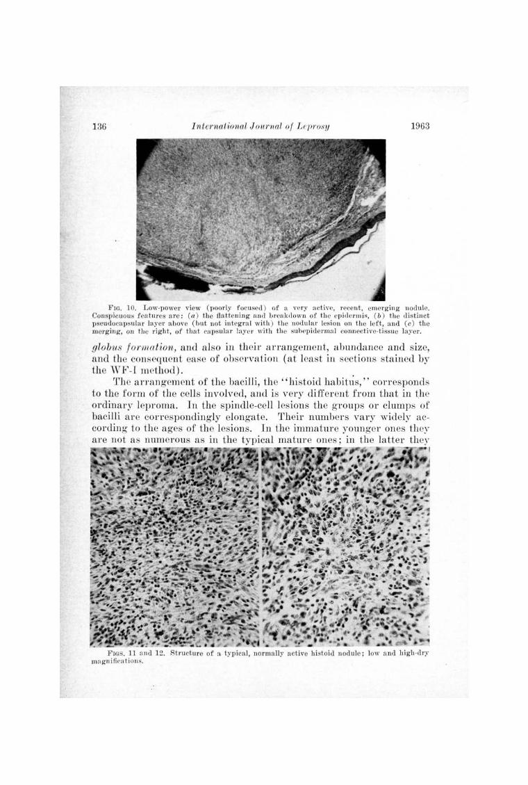
FlO. 10. L ow-p ower view (pood y focused ) of a vc ry active, l'ecent , emerging nod ule. Co nspicuous fell tUJ'es a l'e : (a ) the fla ttening il nd breil krl olVn of the epid erlll is, ( b ) the distin ct pseudocnpsular ]nyel' above (bnt not integral wi t h ) the nodular lesion on the left, nnd (c) t he mer ghlg, on the ri ght, of that capsulal' lnye r wi th the subepicl crmnl co nn ective-tissue layer.
globus fo rmation, and also in their a rrangement, abundance and size, and the consequent ease of observation (at least in sections stained by the vVF -J method) .
The a rrangement of the bacilli, the "histoid habit~s , " corresponds to the fo rm of the cells involved, and is ver y differ ent from that in the ordina ry leproma. In the spindle-cell lesions the groups or clumps of bacilli a re correspondingly elongate. Their number s vary widely according to the ages of the lesions. In the imma ture younger ones they ar e not as num erous as in the typical mature ones ; in the la tter they
I
. -FIGs. 11 fi nd 12. Structure of a typica l, normally active histoid nodule; low and hi gh·d ry
ma gn i fie-a t ions.

31, 2 Wa(~e : JI'istoid V Cl1'iety of L I' ])l'onwtolls L eprosy 137
are very abundant, more so than in the ordinary kind of leproma; later on, as the lesion ages, they become less abundant, so much so that such lesions may be unsuitable for use in making lepromin.
In certain "local r eactional" areas of the subcutaneous nodules the spindle cells are changed to large rounded phagocytes (i.e., of the usual histiocyte type, but lar ge ), and in these particularly " active" ~ cells the ba cillus load is simply incredibl e. It seems impossible for a cell to carry so man y bacilli without disturbance of its morphology, yet usually th e most that can be seen in th e hematoxylin-eosin section is a fin e irregularity of the cytoplasm that sugges ts the minute "vacuolation" of xanthomatous lesion s. , Occasionally coarser vacuolation is seen, always within the cytoplasm but not large enough to penetra te the thickness of a 6-micron section; hence it has no r esemblance to globus fo rmation.
F IG. 13, Structure of an inactive, rath er chronic histoid nodule. The aniline-blue section of this specimen shows mnch less coll agen th an wonld be expected from the appearance of the hematoxylin-eosin section.
In size, the bacilli (as stained) are notably larger than those in the ordinary leproma, even in the sam e section. In consequ ence, a lowpower objective (the 20X especially ) is sufficient for their general observation. This is in sharp contrast with the ordinary lepromatous lesions, to study which the oil immersion lens is needed.
The absence of globus formation is obviously due to the lack of' production by the bacilli of the essential matrix ubstance, or gloeathe "electron transparen t substance" of the electron microscopist., This is evidently a failme, or deviation, of the metabolism of the bacil-Ii, about the cause of which one can only speculate.

138 I nternational J oUf'nal of L ep1'osy 1963
It is to be said, however, that the failure to produce globi is absolutely characteristic but not absolutely invariable. A few histoid specimens have been encountered in which areas of the lesions have undergone the reverse change and produced globi. Areas showing this condition have never been very extensive, and they have not perceptibly affected the character of the rest of the lesion in which they occurred.
TUBERC ULOID " CONTAMI NATION" OF HISTOID LESIONS
~. fea ture of som'e of the specim ens of histoid nodules which caused amazement when first observed and still r emains inexplicable, is the occurrence of definite tuberculoid foci within the lesion substance or in the encircling fibrou s-tissue encapsulation. This "contamination," seen only in nodules of subcutaneous location or origin, is not rare, for it has been found in a fairly large percentage of the specimens. Nothing like it has ever been seen in ordinary lepromatous lesions.
These foci are usually small, less than the diameter of the 20X objective ; in the largest, encounter ed in two specimens, the area of the 20X objective hardly encompasses that of the epithelioid center of the Ie ion.
The smaller foci- but not the larger ones-may be easily overlooked in the hematoxylin-eosin sections, but they are fairly conspicuous in those stained for bacilli. The epithelioid center s are almost fr ee from bacilli, in sharp contrast with the normal abundance in the sur-rounding, unaffected histoid tissue. .
In numbers per specimen block they are often single, not infrequently two to several, only very rarely numerous. N one has ever shown any suggestion of a tendency to multiply, i.e., to form satellite foci in its immediate neighborhood.
No explanation of the occurrence of these tuberculoid foci in these lesion s has been offered by per sons consulted, and none can be offered by me; speculation will not be indulged 'in. It may be remarked, however , that lesions so contaminated cannot be called "borderline," or even" dimorphous,"
DISCUSSION
To appreciate the basically histiocytic nature of the spindle-cell structure of the histoid lesions, and the secondary role of the fibrous tissue element-i.e., to r ealize that the lesions are not fibrou s or fibromatous, as they are likely to be thought to be from the appearance of ordinary hematoxylin-eosin sections-a good differential stain for collagen must be applied routinely to all specimens. That r equires Zenker fixation of the specimens, at least if the Mallory aniline-blue stain is to be used-and also if the structural alterations due to the shrinkage inherent in formalin-fixed tissue is to be avoided, which is greatly to be desired.

31, 2 Wade: Histoid V01"iety of L epromatOll S L ep?'osy 139
Only in the earliest nodular lesions is the spindle-shaped histiocytic element to be seen in pure culture, without any collagenous fibers except in the pseudocapsule '. Infiltration by strands of fibroc:;-tes and, later, advancing fibrosis, arc features of maturing and ag-ing of the nodules.
The demonstration of the bacilli in the- histoid lesions by the method employed in this laboratory exclusively since ] 057 (4) is extraordinarily good. The "histoid habitus" of their arrangement in the ordinary spindl e-cell Ie ion is very distinctive. Tn the histiocytic areas of some of the more active lesions ther e is a simpl y incredible massing of the bacilli. 'rhe contra st between the bacilli seen ill these lesions and those in the ordinary lepromatous lesions is well shown in certain infrequent" mixed" lesions, of which there a I' C only two in m~- collection.
That the bacillus groups and masscs of the histoid lesions do not produce globi- r ecognition of which fact requires recognition of what globi arc- is obviously a matter of metabolism. ~rhe bacilli do not produce the matrix substance, 01' gloea, that characterizes the globus. Beyond that, the explanation i a matter of speculation.
An unexplain ed clinical feature of evident significance is that r eactions of the erythema nodosnm leprosum kind seem not to occur in histoid cases.
Until detailed case r eports can be presented, little can be said about clinical variations of the cases presenting histoid lesions. Several that can properly be called hisfoid leprosy (leproma tous) because the condition was sys temic, have died of it in the course of time. Unfortunately, none has been autopsied. In other cases the histoic1lesions seem to have been local, 'with no sys temic manifestation s, ai1d without particularly unfavorable prognosis.
The not infrequent finding of tuberculoid foci in the nodules of subcutaneous origin, either in the substance ("parenchyma") or the· capsular layer, constitutes a particularly intriguing problem. These foci are evidently of metastatic nature, but from what or where do they originate ~ Here, again, speculation is avoided.
SUMMARY
1. ~rhe "histoid" variety of lepromatous leprosy here discussed is so named because histologically the typical lesions resemble, rather than the inflammatory granuloma that the ordinary leproma is, a tumor developed from a single, spindle-shaped tissue element. The condition is commonly mistaken for flbrosis of ordinary lepromatous lesions. .
2. Lesions of this sort are most likely to be found in old cases which for years had progressed as ordinary lepromatous leprosy, responding to treatment, but had later retrogressed, or relapsed, developing lesions resistant to treatment. They are therefore of a kind seldom seen

140 International J01,wnal of Le'[J1'osy 1963
except in leprosaria, and patients of such kinds in such environment are not likely to arouse sufficient interest to lead to the making of biopsies.
3. The essential clinical feature is nodule formation, notably subcutaneous but also cutaneous. In their manner of growth the nodules are primarily expan ile, and consequently arc usually surrounded by a pscudocapsule derived from the tissue in whi ch they arc located. They arc well vascula,r ized, but they contain no nerve hranches.
4. rrhe "parenchyma" of the earliest nodules is composed solely of spindl e-shaped, hacillated cell s of histiocytic nature, but an irregularly-distributed fibrocy tic " stroma" develops later, and the amount of the fihrou s-tissue elcment within the les ion illcreases with age.
5. Other characteristic and noteworthy histologic features includ e the fr equent r eactional, or activation, change of the parenchyma cells in limitecl areas from the normal spindle shape to typical rounded phagocytes, and the frequen t occu rrence of areas of local softening (" local r eactional" breakdown), even amounting to "absces " format ion but seldom such lesions containing polynuclear leucocytcs.
6. Bacteriologically, the most striking and sign incant feature is the normal absence of globus formation, regardless of the abundance of the bacilli. 'J~heir number s in the active spindle-cell'parenchyma are usually large, and in the rounded phagocytes they are usually extremely numerous, at times incredibly so. The arrangement of the bacilli as elongate clumps in the spindle cells, together with the occurrence of dense, rounded masses in the areas of free phagocytes, constitutes the "histoid habitus " of the bacilli, strikingly differ ent from the arrangement in the ordinary leproma.
7. }~xtraneou s to the histoid lesions in sections of the affected skin, never in association with the subcutaneous nodules, there may be found residual foamy-cell areas, usually small, which are mer ely r esiduae of the previously-existing ordinary lepromatous process. More rarely, there may be found, in association with active histoid skin lesions, more or less active infiltrates of the ordinary lepromatous process ; these infrequent lesions are of double or mixed nature.
8. Finally, but important, is the not infrequent occurrence of definite foci of epithelioid cells located in (i. e., "contaminating ") certain of the histoid lesions-an unexplained, and inexplicable condition. This tuberculoid involvement is, in my experience, confined to the nodules of subcutaneous location or origin.
RESUMEN
1. Varied ad "histoidea" de la lepra lepromatosa aqul discuticla se denomina asf porque, histo16gicamente, las tlpicas lesiones semejan, mas bi en que el granuloma inflamatorio como es el leproma ordinario, un tumor formado de un solo elemento histoideo

31, 2 Wade: Histoid Va1'iety of L ep1'ornatous L ep1'osy 141
fusiforme. La dolencia se confunde comiinmente COn la fibrosis de las ordinarias lesiones leproma to. as.
2. Las lesiones de este genero son mas susceptibles de encontrarse en los casos antiguos que pOl' afios han avanzado como si fueren de la ordinaria lepra lepromatosa, respondiendo al tratamiento, pero mas tarde han involucionado 0 reciclivaclo, manifestanclo lesiones resistentes a la tel'apeutica. Son pOI' 10 tanto de una especies rara vez obscl'nHla exeepto en los leprosario , y los enfermos 'de esos generos no son susceptible. de despel'tar sun ciente interes para conducir a la obtencion de biopsias.
3. La indi pensable caracterfstica clfnica es la formacion de nodulos, notablemente subcutnneo' , pero tambien cutftneos. En su modo de crecimiento, 10 nodulos son pl'imariamente expansibles y pOl' consiguiente uelen estar rodeados pOl' una seudocap.·ul a derivada del tejido en que se hallan situados. E stan bien vascularizados, mas no contienen ramas 11 erviosas .
4. E l parenqu ima de los primel'os nodulos esta compuesto cxclusivamente de celulas baciladas f usiformes de nahu'aleza histiocftica, pel'O se forma mas tarde un "estroma" repal'tido irl'egulamente y la proporcion del elemento de tejido fibroso dentr'o de la lesion aumenta con la edad.
5, Otras ca racterfsticas bistologicas t1picas y dignas de nota comprenden el frecuente cambio r eaccional, 0 estivacion, de las celulas parenqimatosts en zonas limitadas de la fo rlll a normal de huso a t1pico, fagocitos redondeados, y la fl'ecuencia de ZOIHIS de ablandamiento local (desintegracion "reaccional local"), lIegando basta la fOl'lIlac ion de ab:cesso. ," pero conteniendo rara vez esas lesiones leucocitos polinucleal'es.
6. Ba cte1'iologicamente, la caracteristica mas notable y signincativa es la falta n01'mnl de la fo1'macion de globos, independientemente de la ahundancia de los bacilos. Su nlul1el'o en el parenquima fusiforme activo suele ser elevado, y en los fagocitos redondeados suele ser sumamente crecido, y a veces basta increfble. La disposicion de los bac ilos en conglutinaciones alargadas en las eelulas fu siformes, junto con la existp-ncia de espesas masas l'edondeadas en las ~ones de fagocitos libl'es, constituye el " Iuibito bistoideo" de los ba cilos, que es nobJblemente clistinto de su disposicion en ellepl'oma ordinario.
7. Apade de las lesiones bistoideas en los cortes de la piel afecada y jamas asociadas COil 10:; nodulos subcutaneos, pueden encontl'arse zonas de Mikulicz residuales, pOl' 10 general pequefins, que son meramente residuos del ordinario proceso lepromatoso que existio antes. Mas raramente, pueden encontrarse, en asociacion con las lesiones cutaneas bistoideas activas, infiltraciones mas 0 menos activas del ordinal'io proceso lepromatoso; estas lesiones infrecuentes son de naturaleza doble 0 mixta.
8. POl' fin, pero importante, tenemos la existeneia, que no es ram, de focos bien definidos de cClul as epitelioideas situadas en (0 sea "contaminando") ciertas lesiones de las bistoideas-situacion esta inexplicada, e inexplicable. Esta invasion tuherculoidea se halla, en 10 que haya observado el A" limitada a los nodulos subcutaneos en su localizacion U origen.
RESUME
1. Ln variete "bistoi'de" de lepre lepromateuse di scutee dans cet article tire son nom du fait que d'un point de vue bistologique la lesion typique ressemble it une tumeur developpee it partir d'Ull elt~ment tissulaire d'une seule espece, en forme de fuseau, plutOt qu'au granulome inflammatoire qui constitue Ie leprome courant, Cette condition est genernlement prise it tort pOUT de In fibrose SUl'venant dans des Jesions Jepromateu es ordina ires.
2. On a Ie plus de cbances de trouver des lesions de cette espece cbez des vieux cas qui, durant des annees, ont progresse comme une lepre lepl'omateuse ordinaire, repondant a la therapeutique, mais qui plus tard ont repris, ou recidive, developpant des lesions resistantes au traitement. Des lors, ce sont des particularites rarement vues sinon en

142 I nternational J Oll1'1wl of Lepro;;y H)63
leproserie, et les malades de cette especc, dans un tcl cnVil'OnllClll cllt, nc sont p as ::iusceptibles, semble-t-il , d'eveiller assez l'in teret que p our mener au p l'elevenl cnt de biopsies.
3. La pa rt icul al'ite clinique essentiell e est la formation de nodules, p rineipa lelll cnt sous-cutanes, tllai cutanes egalcment. LeUl' typc de croissan cc est tel que ces nodules tendent ava nt tout a se dil ateI' ; en consequence ils sont generalment entoures d'une pseudo-cap ule deri vee du tissu Oll il s sont situcs. Jl s sont bien vascul arises, mais ne cont.i ennent pas de filets nerveux.
4. Le "pa renchyme" des nod ules a leur sta de Ie p lus p rc('occ est compo. e uniquement de ccllules de nature hi stiocytaire, remplics de ba cil1 es, cn for cc de f useau, mais a un stadc ultel'i eur se developpe un "stroma" fibl'ocytaire irl'egulicrelllC'nt di st l'ibue, ct la quan t i~e d'elements ti. sul nires fibrcux (Inns la lesion Img llH'nte nvec Ie tr mps.
5. P a 1'111 i les autl'es caractcristiquc. et p art iculari tes notables d' nn poi nt de vue hi.' tolog iquc, on l'eleve f requemment, dans des zo nes limitees, une modifi cation l'ea r·tionnell e, un ' acti vation des ceHules pa l'cnchymateuses qui dc ce llules ~t il SPl'(·t nOl' lll nl en f useau se tl'ansfor ment en p hagocytes arrondis typiques, ainsi que souvc nt In formati on de regions a structure di ssociee localement, (cffondl'ement " l'eactionnel lo('a 1" ), all ant meme jusqu'u la fo rlll a tion d'abces, n~a i s il est ra re quc de tell es lesions conti cll nen t des leucocytes polYlllo rphonuclea ires.
6. D'un p oin t de vuc bactel'i ologique, la caral'tel'istique In plus f l'fl ppante et la plus signifi cative est l'abscnce l'eguliere de formation en globi, sans egard p OUl' la quantite de ba1illes. Le n0l1lbre de ccux-ci, dans Ie parcnchyme f01'1I1e de ccllules cn Euseau en pleine activite, cst generalment eleve, ct dans lcs phagocytes ronds les bl1cilles sont general el1lent cxt)'cmemcnt norn bl'cux, si nombl'eux a l'occasion que e'en est incroya ble. La disposition nes bacill es cn mnas allonge dans les cellul es en f useau, a insi qui Ie f orm a tion de masses al'l'ondies et denses dans les p hagocytes lib res, constituent I' "habitus h,sto'ide" des bacill es, qui differe de rnaniere f rappante de leur arrangement dans les lepromes ordinail'es.
7. En dehors des Ie ions histoldes, et jamais en associati on avec les nodules souscutanes, on p eut, dans des coupes de p eau malade, tl'ouver des zones r esiduelles, generalement petites, avec cellules spumeuses, qui l'epresentent silllplement les vestiges du processus lepromateux qui a precede. Plus ral'ement, en association avec des lesions histoldes actives de la p eau,on peut tI'ouver des infiltra ts lepromateux ordinaries, p lus ou moins actif s j ces lesions p eu f requentes sont de nature mixte, ou double.
8. E nfin, et ceci e t imp ortant, il n'est pas ra re que surviennent des foyers bien defini s de ccllules epithelio'ides situees dans (o n devrait dire "contaminant") certaines de lesions histoi'des-ce qui constitute une co ndition non expliquee et non explicable. Cette participation de na ture tubel'culo'icle est, d'apres mon experience, l'estl'einte aux nodules s itues dan Ie tissu sous-cutane, ou qui y ont leur Ol'igine.
(Note : Most of these pictures are f rom D. San chez, one of the first of the histoid cases studied and one with an unusually wide variety of uncomplicated lesions.
REFERENCES
1. DHARMENDRA. Kotes On Leprosy. Ne w Delhi : Ministry of H ealth, Government of India, 1960.
2. DINIZ, 0., MARI ANO, J . and RABEfJW, F. E. Atlas de Leprologia. Rio de Janeiro: National Service of H ealth Education, 1960.
3. EWING, J. Neoplastic Diseases. A Treatise on Tumors, 4th Ed. Philadelphia and London: W. B . Saunders Company, 1942, p. 33.
4. MALLORY, F . B. The Principles of Patholog ic Histology. Philadelphia and London: W. B. Saunders Company, 1914, pp. 283 and 285.
5. WADE, H. W . A modifi cation of the Fite formaldehyde (Fite I) method for staining acid-fast bacilli in paraffin sections. Stain Tech. 32 (1957) 287-292.
6. WADE, H . W . Zenker vs formalin fixation for the histopathology of leprosy tissues, and other desirable f eatures of technique. Intel'nat. J. Leprosy 30 (1962) 477-488.


![OFFl AND COUNCILLORS - ILSLila.ilsl.br/pdfs/v31n4a11.pdf · INTERNATIONAL LEPROSY ASSOCIATION 6 l-lillercst Avenue, Pinner, "Jiidc] lcsl'x, England OFFl C~R8 AND COUNCILLORS OFFICERS](https://img.pdfslide.us/doc/110x75/5e048c6a47bcff7c7117d65c/offl-and-councillors-international-leprosy-association-6-l-lillercst-avenue-pinner.jpg)