Embed Size (px)
Citation preview
Int.J.Curr.Microbiol.App.Sci (2014) 3(11): 1025-1039
1025
Review Article
Review on Dengue viral Replication, assembly and entry into the host cells
R.Aruna*
Research and Development Centre, Bharathiar University, Coimbatore, India
*Corresponding author
A B S T R A C T
Introduction
Dengue fever is a mosquito-borne virus
disease of humans. In terms of numbers of
individuals infected, it is by far the most
devastating of all the recognised arthropod-
transmitted virus diseases. Dengue has
become a global problem since the Second
World War and is endemic in more than 110
countries. It is estimated that more than 3
billion humans live in dengue endemic
regions of the world, and currently, more
than 50 million infections occur annually
with at least 500,000 individuals requiring
hospitalisation [1]. Of these, tens of
thousands have a high risk of developing
haemorrhagic disease, potentially with fatal
consequences depending to a large extent on
the quality of the available medical services.
The dengue viruses are positive stranded
RNA viruses in the genus Flavivirus, family
Flaviviridae [2].
There are four distinct dengue virus (DENV)
serotypes that share antigenic relationships
(DENV-1, DENV-2, DENV-3, and DENV-
4), and although infection with one serotype
confers lifelong protection against that
serotype, it does not necessarily protect
against a secondary infection with a
heterologous serotype. Indeed,
nonprotective but cross-reactive antibodies
may enhance disease severity [3]. Currently,
there are no effective vaccines or antiviral
drugs against these viruses. This problem is
being addressed as a matter of urgency as
failure to develop effective DENV control
strategies will inevitably result in a further
increase in the number of infected humans,
as predicted more than a decade ago [4].
This problem is also exacerbated by the
continuing dispersal of these viruses to new
geographic regions.
International Journal of Current Microbiology and Applied Sciences ISSN: 2319-7706 Volume 3 Number 11 (2014) pp. 1025-1039
http://www.ijcmas.com
Dengue fever is a mosquito-borne virus disease of humans. It is estimated that
more than 3 billion humans live in dengue endemic regions of the world, and
currently, more than 50 million infections occur annually with at least 500,000
individuals requiring hospitalization. In this review we will discuss the replication
of dengue virus, involvement of the structural proteins in this process. NS3 and
NS5 methyl transferase ‘s predictive function in the entry of hostcells
K e y w o r d s
Dengue fever,
viral
Replication,
and
assembly
Int.J.Curr.Microbiol.App.Sci (2014) 3(11): 1025-1039
1026
In this review, we will discuss on advances
in structural studies on DENV and its
proteins. We will focuses the relevance of
the functional mechanisms of the viral
proteins, their interactions with host
proteins, and their role in the viral life cycle.
DENV is a member of the Flaviviridae
family and is grouped within the flavivirus
genus together with other pathogenic viruses
including West Nile virus (WNV), Japanese
encephalitis virus (JEV), tick-borne
encephalitis virus (TBEV) and yellow fever
virus (YFV) [5]. The viral genome consists
of a positive-sense RNA of ~11kb. This
RNA encodes 3 structural proteins (C, prM
and E) that form the components of the
virion, and 7 non-structural proteins (NS1,
NS2A/B, NS3, NS4A/B, NS5) involved in
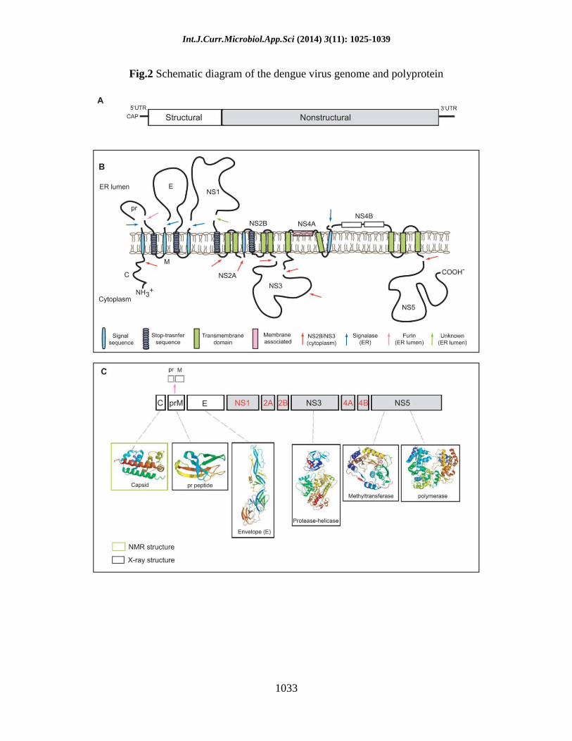
viral RNA replication (Figure 1).
Replication and assembly of dengue virus
particles
A schematic representation of the DENV
genomic RNA and the translation of the
viral proteins are depicted in Fig. 3. After
virus cell entry and uncoating of the
nucleocapsid, the RNA molecule is
translated as a single polyprotein [28].
During this process, the signal- and stop-
transfer sequences of the polyprotein direct
its back-and-forth translocation across the
endoplasmic reticulum (ER) membrane. The
polyprotein is processed co- and
posttranslationally by cellular and virus-
derived proteases into three structural
proteins (C, prM, and E) and seven
nonstructural (NS) proteins (NS1, NS2A,
NS2B, NS3, NS4A, NS4B, and NS5). The E
protein is glycosylated at amino acid residue
Asn67 and Asn153 to assure proper folding
of the protein [23, 29]. Other potential N-
linked glycosylation sites are located in prM
at position 7, 31, and 52 and within NS1 at
position 130 and 207 [30, 31]. Upon protein
translation and folding of the individual
proteins, the NS proteins initiate replication
of the viral genome [28]. The newly
synthesized RNA is subsequently packaged
by the C protein to form a nucleocapsid. The
prM and E proteins form heterodimers that
are oriented into the lumen of the ER. Then,
the prM/E heterodimers associate into
trimers and these oligomeric interactions are
believed to induce a curved surface lattice,
which guides virion budding [25, 26]. It is
unclear how this is synchronized with the
engulfment of the nucleocapsid since no
specific interactions between C and prM/E
proteins have been identified yet [32, 33].
Interestingly, encapsulation of the
nucleocapsid during virus assembly is not
crucial as the formation and release of
capsidless subviral particles has been often
documented [34–37]. Structural analysis of
newly assembled immature virions revealed
that a single virion contains 180 prM/E
heterodimers that project vertically outward
from the viral surface as 60 trimeric spikes
[32, 33, 38]. The immature particles formed
in the ER mature as they travel through the
secretory pathway. The slightly acidic pH
(*5.8–6.0) of the trans-Golgi network
(TGN) triggers dissociation of the prM/E
heterodimers, which leads to the formation
of 90 dimers that lie flat on the surface of
the particle, with prM capping the fusion
peptide of the E protein. This global
structural reorganization of the
glycoproteins enables the cellular
endoprotease furin to cleave prM [39–41].
Furin cleavage occurs at a Arg-X-(Lys/Arg)-
Arg (where X is any amino acid) recognition
sequence and leads to the generation of
membrane-associated M and a „„pr‟‟
peptide. A recent study has shown that the
pr peptide remains associated with the virion
until the virus is secreted to the extracellular
milieu [40]. Both the prM protein as well as
the pr peptide are believed to act as
Int.J.Curr.Microbiol.App.Sci (2014) 3(11): 1025-1039
1027
chaperones stabilizing the E protein during
transit through the secretory pathway,
thereby preventing premature
conformational changes of the E protein that
would lead to membrane fusion. Upon
dissociation of the pr peptide, mature virions
are formed that are able to infect new cells.
Virus assembly and maturation
The structure of DENV was previously
solved through a combination of cryo-
electron microscopy and X-ray
crystallography[6,7,8,9]. Recently, the
structures of intermediates in the assembly
process have also been obtained and these
provide insight into the assembly and
maturation process of DENV [16,17]. In
supernatants of infected cells, the virus is
found either as a mature or immature
particle with a diameter of about 50 nm and
60 nm, respectively. Both particles consist
of an outer glycoprotein shell and an internal
host derived lipid bilayer. Within this
bilayer is an RNA-protein core consisting of
genome RNA and capsid proteins (C). The
glycoprotein shell is well defined and
consists of 180 copies each of an envelope
(E) and membrane protein (prM/M). These
two proteins have different conformations in
the immature and mature DENV particles
and therefore, confer unique structural
features to both forms of particles. In the
immature virion, prM and E form 90
heterodimers that extend as 60 trimeric
spikes from the particle surface (figure 3A).
In the mature virion, E is found as 90
homodimers that lie flat against the viral
surface forming a „smooth‟ protein shell
(figure 3D). The „pr‟ peptide is cleaved from
prM during maturation and M remains in the
mature particle as a transmembrane protein
beneath the E protein shell. The structural
transitions from immature („spiky‟) to
mature („smooth‟) morphology occur while
in transit through the Trans-Golgi Network
(TGN) and are driven predominantly by
conformational changes in the E protein
[8,9,16]. These conformational changes in E
are triggered by low pH (~5.8–6.0) and
occur prior to the maturation cleavage of
prM by a host encoded furin protease (figure
3B) [17]. It was demonstrated that these
conformational changes were reversible
(based on pH). This suggested that the
immature particle could exist reversibly in
either „spiky‟ or „smooth‟ forms depending
on the pH of the cellular environment. This
is a remarkable observation considering that
such reversible conformational changes
require the E protein to toggle between
forming heterodimeric interactions with prM
in spikes that projects vertically outwards
from the particle to forming homodimeric
structures that lie flat against the viral
surface. The molecular reorganization
required for these changes are extensive, the
details of which still remain illusive.
Following maturation however, the mature
particle cannot revert back to its immature
morphology. Once cleaved, the pr peptide
remains associated with E (figure 3C) until
the mature particle is released into the
neutral pH of the extracellular environment.
Therefore, it was proposed that the pr
peptide functioned as a cap-like structure
that protected the fusion peptide on E from
undergoing premature fusion prior to virus
release [19].
Recently, the atomic structures of prM were
solved at pH 5.5 (2.2Å) and 7.0 (2.6Å) [13].
Both structures were similar indicating that
pH did not affect the tertiary structure of
prM. The construct consisted of a prM-E
fusion protein where the prM protein was
fused to the ectodomain of E. The
transmembrane domain of prM was replaced
by a linker. The furin cleavage site was also
mutated. The resulting structure of prM has
a unique fold and consists of 7 antiparallel
β-strands, stabilized with three S-S bonds.
Int.J.Curr.Microbiol.App.Sci (2014) 3(11): 1025-1039
1028
The glycosylation modification at
asparagine 69 was also observed. The
structure of E in this construct was similar to
its prefusion dimeric conformation [17]. As
predicted previously, the pr peptide was
positioned as a 'cap' on E protecting the
hydrophobic fusion loop [18]. These recent
studies on the immature virions and proteins
have provided new details of DENV
assembly and maturation. They have also
opened new avenues that can be explored
toward understanding the molecular
mechanism behind E protein dynamics and
the role of the immature particles in the
virus life cycle. The E protein also provides
the first point of contact between the virus
and the host cell. Several cellular proteins
and carbohydrate molecules that function as
attachment factors mediating viral entry
have been identified, and these molecules
have been shown to interact with the E
protein [18–24]. These attachment factors
assist in concentrating the virus on the cell
surface increasing its access to specific
cellular receptor/s. Structural insight into the
interaction of E with one of these attachment
factors, dendritic-cell-specific ICAM3-
grabbing non-integrin (DC-SIGN) [26] has
been obtained. Unfortunately, a specific
cellular receptor for dengue has not yet been
identified. It is possible that currently
identified attachment molecules could
function as specific cellular receptors for
dengue. However, it would be necessary to
demonstrate that they mediate clathrin-
mediated endocyotsis of the particle and this
has not been shown for any of these putative
receptors to date.
NS3 protease-helicase
NS3 is a multifunctional protein of 618
amino acids that functions both as a
chymotrypsin like serine protease as well as
an RNA helicase and RTPase/NTPase. The
protease domain is N terminal in NS3
(residues 1–180) and cleaves the viral
polyprotein at several sites as depicted in
figure 1. The enzyme consists of 6 β-strands
that form two β-barrels with the catalytic
triad (His-51, Asp-75 and Ser-135)
sandwiched between them. Activity of the
protease is critically dependent upon the
presence of its co-factor, NS2B which is
conserved among the flaviviruses
[28,32,33]. Recently, the structure of the
full-length NS3 of DENV-4 was solved to
3.15Å resolution (figure 4) [34]. This
structure suggests that NS3 is an extended
molecule with the protease domain spatially
oriented on top of subdomains I and II of the
helicase. The NS3 protein included residues
49–66 from the NS2B which were linked to
the N-terminus of fulllength NS3 by a Gly-
Ser linker. However, its protease domain
was inactive but retains the same active site
conformation and overall fold as an active
form of the DENV-2 protease domain that
included an extended region of NS2B
(residues 40–80) [35].
Analysis of these two structures as well as
the structure of the substrate-bound form of
the WNV protease domain [36] indicated
that residues 67–80 of NS2B are critically
important for protease activity of NS3. This
is due to NS2B wrapping around the
protease domain as a 'belt-like' structure and
forming an integral part of the protease
active site. While the central region of NS2B
(residues 67–80) interacts with the protease,
flanking hydrophobic regions of NS2B are
predicted to anchor the NS2B-NS3 complex
in the ER membrane [37]. This geometry
places the protease active site close to
predicted transmembrane domains that must
be cleaved within the polyprotein. The
consensus cleavage site of NS3 requires a
dibasic (Arg/Lys)-Arg motif at the P2 and
P1 positions respectively and a small amino
acid (Gly) at the P1′ position. The substrate
specificity as well as the cis/trans activity of
Int.J.Curr.Microbiol.App.Sci (2014) 3(11): 1025-1039
1029
many of the flavivirus proteases have also
recently been characterized [38,39]. These
studies provided evidence of a required cis-
cleavage of the NS3 helicase domain at a
site hidden from exposure to the protease
domain. This indicated that the NS3 is
structurally dynamic and may indeed require
an extended conformation for its function.
Such a conformation of NS3 could explain
the ability of the NS2B-NS3 protease to
function on sites not readily accessible to it.
These same studies have also suggested that
the cis-trans activity of the protease may
play an important role in controlling the
dynamics of viral protein translation versus
RNA replication by controlling the
availability of viral proteins. The helicase
domain of NS3 (residues 180–618) has
seven structural motifs reminiscent of
superfamily 2 helicases [40]. It has three
subdomains with significant sequence
identity and structural similarity to other
flavivirus helicases [34,41–43]. Subdomains
I and II are also structurally similar to the
corresponding domains in hepatitis C virus
suggesting a common functional mechanism
[44,45], however, the fold of subdomain III
is unique to the flaviviruses, and may be a
site for protein binding. Both subdomains I
(residues 181–326) and II (residues 327–
481) are composed of a central six-stranded
parallel β-sheet, which is flanked by 4 α-
helices. Subdomain III (residues 482–618)
has 4 approximately parallel α-helices
surrounded by three shorter α-helices and
two solvent-exposed antiparallel β-strands.
In the recent structure of full-length NS3
[34], the helicase domain adopts a similar
structure to the isolated DENV helicase
[41], with the exception that several regions
previously disordered in the isolated domain
structure are now visible. However, the
helicase activity of the full-length NS3
protein is ~30-fold higher than the isolated
domain [41] indicating that the protease
domain may influence the enzymatic
activities of the helicase. The protease and
helicase are linked by an interdomain linker
that shares little conservation between the
flaviviruses, but plays an important role in
the association between the two domains in
NS3 (the buried surface area between the
domains is ~1380Å2 and ~380Å2 with and
without the linker, respectively). The
helicase domain of NS3 is also implicated in
interacting with the polymerase, NS5 [46]
and in WNV implicated in virulence [47].
Recently, a residue in the NS3 helicase
domain (W349) was shown to be involved
in virus assembly suggesting that NS3 plays
an additional role in the life cycle following
viral RNA replication [48]. The NS3 protein
has also been implicated in inducing
apoptosis in infected cells [50]. Overall, the
full-length structure of NS3 represents a
significant milestone in flavivirus biology as
crystallization of full-length viral proteins
has long been a challenge.
NS5 methyltransferase-polymerase
The largest (900 residues, 104kDa) and the
most conserved protein in DENV is NS5
(67% sequence identity among DENV
serotypes 1–4). It is also a bifunctional
enzyme with a methyltransferase domain
(MTase; residues 1–296) at its N-terminal
end and a RNA-dependent RNA polymerase
(RdRp; residues 320–900) at its C-terminal
end. The structure of the MTase domain was
previously solved for DENV [50] and
recently solved for WNV [51]. Both
structures have an S-adenosyl-methionine-
dependent MTase core structure that folds
into a α/ β/β sandwich cradled between N-
and C-terminal subdomains (figure 4).
Overall, the two MTase structures are very
similar and this domain has recently been
shown to sequentially catalyze both guanine
N-7 and ribose 2′-O-methylation [50,52].
The primary difference between the two
structures is observed in the SAM-binding
Int.J.Curr.Microbiol.App.Sci (2014) 3(11): 1025-1039
1030
site and the cap-binding site, with the former
having a more open conformation in DENV
versus WNV, and the latter being more open
in WNV [50,51]. It is suggested that the
differences in the SAM-binding site may
reflect two distinct states of the enzyme,
with the closed conformation (in WNV)
representing tight SAM-binding, and the
open state representing the release of the by-
product of the methylation reaction, S-
adenosyl-homocysteine (SAH). Both
enzymes have a highly positively charged
surface at the GTP and SAM binding sites to
accommodate capped RNA substrates. The
crystal structures for both DENV [53] and
WNV [54]polymerases have been solved.
The polymerase domain of NS5 assumes a
structure similar to other RdRp molecules,
and is composed of a canonical right hand
conformation with palm, fingers, thumb
subdomains [55]. It also shares a common
catalytic mechanism for the incorporation of
nucleotides utilizing two metal ions
coordinated by structurally conserved
aspartic acid residues (also known as the
GDD motif). The RdRps differ from DNA-
dependent RNA polymerases by the
existence of the 'finger tips' that connect the
fingers and thumb subdomains to create a
fully encircled active site. The DENV RdRp
displays a more flexible fingers subdomain
and therefore through a rotation of ~8° away
from the thumb subdomain, forms a more
„open‟ conformation compared to WNV
[53,54]. Interestingly, the flavivirus RdRps
have a nuclear localization signal between
residues 320–405. This NLS region in NS5
was previously thought to be a flexible
interdomain linker, but is now known to be a
well-defined structural component of the
RdRp [53- 57]. Specifically, residues 320–
368 are strictly conserved among the
flaviviruses and bind β-importin. These
residues are also implicated in interacting
with NS3 [46,56]. In DENV infections, the
NS5 protein is primarily localized within the
nucleus. However, not all flavivirus RdRps
localize to the nucleus. The rationalization
for a viral RdRp localizing to the nucleus
when its actual enzymatic functions in the
virus life cycle are required in the cytoplasm
is currently unknown but is actively being
investigated [60]. However, these
observations do suggest that apart from its
enzymatic functions, NS5 may also engage
in virus-host interactions and actively
interact with the host environment.
Other viral non-structural proteins
Unfortunately, there is no structural
information available for viral proteins NS1,
NS2A and NS4A/4B. NS1 is a 45kDa
glycoprotein that is translocated into the
lumen of the ER and secreted from the cell
[59–61]. It is implicated in functions within
the viral RNA replication complex [62–64]
as well as in viral defense through inhibition
of complement activation [65]. Although the
protein forms stable oligomers (dimers and
hexamers) in solution, structural analyses of
this protein and has been quite challenging
[66,67]. This is also true for the hydrophobic
proteins NS2A and NS4A/4B. NS2A is a
~22kDa protein that is implicated to form
part of the replication complex [67]. As a
result of an internal NS2B-NS3 cleavage,
two forms are observed, NS2A and NS2Aα
Both forms are important for virus
production [69]. NS4A (16kDa) and NS4B
(27kDa) are integral membrane proteins.
NS4A is proposed to induce membrane
alterations important for virus replication
[31,70]. NS4B is implicated in assisting
viral RNA replication through its direct
interaction with NS3 [71]. It is also
suggested to block IFN α/β-induced signal
transduction [32,69]. Structural analayses of
these three proteins have been unsuccessful
since they all possess multiple
transmembrane hydrophobic segments.
However, the membrane topology of NS4A
Int.J.Curr.Microbiol.App.Sci (2014) 3(11): 1025-1039
1031
and NS4B has been predicted through
biochemical analyses (figure 2B) [31,71].
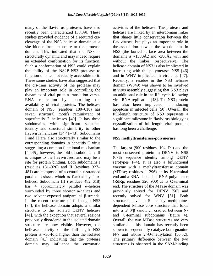
Receptor interaction and viral entry
During natural infection, cells of the
mononuclear phagocyte lineage [monocytes
(MO), macrophages (MØ), and dendritic
cells (DCs)], including the skin-resident
Langerhans cells, are primary targets for
DENV infection [42, 43]. In insects, DENV
was found to initially infect the midgut from
where it spreads and replicates in many
body compartments and organs [44–47].
Also, DENV has been shown to infect
numerous cell lines, including human
(K562, U937, THP-1, HepG2, HUVEC,
ECV304, Raji, HSB-2, Jurkat, LoVo,
KU812), mosquito (C6/36), monkey (Vero,
BS-C-1, CV-1, LLC-MK2), hamster (BHK),
as well as murine MØ (Raw, P388D1, J774)
cell lineages [39, 48–61]. The wide range of
DENV-permissive cells indicates that the
virus must bind to an ubiquitous cell-surface
molecule, or exploit multiple receptors to
mediate infection. Over the last decade,
several candidate receptors and/ or
attachment factors have been identified,
which suggests that DENV is capable of
utilizing multiple molecules to enter the cell.
In mosquito cells, DENV has been shown to
interact with heat-shock protein 70 (Hsp70)
[62], R80, R67 [63], and a 45-kDa protein
[64]. Heparan sulfate [65–67], Hsp90 [62],
CD14 [68], GRP78/BiP [69], and a 37/67-
kDa high-affinity laminin receptor [70] have
been identified as receptors on mammalian
cells. C-type lectin receptors (CLR) are
involved in the interaction of DENV
particles with human myeloid cells [71].
These include DC-specific intracellular
adhesion molecule 3 (ICAM-3)-grabbing
nonintegrin (DC-SIGN, CD209) [72–74],
mannose receptor (MR) [75] and C-type
lectin domain family 5, member A (CLEC5,
MDL-1) [52]. DENV [76–78] as well as
other flaviviruses [79, 80] use clathrin-
mediated endocytosis for cell entry. Using a
single- particle tracking approach, we have
revealed that DENV-2 strain S1 particles
land on the cell surface and migrate in a
diffusive manner toward a pre-existing
clathrin- coated pit [77]. This suggests that
DENV particles move along the cell surface
by rolling over different receptors, or
migrate as virus–receptor complexes. Upon
internalization, the particles are delivered to
Rab5-positive early endosomes, which
mature into Rab7-positive late endosomes,
where membrane fusion primarily occurs
[77]. A recent report also demonstrated that
DENV, depending on the serotype and/or
the target cell type used, is able to utilize an
alternative internalization pathway,
independent of clathrin, caveolae, and lipid
rafts [81]. The subcellular organelle from
which membrane fusion occurs is most
likely dependent on the pH-dependent
membrane fusion properties of the virus and
may therefore vary between individual
DENV strains [77, 78]. Numerous
functional and structural studies have been
undertaken to unravel the molecular
mechanisms involved in the membrane
fusion process of the virus [27, 82–85]. It is
postulated that the acidic pH in endosomes
triggers dissociation of E homodimers,
which then leads to the outward projection
of domain II and exposure of the
hydrophobic fusion peptide to the target
membrane [86]. Subsequently, the
hydrophobic residues in the fusion loop
would insert into the outer leaflet of the
target membrane, triggering the assembly of
E trimers. Next, domain III is assumed to
shift and fold back toward the fusion peptide
into a hairpin-like conformation. This
folding-back mechanism would force the
target membrane and the viral membrane to
bend towards each other and eventually to
fuse, releasing the nucleocapsid into the cell
cytosol.
Int.J.Curr.Microbiol.App.Sci (2014) 3(11): 1025-1039
1032
Fig.1 Life cycle of Dengue virus. Adapted from H. M. van der Schaar
Int.J.Curr.Microbiol.App.Sci (2014) 3(11): 1025-1039
1033
Fig.2 Schematic diagram of the dengue virus genome and polyprotein
Int.J.Curr.Microbiol.App.Sci (2014) 3(11): 1025-1039
1034
References
1. Pinheiro. FP, Corber.SJ, “Global
situation of dengue and dengue
haemorrhagic fever, and its
emergence in the Americas,”12
BioMed Research International
World Health Statistics Quarterly,
vol. 50, no. 3-4, pp. 161–169, 1997.
2. Gubler. DJ, “The global
emergence/resurgence of arboviral
diseases as public health problems,”
Archives of Medical Research, vol.
33, no. 4, pp. 330–342, 2002.
3. Halstead.SD, “Dengue,” The
Lancet, vol. 370, no. 9599, pp. 1644–
1652, 2007.
4. De A. Zanotto.PA, Gould.EA,
Gao.GF, Harvey.PH, Holmes.EC,
“Population dynamics of flaviviruses
revealed by molecular phylogenies,”
Proceedings of the National
Academy of Sciences of the United
States of America, vol. 93, no. 2, pp.
548–553, 1996.
5. Kuhn, RJ. The Flaviviruses. In:
Acheson, NH., editor. Fundamentals
of Molecular Virology. 2004. In
press
6. Ma L, Jones CT, Groesch TD, Kuhn
RJ, Post CB. Solution structure of
dengue virus capsid protein reveals
another fold. Proc Natl Acad Sci U S
A 2004;101:3414–3419.
[PubMed:14993605]
7. Modis Y, Ogata S, Clements D,
Harrison SC. A ligand-binding
pocket in the dengue virus envelope
glycoprotein. Proc Natl Acad Sci
USA 2003;100:6986–6991.
[PubMed: 12759475]
8. Modis Y, Ogata S, Clements D,
Harrison SC. Structure of the
Dengue virus envelope protein after
fusion. Nature 2004;427:313–319.
[PubMed: 14737159]
9. Modis Y, Ogata S, Clements D,
Harrison SC. Variable surface
epitopes in the crystal structure of
dengue virus type 3 envelope
glycoprotein. Journal of Virology
2005;79:1223–1231.
[PubMed:15613349]
10. Kuhn RJ, Zhang W, Rossmann MG,
Pletnev SV, Corver J, Lenches E,
Jones CT, Mukhopadhyay S,
Chipman PR, Strauss EG, et al.
Structure of dengue virus:
implications for flavivirus
organization, maturation, and fusion.
Cell 2002;108:717–725. [PubMed:
11893341]
11. Mukhopadhyay S, Kuhn RJ,
Rossmann MG. A structural
perspective of the Flavivirus life
cycle. Nat Rev Microbiol 2005;3:13–
22. [PubMed: 15608696]
12. Zhang W, Chipman PR, Corver J,
Johnson PR, Zhang Y,
Mukhopadhyay S, Baker TS, Strauss
JH, Rossmann MG, Kuhn RJ.
Visualization of membrane protein
domains by cryo-electron
microscopy of dengue virus. Nat
Struct Biol 2003;10:907–912.
[PubMed: 14528291]
13. Zhang Y, Corver J, Chipman PR,
Pletnev SV, Sedlak D, Baker TS,
Strauss JH, Kuhn RJ, Rossmann
MG. Structures of immature
flavivirus particles. J EMBO
2003;22:2604–2613.
14. Zhang Y, Zhang W, Ogata S,
Clements D, Strauss JH, Baker TS,
Kuhn RJ, Rossmann MG.
Conformational changes of the
flavivirus E glycoprotein. Structure
2004;12:1607–1618.
[PubMed:15341726]
15. Li L, Lok S-M, Yu I-M, Zhang Y,
Kuhn R, Chen J, Rossmann MG.
Structure of the flavivirus precursor
Int.J.Curr.Microbiol.App.Sci (2014) 3(11): 1025-1039
1035
membrane-envelope protein complex
and its implication for maturation.
Science. 2008submitted
16. Yu I-M, Zhang W, Holdaway HA,
Li L, Kostyuchenko VA, Chipman
PR, Kuhn R, Rossmann MG, Chen J.
Structure of immature dengue virus
at low pH primes proteolytic
maturation. Science. 2008submitted
17. Navarro-Sanchez E, Altmeyer R,
Amara A, Schwartz O, Fieschi F,
Virelizier JL, Arenzana-Seisdedos F,
Despres P. Dendritic-cell-specific
ICAM3-grabbing non-integrin is
essential for the productive infection
of human dendritic cells by
mosquito-cell-derived dengue
viruses. EMBO Rep 2003;4:723–
728. [PubMed: 12783086]
18. Tassaneetrithep B, Burgess TH,
Granelli-Piperno A, Trumpfheller C,
Finke J, Sun W, Eller MA,
Pattanapanyasat K, Sarasombath S,
Birx DL, et al. DC-SIGN (CD209)
mediates dengue virus infection of
human dendritic cells. J Exp Med
2003;197:823–829. [PubMed:
12682107]
19. Ren J, Ding T, Zhang W, Song J,
Ma W. Does Japanese encephalitis
virus share the same cellular receptor
with other mosquito-borne
flaviviruses on the C6/36 mosquito
cells? Virol J 2007;4:83. [PubMed:
17803826]
20. Krishnan MN, Sukumaran B, Pal U,
Agaisse H, Murray JL, Hodge TW,
Fikrig E. Rab 5 is required for the
cellular entry of dengue and West
Nile viruses. J Virol 2007;81:4881–
4885. [PubMed: 17301152]Epub
2007 Feb 4814
21. Jindadamrongwech S, Thepparit C,
Smith DR. Identification of GRP 78
(BiP) as a liver cell expressed
receptor element for dengue virus
serotype 2. Arch Virol
2004;149:915–927. [PubMed:
15098107]
22. Liu H, Chiou SS, Chen WJ.
Differential binding efficiency
between the envelope protein of
Japanese encephalitis virus variants
and heparan sulfate on the cell
surface. J Med Virol 2004;72:618–
624. [PubMed: 14981764]
23. Miller JL, Dewet BJ, Martinez-
Pomares L, Radcliffe CM, Dwek
RA, Rudd PM, Gordon S. The
Mannose Receptor Mediates Dengue
Virus Infection of Macrophages.
PLoS Pathog 2008;4:e17. [PubMed:
18266465]
24. Falgout B, Markoff L. Evidence that
flavivirus NS1-NS2A cleavage is
mediated by a membranebound host
protease in the endoplasmic
reticulum. J Virol 1995;69:7232–
7243. [PubMed: 7474145]
25. Falgout B, Pethel M, Zhang Y-M,
Lai C-J. Both nonstructural proteins
NS2B anbd NS3 are required for the
proteolytic processing of Dengue
virus nonstructural proteins. J Virol
1991;65:2467–2475.[PubMed:
2016768]
26. Chambers TJ, McCourt DW, Rice
CM. Production of yellow fever
virus proteins in infected cells:
identification of discrete polyprotein
species and analysis of cleavage
kinetics using region-specific
polyclonal antisera. Virology
1990;177:159–174. [PubMed:
2353452]
27. Mackenzie JM, Jones MK,
Westaway EG. Markers for trans-
Golgi membranes and the
intermediate compartment localize to
induced membranes with distinct
replication functions in flavivirus-
infected cells. Journal of Virology
Int.J.Curr.Microbiol.App.Sci (2014) 3(11): 1025-1039
1036
1999;73:9555–9567. [PubMed:
10516064]
28. Miller S, Kastner S, Krijnse-Locker
J, Buhler S, Bartenschlager R. The
non-structural protein 4A of dengue
virus is an integral membrane protein
inducing membrane alterations in a
2K-regulated manner. J Biol Chem
2007;282:8873–8882. [PubMed:
17276984]
29. Chambers TJ, Nestorowicz A,
Amberg SM, Rice CM. Mutagenesis
of the yellow fever virus NS2B
protein: effects on proteolytic
processing, NS2B-NS3 complex
formation, and viral replication. J
Virol 1993;67:6797–6807. [PubMed:
8411382]
30. Wu CF, Wang SH, Sun CM, Hu ST,
Syu WJ. Activation of dengue
protease autocleavage at the NS2B-
NS3 junction by recombinant NS3
and GST-NS2B fusion proteins. J
Virol Methods 2003;114:45–54.
[PubMed: 14599678]
31. Luo D, Xu T, Hunke C, Gruber G,
Vasudevan SG, Lescar J. Crystal
structure of the NS3 proteasehelicase
from dengue virus. J Virol
2008;82:173–183. [PubMed:
17942558]
32. Erbel P, Schiering N, D‟Arcy A,
Renatus M, Kroemer M, Lim SP,
Yin Z, Keller TH, Vasudevan SG,
Hommel U. Structural basis for the
activation of flaviviral NS3 proteases
from dengue and West Nile virus.
Nat Struct Mol Biol 2006;13:372–
373. [PubMed: 16532006]
33. Aleshin AE, Shiryaev SA, Strongin
AY, Liddington RC. Structural
evidence for regulation and
specificity of flaviviral proteases and
evolution of the Flaviviridae fold.
Protein Sci 2007;16:795– 806.
[PubMed: 17400917]
34. Clum S, Ebner KE, Padmanbhan R.
Cotranslational membrane insertion
of the serine proteinase precursor
NS2B-NS3(Pro) of dengue virus
type 2 is required for efficient in
vitro processing and is mediated
through the hydrophobic regions of
NS2B. J Biol Chem
1997;272:30715–30723. [PubMed:
9388208]
35. Bera AK, Kuhn RJ, Smith JL.
Functional characterization of cis
and trans protease activity of the
flavivirus NS2B/NS3 protease. J
Biol Chem 2007;282:12883–12892.
[PubMed: 17337448]
36. Shiryaev SA, Ratnikov BI, Aleshin
AE, Kozlov IA, Nelson NA, Lebl M,
Smith JW, Liddington RC, Strongin
AY. Switching the substrate
specificity of the two-component
NS2B-NS3 flavivirus proteinase by
structure-based mutagenesis. J Virol
2007;81:4501–4509. [PubMed:
17301157]
37. Gorbalenya AE, Koonin EV.
Helicases: Amino acid
sequencecomparisons and structure-
function relationships. Current
Opinions in Structural Biology
1993;34:419–429.
38. Wu J, Bera K, Kuhn RJ, Smith JL.
Structure of the flavivirus helicase:
implications for catalytic activity,
protein interactions, and proteolytic
processing. J Virol 2005;79:10268–
10277. [PubMed: 16051820]
39. Yamashita T, Unno H, Mori Y, Tani
H, Moriishi K, Takamizawa A, Agoh
M, Tsukihara T, Matsuura Y. Crystal
structure of the catalytic domain of
Japanese encephalitis virus NS3
helicase/nucleoside triphosphatase at
a resolution of 1.8 A. Virology
2008;373:426–436. [PubMed:
18201743]
Int.J.Curr.Microbiol.App.Sci (2014) 3(11): 1025-1039
1037
40. Xu T, Sampath A, Chao A, Wen D,
Nanao M, Chene P, Vasudevan SG,
Lescar J. Structure of the Dengue
virus helicase/nucleoside
triphosphatase catalytic domain at a
resolution of 2.4 A. J Virol
2005;79:10278–10288. [PubMed:
16051821]
41. Yao N, Hesson T, Cable M, Hong Z,
Kwong AD, Le HV, Weber PC.
Structure of the hepatitis C virus
RNA helicase domain. Nat Struct
Biol 1997;4:463–467. [PubMed:
9187654]
42. Kim JL, Morgenstern KA, Griffith
JP, Dwyer MD, Thomson JA,
Murcko MA, Lin C, Caron PR.
Hepatitis C virus NS3 RNA helicase
domain with a bound
oligonucleotide: the crystal
structureprovides insights into the
mode of unwinding. Structure
1998;6:89–100. [PubMed: 9493270]
43. Kapoor M, Zhang L, Ramachandra
M, Kusukawa J, Ebner KE,
Padmanabhan R. Association
between NS3 and NS5 proteins of
dengue virus type 2 in the putative
RNA replicase is linked to
differential phosphorylation of NS5.
J Biol Chem 1995;270:19100–
19106. [PubMed: 7642575]
44. Brault AC, Huang CY, Langevin
SA, Kinney RM, Bowen RA, Ramey
WN, Panella NA, Holmes EC,
Powers AM, Miller BR. A single
positively selected West Nile viral
mutation confers increased
virogenesis in American crows. Nat
Genet 2007;39:1162–1166.
[PubMed: 17694056]
45. Patkar CG, Kuhn RJ. Yellow Fever
Virus NS3 Plays an Essential Role in
Virus Assembly Independent of Its
Known Enzymatic Functions. J Virol
2008;82:3342–3352. [PubMed:
18199634]
46. Ramanathan MP, Chambers JA,
Pankhong P, Chattergoon M,
Attatippaholkun W, Dang K, Shah
N, Weiner DB. Host cell killing by
the West Nile Virus NS2B-NS3
proteolytic complex: NS3 alone is
sufficient to recruit caspase-8-based
apoptotic pathway. Virology
2006;345:56–72.
[PubMed:16243374]
47. Egloff MP, Benarroch D, Selisko B,
Romette JL, Canard B. An RNA cap
(nucleoside-2′-O)- methyltransferase
in the flavivirus RNA polymerase
NS5: crystal structure and functional
characterization. Embo J
2002;21:2757–2768. [PubMed:
12032088]
48. Zhou Y, Ray D, Zhao Y, Dong H,
Ren S, Li Z, Guo Y, Bernard KA,
Shi PY, Li H. Structure and function
of flavivirus NS5 methyltransferase.
J Virol 2007;81:3891–3903.
[PubMed: 17267492]
49. Ray D, Shah A, Tilgner M, Guo Y,
Zhao Y, Dong H, Deas TS, Zhou Y,
Li H, Shi PY. West Nile virus 5′-cap
structure is formed by sequential
guanine N-7 and ribose 2′-O
methylations by nonstructural
protein 5. J Virol 2006;80:8362–
8370. [PubMed: 16912287]
50. Yap TL, Xu T, Chen YL, Malet H,
Egloff MP, Canard B, Vasudevan
SG, Lescar J. Crystal structure of the
dengue virus RNA-dependent RNA
polymerase catalytic domain at 1.85-
angstrom resolution. J Virol
2007;81:4753–4765. [PubMed:
17301146]
51. Malet H, Egloff MP, Selisko B,
Butcher RE, Wright PJ, Roberts M,
Gruez A, Sulzenbacher G, Vonrhein
C, Bricogne G, et al. Crystal
Int.J.Curr.Microbiol.App.Sci (2014) 3(11): 1025-1039
1038
structure of the RNA polymerase
domain of the West Nile virus non-
structural protein 5. J Biol Chem
2007;282:10678–10689. [PubMed:
17287213]
52. Ferrer-Orta C, Arias A, Escarmis C,
Verdaguer N. A comparison of viral
RNA-dependent RNA polymerases.
Curr Opin Struct Biol 2006;16:27–
34. [PubMed: 16364629]
53. Johansson M, Brookes AJ, Jans DA,
Vasudevan SG. A small region of the
dengue virus-encoded RNA-
dependent RNA polymerase, NS5,
confers interaction with both the
nuclear transport receptor importin-b
and the viral helicase, NS3. J Gen
Virol 2001;82:735–745. [PubMed:
11257177]
54. Brooks AJ, Johansson M, John AV,
Xu Y, Jans DA, Vasudevan SG. The
interdomain region of dengue NS5
protein that binds to the viral
helicase NS3 contains independently
functional importin b1 and importin
a/b-recognised nuclear localisation
signals (NLSs). J Biol Chem
2002;277:36399–36407. [PubMed:
12105224]
55. Pryor MJ, Rawlinson SM, Butcher
RE, Barton CL, Waterhouse TA,
Vasudevan SG, Bardin PG, Wright
PJ, Jans DA, Davidson AD. Nuclear
localization of dengue virus
nonstructural protein 5 through its
importin alpha/beta-recognized
nuclear localization sequences is
integral to viral infection. Traffic
2007;8:795–807. [PubMed:
17537211]Epub 2007 May 2030
56. Mason PW. Maturation of Japanese
encephalitis virus glycoproteins
produced by infected mammalian
and mosquito cells. Virology
1989;169:354–364. [PubMed:
2523178]
57. Schlesinger JJ, Brandriss MW,
Putnak JR, Walsh EE. Cell surface
expression of yellow fever virus non-
structural glycoprotein NS1:
consequences of interaction with
antibody. J Gen Virol 1990;71(Pt
3):593–599. [PubMed: 2138210]
58. Noisakran S, Dechtawewat T,
Rinkaewkan P, Puttikhunt C,
Kanjanahaluethai A, Kasinrerk W,
Sittisombut N, Malasit P.
Characterization of dengue virus
NS1 stably expressed in 293T cell
lines. J Virol Methods 2007;142:67–
80. [PubMed: 17331594]Epub 2007
Feb 2028
59. Westaway EG, Khromykh AA,
Mackenzie JM. Nascent flavivirus
RNA colocalized in situ with double-
stranded RNA in stable replication
complexes. Virology 1999;258:108–
117. [PubMed: 10329573]
60. Lindenbach BD, Rice CM. trans-
Complementation of yellow fever
virus NS1 reveals a role in early
RNA replication. J Virol
1997;71:9608–9617. [PubMed:
9371625]
61. Lindenbach BD, Rice CM. Genetic
interaction of flavivirus nonstructural
proteins NS1 and NS4A as a
determinant of replicase function. J
Virol 1999;73:4611–4621. [PubMed:
10233920]
62. Chung KM, Nybakken GE,
Thompson BS, Engle MJ, Marri A,
Fremont DH, Diamond
MS.Antibodies against West Nile
Virus nonstructural protein NS1
prevent lethal infection through Fc
gamma receptor-dependent and -
independent mechanisms. J Virol
2006;80:1340–1351. [PubMed:
16415011]
63. Flamand M, Megret F, Mathieu M,
Lepault J, Rey FA, Deubel V.
Int.J.Curr.Microbiol.App.Sci (2014) 3(11): 1025-1039
1039
Dengue virus type 1 nonstructural
glycoprotein NS1 is secreted from
mammalian cells as a soluble
hexamer in a glycosylationdependent
fashion. J Virol 1999;73:6104–6110.
[PubMed: 10364366]
64. Winkler G, Maxwell SE, Ruemmler
C, Stollar V. Newly synthesized
dengue-2 virus nonstructural protein
NS1 is a soluble protein but becomes
partially hydrophobic and
membrane-associated after
dimerization. Virology
1989;171:302–305. [PubMed:
2525840]
65. Chambers TJ, McCourt DW, Rice
CM. Yellow fever virus proteins
NS2A, NS2B, and NS4B:
identification and partial N-terminal
amino acid sequence analysis.
Virology 1989;169:100–109.
[PubMed: 2922923]
66. Kummerer BM, Rice CM.
Mutations in the yellow fever virus
nonstructural protein NS2A
selectively block production of
infectious particles. J Virol
2002;76:4773–4784. [PubMed:
11967294]
67. Roosendaal J, Westaway EG,
Khromykh A, Mackenzie JM.
Regulated cleavages at the West Nile
virus NS4A-2K-NS4B junctions play
a major role in rearranging
cytoplasmic membranes and Golgi
trafficking of the NS4A protein. J
Virol 2006;80:4623–4632. [PubMed:
16611922]
68. Umareddy I, Chao A, Sampath A,
Gu F, Vasudevan SG. Dengue virus
NS4B interacts with NS3 and
dissociates it from single-stranded
RNA. J Gen Virol 2006;87:2605–
2614. [PubMed: 16894199]
69. Munoz-Jordan JL, Sanchez-Burgos
GG, Laurent-Rolle M, Garcia-Sastre
A. Inhibition of interferon signaling
by dengue virus. Proc Natl Acad Sci
U S A 2003;100:14333–14338.
[PubMed: 14612562]
70. Munoz-Jordan JL, Laurent-Rolle M,
Ashour J, Martinez-Sobrido L,
Ashok M, Lipkin WI, Garcia- Sastre
A. Inhibition of alpha/beta interferon
signaling by the NS4B protein of
flaviviruses. J Virol 2005;79:8004–
8013. [PubMed: 15956546]
71. Miller S, Sparacio S, Bartenschlager
R. Subcellular localization and
membrane topology of the Dengue
virus type 2 Non-structural protein
4B. J Biol Chem 2006;281:8854–
8863. [PubMed:16436383]