Embed Size (px)
Citation preview
International Conference on Nanotheranostics
ICoN 2013
Short Course Nanotheranostics: all-in-one personalized medicine
28 September 2013
Golden Bay Beach Hotel Larnaca, Cyprus
2013 International Conference on Nanotheranostics (ICoN 2013)
Short Course: Nanotheranostics: all-in-one personalized medicine Session Chairs: George Potamitis, Chrysa Tziakouri-Shiakalli, Cyprus Medical Association
This short course will provide an overview of the major concepts behind the newly created field of nanotheranostics. Nanotheranostic agents have a number of significant advantages over current approaches: (i) Nanotheranostic agents can be customized to the disease and personalized to the patient. (ii) Active targeting and localization allows for better treatment with much less intense side effects compared to current regimens. (iii) The integration of therapy and monitoring provides real-time information on whether or not the specific treatment regimen is working for the specific patient. Given these attributes, it is not surprising that the field of nanotheranostics is considered the future of treatment of highly inhomogeneous and variable diseases such as cancer and chronic inflammatory disorders.
11.30-11.45 Introduction Going to the lower limits: nanotechnology and nanomedicine Theranostics: all-in-one personalized medicine Andreani Odysseos, EFB, EPOS-Iasis R&D Ltd, Cyprus
11.45-12.05 Theranostic Nanoparticles Rena Bizios, University of Texas at San Antonio, USA
12.05-12.25 Clinical applicability of Optical Imaging Costas Pitris, University of Cyprus, Cyprus
12.25-12.45 Nanotheranostics at the clinical fore Image-guided therapy: paving the way of nanotheranostic agents to the clinic Monitoring therapy by imaging Andreani Odysseos, EFB, EPOS-Iasis R&D Ltd, Cyprus
12.45-13.00 Image-guided quantification of drug delivery: a revolution in Radiology (Positron Emission Tomography (PET) and Magnetic Resonance Imaging (MRI)) Radiation-based therapies Costas Pitris, University of Cyprus, Cyprus
13.00-13.30 Discussion of Clinical Challenges and Prospects
9/27/2013
1
Special Short Course:
Nanotheranostics: all‐in‐one personalized medicine
http://www.epos‐iasis.com/IAPP/Education/
“ La médicine est un art fragilappuyé sur des sciences solides ”
Claude BernardFrench Physiologist (1813‐1878)
Going to the lower limits: nanotechnology and nanomedicine
ICoN2013‐All‐in‐one personalized medicine

9/27/2013
2
…An introduction to Nanotechnology…
…Big events happen in small worlds…
The Nano‐world: a world of wonders, synthesis, complementarity , endurance and promise
Nanobioetechnology and NanoMedicineThe First Promise
• Richard Feynman's lecture “There's Plenty of Room at the Bottom”, a description of atomic scale machines: the birth of Nanotechnology
• Tiny machines, self‐assembling DNA, molecular machines : the conception of NanoBioTechnology
“…I want to build a billion tiny factories, models of eachother, which are manufacturing simultaneously. . . Theprinciples of physics, as far as I can see, do not speakagainst the possibility of maneuvering things atom byatom. It is not an attempt to violate any laws; it issomething, in principle, that can be done; but inpractice, it has not been done because we are toobig”.
Richard Feynman, Nobel Prize winner in Physics,1965
ICoN2013‐All‐in‐one personalized medicine
Fantastic Voyage: A midget submarine swam in the human circulatory system to destroy a life‐threatening clot
…An introduction to NanoBio technology and NanoMedicine…
ICoN2013‐All‐in‐one personalized medicine
9/27/2013
3
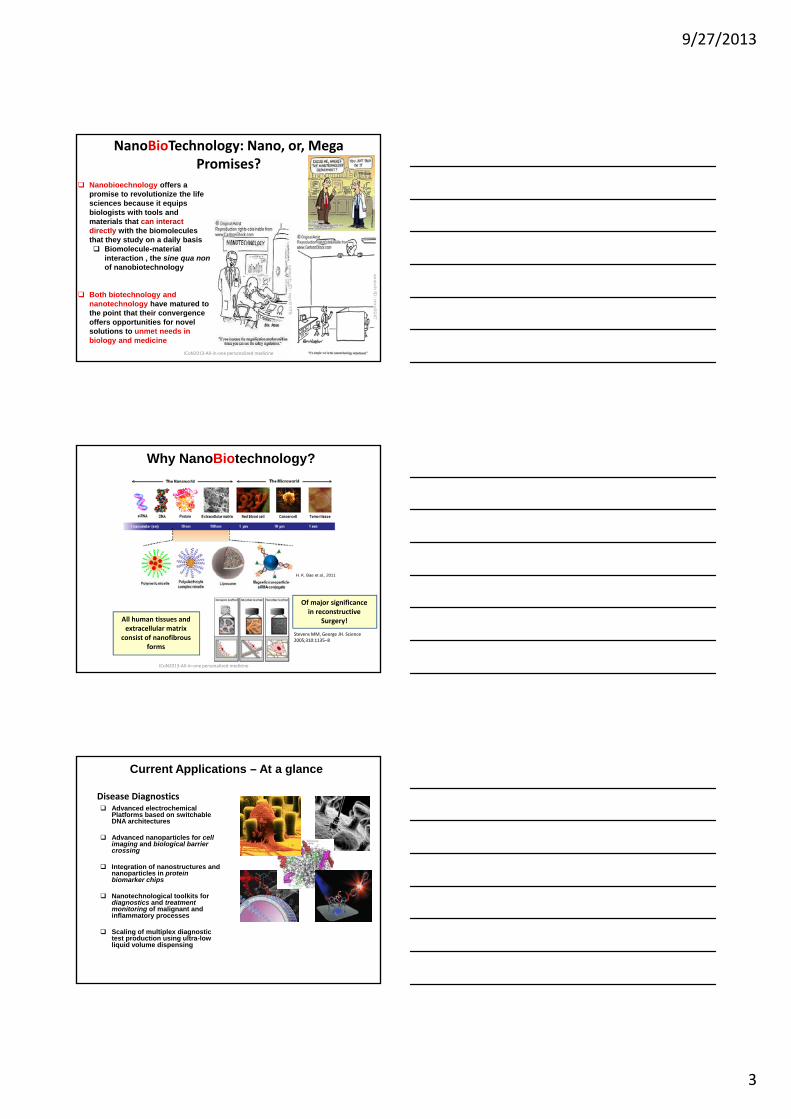
Nanobioechnology offers a promise to revolutionize the life sciences because it equips biologists with tools and materials that can interact directly with the biomoleculesthat they study on a daily basis
Biomolecule material
NanoBioTechnology: Nano, or, Mega Promises?
Biomolecule-material interaction , the sine qua nonof nanobiotechnology
Both biotechnology and nanotechnology have matured to the point that their convergence offers opportunities for novel solutions to unmet needs in biology and medicine
ICoN2013‐All‐in‐one personalized medicine
Why NanoBiotechnology?
H. K. Bae et al., 2011
All human tissues and extracellular matrix
consist of nanofibrousforms
Stevens MM, George JH. Science 2005;310:1135–8
ICoN2013‐All‐in‐one personalized medicine
Of major significance in reconstructive
Surgery!
Current Applications – At a glance
Disease DiagnosticsAdvanced electrochemical Platforms based on switchable DNA architectures
Advanced nanoparticles for cell imaging and biological barrier crossing
Integration of nanostructures and gnanoparticles in protein biomarker chips
Nanotechnological toolkits for diagnostics and treatment monitoring of malignant and inflammatory processes
Scaling of multiplex diagnostic test production using ultra-low liquid volume dispensing
9/27/2013
4
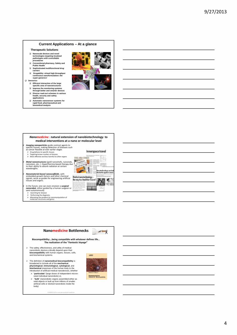
Current Applications – At a glanceTherapeutic Solutions
Nanoscale devices and novel technologies targeting localized pathologies with controllable proceduresConventional pharmacy, Safety and Public HealthSophisticated multifunctional drug carriersDrugability: virtual high throughput g y g g pcontinuous nanoformulation: the super-generics!
SensoricsEfficient interaction of the large specific area of nanostructuresImprove the monitoring systems through better and smarter devicesDiverse read-out schemes in various health, security and safety applications Automatic nanosensor systems for rapid food, pharmaceutical and biomedical analysis
Nanomedicine : natural extension of nanobiotechnology to medical interventions at a nano or molecular level
Imaging nanoparticles guide contrast agents to specific tissues, making detection of diseases such as cancer feasible at ever earlier stages
Drug delivery to specific tissuesTargeting known markers of disease,More effective and less harmful to other organs
Metal nanostructures (gold nanoshells, nanorods, nanostars, etc, ) ‐Hyperthermia‐based therapy due to their ability to absorb radiation at certain ywavelengths.
Nanomaterial‐based nanoscaffolds, with embedded growth factors and other chemical signals, serve as guides for engineering artificial tissues and organs.
In the future, one can even envision a surgical nanorobot, either guided by a human surgeon or semi‐autonomously
Searching for diseasePerforming the diagnosis, Alleviating the problem by nanomanipulation of molecular structures and genes
Nanomedicine Bottlenecks
Biocompatibility:…being compatible with whatever defines life…The realization of the “Fantastic Voyage”
The safety, effectiveness, and utility of medical nanorobotic devices critically depend upon their biocompatibility with human organs, tissues, cells, and biochemical systems.
Th d fi iti f di l bi tibilit iThe definition of nanomedical biocompatibility is broadened to include all of the mechanical, physiological, immunological, cytological, and biochemical responses of the human body to the introduction of artificial medical nanodevices, whether
“particulate” (large doses of independent micron-sized individual nanorobots) or,“bulk” (nanorobotic organs assembled either as solid objects or built up from trillions of smaller artificial cells or docked nanorobots inside the body)
ICoN2013‐All‐in‐one personalized medicine
9/27/2013
5
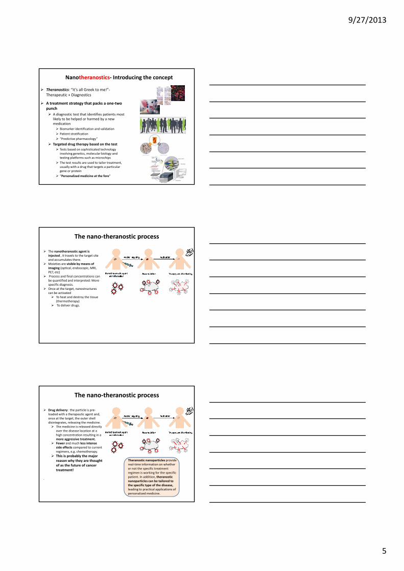
Theranostics: “it’s all Greek to me!”‐Therapeutic + Diagnostics
A treatment strategy that packs a one‐two punch
A diagnostic test that identifies patients most likely to be helped or harmed by a new medication
Nanotheranostics‐ Introducing the concept
Biomarker identification and validation
Patient stratification
“Predictive pharmacology”
Targeted drug therapy based on the test Tests based on sophisticated technology involving genetics, molecular biology and testing platforms such as microchips
The test results are used to tailor treatment, usually with a drug that targets a particular gene or protein
“Personalized medicine at the fore”
The nanotheranostic agent is injected , it travels to the target site and accumulates there.Moieties are visible by means of imaging (optical, endoscopic, MRI, PET, etc) Process and final concentrations can be quantified and interpreted: More specific diagnosis
The nano‐theranostic process
specific diagnosis. Once at the target, nanostructures can be activated
To heat and destroy the tissue (thermotherapy)To deliver drugs.
Drug delivery: the particle is pre‐loaded with a therapeutic agent and, once at the target, the outer shell disintegrates, releasing the medicine.
The medicine is released directly over the disease location at a high concentration resulting in a more aggressive treatment.Fewer and much less intense
The nano‐theranostic process
Fewer and much less intense side effects compared to current regimens, e.g. chemotherapy. This is probably the major reason why they are thought of as the future of cancer treatment!
.
Theranostic nanoparticles provide real‐time information on whether or not the specific treatment regimen is working for the specific patient. In addition, theranosticnanoparticles can be tailored to the specific type of the disease,leading to practical applications of personalized medicine.
9/27/2013
6
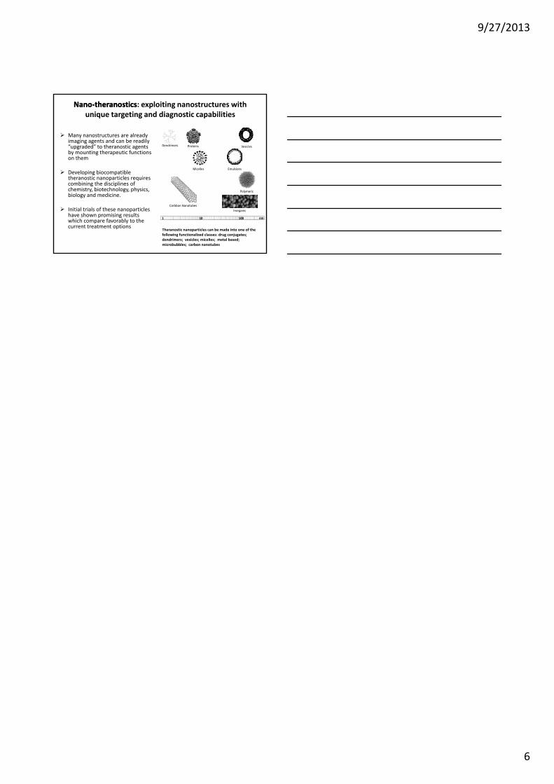
NanoNano‐‐theranosticstheranostics: exploiting nanostructures with unique targeting and diagnostic capabilities
Dendrimers Proteins
Micelles
Vesicles
Emulsions
Many nanostructures are already imaging agents and can be readily “upgraded” to theranostic agents by mounting therapeutic functions on them
Developing biocompatible
1 10 100 nm
Carbbon Nanotubes
Polymeric
Inorganic
Developing biocompatible theranostic nanoparticles requires combining the disciplines of chemistry, biotechnology, physics, biology and medicine.
Initial trials of these nanoparticles have shown promising results which compare favorably to the current treatment options
Theranostic nanoparticles can be made into one of the following functionalized classes: drug conjugates; dendrimers; vesicles; micelles; metal based; microbubbles; carbon nanotubes
9/27/2013
1
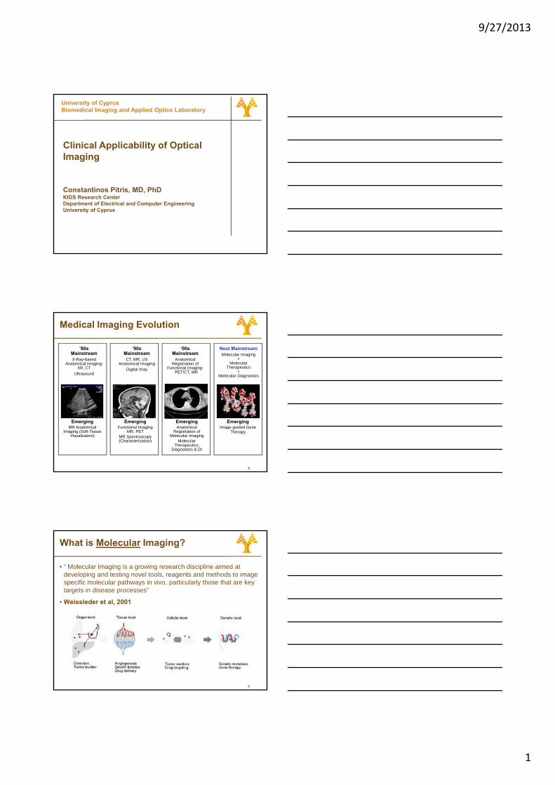
University of CyprusBiomedical Imaging and Applied Optics Laboratory
Clinical Applicability of Optical Imaging
Constantinos Pitris, MD, PhDKIOS Research CenterDepartment of Electrical and Computer EngineeringUniversity of Cyprus
Medical Imaging Evolution
’80sMainstreamX-Ray-based
Anatomical Imaging: XR, CT
Ultrasound
’90sMainstreamCT, MR, US
Anatomical ImagingDigital Xray
‘00sMainstream
Anatomical Registration of
Functional Imaging:PET/CT, MR
Next MainstreamMolecular Imaging
+Molecular
Therapeutics+
Molecular Diagnostics
2
EmergingFunctional Imaging
MR, PET MR Spectroscopy (Characterization)
EmergingMR Anatomical
Imaging (Soft-Tissue Visualization)
EmergingAnatomical
Registration of Molecular Imaging
Molecular Therapeutics,
Diagnostics & DI
EmergingImage-guided Gene
Therapy
What is Molecular Imaging?
• “ Molecular imaging is a growing research discipline aimed at developing and testing novel tools, reagents and methods to image specific molecular pathways in vivo, particularly those that are key targets in disease processes”
• Weissleder et al, 2001
3
9/27/2013
2
What is Molecular Imaging?
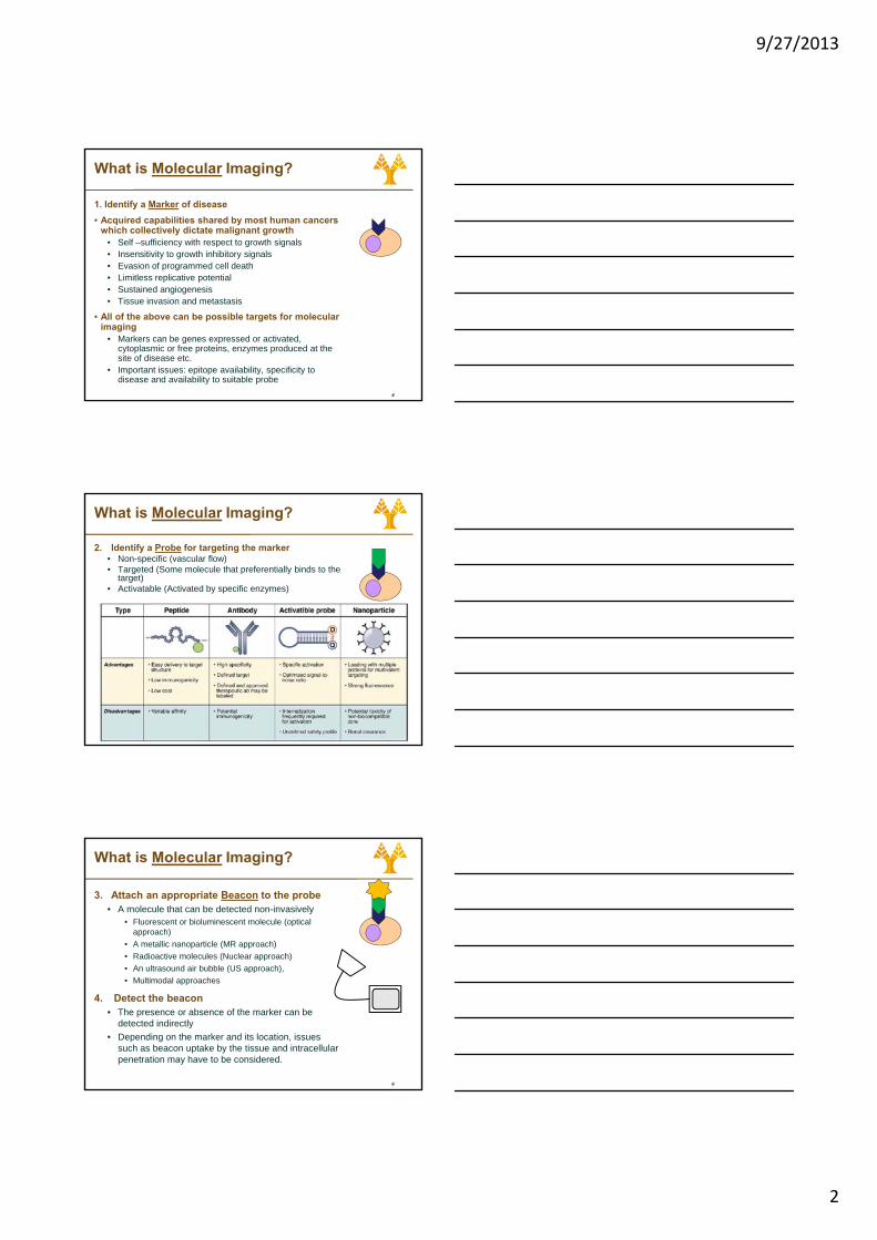
1. Identify a Marker of disease• Acquired capabilities shared by most human cancers
which collectively dictate malignant growth• Self –sufficiency with respect to growth signals• Insensitivity to growth inhibitory signals• Evasion of programmed cell death
Li itl li ti t ti l
4
• Limitless replicative potential• Sustained angiogenesis• Tissue invasion and metastasis
• All of the above can be possible targets for molecular imaging
• Markers can be genes expressed or activated, cytoplasmic or free proteins, enzymes produced at the site of disease etc.
• Important issues: epitope availability, specificity to disease and availability to suitable probe
What is Molecular Imaging?
2. Identify a Probe for targeting the marker • Non-specific (vascular flow)• Targeted (Some molecule that preferentially binds to the
target)• Activatable (Activated by specific enzymes)
5
What is Molecular Imaging?
3. Attach an appropriate Beacon to the probe• A molecule that can be detected non-invasively
• Fluorescent or bioluminescent molecule (optical approach)
• A metallic nanoparticle (MR approach)• Radioactive molecules (Nuclear approach)
6
• An ultrasound air bubble (US approach),• Multimodal approaches
4. Detect the beacon • The presence or absence of the marker can be
detected indirectly• Depending on the marker and its location, issues
such as beacon uptake by the tissue and intracellular penetration may have to be considered.
9/27/2013
3
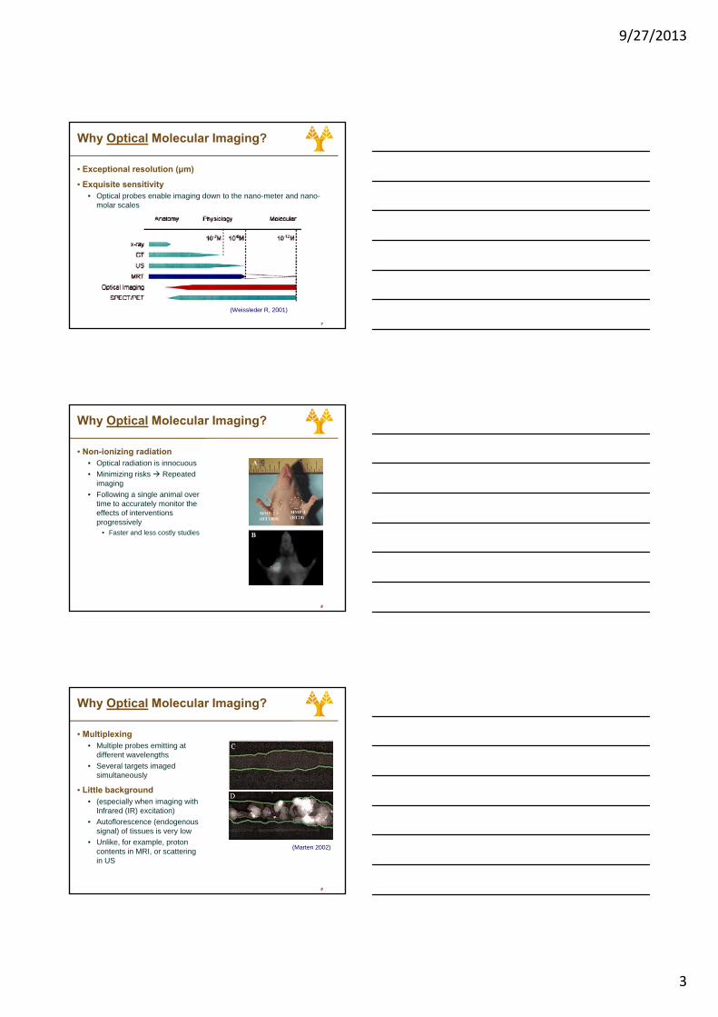
Why Optical Molecular Imaging?
• Exceptional resolution (μm)
• Exquisite sensitivity• Optical probes enable imaging down to the nano-meter and nano-
molar scales
7
(Weissleder R, 2001)
Why Optical Molecular Imaging?
• Non-ionizing radiation • Optical radiation is innocuous• Minimizing risks Repeated
imaging• Following a single animal over
time to accurately monitor the
8
effects of interventions progressively
• Faster and less costly studies
Why Optical Molecular Imaging?
• Multiplexing• Multiple probes emitting at
different wavelengths• Several targets imaged
simultaneously
• Little background
9
• Little background • (especially when imaging with
Infrared (IR) excitation)• Autoflorescence (endogenous
signal) of tissues is very low• Unlike, for example, proton
contents in MRI, or scattering in US
(Marten 2002)
9/27/2013
4
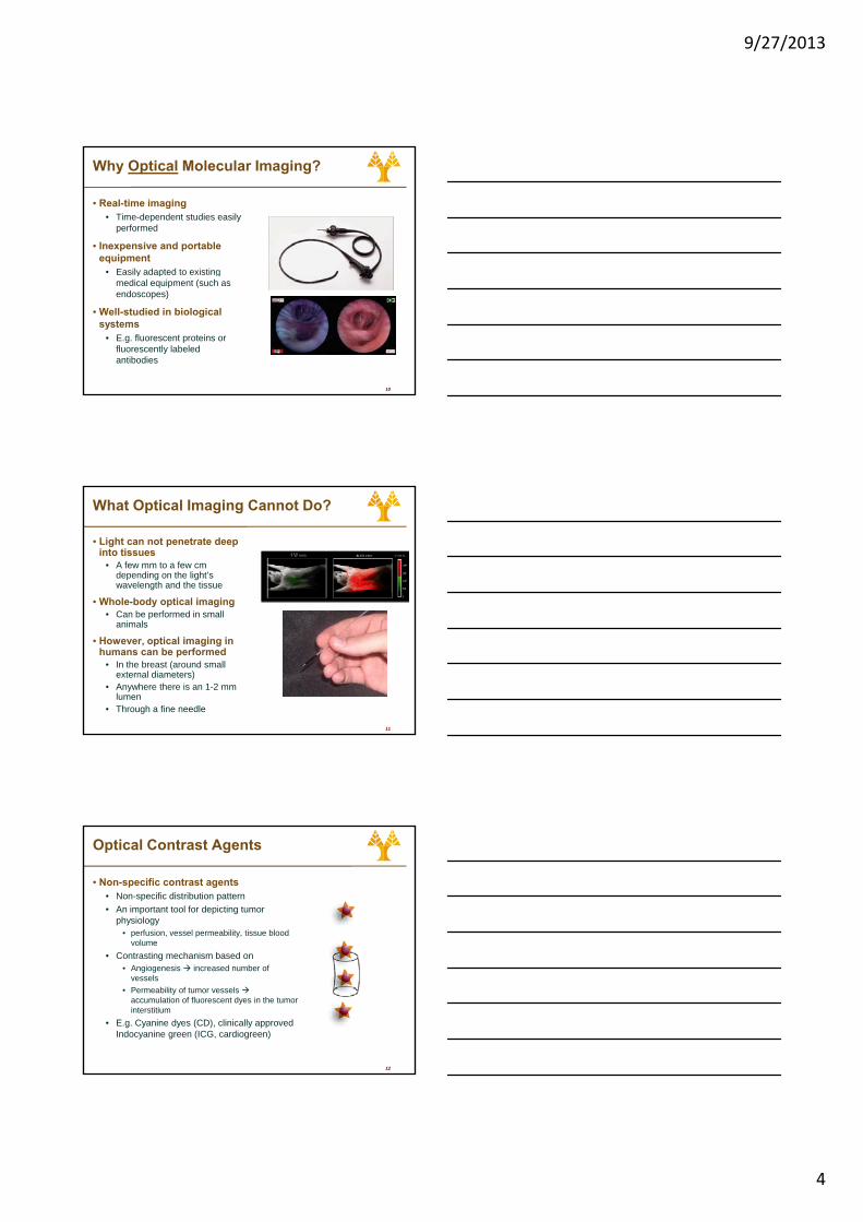
Why Optical Molecular Imaging?
• Real-time imaging • Time-dependent studies easily
performed
• Inexpensive and portable equipment
• Easily adapted to existing
10
• Easily adapted to existing medical equipment (such as endoscopes)
• Well-studied in biological systems
• E.g. fluorescent proteins or fluorescently labeled antibodies
What Optical Imaging Cannot Do?
• Light can not penetrate deep into tissues
• A few mm to a few cm depending on the light’s wavelength and the tissue
• Whole-body optical imaging
11
• Can be performed in small animals
• However, optical imaging in humans can be performed
• In the breast (around small external diameters)
• Anywhere there is an 1-2 mm lumen
• Through a fine needle
Optical Contrast Agents
• Non-specific contrast agents• Non-specific distribution pattern• An important tool for depicting tumor
physiology• perfusion, vessel permeability, tissue blood
volume
12
• Contrasting mechanism based on• Angiogenesis increased number of
vessels• Permeability of tumor vessels
accumulation of fluorescent dyes in the tumor interstitium
• E.g. Cyanine dyes (CD), clinically approved Indocyanine green (ICG, cardiogreen)
9/27/2013
5
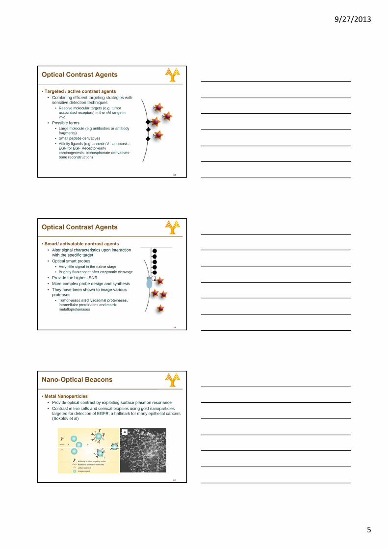
Optical Contrast Agents
• Targeted / active contrast agents• Combining efficient targeting strategies with
sensitive detection techniques• Resolve molecular targets (e.g. tumor
associated receptors) in the nM range in vivo
P ibl f
13
• Possible forms• Large molecule (e.g antibodies or antibody
fragments)• Small peptide derivatives• Affinity ligands (e.g. annexin V - apoptosis ;
EGF for EGF Receptor-early carcinogenesis; biphosphonate derivatives-bone reconstruction)
Optical Contrast Agents
• Smart/ activatable contrast agents• Alter signal characteristics upon interaction
with the specific target• Optical smart probes
• Very little signal in the native stage• Brightly fluorescent after enzymatic cleavage
14
• Provide the highest SNR• More complex probe design and synthesis• They have been shown to image various
proteases • Tumor-associated lysosomal proteinases,
intracellular proteinases and matrix metalloproteinases
Nano-Optical Beacons
• Metal Nanoparticles• Provide optical contrast by exploiting surface plasmon resonance• Contrast in live cells and cervical biopsies using gold nanoparticles
targeted for detection of EGFR, a hallmark for many epithelial cancers (Sokolov et al)
15
9/27/2013
6
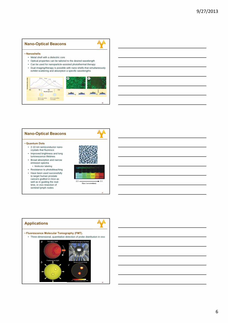
Nano-Optical Beacons
• Nanoshells• Metal shell with a dielectric core• Optical properties can be tailored to the desired wavelength• Can be used for nanoparticle-assisted photothermal therapy• Dual imaging/therapy is possible with nano-shells that simultaneously
exhibit scattering and absorption a specific wavelengths
16
exhibit scattering and absorption a specific wavelengths
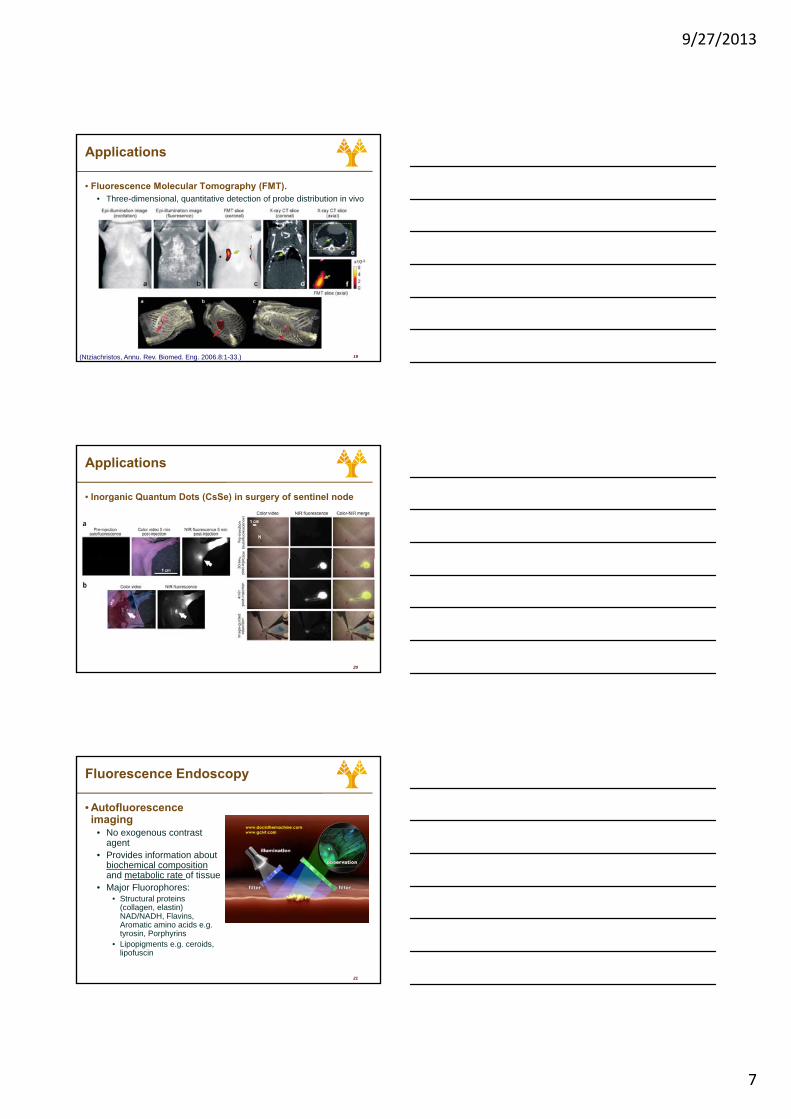
Nano-Optical Beacons
• Quantum Dots• 2-10 nm semiconductor nano-
crystals that fluoresce• Improved brightness and long
luminescence lifetimes• Broad absorption and narrow
17
emission spectra• Multicolor labeling
• Resistance to photobleaching• Have been used successfully
to target human prostate cancers grafted in mice as well as in guiding the real-time, in vivo resection of sentinel lymph nodes
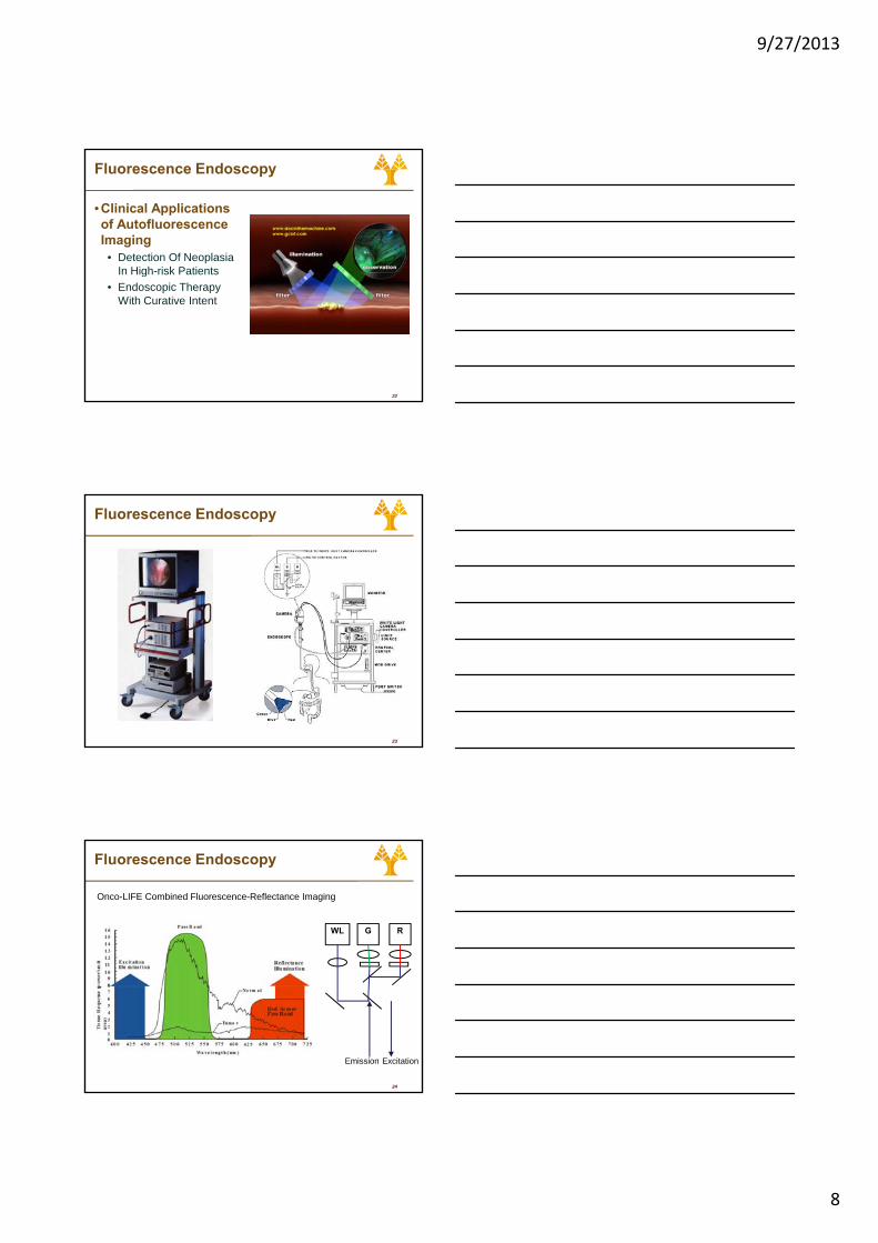
Applications
• Fluorescence Molecular Tomography (FMT).• Three-dimensional, quantitative detection of probe distribution in vivo
18
9/27/2013
7
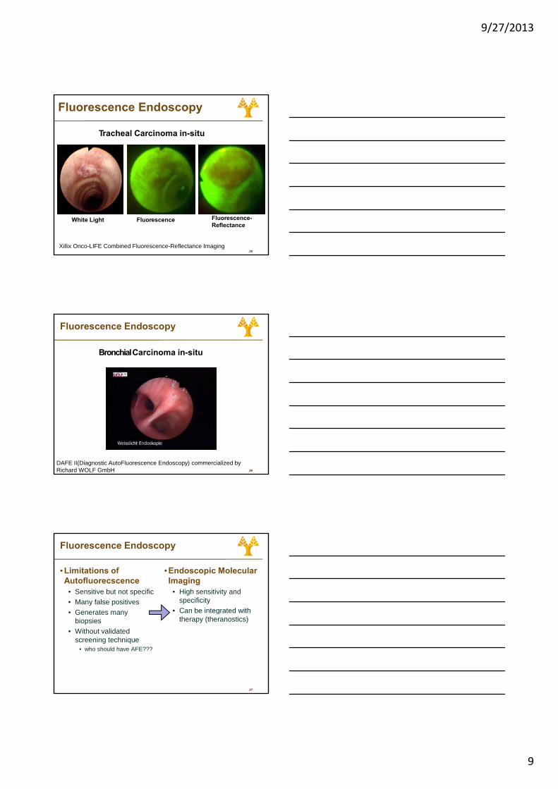
Applications
• Fluorescence Molecular Tomography (FMT).• Three-dimensional, quantitative detection of probe distribution in vivo
19(Ntziachristos, Annu. Rev. Biomed. Eng. 2006.8:1-33.)
Applications
• Inorganic Quantum Dots (CsSe) in surgery of sentinel node
20
Fluorescence Endoscopy
• Autofluorescenceimaging
• No exogenous contrast agent
• Provides information about biochemical composition
21
pand metabolic rate of tissue
• Major Fluorophores:• Structural proteins
(collagen, elastin) NAD/NADH, Flavins, Aromatic amino acids e.g. tyrosin, Porphyrins
• Lipopigments e.g. ceroids, lipofuscin
9/27/2013
8
Fluorescence Endoscopy
• Clinical Applications of AutofluorescenceImaging
• Detection Of NeoplasiaIn High-risk Patients
22
g• Endoscopic Therapy
With Curative Intent
Fluorescence Endoscopy
23
Fluorescence Endoscopy
WL G R
Onco-LIFE Combined Fluorescence-Reflectance Imaging
24
ExcitationEmission
9/27/2013
9
Fluorescence Endoscopy
Tracheal Carcinoma in-situ
25
Fluorescence-Reflectance
White Light Fluorescence
Xillix Onco-LIFE Combined Fluorescence-Reflectance Imaging
Fluorescence Endoscopy
Bronchial Carcinoma in-situ
26
DAFE II(Diagnostic AutoFluorescence Endoscopy) commercialized by Richard WOLF GmbH
Fluorescence Endoscopy
• Limitations of Autofluorecscence
• Sensitive but not specific• Many false positives• Generates many
• Endoscopic Molecular Imaging
• High sensitivity and specificity
• Can be integrated with
27
Generates many biopsies
• Without validated screening technique
• who should have AFE???
gtherapy (theranostics)
9/27/2013
10
Applications
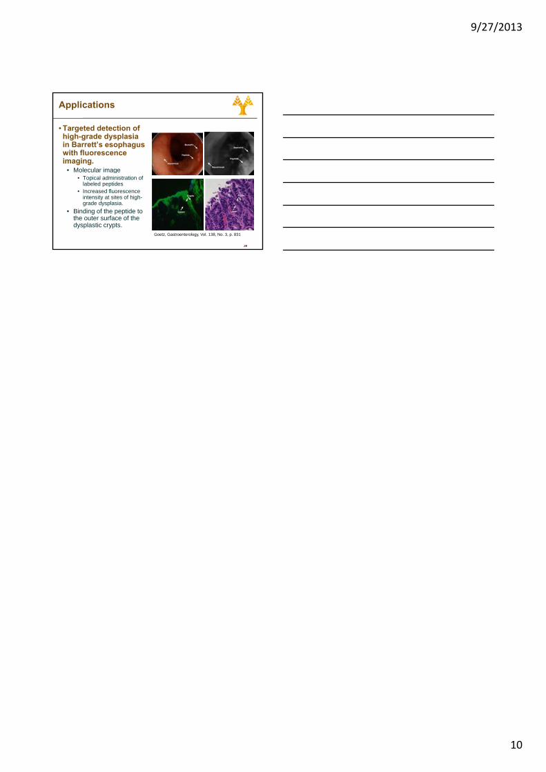
• Targeted detection of high-grade dysplasia in Barrett’s esophagus with fluorescence imaging.
• Molecular image
28
Molecular image • Topical administration of
labeled peptides • Increased fluorescence
intensity at sites of high-grade dysplasia.
• Binding of the peptide to the outer surface of the dysplastic crypts.
Goetz, Gastroenterology, Vol. 138, No. 3, p. 831
9/27/2013
1
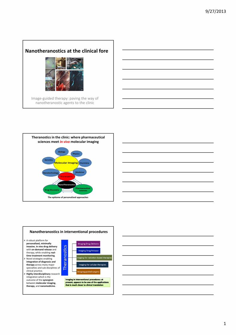
Nanotheranostics at the clinical fore
Image‐guided therapy: paving the way of nanotheranostic agents to the clinic
Theranostics in the clinic: where pharmaceutical sciences meet In vivo molecular imaging
Molecular ImagingGenetics
PhysicsBiology
Chemistry
Nanotechnology Medicine
Theranostics
PharmaceuticalSciencesDrug Discovery
NanoTheranostics
The epitome of personalized approaches
Nanotheranostics in interventional procedures
A robust platform for personalized, minimally invasive, in vivo drug delivery with on‐demand release and therapy, while enabling real‐time treatment monitoring. Novel strategies enabling
Imaging in interventional procedures: at Imaging in interventional procedures: at present, appears to be one of the applications present, appears to be one of the applications that is much closer to clinical translationthat is much closer to clinical translation
integration of diagnosis and therapy across many major specialties and sub‐disciplines of clinical practice. Highly interdisciplinary research integration which is the outcome of the synergismbetween molecular imaging, therapy, and nanomedicine.
9/27/2013
2
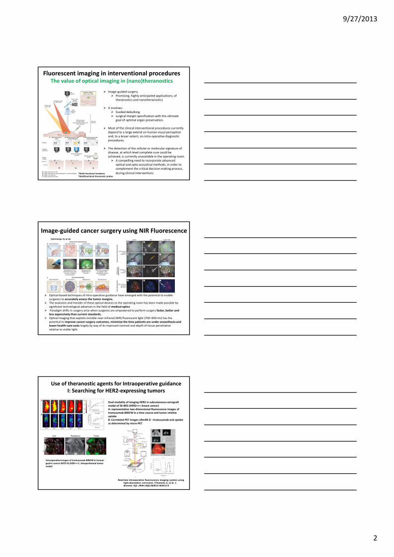
Fluorescent imaging in interventional procedures The value of optical imaging in (nano)theranostics
Image‐guided surgery.Promising, highly anticipated applications, of theranostics and nanotheranostics
It involves: Guided debulkingsurgical margin specification with the ultimate goal of optimal organ preservation.
Most of the clinical interventional procedures currently depend to a large extend on human visual perception and, to a lesser extent, on intra‐operative diagnostic procedures.
The detection of the cellular or molecular signature of disease, at which level complete cure could be achieved, is currently unavailable in the operating room.
A compelling need to incorporate advanced optical and opto‐acoustical methods, in order to complement the critical decision making process, during clinical interventions *Multi‐functional hardware
*Multifunctional theranostic probes
Image‐guided cancer surgery using NIR Fluorescence(Vahrmeijer AL et al)
Optical‐based techniques of intra‐operative guidance have emerged with the potential to enable surgeons to accurately assess the tumor margins. The evolution and transfer of these optical devices to the operating room has been made possible by significant technological advances in the field of medical opticsParadigm shifts in surgery arise when surgeons are empowered to perform surgery faster, better and less expensively than current standards. Optical imaging that exploits invisible near‐infrared (NIR) fluorescent light (700–900 nm) has the potential to improve cancer surgery outcomes, minimize the time patients are under anaesthesia and lower health‐care costs largely by way of its improved contrast and depth of tissue penetration relative to visible light.
Use of theranostic agents for Intraoperative guidanceI: Searching for HER2‐expressing tumors
Dual modality of imaging HER2 in subcutaneous xenograftmodel of SK‐BR3 (HER2+++ breast cancer)A: representative two‐dimensional fluorescence images of trastuzumab‐800CW in a time course and tumor relative uptakeB: Correlated PET images after89 Zr –trastuzumab and uptake as determined by micro‐PET
Intraoperative images of trastuzumab‐800CW in human gastric cancer KATO‐III (HER+++) intraperitoneal tumor model
Real-time intraoperative fluorescence imaging system using light-absorption correction. (Themelis G, et al. J. Biomed. Opt. 2009;14(6):064012-064012-9.
9/27/2013
3
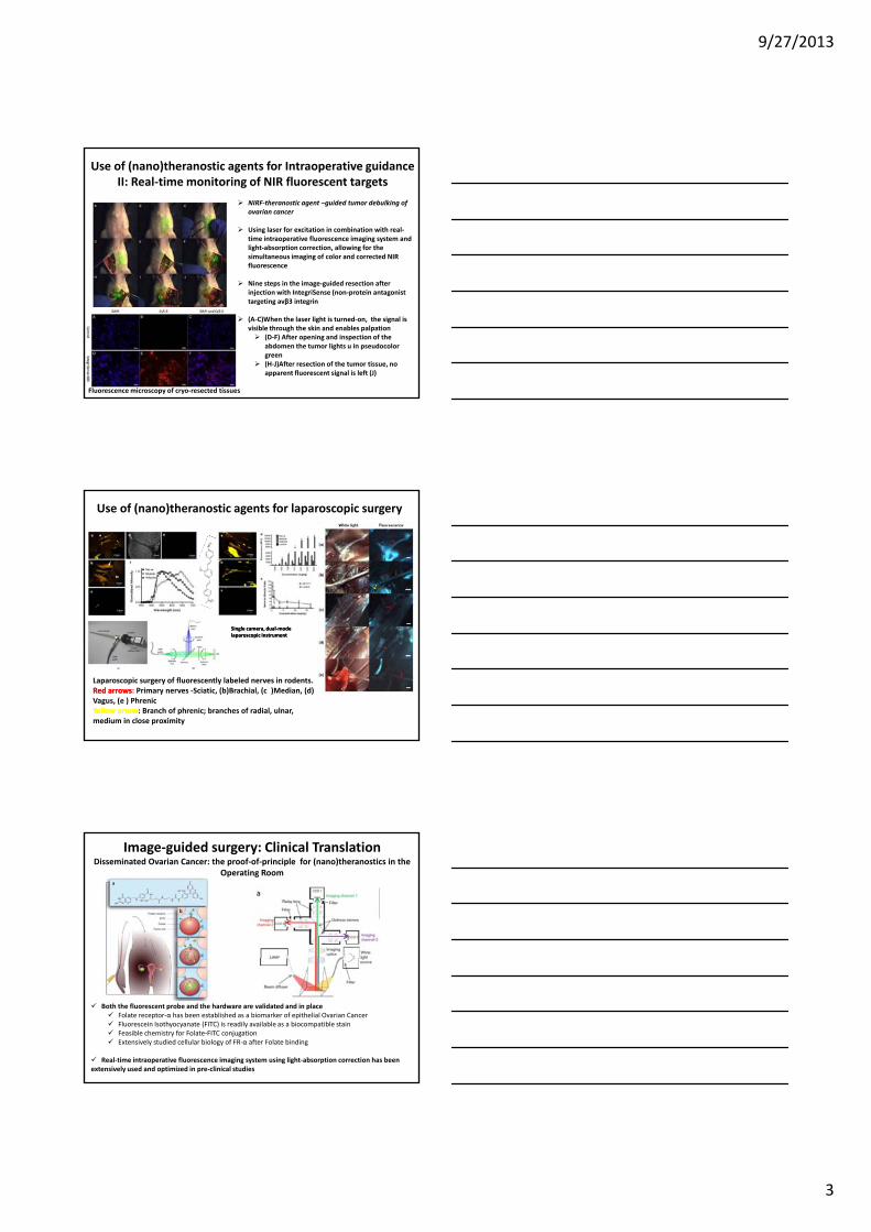
Use of (nano)theranostic agents for Intraoperative guidanceII: Real‐time monitoring of NIR fluorescent targets
NIRF‐theranostic agent –guided tumor debulking of ovarian cancer
Using laser for excitation in combination with real‐time intraoperative fluorescence imaging system and light‐absorption correction, allowing for the simultaneous imaging of color and corrected NIR fluorescence
Nine steps in the image‐guided resection after injection with IntegriSense (non‐protein antagonist targeting avβ3 integrin
(A‐C)When the laser light is turned‐on, the signal is visible through the skin and enables palpation
(D‐F) After opening and inspection of the abdomen the tumor lights u in pseudocolorgreen(H‐J)After resection of the tumor tissue, no apparent fluorescent signal is left (J)
Fluorescence microscopy of cryo‐resected tissues
Use of (nano)theranostic agents for laparoscopic surgery
Single camera, dualSingle camera, dual‐‐mode mode laparoscopic instrumentlaparoscopic instrument
Laparoscopic surgery of fluorescently labeled nerves in rodents.Red arrowsRed arrows: Primary nerves ‐Sciatic, (b)Brachial, (c )Median, (d) Vagus, (e ) PhrenicYellow arrowYellow arrow: Branch of phrenic; branches of radial, ulnar, medium in close proximity
Image‐guided surgery: Clinical TranslationDisseminated Ovarian Cancer: the proof‐of‐principle for (nano)theranostics in the
Operating Room
Both the fluorescent probe and the hardware are validated and in placeFolate receptor‐α has been established as a biomarker of epithelial Ovarian CancerFluorescein Isothyocyanate (FITC) is readily available as a biocompatible stainFeasible chemistry for Folate‐FITC conjugationExtensively studied cellular biology of FR‐α after Folate binding
Real‐time intraoperative fluorescence imaging system using light‐absorption correction has beenextensively used and optimized in pre‐clinical studies
9/27/2013
4
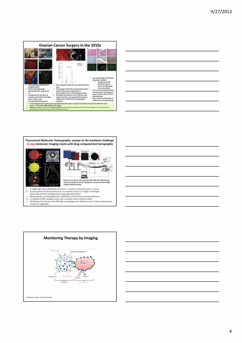
Ovarian Cancer Surgery in the 2010s
FITC‐conjugate Folate Acid was administered IV (a,b) Color image (a) with the corresponding tumor‐specific fluorescence image (b) of a representative area in the abdominal cavity.Scoring(c) was based on three different color images and their corresponding fluorescence images (FLI); P<0.001 by five independent surgeons.
“Intraoperative multispectral imaging system”Patient with high‐grade, disseminated OC, stage III, FR‐α++Intraoperative snap‐shots of simultaneously detected images: (d,f) in colour; (e,g) corresponding fluorescence
Immunohistologic Verification: the golden standard
Fibrothecoma; (b) borderline serous tumor; (c) High‐grade serous carcinoma
H+E vs Immunohistochemistry vs Fluorescence microscopy of frozen sections after FITC‐FR‐α administration.Data verify the assessment of visible lesions intraoperatively
Functionalization of the probe with a potential therapeutic target is expected to introduce theranostics) within the same session HIF; VEGR, ERBB, HSP90; CXCL; MMPsOdysseos A, Pitris C et al: U.S Pr. Patent‐61,382,875: Multimodal and Multifunctional Theranostic Agents for In vivo Detection, Monitoring and Targeted Therapy of TKR‐mediated Disorders
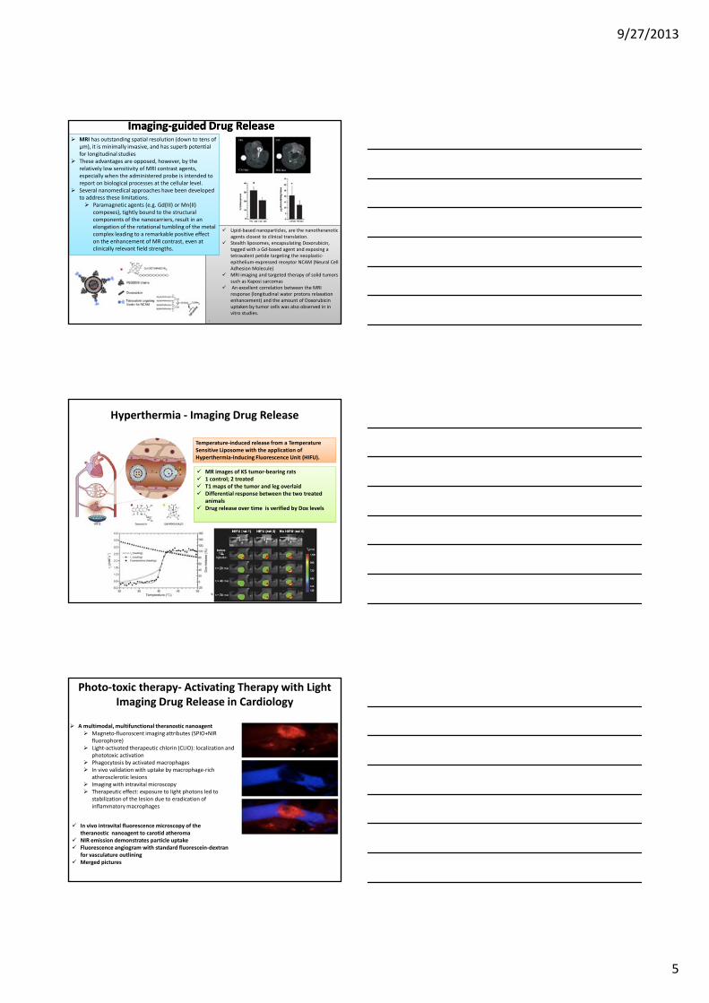
Fluorescent Molecular Tomorgraphy: answer to the hardware challenge In vivo molecular imaging meets with drug computerized tomography
a. A single light source illuminates into tissue –excitation of fluorochromes in tissuesb. In each position the fluorochromes act as secondary sources at a higher wavelength
-Both collected from multiple points using appropriate filtersc. Measurements are tomographically combined to yield 3D fluorochrome distribution d. A cylindrical FMT imaging system: blue-excitation, black-collection fiberse. Modelling of the distance that NIR light can propagate into different tissues ( before attenuation by
an order of magnitude)
Based on (a) the use of target-specific molecular fluorescent reporters (probes) and (b) volumetric reconstruction of light emitted from the probe
Monitoring Therapy by Imaging
(Drug Discovery Today. V17,17/18, Sept 2012)
9/27/2013
5
MRI has outstanding spatial resolution (down to tens of μm), it is minimally invasive, and has superb potential for longitudinal studiesThese advantages are opposed, however, by the relatively low sensitivity of MRI contrast agents, especially when the administered probe is intended to report on biological processes at the cellular level. Several nanomedical approaches have been developed to address these limitations.
Paramagnetic agents (e.g. Gd(III) or Mn(II) compexes), tightly bound to the structural components of the nanocarriers, result in an
ImagingImaging‐‐guided Drug Releaseguided Drug Release
Lipid‐based nanoparticles, are the nanotheranoticagents closest to clinical translation. Stealth liposomes, encapsulating Doxorubicin, tagged with a Gd‐based agent and exposing a tetravalent petide targeting the neoplastic‐epithelium‐expressed receptor NCAM (Neural Cell Adhesion Molecule)MRI imaging and targeted therapy of solid tumors such as Kaposi sarcomas An excellent correlation between the MRI response (longitudinal water protons relaxation enhancement) and the amount of Doxorubicin uptaken by tumor cells was also observed in in vitro studies.
.
pelongation of the rotational tumbling of the metal complex leading to a remarkable positive effect on the enhancement of MR contrast, even at clinically relevant field strengths.
Hyperthermia ‐ Imaging Drug Release
Temperature‐induced release from a Temperature Sensitive Liposome with the application of Hyperthermia‐Inducing Fluorescence Unit (HIFU).
MR images of KS tumor‐bearing rats1 control; 2 treatedT1 maps of the tumor and leg overlaidDifferential response between the two treated animalsanimalsDrug release over time is verified by Dox levels
Photo‐toxic therapy‐ Activating Therapy with LightImaging Drug Release in Cardiology
A multimodal, multifunctional theranostic nanoagentMagneto‐fluoroscent imaging attributes (SPIO+NIR fluorophore)Light‐activated therapeutic chlorin (CLIO): localization and phototoxic activationPhagocytosis by activated macrophagesIn vivo validation with uptake by macrophage‐rich atherosclerotic lesionsImaging with intravital microscopyTherapeutic effect: exposure to light photons led to stabilization of the lesion due to eradication of inflammatory macrophages
In vivo intravital fluorescence microscopy of the theranostic nanoagent to carotid atheromaNIR emission demonstrates particle uptakeFluorescence angiogram with standard fluorescein‐dextran for vasculature outliningMerged pictures
9/27/2013
1
University of CyprusBiomedical Imaging and Applied Optics Laboratory
Image-guided quantification of drug delivery and development
Constantinos Pitris, MD, PhDKIOS Research CenterDepartment of Electrical and Computer EngineeringUniversity of Cyprus
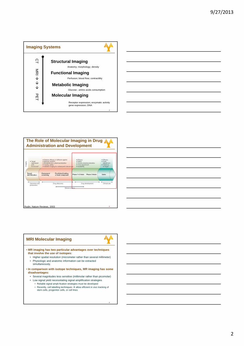
The Ultimate Goal of Molecular Imaging
• Personalized (patient-tailored) medicine
• To adapt the treatment to the patient specific characteristics
2
• From : one treatment for all• To: one patient - one treatment
• This requires• Knowledge of the underlying
molecular defects of the cancer• Systems to effectively and
efficiently identify those defects, deliver, and monitor the therapy
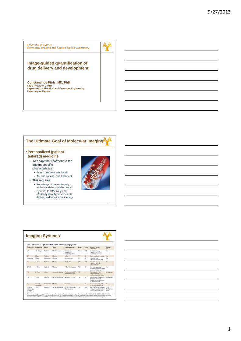
Imaging Systems
3
9/27/2013
2
Imaging Systems
Functional ImagingAnatomy; morphology; density
CT
MR
I
Structural Imaging
4
Metabolic Imaging
Molecular Imaging
Perfusion; blood flow; contractility
Glucose ; amino acids consumption
Receptor expression; enzymatic activitygene expression; DNA
PET
The Role of Molecular Imaging in Drug Administration and Development
5Rudin, Nature Reviews, 2003
MRI Molecular Imaging
• MR imaging has two particular advantages over techniques that involve the use of isotopes:
• Higher spatial resolution (micrometer rather than several millimeter)• Physiologic and anatomic information can be extracted
simultaneously.
• In comparison with isotope techniques MR imaging has some
6
• In comparison with isotope techniques, MR imaging has some disadvantages:
• Several magnitudes less sensitive (millimolar rather than picomolar)• Low signal yield necessitating signal amplification strategies
• Reliable signal ampli-fication strategies must be developed• Recently, cell labelling techniques allow efficient in vivo tracking of
stem cells, progenitor cells, or cell lines
9/27/2013
3
MRI Molecular Imaging
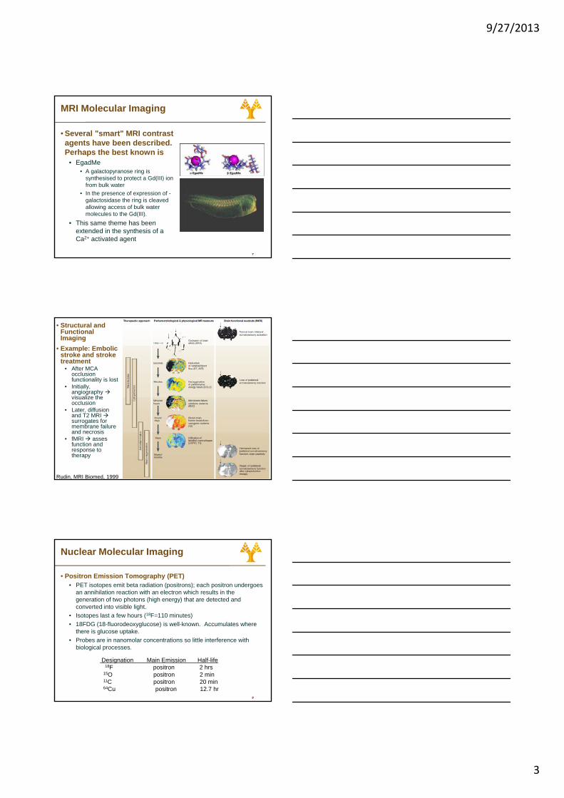
• Several "smart" MRI contrast agents have been described. Perhaps the best known is
• EgadMe• A galactopyranose ring is
th i d t t t Gd(III) i
7
synthesised to protect a Gd(III) ion from bulk water
• In the presence of expression of -galactosidase the ring is cleaved allowing access of bulk water molecules to the Gd(III).
• This same theme has been extended in the synthesis of a Ca2+ activated agent
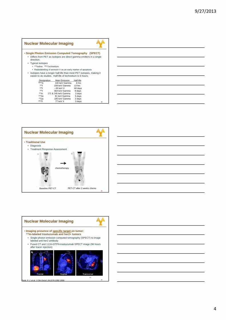
• Structural and Functional Imaging
• Example: Embolic stroke and stroke treatment
• After MCA occlusion functionality is lost
• Initially, angiography visualize the
8
visualize the occlusion
• Later, diffusion and T2 MRI surrogates for membrane failure and necrosis
• fMRI asses function and response to therapy
Rudin, MRI Biomed, 1999
Nuclear Molecular Imaging
• Positron Emission Tomography (PET)• PET isotopes emit beta radiation (positrons); each positron undergoes
an annihilation reaction with an electron which results in the generation of two photons (high energy) that are detected and converted into visible light.
• Isotopes last a few hours (18F=110 minutes)
9
• 18FDG (18-fluorodeoxyglucose) is well-known. Accumulates where there is glucose uptake.
• Probes are in nanomolar concentrations so little interference with biological processes.
Designation Main Emission Half-life 18F positron 2 hrs
15O positron 2 min 11C positron 20 min64Cu positron 12.7 hr
9/27/2013
4
Nuclear Molecular Imaging
• Single Photon Emission Computed Tomography (SPECT)• Differs from PET as isotopes are direct gamma emitters in a single
direction. • Typical isotopes
• 123Iodine 99mTechnetium.• Radiolabelling of annexin-V as an early marker of apoptosis
10
• Isotopes have a longer half-life than most PET isotopes, making it easier to do studies. Half-life of technetium is 6 hours.
Designation Main Emission Half-life 99mTc 140 keV Gamma 6 hrs
123I 159 keV Gamma 13 hrs125I 30 keV X 60 days131I 364 keV Gamma 8 days
111In 171 & 245 keV Gamma 3 days133Xe 81 keV Gamma 5 days67Ga 185 keV Gamma 3 days201Tl 77 keV X 3 days
A B
Nuclear Molecular Imaging
• Traditional Use• Diagnosis• Treatment Response Assessment
11
Baseline PET-CT PET-CT after 2 weeks chemo
chemotherapy
Nuclear Molecular Imaging
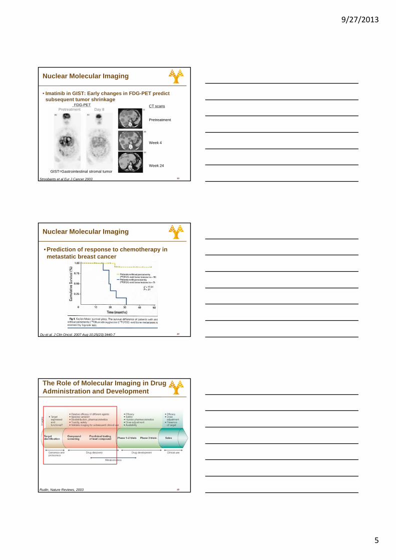
• Imaging presence of specific target on tumor: 111In-labeled trastuzumab and her2+ tumors
• Single-photon emission computed tomography (SPECT) to image labeled anti-her2 antibody
• Fused CT and 111In-DTPA-trastuzumab SPECT image (96 hours after tracer injection)
12
12
Perik, P. J. et al. J Clin Oncol; 24:2276-2282 2006
9/27/2013
5
Nuclear Molecular Imaging
• Imatinib in GIST: Early changes in FDG-PET predict subsequent tumor shrinkage
FDG-PETPretreatment Day 8
CT scans
Pretreatment
13Stroobants et al Eur J Cancer 2003
Week 4
Week 24GIST=Gastrointestinal stromal tumor
Nuclear Molecular Imaging
• Prediction of response to chemotherapy in metastatic breast cancer
14Du et al. J Clin Oncol. 2007 Aug 10;25(23):3440-7
The Role of Molecular Imaging in Drug Administration and Development
15Rudin, Nature Reviews, 2003
9/27/2013
6
Molecular Imaging in Drug Development
• “Up to 70% of the experiments in pharmaceutical research and development result in an image as an output” (R. Dunkle 2003)
16
• Can it improve the drug development process?
• In what specific activities could it be most useful?
Molecular Imaging in Drug Development
• Determine desirable, pharmacological effects or undesirable side effects on the molecular level
• i.e., the effect of the drug candidate on in vivo biochemistry and physiology
• Evaluate the interaction of a drug or drug candidate with the desired target including dose occupancy relationships and
17
desired target including dose occupancy relationships and kinetic information
• e.g., receptor, enzyme or transport system,
• Quantify the delivery of a drug to a specific target
• Examine the absorption, distribution, metabolism and elimination of the labeled drug candidate
Molecular Imaging in Drug Development
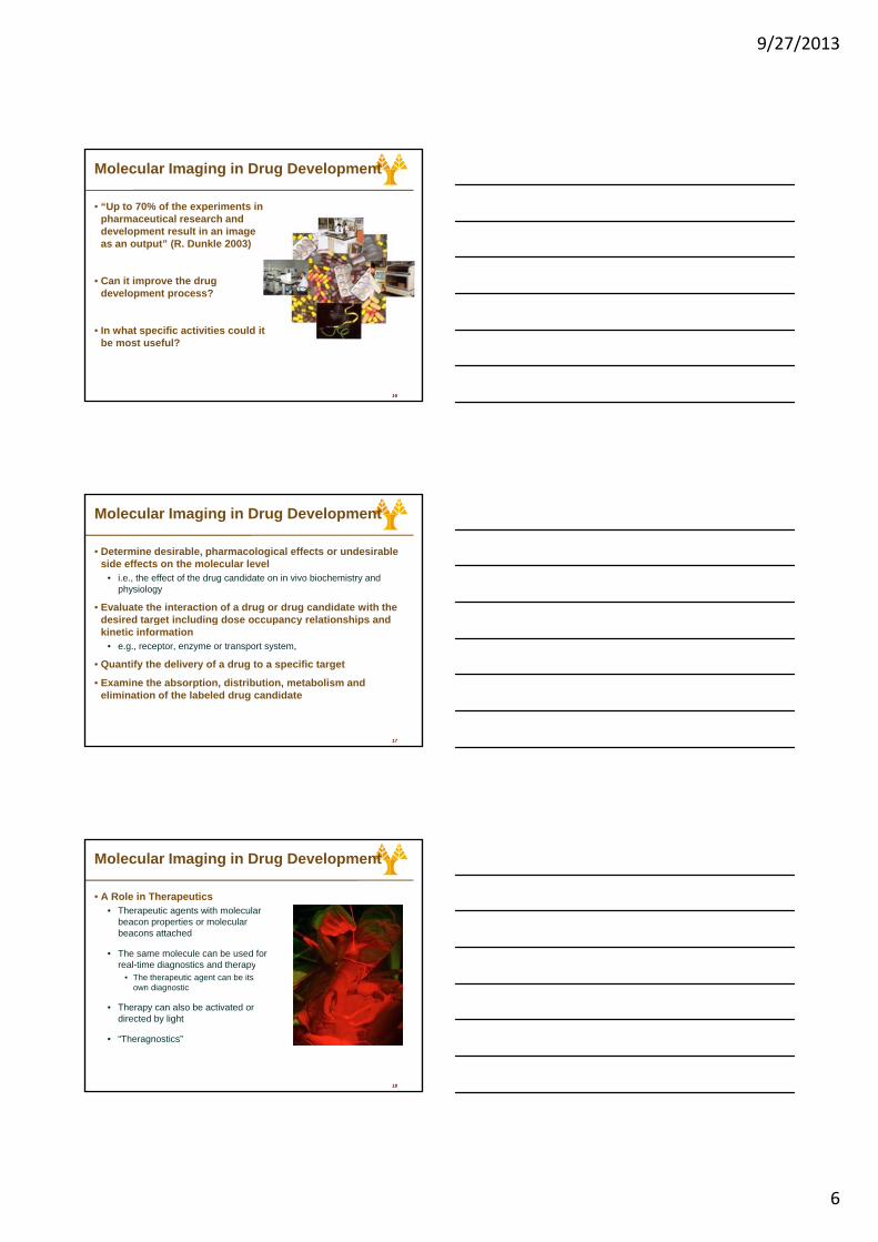
• A Role in Therapeutics• Therapeutic agents with molecular
beacon properties or molecular beacons attached
• The same molecule can be used for real-time diagnostics and therapy
18
real-time diagnostics and therapy• The therapeutic agent can be its
own diagnostic
• Therapy can also be activated or directed by light
• “Theragnostics”
9/27/2013
7
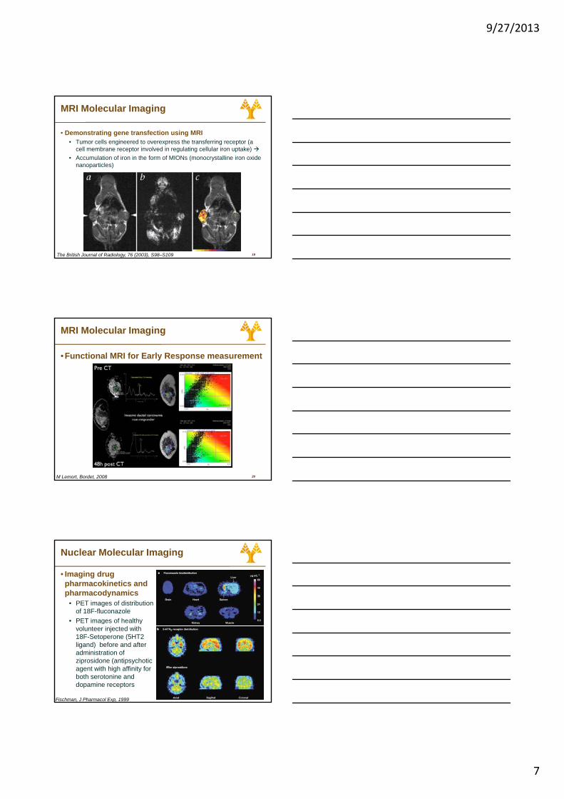
MRI Molecular Imaging
• Demonstrating gene transfection using MRI• Tumor cells engineered to overexpress the transferring receptor (a
cell membrane receptor involved in regulating cellular iron uptake) • Accumulation of iron in the form of MIONs (monocrystalline iron oxide
nanoparticles)
19The British Journal of Radiology, 76 (2003), S98–S109
MRI Molecular Imaging
• Functional MRI for Early Response measurement
20M Lemort, Bordet, 2008
Nuclear Molecular Imaging
• Imaging drug pharmacokinetics and pharmacodynamics
• PET images of distribution of 18F-fluconazole
• PET images of healthy
21
• PET images of healthy volunteer injected with 18F-Setoperone (5HT2 ligand) before and after administration of ziprosidone (antipsychotic agent with high affinity for both serotonine and dopamine receptors
Fischman, J Pharmacol Exp, 1999
9/27/2013
8
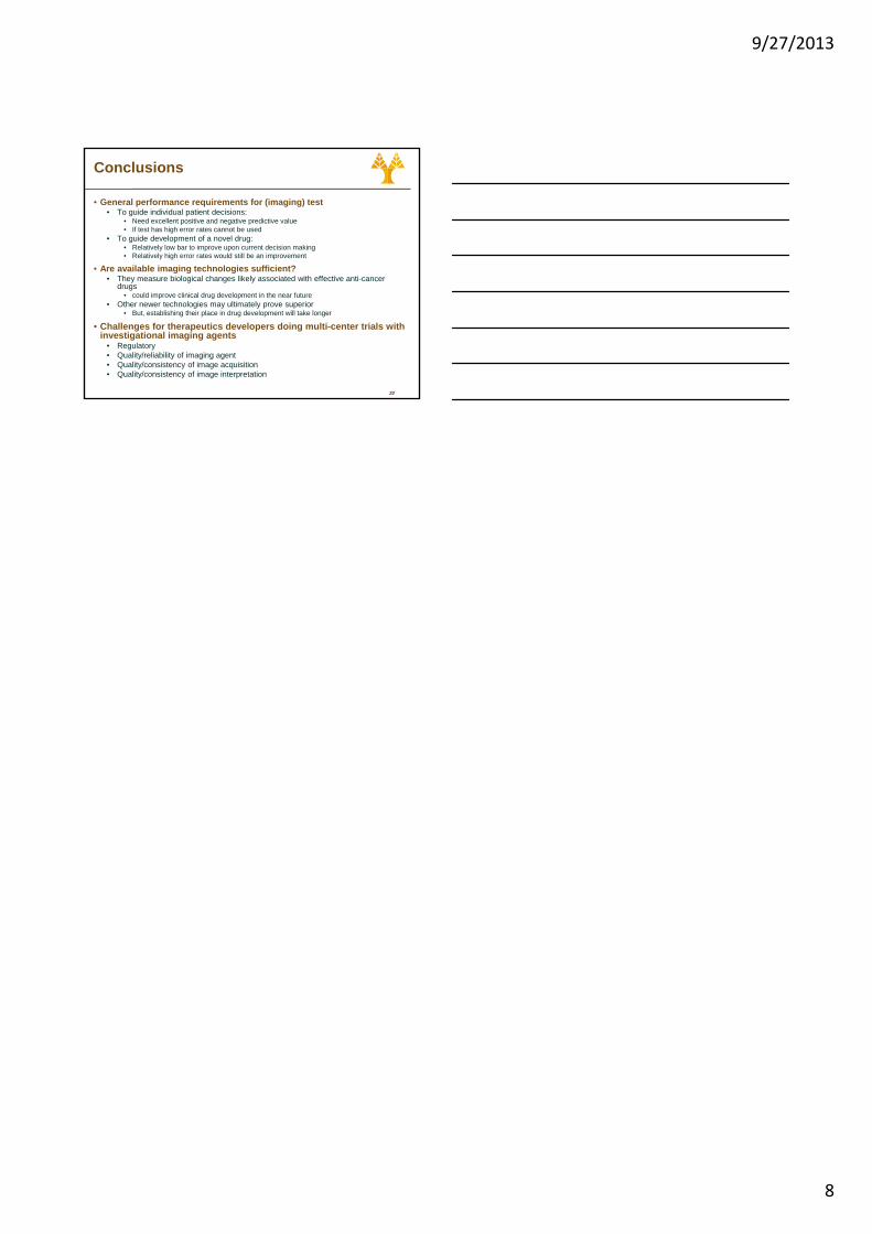
Conclusions
• General performance requirements for (imaging) test• To guide individual patient decisions:
• Need excellent positive and negative predictive value• If test has high error rates cannot be used
• To guide development of a novel drug:• Relatively low bar to improve upon current decision making • Relatively high error rates would still be an improvement
• Are available imaging technologies sufficient?
22
• They measure biological changes likely associated with effective anti-cancer drugs
• could improve clinical drug development in the near future• Other newer technologies may ultimately prove superior
• But, establishing their place in drug development will take longer
• Challenges for therapeutics developers doing multi-center trials with investigational imaging agents
• Regulatory• Quality/reliability of imaging agent• Quality/consistency of image acquisition• Quality/consistency of image interpretation

9/27/2013
1
…Lessons learned and progress made during more than 40 years of environmental and bioethics regulation can serve as a guidepost for addressing nanobiotechnology regulation and oversight issues…
…a Nanomedicine sub‐committee at European Medicines Agency
New Bioethical Standards at the Fore???
Are we at the dawning of a “second Industrial Revolution”
Nanotheranostics appears to hold almost limitless potential for beneficial applications
This tremendous upside comes with a host of governance and oversight challenges
These regulatory challenges are unique to nanobiotechnologyand the existing apparatus of the regulatory state is inadequate to address the novel problems that arise
The statutes that define the current approaches to environmental health and safety protection were written prior to the emergence of nanobiotechnology and must be rewritten, reinterpreted or applied in novel ways to address the new realities posed by the nanobiotechnology revolution
The problems most centrally associated with the emergence of nanobiotechnology are pervasive throughout the field of environmental regulation
ComplexityUncertainty Dysfunctional mix of regulatory gaps and overlapping agency authorities
“ Δεν μπορείς να μπεις δυό
φορές στο ίδιο ποτάμι”
Ηράκλειτος o Εφέσιος
“You cannot enter the same river
twice”
Heraclitus of Ephesus
9/27/2013
2
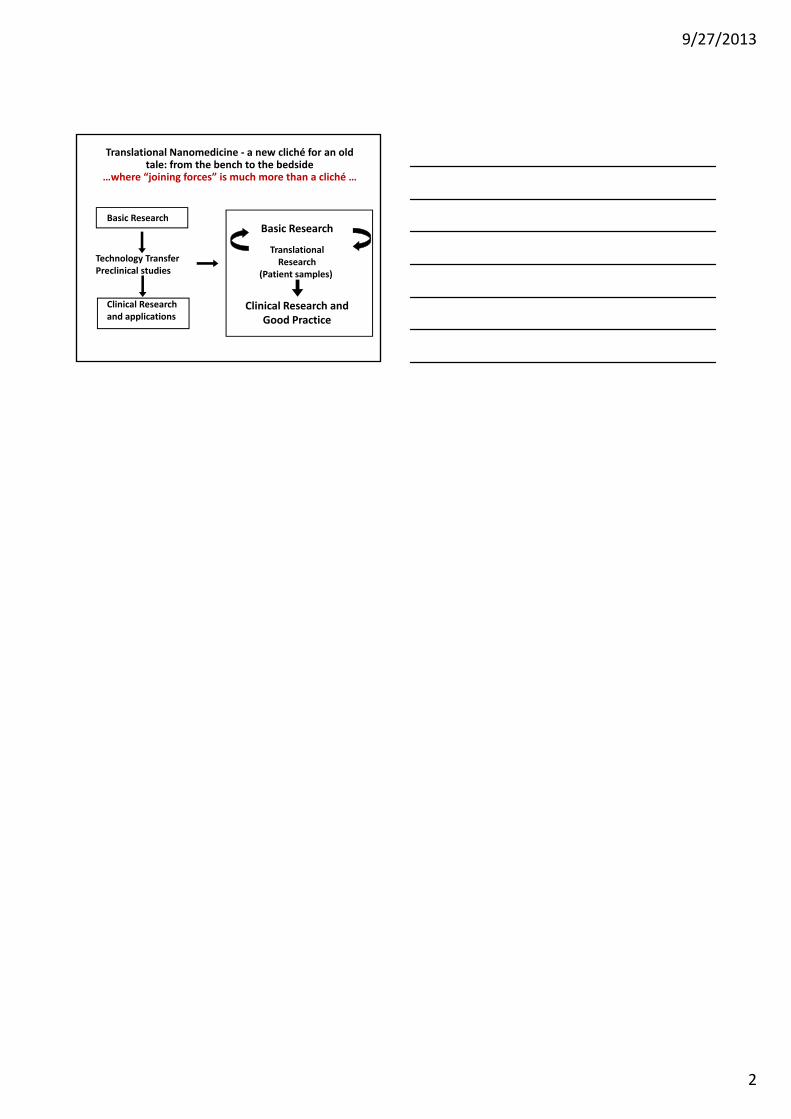
Translational Nanomedicine ‐ a new cliché for an old tale: from the bench to the bedside
…where “joining forces” is much more than a cliché …
Basic ResearchBasic Research
Translational
Clinical Research and applications
Technology TransferPreclinical studies
TranslationalResearch
(Patient samples)
Clinical Research andGood Practice