Embed Size (px)
Citation preview
International clinical guideline for the management of classicalgalactosemia: diagnosis, treatment, and follow-up
Lindsey Welling1 & Laurie E. Bernstein2& Gerard T. Berry3,4 & Alberto B. Burlina5 &
François Eyskens6 & Matthias Gautschi7 & Stephanie Grünewald8&
Cynthia S. Gubbels3,4 & Ina Knerr9 & Philippe Labrune10 & Johanna H. van der Lee11 &
Anita MacDonald12& Elaine Murphy13 & Pat A. Portnoi14 & Katrin Õunap15,16
&
Nancy L. Potter17 & M. Estela Rubio-Gozalbo18 & Jessica B. Spencer19 & Inge Timmers20 &
Eileen P. Treacy21 & Sandra C. VanCalcar22 & Susan E.Waisbren23&AnnetM. Bosch1,24
&
On behalf of the Galactosemia Network (GalNet)
Received: 16 February 2016 /Revised: 17 August 2016 /Accepted: 29 September 2016# The Author(s) 2016. This article is published with open access at Springerlink.com
Abstract Classical galactosemia (CG) is an inborn error ofgalactose metabolism. Evidence-based guidelines for thetreatment and follow-up of CG are currently lacking, and
treatment and follow-up have been demonstrated to varyworldwide. To provide patients around the world the samestate-of-the-art in care, members of The Galactosemia
Communicated by: Georg Hoffmann
The online version of this article (doi:10.1007/s10545-016-9990-5)contains supplementary material, which is available to authorized users.
* Annet M. [email protected]
On behalf of the Galactosemia Network (GalNet)
1 Department of Pediatrics, Emma Children’s Hospital, AcademicMedical Center, Meibergdreef 9, 1105AZ Amsterdam, The Netherlands
2 Section of Clinical Genetics and Metabolism, Inherited MetabolicDisease Nutrition Department, University of Colorado–DenverSchool of Medicine, The Children’s Hospital Colorado, Aurora, CO,USA
3 Division of Genetics and Genomics, Boston Children’s Hospital,Harvard Medical School, Boston, MA, USA
4 Broad Institute of MIT and Harvard, Cambridge, MA, USA5 Department of Pediatrics, Metabolic Unit, University Hospital,
University of Padova, Padova, Italy6 Department of Metabolic Disorders in Children, Antwerp University
Hospital UZA, Edegem, Belgium7 University Children’s Hospital, Pediatric Endocrinology, Diabetes
and Metabolism, and Institute of Clinical Chemistry, Inselspital,University of Bern, Bern, Switzerland
8 Metabolic Unit, Great Ormond Street Hospital and Institute of ChildHealth, University College London, London, UK
9 National Centre for Inherited Metabolic Disorders, Temple St.Children’s University Hospital, Dublin, Ireland
10 Department of Pediatrics, APHP, Hopital Antoine Béclère, CedexClamart, France
11 Pediatric Clinical Research Office, Emma Children’s Hospital,Academic Medical Center, Amsterdam, The Netherlands
12 BirminghamChildren’s Hospital, Steelhouse Lane, Birmingham,UK13 Charles Dent Metabolic Unit, National Hospital for Neurology and
Neurosurgery, Queen Square, London, UK14 Medical Advisory Panel, Galactosemia Support Group UK, West
Midlands, UK15 Department of Pediatrics, University of Tartu, Tartu, Estonia16 Department of Genetics, Tartu University Hospital, Tartu, Estonia17 Department of Speech and Hearing Sciences, Washington State
University, Spokane, WA, USA18 Department of Pediatrics and Laboratory Genetic Metabolic
Diseases, Maastricht University Medical Centre,Maastricht, The Netherlands
19 Department of Gynecology and Obstetrics, School of Medicine,Emory University, Atlanta, Georgia
20 Department of Cognitive Neuroscience, Maastricht University,Maastricht, The Netherlands
Network (GalNet) developed an evidence-based and interna-tionally applicable guideline for the diagnosis, treatment, andfollow-up of CG. The guideline was developed using theGrading of Recommendations Assessment, Development,and Evaluation (GRADE) system. A systematic review ofthe literature was performed, after key questions were formu-lated during an initial GalNet meeting. The first author andone of the working group experts conducted data-extraction.All experts were involved in data-extraction. Quality of thebody of evidence was evaluated and recommendations wereformulated. Whenever possible recommendations were evi-dence-based, if not they were based on expert opinion.Consensus was reached by multiple conference calls, consen-sus rounds via e-mail and a final consensus meeting.Recommendations addressing diagnosis, dietary treatment,biochemical monitoring, and follow-up of clinical complica-tions were formulated. For all recommendations but one, fullconsensus was reached. A 93% consensus was reached on therecommendation addressing age at start of bone densityscreening. During the development of this guideline, gaps ofknowledge were identified in most fields of interest, foremostin the fields of treatment and follow-up.
Introduction
Classical galactosemia (CG, MIM 230400) is an autosomalrecessive inborn error of galactose metabolism caused by aprofound (absent or barely detectable) deficiency ofgalactose-1-phosphate-uridyltransferase (GALT; EC 2.7.7.12),which leads to the accumulation of the metabolites galactose-1-phosphate (Gal-1-P), galactitol, and galactonate. The humanGALT gene maps to chromosome 9p13 (Flanagan 2009). Theincidence of CG widely varies worldwide, with an estimatedincidence of 1:19,000 to 1:44,000 in Europe (with a higherincidence in the Irish Traveller population) and the USA(Bosch 2006; Ounap et al 2010; Waisbren et al 2012; Cosset al 2013). After the ingestion of galactose from breast milkor infant formula, affected neonates develop a life-threatening
illness with feeding difficulties, liver failure, E. coli sepsis, andbilateral cataract in the first weeks of life. While the acutesymptoms resolve rapidly upon initiation of a lactose-free andgalactose-restricted diet, such as a soy-based formula, manypatients, irrespective of the severity of the illness in the new-born period (Hughes et al 2009), suffer from long-term com-plications such as cognitive deficits, speech and language def-icits, neurological abnormalities, and hypergonadotropichypogonadism in females (Donnell et al 1961; Komrowerand Lee 1970; Kaufman et al 1981; Waisbren et al 1983;Kaufman et al 1994). The phenotypic spectrum of the diseaseis extremely wide, varying from normal development to severecomplications affecting independence. It is debated whetherthese complications are progressive. The disease mechanismis not fully understood. Endogenous production of galactoseis significant, causing a persistent elevation of Gal-1-P andgalactitol in patients with CG, even on a galactose restricteddiet (Ning et al 2001; Schadewaldt et al 2004; Huidekoper et al2005). Elevated Gal-1-P levels competitively inhibit severalmetabolic pathways including those involved in thegalactosylation of proteins and lipids (Fridovich-Keil andWalter 2008). Both Gal-1-P and galactitol levels have a highinter- and intra-personal variability and do not seem to predictoutcome, limiting their usefulness for biochemical monitoring(Hutchesson et al 1999).
The UK Galactosemia Steering Group established gen-eral national recommendations (Walter et al 1999), but didso over a decade ago and without a formal assessment ofthe evidence. No other guidelines, meeting current stan-dards of evidence-based medicine, have been published todate. Treatment and follow-up of CG vary significantlyworldwide (Jumbo-Lucioni et al 2012). To provide pa-tients around the world the same state-of-the-art care, wedeveloped an evidence-based and internationally applica-ble guideline. This guideline addresses all importanttopics with regard to diagnosis, treatment, and follow-upof CG, and can be used as a reference. The authors havechosen not to address newborn screening (NBS) in thisguideline. Additionally, a summary of all recommenda-tions is provided as a supplement for easy use in clinicalsetting. The target users of this guideline are medical doc-tors, dieticians, psychologists, speech and language ther-apists, and other multidisciplinary team members in-volved in care for patients with CG. At this time we pro-pose that this guideline may be applied to all patients witha GALT enzyme activity below 15 %. While CG is de-fined by a profound impairment of GALT enzyme activity(absent or barely detectable) and/or the presence of twonull or severe missense variations, through newbornscreening patients with low but not profoundly deficientGALT enzyme activities up to 15 % are detected. Futureresearch is necessary for evidence based advise on treat-ment in these children.
21 National Centre for Inherited Metabolic Disorders, Temple St.Children’s University Hospital and Mater Misericordiae UniversityHospital, Dublin, Ireland
22 Department of Molecular andMedical Genetics, School ofMedicine,Oregon Health and Science University, Portland, OR, USA
23 Division of Genetics and Genomics, Boston Children’s Hospital andHarvard Medical School, Boston, MA, USA
24 Department of Pediatrics, Emma Children’s Hospital, AcademicMedical Center, room H7-270, ZIP code: 22660, 1100DD Amsterdam, The Netherlands
Methodology
The Grading of Recommendat ions Assessment ,Development, and Evaluation (GRADE) system was used tomethodologically design and develop this guideline (TheGRADE Working Group 2009).
Guideline participants and key questions
The development of this guideline was initiated by theGalactosemia Network (GalNet). Important topics and prob-lems in the field of diagnosis, treatment, and follow-up of CGwere explored. Ten different fields of interest were identified:1) diagnostics, 2) biochemical follow-up, 3) dietary manage-ment, 4) cognitive development, 5) speech and language de-velopment, 6) neurological complications, 7) psychosocial de-velopment and mental health, 8) endocrinology and fertility,9) bone health, and 10) ophthalmological complications. Atthe start of the GalNet in 2012, all Society for the Study ofInborn Errors of Metabolism (SSIEM) members were invitedto participate in the network. All who expressed their interestin the GalNet were invited to a first meeting in January 2014in Maastricht (the Netherlands), where key questions in eachfield of interest were formulated by the experts of the GalNet,in collaboration with representatives of the EuropeanGalactosemia Society (patient organization) (Table 1).Experts attending this meeting were invited to participate inguideline development, and a 21-member guideline expertpanel was formed. Based on their specialty, experts from thispanel participated in working groups focusing on key ques-tions related to their field of interest.
Information sources and search strategy
The first author and an experienced clinical librarian conduct-ed formalized literature searches, using a different search strat-egy for each set of key questions belonging to a specific fieldof interest (for example: ‘Bone health’). Databases searchedincluded MEDLINE, EMBASE, PsychInfo, Web of Science,and Cochrane library, as applicable per set of key questions.No filters were used for the searches. Search strategies areprovided in Supplement 1.
Eligibility criteria of studies
Study design Studies with the following design were includ-ed: randomized controlled trials (RCT), non-RCT, cohortstudies, case–control studies, case series, cross-sectional stud-ies, and experimental studies. Case reports and conferenceabstracts were excluded. Studies in humans and in vitro stud-ies with human tissue were included, animal studies wereexcluded.
Characteristics Studies published in any year and written inEnglish were included. Studies reported in any other languagewere excluded. Full-text version of the articles had to beavailable.
Study selection
Titles of the identified articles were screened (by first and lastauthor) and immediately discarded when clearly not on thetopic or not meeting the inclusion criteria. Abstracts of theremaining articles were read (by first and last author) andrelevant articles meeting the inclusion criteria were included.When necessary the entire article was read (by first author)before deciding to include or exclude the article.
Data-extraction
The first author and one of the working group experts con-ducted data-extraction (identification of key data elements)per manuscript. All experts were involved in data-extractionfor one or multiple key questions. Evidence was summarizedper recommendation (see Summary of evidence Tables,provided in Supplement 2). Based on this summary, each rec-ommendation was categorized as “supported by evidence” oras “expert opinion”. If the recommendation was categorizedas ‘expert opinion’, this was mentioned after the statement.
Critical appraisal and risk of bias assessment
Risk of bias was assessed with the appropriate checklist fromSIGN when available. To our knowledge no standardized crit-ical appraisal checklists exist to date for articles with a descrip-tive study design (case series, cross-sectional studies, experi-mental studies). Therefore we did not formally assess risk ofbias, but did acknowledge the low level of evidence availablein these observational, descriptive studies. We recognized inadvance that almost all evidence in the field of galactosemia isfrom descriptive studies. This assumption was confirmed.Thus, the body of evidence in our guideline was uniformlyrated as ‘low to very low’ in terms of the GRADE system.Individual studies were not assigned a level of evidence.Major issues as noted by the investigators were reported inthe ‘Remarks’ section of the Summary of evidence Table andwere taken into account when making recommendations.
Strength of recommendation
The body of evidence for each recommendation was ‘low tovery low’. Accordingly all recommendations (also the recom-mendations labeled ‘expert opinion’) were assigned a ‘discre-tionary’ strength of recommendation. Only if highly consistentresults were found across multiple studies, and if experts hadconfidence in these results, was the strength of recommendation
upgraded to ‘strong’. The strength of recommendation is men-tioned after the recommendation: Strong recommendation: ++;discretionary recommendation: +. The body of evidencesupporting a recommendation is presented in the Summary ofevidence Tables (Supplement 2). Evidence is summarized perkey question or set of key questions, and not per recommenda-tion, due to overlap in evidence for multiple key questions. Also,in some cases, multiple recommendations were formulatedbased on one key question.
Consensus procedures
Experts in speech and language, gynecology, psychology,and nutrition participated in separate working groups that
developed recommendations and achieved consensus ontopics related to their discipline. An 11-person clinicalconsensus committee comprised of physicians overseeingcare of patients with CG (AB, AMB, FE, IK, EPT, EM,GTB, KO, MERG, MG, PL) not only participated in oneor more working groups, but also participated in the con-sensus process of the recommendations of all the othertopics. After the recommendations from each workinggroup were completed on specific topics, and the workinggroup members all agreed with the recommendations, theclinical consensus committee reviewed them to identifypotential disagreements. The first and last author mademinor revisions and incorporated major revisions for re-view by the specific working group as well as the clinical
Table 1 Key questions
Field of interest Key questions
Diagnostics (recommendations 1 to 3) • What is the gold standard for the diagnosis of Classical Galactosemia?(enzyme activity and GALT gene mutation analysis, is enzyme alone enough,is mutation alone enough?)
• Who needs to be treated? (cut-off enzyme activity?)
Diet (recommendations 4 to 7) • What is the safe amount of dietary galactose (for the different age groups)?• Based on the answer to above question: should fruit/vegetables/mature
cheese/offal/legumes be restricted in the diet?• Should the diet be evaluated regularly for deficiencies?
Which deficiencies and how frequently?
Biochemical follow-up (recommendations 8 to 11) • What parameters need to be followed until stabilization in the first yearof life and how frequently?
• What (if any) parameters need to be followed up after age 1 year?What is the value of the parameters? At what ages, with what frequency?
Developmental follow-up (recommendations 12 to 14) • Should IQ be tested? If so, how? At what ages?• Should executive functions be tested? If so how? At what ages?
Speech and language (recommendations 15 to 17) • Should speech and language be evaluated? If so, how? At what ages?• What treatment should be advised in case of speech and language disorders?
Neurology (recommendations 18 to 20) • Should patients be screened for neurological pathology? (ataxia, tremor)How? What age and frequency?
• Should MRI scan be included in the follow-up of patients?
Psychosocial development/mental health(recommendations 21 to 23)
• Should patients be screened for psychosocial deficits? How? What ages?• Should patients be screened for mental health issues? How? What ages?• Should Quality of Life (QoL) be regularly evaluated?
Endocrinology/fertility follow-up (recommendations 24 to 33) • How should girls be screened for endocrine dysfunction, and at what ages?(What markers? Is there a role for ultrasound/MRI?)
• When should hormonal supplementation be started?Which supplementation is best? Up to what age?
• What should be the endocrine follow-up in females at adult age?• Is there a need for endocrine follow-up in males?• Counselling fertility: what do we say?• Fertility preservation: what do we recommend?
Bone (recommendations 34 to 37) • Should bone health be assessed? How? From what age? How frequently?• What is the clinical relevance of a decrease of -2SD in bone mass?
(later in the process this key question was omitted, because this is a generalquestion not concerning CG)
• What is advised treatment for bone mass below -1 SD, bone mass -2SD?• Which bone parameters are relevant for follow-up and treatment assessment?
Ophthalmological complications (recommendations 38 to 40) • In the newborn period which patients need ophthalmological examination?•Which patients need ophthalmological follow-up? At what age, with what frequency?
consensus committee. A third review took place at a finalin-person consensus meeting, to which all members of theclinical consensus committee were invited. During thisf ina l mee t ing a media tor gu ided the sess ions .Recommendations that were adapted were sent for ap-proval to all experts of the relevant working groups andto Clinical Consensus Committee members not presentduring the meeting. All authors endorsed the final manu-script prior to its submission.
External review
This guideline was externally reviewed by two independentexperts; a pediatric neurologist and internal medicine specialistfor endocrinology and metabolic disorders, both with experi-ence in CG and rare disorders. In addition, representatives ofthe European Galactosemia Patient Society reviewed the guide-line. The goal of this review by independent experts was toimprove the quality of the guideline and to assess applicabilityand feasibility. This external review was undertaken with open-ended questions. The main findings of the reviewers were 1)Lengthy but easy to read manuscript 2) Clear, concise, andfeasible recommendations 3) Suggestions to improve qualityand readability of the text. The suggestions of the reviewerswere taken into account by incorporating major revisions toseveral paragraphs, to shorten the text and improve the quality.
Implementation of this guideline
This guideline is aimed for worldwide adoption and implemen-tation. During the development of the guideline, it was recog-nized that not all centers would have state-of-the-art facilities ortest instruments. Thus, alternatives are provided. All participat-ing experts, the GalNet (www.galactosemianetwork.org/) aswell as the European Galactosemia Society (www.galactosaemia.eu/) and the USA Galactosemia Foundation(www.galactosemia.org/), have agreed to be involved in theimplementation of this guideline. A short version of allrecommendations, easy to utilize in the clinic, is provided as asupplement.
Results
Study selection process
The results of the different search strategies and the results ofthe study selection processes are presented in Supplement 1.
Risk of bias assessment
Only one study was identified for which an appropriate criticalappraisal and risk of bias assessment checklist from SIGNwas
available. This study, a RCT, was scored to be of high qualitywith low risk of bias (Panis et al 2006b). Two studies with adescriptive study design were excluded as evidence (Pesceand Bodourian 1982, Milánkovics et al 2010), as determinedby the authors, for reasons reported in the Summary of evi-dence Tables (Supplement 2).
Consensus procedures
E-mail rounds
A total of 40 recommendations were formulated. After theclinical consensus committee reviewed the recommendationsvia one or two e-mail rounds, a 100 % consensus was reachedfor all recommendations with regard to dietary management,diagnostics, neurology, and speech and language. Less than afull consensus was reached for one recommendation in eachof the fields of bone health, developmental follow-up andendocrinology/fertility follow-up, and for two recommenda-tions in each of the fields of biochemical follow-up, cataract,and psychosocial development/mental health.
Final consensus meeting
The final consensus meeting took place in October 2015 inAmsterdam, the Netherlands. Nine of 11 clinical consensuscommittee members attended the meeting. All nine recom-mendations for which no consensus was reached during thee-mail rounds were discussed, adapted, and adopted duringthe meeting. All members attending the meeting agreed toeight of these nine recommendations, and the two membersnot attending the meeting provided consensus for adoption ofall nine recommendations later via e-mail. Experts that werenot part of clinical consensus committee, but who were in-volved in formulating these nine recommendations, were alsocontacted afterward via e-mail and all gave consensus for therecommendations that were adapted during the final consen-sus meeting (and adopted by the clinical consensus committeemembers attending this meeting). One member did not pro-vide consensus for one recommendation (#35) with regard tobone health. Therefore a 93 % consensus was reached for thisparticular recommendation.
Recommendations
Diagnosis
CG is defined by a profound impairment of GALT enzymeactivity (absent or barely detectable) and/or the presence oftwo null or severe missense variations. Untreated patientsdemonstrate a multi-organ toxicity in the newborn period thatis lactose intake- and duration-dependent. Through newborn
screening (NBS), patients with low but not profoundly defi-cient GALTenzyme activities up to 15 % are detected who donot demonstrate the p.S135L variation (c.404C>T(p.Ser135Leu)) or Duarte genotypes. Future research is nec-essary for evidence-based advise on treatment in these chil-dren. At this time we propose that this guideline may be ap-plied to all patients with a GALTenzyme activity below 15%.
In some countries, CG patients are identified through NBS,but other countries chose not to include CG in their NBS pro-gram due to ongoing uncertainties about the balance betweenrisks and benefits (Jumbo-Lucioni et al 2012). NBS preventsdevelopment of critical illness and death, but it probably doesnot change frequency of long-term complications. Varela-Lemaet al recently concluded that existing evidence remains insuffi-cient to establish the appropriateness of NBS for CG (Varela-Lema et al 2016). NBS is not further addressed in this guideline.
The most commonly used methods to diagnose CG, afterclinical suspicion or identification through NBS, are measure-ment of GALT enzyme activity in red blood cells (RBC), and(confirmation by) GALT genetic analysis. Usually GALT ac-tivity is expressed as the percentage of the activity of healthynon-carrier controls. In the database of Calderon et al, lastupdated in January 2013 (www.arup.utah.edu/database/GALT/GALT_welcome.php), 336 different variations hadbeen reported (Calderon et al 2007). Only one study reportson the diagnostic process in CG with a combination of GALTenzyme activity measurement and genetic analysis of the mostcommon variations in the GALT gene (Calderon et al 2007).Detection of genetic variations accorded with enzyme activitymeasurement in 93 % of samples, increasing to 99 % aftersamples with discordant results were fully sequenced. Formeasurement of GALT enzyme activity most laboratoriesuse radioactive assays, which are laborious and/or are incapa-ble of measuring low enzyme activity (Li et al 2011). Othermethods have been developed, including assays using ultraperformance liquid chromatography-tandem mass spectrome-try, liquid chromatography-tandem mass spectrometry, andreversed-phase high performance liquid chromatography withUV detection (Xu et al 1995; Ko et al 2010; Li et al 2010;Lindhout et al 2010; Li et al 2011). Measurement of GALTactivity in RBC is unreliable after blood transfusion, and ge-netic analysis or enzyme activity measurement in lympho-cytes should be performed. Supportive diagnostic methods(before the final diagnosis is made) include measurements oftotal blood galactose, RBC Gal-1-P, and/or urinary galactitol.
Recommendation #1 (+)
Clinicians should confirm the diagnosis of CG by the mea-surement of GALT enzyme activity in red blood cells (absentor significantly decreased), and/or GALT gene analysis. It isenough to confirm the diagnosis by genetic analysis only, ifthe detected variations are reported as disease causing in
genetic variation databases (Calderon et al 2007;http://www.arup.utah.edu/database/galt/galt_welcome.php)and the biological parents each carry one variation.
Treatment
There is worldwide consensus that patients with the classicalform of galactosemia should be treated with a galactose-restricted diet (Jumbo-Lucioni et al 2012).
p.S135L variant
A well-known variant with a GALT activity <15 %, is thep.S135L variation. Worldwide patients homozygous for thisvariation, which is most often seen in people of African de-scent, are treated with a galactose-restricted diet. Homozygouspatients have RBC GALT activities with values between 0.2and 1.7 % of normal activity, with enzyme activities of up to10 % in other tissues such as liver and intestinal mucosa, andmay have better long-term clinical outcomes than patientswith CG (Elsas et al 1994; Wang et al 1998). Genotype wasnot confirmed in the patients in the cited studies.
One study showed a lower galactitol excretion in four pa-tients with p.S135L/p.S135L than in patients homozygous forthe p.Q188R (c.563A>G (p.Gln188Arg)) variation andp.Q188R/other patients, but the levels were still above the ref-erence range (Palmieri et al 1999). In vivo galactose oxidationcapacity in patients with p.S135L/p.S135L is comparable tohealthy controls (Berry et al 1995; Lai et al 1996; Berry et al1997; Berry et al 2000). In vitro galactose oxidation capacity inlymphoblastic cells lines of two patients homozygous forp.S135L was significantly higher than in patients homozygousfor p.Q188R, but reduced compared to control cells (Yager et al2001), and after incubation with 1-13C galactose Gal-1-P levelsin p.Q188R/p.Q188R and p.S135L/p.S135L lymphoblasticcells were fully comparable to control cells (Wehrli et al2002). There is no difference in the UDPgal and UDPglu levelsbetween p.S135/p.S135L and p.Q188R/p.Q188R cells (Wehrliet al 2002). IgG N-glycans from one pediatric patient withp.S135L/p.S135L (on a galactose intake of 300 mg/day)showed decreased galactosylation in comparison with healthychildren, similar to p.Q188R/p.Q188R patients (a galactoseintake of <50 mg/day) (Coss et al 2014). This is indicative ofongoing N-glycan processing defects in these patients.
Duarte galactosemia
Patients with Duarte variant galactosemia (DG) have oneGALT allele that is severely impaired, and a second GALTallele (Duarte-2, D2) that is partially impaired. At least fivesequence changes on D2 alleles have been demonstrated sofar: a p.N314D (c.940A>G (p.Asn314Asp) missense substitu-tion, three intronic base changes, and a 4 bp deletion in the 50
proximal sequence (Kozak et al 1999; Carney et al 2009). DGis associated with a mean residual enzyme activity of 14–25%(Fridovich-Keil et al 2014), in contrast to most patients homo-zygous for the classical p.Q188R variation, who usually havea severely deficient (<1 %) residual GALT enzyme activity(Wang et al 1998). Individuals with DG have a galactose oxi-dation capacity comparable to healthy controls (Berry et al1995; Lai et al 1996; Berry et al 1997; Berry et al 2000).Children with the DG variant are not known to present withclinical symptoms, but are detected by newborn screening, andsince the start of these programs there is debate in the USAabout whether or not these children need treatment and/or fol-low-up. Long-term clinical outcome in untreated affected indi-viduals is assumed to be normal, but data are scarce with alimited number of studies with regard to clinical outcome andbiochemical follow-up of DG. To our knowledge, currently themost common practice in Europe is not to treat and follow-upindividuals with DG, while in the USA some metabolic centersprescribe a galactose-restricted diet in the first year of life(Ficicioglu et al 2008; Fridovich-Keil et al 2014).
Three papers reported on neonatal symptoms in DG vari-ants (genotype confirmed). One paper reported mild unspeci-fied symptoms in DG variants, however, this manuscript wasexcluded as evidence as the five reported DG variant childrensuffered frommultiple pathologies such as cardiac disease anddysmorphic features (Milánkovics et al 2010). Two other pa-pers reported no symptoms and no abnormalities of liver func-tion (Badawi et al 1996; Ficicioglu et al 2008). Reports oflong-term outcomes in DG indicate normal IQ scores, lan-guage skills, FSH values, and ophthalmologic examinationsin untreated children aged 1–6 years with DG, as well as inthose treated with a galactose-restricted diet in the first year oflife (Ficicioglu et al 2008; Ficicioglu et al 2010). Levels ofFSH in female children with DG (up to 10.5 years) are com-parable to healthy controls (Badik et al 2011). One study re-ported a higher percentage of children with DG enrolled inspecial education services, primarily speech and language,compared to the general population, but these results werenot significant (Powell et al 2009), and detailed informationabout the nature of the special educational services was notavailable for all the children with DG. A pilot study assesseddevelopmental outcome in ten children with DG compared tofive unaffected siblings from the same group of families (allchildren aged 6–11 years) (Lynch et al 2015). In this smallsample, some differences in socio-emotional development, indelayed recall, and in auditory processing speed between chil-dren with DG and the unaffected siblings were found.
During the first year of life, children with DG who are un-treated have significantly higher levels of RBC Gal-1-P,galactitol, and galactonate when compared to those childrenwith DG started on a diet after diagnosis (who have levels ofGal-1-P and galactitol within the reference range at the age of4 weeks) and also have higher levels than patients with CG on a
galactose-restricted diet (Ficicioglu et al 2005; Ficicioglu et al2008). In children with DG,who are untreated, levels of Gal-1-Pand galactitol gradually decrease to a level within the referencerange at the age of 1 year without intervention (Ficicioglu et al2008). After children with DG have RBCGal-1-P values withinthe reference range, they still demonstrate increased levels ofother galactose metabolites, including RBC galactitol(<10 years) and RBC galactonate (1–6 years), that correlate withgalactose intake (Schwarz et al 1985; Ficicioglu et al 2010).
Recommendation #2 (expert opinion, +)
Clinicians should treat patients with a red blood cell GALTenzyme activity below 10 % and/or pathologic variations onboth alleles of the GALT gene, including p.S135L, with agalactose-restricted diet. There is not enough evidence to con-clude whether patients with 10–15 % red blood cell residualGALT activity should or should not be treated.
Recommendation #3 (expert opinion, +)
We recommend not to treat patients with the Duarte variant.
Dietary management
Ingestion of galactose derived from lactose in breast milk orwhey-based formula causes life-threatening symptoms in thefirst weeks of life in patients with a severe deficiency of theGALTenzyme activity. These symptoms quickly resolve uponinitiation of a galactose-restricted diet. While in some coun-tries CG is part of the newborn screening panel, many patientswill have presented with symptoms before referral for abnor-mal newborn screening. For most infants, the galactose-restricted diet includes discontinuation of breast milk orwhey-based infant formulas and initiation of a soy-based for-mula, but an elemental formula may also be chosen (Jumbo-Lucioni et al 2012). There is an ongoing debate about thesafety of soy-based formulas, due to the mild estrogenicityof soy. However, a recent review and meta-analysis demon-strated no effects on long-term growth, bone health and met-abolic, reproductive, endocrine, immune and neurologicalfunctions, and neurocognitive parameters in non-galactosemic children treated with soy-based formulas(Vandenplas et al 2014). Elemental formulas containing L-amino acids are more expensive than soy-based formulasand, at this time, there is no evidence that consuming an ele-mental formula provides a clinical benefit for infants with CG.Casein hydrolysate formulas, containing medium-chain fattyacids, may be beneficial for infants with significant liver dis-ease. Casein protein hydrolysate formula (derived from cow’smilk) does contain traces of residual lactose (<10 mg/100 mL), but this is considered safe in CG. In contrast,whey-based hydrolysates contain more residual lactose and
are not advocated for infants with CG. Due to the high galac-tose content of all animal milks and other dairy products(cow’s milk contains 2400 mg galactose/100 mL) all clinicseliminate these products from the diet (Jumbo-Lucioni et al
2012; Adam et al 2015), but extent of galactose and lactoserestriction varies between countries and even from clinic-to-clinic within the same country. Also there is a variation in theextent of restriction of less obvious sources of galactose (e.g.,
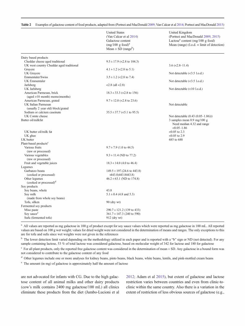
Table 2 Examples of galactose content of food products, adapted from (Portnoi andMacDonald 2009; VanCalcar et al 2014; Portnoi andMacDonald 2015)
United States(Van Calcar et al 2014)Galactose content(mg/100 g food)a
Mean ± SD (rangeb)
United Kingdom(Portnoi and MacDonald 2009, 2015)Lactosee content (mg/100 g food)Mean (range) (l.o.d. = limit of detection)
Dairy based productsCheddar cheese aged traditional 9.5 ± 17.9 (<2.8 to 104.3)UK west country Cheddar aged traditional 3.6 (<2.8–11.4)Gruyere 4.1 ± 1.2 (<2.8 to 5.1)UK Gruyere Not detectable (<3.5 l.o.d.)Emmentaler/Swiss 3.5 ± 1.2 (<2.8 to 7.4)UK Emmentaler Not detectable (<3.5 l.o.d.)Jarlsberg <2.8 (all <2.8)UK Jarlsberg Not detectable (<10 l.o.d.)American Parmesan, brick(aged >10 month) momo)months)
18.3 ± 33.3 (<2.8 to 156)
American Parmesan, grated 9.7 ± 12.0 (<2.8 to 23.6)UK Italian Parmesan(usually 2 year old) block/grated
Not detectable
Sodium or calcium caseinate 35.5 ± 37.7 (<5.1 to 95.5)UK Comte cheese Not detectable (0.43 (0.05–1.86)))
Butter oil/milkfat 3 samples mean 0.9 mg/100 gNeed median 4.32 and range<0.05–1.86
UK butter oil/milk fat <0.05 to 2.3UK ghee <0.05 to 2.9
UK butter 685 to 688Plant-based productsc
Various fruits(raw or processed)
9.7 ± 7.9 (1.0 to 44.5)
Various vegetables(raw or processed)
9.3 ± 11.4 (ND to 77.2)
Fruit and vegetable juices 18.3 ± 14.0 (4.0 to 46.4)LegumesGarbanzo beans(cooked or processed)
149.5 ± 197.(24.6 to 443.8)t443.8)443.8443.8)
Other legumes(cooked or processed)d
46.2 ± 63.1 (ND to 174.8)
Soy productsSoy beans, whole 43.8Soy milk(made from whole soy beans)
5.1 ± 0.4 (4.8 and 5.3)
Tofu, silken 90 (dry wt)Fermented soy productsMiso paste 290.7 ± 121.2 (139 to 433)Soy saucea 361.7 ± 147.3 (240 to 590)Sufu (fermented tofu) 912 (dry wt)
a All values are reported as mg galactose in 100 g of product except for soy sauce values which were reported as mg galactose in 100 mL. All reportedvalues are based on 100 g wet weight; values for dried weight were not considered in the determination of means and ranges. The only exceptions to thisare for tofu and sufu since wet weights were not given in the referencesb The lower detection limit varied depending on the methodology utilized in each paper and is reported with a “b” sign or ND (not detected). For anysample containing lactose, 53 % of total lactose was considered galactose, based on molecular weight of 342 for lactose and 180 for galactosec For all plant products, only the reported free galactose content was considered in the determination of mean ± SD. Any galactose in a bound form wasnot considered to contribute to the galactose content of any foodd Other legumes include one or more analyses for kidney beans, pinto beans, black beans, white beans, lentils, and pink-mottled cream beanse The amount (in mg) of galactose is approximately half the amount of lactose
fruits and vegetables that contain free galactose or foods con-taining trace amounts of lactose).
Recently, Van Calcar et al reviewed the available literatureon the galactose content of fruits, vegetables, legumes, dairyproducts, aged cheeses, and caseinates (Van Calcar et al2014). In this section of the guideline we refer to Table 2summarizing the reported galactose contents of food products,as reviewed by Van Calcar et al 2014 and Portnoi andMacDonald 2009. The free galactose content of most freshor processed fruits, vegetables, and legumes is less than50 mg/100 g serving (Van Calcar et al 2014), and an adult dietenriched in fruits and vegetables was found to contain only54 mg of galactose per day (Berry et al 1993). This galactoseintake is negligible compared to the endogenous galactoseproduction in humans, which is thought to contribute to de-velopment of long-term complications. The endogenous pro-duction is strongly age-dependent, with the highest productionin newborns (>24.8 mg/kg/day) decreasing to a minimum of8.4 mg/kg/day in adults (Berry et al 1995; Berry et al 1997;Ning et al 2000; Schadewaldt et al 2004; Berry et al 2004;Huidekoper et al 2005; Schadewaldt et al 2014). Thus, for a70 kg adult, endogenous galactose production would be morethan 580 mg/day. In addition, endogenous production of ga-lactose does not appear to be affected by exogenous intake ofgalactose (Huidekoper et al 2005). The disparity between di-etary intake and endogenous production has prompted manycountries to recommend a galactose-restricted diet withoutrestrictions of fruit, vegetables, and legumes. There is no ev-idence to suggest that consumption of these minor sources ofgalactose has any adverse effects on long-term clinical status(Bosch et al 2004a; Krabbi et al 2011; Van Calcar et al 2014).Importantly, Portnoi and MacDonald 2009 and Van Calcaret al 2014 demonstrated that the galactose content is low oreven negligible in various aged cheeses including Gruyere,mature Parmesan, and Emmentaler cheese (alternative spel-lings are Emmenthaler, Emmental, Emmenthal) produced inboth Europe and North America, although the galactose con-tent in the same type of cheese can vary due to variation inmaturation and other biological and processing factors.Cheese is an excellent source of calcium, and many clinicsallow and encourage including aged cheese in the diet of pa-tients with galactosemia (Portnoi and MacDonald 2009).However, low-lactose milk aimed at the lactose-intolerantpopulation is contra-indicated in patients with CG. In theseproducts lactose has been hydrolyzed to glucose and galactoseby addition of lactase, but still contains considerable galactosecontent. There is a continuing debate as to whether galactoserestriction could be relaxed further with increasing age, espe-cially since there is concern that an overly strict galactoserestriction might be harmful (Coss et al 2012). In adolescentsand adults with CG, intake of oral galactose of up to 200 mgover 3 weeks, 600 mg over 6 weeks, and 4000 mg galactoseover 14 weeks had no effect on RBC Gal-1-P concentrations
and these subjects did not develop any clinical manifesta-tions over this short time frame (Berry et al 1993; Boschet al 2004a; Coss et al 2012). Coss et al further demonstrat-ed that patients with more severe complications have moreabnormal IgG glycan patterns and, with exposure up to2000 mg galactose/day for 16 weeks, the abnormal glyco-sylation of serum IgG improved in some of the subjects;however, the improvement in glycosylation was highly in-dividual, especially at higher galactose intakes (Coss et al2012). There are also two published case reports of adults,both homozygous for the p.Q188R variation, who havebeen off-diet since 3 years of age. These patients ingestedapproximately 2500 and 9000 mg of galactose per day, yettheir clinical outcome and biochemical parameters werecomparable to those seen in treated adult patients (Leeet al 2003; Panis et al 2006a). However, experience is lim-ited and there is little evidence to support the safety ofdiscontinuation of the galactose-restricted diet (Table 3).
Recommendation #4 (++)
Clinicians should immediately commence a galactose-restricted diet (e.g., soy-based, casein hydrolysate or elemen-tal formula) if classical galactosemia is suspected in an infant,without waiting for confirmation of the diagnosis.
Recommendation #5 (expert opinion, +)
We recommend treating patients with classical galacto-semia with a life-long galactose-restricted diet that only elim-inates sources of lactose and galactose from dairy products,but permits galactose from non-milk sources that contributeminimal dietary galactose. Within this definition we acceptthat small amounts of galactose are present in specific maturecheeses and caseinates. At present there is insufficient evi-dence to support a specific age-related recommendation forthe quantity of galactose allowed in the diet.
Recommendation #6 (+)
We recommend allowing any amount and type of fruits,vegetables, legumes, unfermented soy-based products, maturecheeses (with galactose content <25 mg/100 g), and the foodadditives sodium or calcium caseinate, in the diet for classicalgalactosemia. Although higher in galactose, all fermentedsoy-based products can be allowed in the small quantities thatare typically used in the diet.
The opinion about whether to restrict offal in the diet isdivided; its galactose content is unknown, but there is nodirect evidence of harm. It is a theoretical risk only, there-fore it has been decided to put offal in the ‘in moderation’section in the ‘Current diet restriction for classical galac-tosemia’ table.
Deficiencies
With elimination of dairy products from the diet, patients withCG are at risk for calcium and vitamin D deficiency. Themajority of studies report serum calcium levels in the refer-ence range in children and adolescent patients with CG(Rubio-Gozalbo et al 2002; Panis et al 2004; Gajewska et al2006; Gajewska et al 2008). Only one study with five patientswith CG reports significantly lowered concentrations of calci-um compared to healthy controls, but the age of these patientswas not reported (El-Bassyouni et al 2006). Vitamin D isrequired for optimal calcium utilization, and normal concen-trations of both 1,25-OH vitamin D and 25-OH-vitamin Dhave been measured in children and adolescents with CG
(Rubio-Gozalbo et al 2002; Panis et al 2004; Gajewska et al2006; Gajewska et al 2008). However, in 80 % of adults withCG, 25-OH vitamin D levels were reported to be below thereference range (Waisbren et al 2012). While some studiesreport an adequate daily intake of calcium and vitamin D inchildren and adolescents (Rubio-Gozalbo et al 2002; Paniset al 2004), others report deficient intakes of calcium(Wiesmann et al 1995; Rutherford et al 2002). One studyreported that 75 % of adult patients have an intake of vitaminD below the daily recommended intake (Waisbren et al 2012).
Recommendation #7 (+)
We recommend an annual dietary assessment of calciumand vitamin D intake with measurement of plasma total 25-OH-vitamin D levels. Both calcium and vitamin D should besupplemented as necessary following the age-specific recom-mendations for the general population.
Biochemical follow-up
Biochemical monitoring in CG is aimed at the follow-up ofabnormal parameters of galactose metabolism and evaluationof adherence to the galactose-restricted diet. Currently, moni-toring varies widely between centers, and markers measuredmost frequently include blood galactose, RBCGal-1-P, and/orurinary galactitol levels (Jumbo-Lucioni et al 2012). Levels ofRBC Gal-1-P and urinary galactitol are raised at birth, de-crease rapidly after initiation of a galactose-restricted diet,and then stabilize, but remain elevated compared to healthycontrols (Waggoner et al 1990; Schweitzer et al 1993;Hutchesson et al 1999; Palmieri et al 1999; Ning et al 2000;Yager et al 2003; Schadewaldt et al 2004; Krabbi et al 2011).There are serious doubts regarding the usefulness of thesemarkers in monitoring the disease and adherence to the diet.There is no clear association/correlation between galactosemetabolites and other markers and the development of bothacute and long-term complications in patients with CG(Waggoner et al 1990; Schweitzer et al 1993; Cleary et al1995; Hughes et al 2009). There are no prospective longitu-dinal studies assessing the predictive value of these markersfor the development of long-term complications. Also, severalstudies have demonstrated no clear increase in RBC Gal-1-Pand urinary galactitol levels after short-term oral galactoseloading (up to 4000 mg) was given to patients, thusquestioning the usefulness of these markers in monitoring(short-term) adherence to the diet (Berry et al 1993; Boschet al 2004a; Coss et al 2012). It also has been reported thatblood Gal-1-P and urinary galactitol have a high biologicalvariability with high inter- and intra-individual variation, mak-ing single measurements of little value (Hutchesson et al1999). Gal-1-P is useful in detecting gross dietary deviationsand acute intoxication (Bosch et al 2004a). There is (limited)
Table 3 Current diet restriction for classical galactosemia (adaptedfrom Bernstein et al, Children’s Hospital Colorado in collaboration withthe Galactosemia Foundation Task Force) (Bernstein et al 2014)
Allowed foods and ingredients*
Soy-based infant formulas containing soy protein isolate,amino acid-based elemental infant formulas
All fruits, vegetables and their juices, pickled fruit and vegetables
All legumes (e.g., navy beans, kidney beans, garbanzobeans/chick peas, soybeans)
Soy-based products that are not fermented (soy milk, tofu,textured soy protein, hydrolyzed vegetable protein,soy protein concentrate, meat analogs)
Aged cheeses1: Jarlsberg, Emmentaler, Swiss, Gruyere,Tilsiter, mature Parmesan, mature Cheddar cheese
Sodium and calcium caseinate
All cacao products except milk chocolate
Eggs
Additional ingredients: natural and artificial flavorings,all gums, including carrageenan
Foods used in moderation *
Soy sauce, soy products that are fermented(e.g., miso, natto, tempeh, sufu)
Meat by-products
Offal
Restricted foods and ingredients*
Breast milk, all milk-based infant formulas
Processed meats using lactose
All milk-based foods and beverages, including low lactose milk,except for caseinates and aged cheeses, listed above
All milk-based ingredients including buttermilk solids, casein,dry milk protein, dry milk solids, hydrolyzed whey protein,hydrolyzed casein protein, lactose, lactalbumin, whey
All cheese and cheese-based products except those listed above
Butter
1 Galactose content and consequently allowed types of cheese may varyin different countries
* All manufactured foods need to be checked for the presence of milk byreading food ingredient labels
evidence that monitoring of galactosylation may be an effec-tive parameter in the future (Coss et al 2012; Knerr et al 2015).At this time Gal-1-P measurement, using each patient as hisown reference, appears the best parameter for monitoring pa-tients. There is, however, a strong need for improved moni-toring biomarkers.
Recommendation #8 (++)
In the first year of life clinicians should measure red bloodcell Gal-1-P levels at diagnosis, and after 3 and 9 months ofdietary galactose restriction.
Recommendation #9 (expert opinion, +)
We recommend measuring red blood cell Gal-1-P levelsyearly after the first year of life until an individual baselinehas been established.
Recommendation #10 (expert opinion, +)
We recommend measuring red blood cell Gal-1-P levels incase of increase in galactose intake and concern aboutintoxication.
Recommendation #11 (expert opinion, +)
The clinical utility of serial blood or urinary galactitol mea-surement is limited.
Long-term complications
Cognitive development
Despite initiation of a lactose- and galactose-restricted dietearly in life, patients are at risk for decreased intellectual abil-ity. Therefore, intellectual quotient (IQ) tests are frequentlyperformed in the follow-up of patients with CG. IQ scorescan also serve as a baseline for interpreting deficits in otherareas; for example, poor executive functioning is common inchildren with low IQ. In addition, periodic evaluation of cog-nitive abilities can serve to assess the following: adequacy oftreatment in preventing cognitive decline, the risk for devel-opmental delay in specific domains (such as verbal abilities,reasoning abilities, memory, and processing speed), and thepotential need for early intervention and special educationservices. In CG, most studies report poor cognitive outcomeswith mean IQ scores below average or in the low averagerange, but there is considerable variability between individualpatients and scores range from very low to above average(Donnell et al 1961; Komrower and Lee 1970; Waggoneret al 1990; Schweitzer et al 1993; Kaufman et al 1994;Kaufman et al 1995; Hansen et al 1996; Rasmussen et al
1996; Badawi et al 1996; Shield et al 2000; Widhalm et al2002; Antshel et al 2004; Doyle et al 2010; Schadewaldt et al2010; Coss et al 2013; Rubio-Agusti et al 2013). The reportedpercentage of patients with an IQ score below 85 varies from45 to 72 % (Schweitzer et al 1993; Rasmussen et al 1996;Shield et al 2000; Hoffmann et al 2011). A subject of debatein the follow-up of CG is whether the cognitive impairment isprogressive. Some studies report a negative correlation be-tween age and performance (Komrower and Lee 1970;Waggoner et al 1990; Doyle et al 2010). This finding maybe an artifact of these studies’ cross-sectional design and notmeasuring scores within the same patients over time. Othercross-sectional studies reported no significant difference in IQscores between older and younger patients (Hoffmann et al2011; Coss et al 2013). In longitudinal studies, no deteriora-tion of cognitive function over time was reported duringchildhood/adolescence (Fishler et al 1966; Waggoner et al1990; Manis et al 1997) and adulthood (Waggoner et al1990; Schadewaldt et al 2010). A number of studies reportsignificantly lower IQ scores in females compared to males(Komrower and Lee 1970; Waggoner et al 1990), yet otherstudies could not confirm this finding (Fishler et al 1966;Kaufman et al 1995; Hoffmann et al 2011). Mean IQ scoresand range of IQ for patients with DG (aged 1–6 years), both onan early-initiated lactose-free diet and on a regular diet, werefound to be comparable to the general population (Ficiciogluet al 2008).
Recommendation #12 (++)
Clinicians should refer patients for testing of developmen-tal quotient (DQ) and intellectual quotient (IQ), to obtain awell-validated measure of development and cognitive abili-ties. At minimum, testing should be done at:
Age 2–3 years: to assess early speech/language and motordevelopment in time for early intervention, using a standard-ized test instrument such as the Bayley Scales of Infant andToddler Development (BSID) or a similar measure.
Age 4–5 years: to assess school readiness and need for oc-cupational therapy and speech-language therapy, using a stan-dardized test instrument such as the Wechsler Preschool andPrimary Scale of Intelligence (WPPSI) or a similar measure.
Age 8–10 years: to assess cognitive development, specificareas of strengths and weaknesses, and the need for special ther-apies, using a standardized test instrument such as the WechslerIntelligence Scale for Children (WISC) or a similar measure.
Age 12–14 years: to assess cognitive development andspecific areas of strengths and weaknesses, and to assess theneed for special therapies, using a standardized test instrumentsuch as the Wechsler Intelligence Scale for Children (WISC)or a similar measure.
Age 15 years and older: according to needs, specificquestions.
(consider combining these assessments with speech andlanguage screening, recommendation #15, and psychosocialdevelopment screening, recommendation #21)
Recommendation #13 (expert opinion, +)
For obtaining a measure of functioning when formalizedtesting is not possible or when additional assessments are need-ed between formalized testing points, we recommend using avalidated parent/informant questionnaire, such as the AdaptiveBehavior Assessment System (ABAS) or a similar measure.
There is no correlation between IQ and the time of initia-tion of dietary treatment as long as treatment is started withinthe first 8 weeks of life (Schweitzer et al 1993; Schadewaldtet al 2010). Correlation with genotype is unclear as somestudies suggest that IQ is not correlated to the p.Q188R vari-ation (Lee 1972; Kaufman et al 1994; Cleary et al 1995), whileanother study reports that patients with homozygosity forp.Q188R have lower IQ scores than patients with less com-mon genotypes (Shield et al 2000).
Executive functions Only a few studies have evaluated theexecutive function of patients with CG. Mean scores of exec-utive functioning in adult patients with CG are below theaverage, but there is considerable variability between individ-ual patients (Doyle et al 2010). Overall, 15 % of adult patientsdemonstrate deficits in executive functioning as self-reportedon the Behavior Rating Inventory of Executive Function(BRIEF) (Waisbren et al 2012). As evidenced by direct eval-uation of children and parent responses on the BRIEF, chil-dren with CG exhibit less well-developed executive function-ing compared to peers (Antshel et al 2004). Sustained atten-tion and information processing may also be impaired(Widhalm et al 2002).
Recommendation #14 (expert opinion, +)
We recommend a clinical assessment of executive function,if feasible in the clinic, with specific attention to processingspeed and visual spatial comprehension. In children (8–10 years) as a first screening use the Behavior RatingInventory of Executive Function (BRIEF), and in adolescents(12–14 years) and in young adults (18–20 years) use theCambridge Neuropsychological Test Automated Battery(CANTAB), the Amsterdam Neuropsychological Tasks pro-gram (ANT) or a similar measure, with follow-up, as needed.
Speech and language impairment
Various speech and language disorders have been reported inCG. Language is the tool by which we communicate thoughts,feelings, and ideas either spoken or written, and speech is thetool by which we verbally communicate with others. In some
reports on speech and language disorders in CG the disorder iswell-defined such as childhood apraxia of speech (also calleddevelopmental verbal dyspraxia, a motor speech disorder withproblems saying sounds, syllables, and words), articulation dis-orders, dysarthria, and receptive language disorders. In manyreports more general speech/language delays or disorders arereported, relating primarily to producing rather than under-standing speech and language. Few studies have used standard-ized and validated test instruments, but overall 24–88 % ofchildren and adults with CG are reported to have a speechand language disorder (Waggoner et al 1990; Nelson et al1991; Schweitzer et al 1993; Hansen et al 1996; Rasmussenet al 1996; Webb et al 2003; Potter et al 2008; Hughes et al2009; Hoffmann et al 2011; Shriberg et al 2011; Waisbren et al2012; Coss et al 2013; Rubio-Agusti et al 2013). Speech motorfunction may be affected as well, specifically reduced tonguestrength (73%), decreased breath support for speech (32–64%)(Waisbren et al 2012; Potter et al 2013), and disturbed vocalquality to laryngeal insufficiency (33 % of children with CGand speech disorders) (Potter 2011). Almost 10 % of patientswith CG are affected by vocal tremors of unknown origin(Potter 2011). Children with CG and a history of speech disor-ders also have a four- to sixfold greater relative risk for co-occurring language disorders (Potter et al 2008). Patients withCG show difficulties in language production tasks, both behav-iorally (less accurate and slower) and in their brain’s signaturemeasured by functional magnetic resonance imaging (fMRI)and by event-related potentials (ERPs), compared to healthycontrols. The ERP differences continue throughout consecutivelinguistic preparation phases, which indicates an affected lexi-cal access and impaired syntactic planning (Timmers et al2012), while the fMRI findings point toward both affectedlinguistic preparation and motor speech planning (Timmerset al 2015a). Many individuals with CG (up to 86 %) havereceived speech therapy, with most children receiving directspeech therapy from a speech and language therapist or, lessfrequently, indirect speech therapy with the speech and lan-guage therapist working with the child’s family (Waggoneret al 1990; Schweitzer et al 1993; Potter et al 2008; Timmerset al 2012). There is no evidence in the literature addressingtherapeutic options or therapeutic efficacy. In CG, languagedisorders and type of language disorder are associated with,but not fully explained by, cognitive function (Waggoner et al1990; Nelson et al 1991; Hoffmann et al 2011). More than half(56 %) of patients with CG with average cognition and most(88 %) patients with CG with borderline-low cognition haveco-occurring speech and language disorders (Potter et al 2008).Nelson et al (1991) reported that the presence or severity ofCAS is not related to age at start of diet, the presence of neo-natal symptoms, gender or age at the time of speech evaluation.Another study reported that the number of days consumingmilk prior to diagnosis is associated with poorer speech out-comes in males, not females, with CG (Potter et al 2013).
Patients with the p.Q188R/p.Q188R genotype are reported tobe at greater risk for speech and language impairment thanparticipants with p.Q188R/other genotypes (Potter et al 2008,Robertson et al 2000). Two-thirds of children with CG andspeech disorders have co-occurring coordination disordersand children with CG and CAS or dysarthria have poorer bal-ance and manual dexterity (Potter et al 2013).
Recommendation #15 (++)
All children with CG should be screened for speech andlanguage delay at ages 7–12 months, 2, 3, and 5 years (con-sider combining with screening for cognitive disorders, rec-ommendation #12). If children show low or borderline speechand language development, full assessments should beconducted.
Recommendation #16 (expert opinion, +)
We recommend that an assessment of speech and languageincludes hearing screening, a brief assessment of pre-linguisticcommunication (<2 years of age) and expressive, receptive,and pragmatic language use, structure-function examination,motor speech (observation of respiration, resonance, voice,articulation), and speech intelligibility for all children notmeeting age appropriate milestones. We recommend a cogni-tive evaluation as well if a disorder is suspected.
Recommendation #17 (expert opinion, +)
For children who are not meeting age appropriate speech orlanguage milestones, we recommend treatment based onguidelines for treatment of speech, language, and voice disor-ders in the general population. Therapy should begin duringthe first year of life and include modeling and training ofgestural communication to increase infant and toddler lan-guage development. Play-based milieu for language develop-ment is recommended during the second year of life.Individual speech therapy focused on high repetition of asmall number of targets should begin during the second yearof life and continue as needed throughout the preschool andelementary school years. Respiration, phonation, and reso-nance deficits should also be addressed.
Neurological complications
Patients with CG are at risk for central nervous system dys-function, not onlymanifesting itself as neurological symptomsand motor problems, but also as cognitive impairment, speechand language problems, and psychiatric symptoms. The latterthree are discussed in a separate part of this guideline.
Affected newborns may develop encephalopathy and signsof increased intracranial pressure with cerebral edema after
ingestion of galactose (Huttenlocher et al 1970; Belman et al1986; Najafi et al 2005). In the long-term it is known thatpatients may develop neurological complications, with an over-all frequency of motor dysfunction of up to 66 % (Hughes et al2009; Milánkovics et al 2010; Rubio-Agusti et al 2013;Viggiano et al 2015). The studies addressing specific neurolog-ic symptoms all report on different symptoms and have veryheterogeneous populations with large variation in sample sizes,making it difficult to reliably estimate the percentage of patientsthat suffer from each of these symptoms. Frequently notedsigns are mild to severe ataxia, tremor, dystonia, dysarthria,and dysmetria (Waggoner et al 1990; Nelson et al 1992;Schweitzer et al 1993; Kaufman et al 1994; Kaufman et al1995; Dubroff et al 2008; Krabbi et al 2011; Waisbren et al2012; Coss et al 2013; Rubio-Agusti et al 2013). Epilepsy isreported in a minority of patients (Schweitzer et al 1993;Krabbi et al 2011; Rubio-Agusti et al 2013; Aydin-Özemiret al 2014). One study, that followed 22 patients with CG di-agnosed early, did not find any cases with ataxia or dysmetria inyoung patients aged 0–6 years (Karadag et al 2013). One studyreported eye movement abnormalities and pyramidal signs insome patients (including brisk tendon reflexes and clonus, ex-tensor plantar response, spastic paraparesis, and pseudobulbarsigns) (Rubio-Agusti et al 2013).
Recommendation #18 (++)
Clinicians should screen patients with CG for neurologicalinvolvement by clinical examination from the age of 2–3 years. Such screening should include examination for ataxia,tremor, dysmetria, and dystonia. If a specific neurological def-icit is noted, monitoring of progression with a designated scaleis advised. It is suggested to screen adult patients annually andto record progression, if any. Pediatric patients could bescreened more frequently (every 6 months) in order to identifypotentially modifiable neurological problems.
Recommendation #19 (+)
We recommend asking patients or caregivers about onset ofseizure and seizure-like activity since previous examinationand perform an EEG, if indicated.
Cerebral imagingAn abnormal white matter signal is presentin the majority of MRI scans (>75 %) of patients with CG,indicative of abnormal myelination (Nelson et al 1992;Kaufman et al 1995; Moller et al 1995; Wang et al 2001;Hughes et al 2009; Krabbi et al 2011). Additional reportedMRI findings are focal white matter lesions, white matter vol-ume loss, (mild) cerebral atrophy, enlargement of the fourthventricle and cerebellar sulci (suggesting cerebellar atrophy),and enlargement of lateral ventricles (Nelson et al 1992;Kaufman et al 1995; Moller et al 1995; Wang et al 2001;
Hughes et al 2009; Krabbi et al 2011; Rubio-Agusti et al 2013;Timmers et al 2015b). One study followed patients over timewith MRI scans (Nelson et al 1992). All eight patients youn-ger than 1 year of age had normal white matter signaling. Allpatients over the age of 1 year either had an abnormal periph-eral myelin pattern, or developed abnormalities within 1–2 years without apparent progression in time. From this study22/63 patients had mild lateral ventricular enlargement at theinitial MRI. This enlargement was unchanged in four patientsthat had follow-up MRIs 1–2 years later. Of the 20/63 patientswithout lateral ventricular enlargement that had follow-up ex-aminations, none developed lateral ventricular enlargement.In some studies only patients with neurological symptomshad MRI examination, and in other studies clinical statuswas not reported. It is unclear if the abnormalities on MRI-scans are representative for the whole population of patientswith CG and if the findings are correlated with clinical symp-toms. One study also performed magnetic resonance spectros-copy (MRS) in addition to brain MRI to assess in vivo brainmetabolism (Moller et al 1995). MRS showed a normal spec-trum of metabolites, with no indications of elevated Gal-1-Plevels or an impairment of energy supply in the brain. Norelationship between MRS and clinical data was found. Astudy on white matter microstructure pathology using neuriteorientation dispersion and density imaging (NODDI) showedextensive white matter abnormalities with a lower neurite den-sity index and increased orientation dispersion index in theregions involved with higher order cognitive functions andlanguage and motor functions (Timmers et al 2015b).
Recommendation #20 (expert opinion, +)
We do not recommend routine brain and spinal cord imag-ing in the follow-up of patients with CG. In those patients withsignificant or progressive neurological symptoms and signs,imaging may be warranted to (1) determine if a second con-dition is present or (2) further define the development andprogression of neuroradiology findings in individual patients.
Psychosocial development
Due to the chronicity of the disease, the need to adhere to alife-long diet and the impact of the long-term complications,patients with CG are at risk for problems in psychosocialdevelopment, including personality, social relationships, andemotional well-being.
Psychosexual and social development, marital statusSpecific testing of social and psychosocial milestones in adultpatients showed that patients achieved fewer developmentalmilestones in the psychosexual and social domain (havingfriends and engaging in social activities) (Bosch et al 2009;Gubbels et al 2011a). Multiple studies assessed marital status
in adult patients with CG, with the percentage of patientsmarried or living in stable partnership varying between 13and 57 % (Bhat et al 2005; Bosch et al 2009; Waisbren et al2012; Hoffmann et al 2012). Bosch et al 2009 reported that thepercentage of patients married (14.3 %) was significantly low-er compared to the reference population (39.1 %). Hoffmannet al 2012 found a difference in marital status between sexeswith more married females (57 %) compared to males (11 %),with the percentage of married males much lower than in thereference population (47.2 %). In one study assessing malesonly, 5 % of patients were married, compared to 30.9 % in thereference group of males that did not differ in age, though thisdifference was not statistically significant (Gubbels et al2011a). Few patients with CG had children (0–5 %), while adesire to have children was reported by nearly half of patients(Bhat et al 2005; Hoffmann et al 2012). The percentage ofpatients actually trying to conceive a child or the percentagesuccessful was not reported. While primary ovarian insuffi-ciency (present in the majority of female patients) might be animportant contributing factor to the minority of females hav-ing children, there is no evidence of decreased fertility inmales (see the Endocrinology and fertility part of this guide-line) (Rubio-Gozalbo et al 2006; Gubbels et al 2011a). Thepercentage of patients trying to conceive might also be low,due to the fact that fertility counselling in the recent past wasoften focused on the expected low chances of achievingpregnancy.
Educational attainment and employment Bosch et al re-ported that 44 % of children with CG, aged 6–11 years,attended special schools as opposed to 3 % of the generalpopulation (Bosch et al 2004b). Educational attainment wassignificantly lower than the general population with 61.5 %completing basic school and low vocational training only,compared with 27.2 % of the general population (Boschet al 2004b). Lower education attainment was confirmed inanother study which reported that fewer individuals with CG(10.8 %) had a school leaving certificate, compared to 3.5 %in the general population, and a minority achieved a universityentrance diploma (8.1 %) compared to the general population(28.6 %). However, a higher percentage of individuals withCG (81 %) earned a secondary school degree compared to thegeneral population (59.5 %) (Hoffmann et al 2012). In adult-hood, up to 30 % of individuals with CG were unemployed,with no differences between males and females (Bosch et al2009; Waisbren et al 2012; Hoffmann et al 2012). A recentsurvey of 60 adult patients with galactosemia in the UK re-vealed that 58 % were in paid employment, compared with74 % of the general population (unpublished data, CharlesDent Metabolic Unit, UK, 2015).
Psychiatric symptoms and emotional problems Patientswith CG have been reported to suffer more frequently from
psychiatric symptoms and emotional disturbance, includingdepression, anxiety, obsessive-compulsive disorder, and au-tism spectrum disorder (Lee 1972; Rubio-Agusti et al 2013).Parents of children with CG report their children exhibit moreinternalizing symptoms (e.g., depression and anxiety) withoutelevated levels of dysphoric mood compared to controls(Antshel et al 2004). Depression (as detected with the BeckDepression Inventory), was present in 12 % of adult patients,and anxiety (as measured with the Beck Anxiety Inventory)was present in 52 % (Waisbren et al 2012).
Coping with CG Coping with CG by patients was assessed intwo studies, both using condition specific but non-validatedquestionnaires. It was demonstrated that over 75 % of the pa-tients rated their coping with CG as ‘very good’ or ‘good’. Theremainder had difficulties in coping with their condition. CGwas seen as a burden by 39 % of patients (Bosch et al 2004b;Hoffmann et al 2012). Many patients (42 %) had a problemmaintaining the diet, and patients reported that ‘diet/nutrition’was the primary aspect of life influenced by galactosemia,followed by ‘school/work’ and ‘friends/leisure’. Many parentsof patients with CG (60 %) considered it a burden to take careof a child with CG, and many believed that CG influenced theirrelationship with their child. More than half of parents of girlsfrequently worried about possible infertility. However, a highpercentage of parents (86 %) believed that one could live agood life with this disorder (Bosch et al 2004b) and only7.7 % of adults with CG reported that galactosemia had a neg-ative effect on family life (Hoffmann et al 2012).
Screening for psychosocial deficits Currently no universaland validated checklist is available to screen for psychosocialdeficits.
Recommendation #21 (expert opinion, +)
We recommend screening children for psychosocial defi-cits, including autism spectrum disorders, sensory integrationproblems, depression and anxiety, using standardized ques-tionnaires such as the Behavior Assessment System forChildren, Second Edition (BASC-2) in English or a similartool in other languages. We recommend performing thisscreening at age 2 years in combination with screening forspeech and language delays (see recommendation #15) andto combine this screening with developmental testing at ages4–5, 8–10, and 12–14 years (see recommendation #12).
Recommendation #22 (+)
We recommend screening adults for mental health issueswith validated questionnaires that include brief scales for anx-iety and depression, such as the NIH PROMIS Questionnaires,Beck Anxiety Inventory (BAI), Beck Depression Inventory
(BDI) or similar measures. With adults, we recommenddiscussing living situations, work and/or educational situations,satisfaction with social relationships, and sexual intimacy dur-ing outpatient clinic visits and to refer for professional consul-tation, if necessary.
Health-related quality of life (HRQoL) Three studies reporton HRQoL in patients with CG. One study demonstrated thathaving CG negatively affects HRQoL of both children andadults (Bosch et al 2004b). Children aged 6–15 years had alower HRQoL in the domains of cognitive function and ofmotor function. Patients >16 years of age reported significant-ly lower scores in the domains of cognitive function and socialfunction. Hoffmann et al showed that adult patients with CGscored significantly lower on the domains ‘positive mood’ and‘social well-being’ when compared to the general populationand compared to PKU patients (Hoffmann et al 2012). Finally,a study evaluating HRQoL in patients >6 years of age reportedthat patients with CG did not differ from their peers in theirphysical activities, mobilization, overall health, and their self-esteem, but that they did have difficulties in their relationshipswith others. Despite feeling ‘not as good as most people’, allpatients had been happy at some point in the 4 weeks preced-ing the interview (Lambert and Boneh 2004). The HRQoL ofparents of children with CG did not differ from the HRQoL ofparents of healthy children (ten Hoedt et al 2011).
Recommendation #23 (expert opinion, +)
We do not recommend routine health-related quality of life(HRQoL) evaluations.
Fertility Over 80 % of females with CG develop primaryovarian insufficiency (POI) (Kaufman et al 1981; Kaufmanet al 1988; Waggoner et al 1990; Sanders et al 2009; Rubio-Gozalbo et al 2010; Fridovich-Keil et al 2011; Coss et al 2013;Rubio-Agusti et al 2013). The clinical spectrum of POI inwomen with CG varies from primary amenorrhea with orwithout lack of development of secondary sexual charac-teristics, to normal pubertal development followed by ir-regular menses, oligomenorrhea or secondary amenorrhea.Most patients experience subfertility, and often show a di-minished ovarian reserve. Many females need puberty in-duction and/or hormone replacement therapy to preventsequelae of POI (Gubbels et al 2008). The mechanismsunderlying POI and the timing of the ovarian damage inCG are not understood to date. Possible underlying patho-physiological mechanisms of POI in CG include directtoxicity of accumulated galactose or one of its metabolites,abnormal glycosylation of glycoproteins or glycolipids (in-cluding hormones such as follicle stimulating hormone(FSH)), and wrongful activation of follicular apoptosis(Liu et al 2000; Forges et al 2006; Rubio-Gozalbo et al
2010; Fridovich-Keil et al 2011). One study reported thatfemale patients with CG have additional FSH isoformsbesides normal acidic FSH isoform, (Prestoz et al 1997),but another study did not confirm these findings (Gubbelset al 2011b). FSH-inactivity seems not to be a probablecause of POI, as most female patients with CG do notrespond significantly to stimulation with exogenous FSHand/or luteinizing hormone (LH) (O’Herlihy and Danks1985; Gubbels et al 2013a).
Biomarkers for POI Corresponding to the incidence of POI,most (>80 %) female patients with CG demonstrate raisedlevels of FSH (Waggoner et al 1990; Guerrero et al 2000),while estradiol levels are decreased in adolescent girls andwomen (Rubio-Gozalbo et al 2006). Levels of anti-Müllerian hormone (AMH), a marker for ovarian reservewhich is produced by granulosa cells of (healthy) pre-antraland small antral follicles of the ovary (Broer et al 2014), arereported to be abnormally low among girls and women withCG across all age groups, even in patients <1 years of age(Sanders et al 2009; Gubbels et al 2013a; Spencer et al2013). However there is no significant difference inAMH levels between girls with CG with spontaneousmenarche compared to hormone replacement therapyassisted menarche (Spencer et al 2013). Female patientswith CG demonstrated normal gonadotropin levels thatincrease as POI becomes apparent, normal levels of pro-lactin, total cholesterol, triglycerides, high-density lipo-protein (HDL) cholesterol, low-density lipoprotein(LDL) cholesterol, thyroid-stimulating hormone (TSH),thyroid-binding globulin (TBG), free thyroxine (FT4),basal testosterone, free testosterone, andostenedione,and dehydroepiandrosterone sulfate(DHEAS) (Kaufmanet al 1981; O’Herlihy and Danks 1985; Rubio-Gozalboet al 2006). Also, no ovarian antibodies were found(Kaufman et al 1981).
Imaging Ovaries of girls and women with CG measured onMRI were significantly smaller when compared to age-matched controls, but did not differ significantly from post-menopausal controls (Gubbels et al 2013a). In more than halfof patients with CG ovaries could not be visualized on ultra-sound, compared to 10 % in healthy controls. On ultrasoundmost patients had antral follicle counts below the control range(Spencer et al 2013).
Recommendation #24 (++)
Girls with CG should be screened for hypergonadotropichypogonadism if they reach the age of 12 years with insuffi-cient secondary sex characteristics or if they reach the age of14 years with no regular menses. Screening should includefollicle-stimulating hormone and 17-beta-estradiol.
Recommendation #25 (expert opinion, +)
We recommend considering follicle stimulating hormonelevel, growth, and psychosocial maturity of the individual girl,for determination of age at start of treatment. For pubertyinducement, a low dose estrogen in a step-wise escalatingdose is used, then later combined with cyclic progesteronefor regular withdrawal bleeds. We recommend consideringreferral to a pediatric endocrinologist.
Recommendation #26 (expert opinion, +)
We recommend not using anti-Müllerian hormone andovarian imaging routinely for follow-up as these have notbeen shown to accurately predict pubertal development orfertility outcome.
Recommendation #27 (+)
We do not recommend endocrine follow-up for DuarteGalactosemia, as there is no evidence that the ovaries areaffected.
Recommendation #28 (expert opinion, +)
We recommend that girls and women with CG, who havegone through puberty and established regular menstrual pe-riods, should be monitored annually for menstrual abnor-malities, secondary amenorrhea, and symptoms of prima-ry ovarian insufficiency (POI). Changes in menses or POIsymptoms should be evaluated with a serum follicle-stimulating hormone level. Anti-Müllerian hormone mea-surement is not helpful in determining which women willundergo POI, but may be helpful in identifying women atrisk for imminent POI when it is undetectable. Imaging bypelvic ultrasound or MRI is not recommended unless oth-erwise clinically indicated.
Recommendation #29 (expert opinion, +)
We recommend that women with hypergonadotropichypogonadism, or primary ovarian insufficiency should be pro-vided counseling and support about their reproductive optionsand management of irregular or absent menses. Hormone re-placement therapy should be initiated with the onset of second-ary amenorrhea to reduce the risk of osteoporosis and othercomplications of primary ovarian insufficiency.
Recommendation #30 (++)
We recommend considering a referral to a reproductiveendocrinologist for women who desire pregnancy and havebeen unable to conceive naturally, or for women who desire
additional counseling about fertility treatment options includ-ing oocyte donation.
Recommendation #31 (expert opinion, +)
We recommend providing counseling about adequate birthcontrol methods for women who do not desire pregnancy.While combined oral or transdermal contraceptives may pro-vide cycle control, bone protection, and attenuate hot flashes,they may fail to provide adequate birth control in women withvery elevated follicle-stimulating hormone levels. An intra-uterine device may provide the lowest failure rate.
Risk factors for POI The risk of POI in CG is positivelyassociated with higher mean Gal-1-P levels after 1 year ofdietary treatment, reduced galactose oxidation capacity, andhomozygosity for the p.Q188R variation (16-fold increasedrisk) compared to heterozygosity for this variation or two dif-ferent variations (Guerrero et al 2000). Homozygosity for thep.Q188R variation, however, is not predictive for the devel-opment of primary amenorrhea versus secondary amenorrhea(Kaufman et al 1994). Because of the high frequency of themild p.S135L variation in African American patients, thereis an association between ethnicity and the outcome of POI,with a high proportion of the Caucasian females but noAfrican American females diagnosed with POI (Guerreroet al 2000). Residual GALT activity might be a modifier ofovarian function as well, as female patients with >0.4 %predicted wild-type GALT activity are more likely to showAMH levels of >0.1 ng/ml when compared to girls with<0.4 % GALT activity (Spencer et al 2013). There is noassociation between POI and the age at initiation of dietarytreatment, degree of dietary control, highest erythrocyteGal-1-P level (Guerrero et al 2000), and urinary galactitollevels (Kaufman et al 1981).
Pregnancy, counselling and fertility preservation Chancesof pregnancy are reduced in POI, but pregnancies inwomen with CG have been reported, and appear not tobe as rare as is generally assumed (reviewed by Gubbelset al 2008). As recommendations on fertility preservationwere lacking, Van Erven et al issued recommendations forphysicians based on current knowledge concerning galac-tosemia and fertility preservation (Van Erven et al 2013).Oocyte donation may be used to establish pregnancy inwomen with CG and POI, but has some psychologicaldisadvantages (Sauer et al 1991). Three fertility preserva-tion techniques are currently offered to patients in need offertility preservation: ovarian tissue, mature oocyte and/orembryo cryopreservation. The application of fertility pres-ervation in these patients is complicated however, becausethe underlying mechanisms and onset of POI in CG arenot fully understood. Also the experience with fertility
preservation is mainly derived from cancer patients withpreviously unaffected ovaries, and some procedures likecryopreservation of ovarian tissue are still experimental(Van Erven et al 2013).
Recommendation #32 (expert opinion, +)
Fertility preservation may not be successful. Currently,fertility preservation techniques are not yet readily used ineveryday practice. We recommend fertility preservationshould only be offered with appropriate institutional re-search ethics review board approval to girls with classicalgalactosemia at a young pre-pubertal age.
Males In contrast to female patients, fertility does notseem to be affected in males, but there is a paucity of dataabout reproductive function and male patients fatheringchildren, possibly due to psychosocial rather than biolog-ical reasons. One study reports a confirmed delayed onsetof puberty in one of 18 males over 12 years of age(Schweitzer et al 1993). A higher rate of cryptorchidism(Rubio-Gozalbo et al 2006; Gubbels et al 2011b), lowersemen volumes as a group, and sperm concentrations inindividuals are reported (Gubbels et al 2011b). Malesdemonstrated normal levels of gonadotropins, total cho-lesterol, triglycerides, HDL cholesterol, LDL cholesterol,prolactin, TSH, TBG, FT4, basal testosterone, free testos-terone, androstenedione, and DHEAS (Rubio-Gozalboet al 2006; Gubbels et al 2013b).
Recommendation #33 (+)
We do not recommend routine endocrinology follow-up inmales.
Bone health
Patients with CGmight be at risk for impaired bone health dueto various reasons, such as restrictions in dietary intake, de-creased physical activity in some patients, POI in females, andcurrently unknown pathophysiologic factors intrinsic to thedisease. The preferred and most frequently used method tomeasure BMD in children is dual-energy X-ray absorptiome-try (DXA) (Crabtree et al 2014). Other types of bone densi-tometry measurements, such as quantitative computed tomog-raphy (CT) and quantitative ultrasound, are also available.BMD can be measured at different sites and is frequentlyreported as a T- or Z-score, which are both units of standarddeviation. In children the preferred sites to measure BMD arethe lumbar spine and the total body less head (TBLH); inadults the lumbar spine, total hip and/or femoral neck (ISCD2015). Two-dimensional cross-sectional area measurements(areal BMD (aBMD)) or estimated volumetric BMD
(vBMD) measurements can be performed. The diagnosis ofosteoporosis in children, premenopausal women, and malesunder 50 years of age should be based on densitometriccriteria combined with fracture history. The definition of os-teoporosis for these groups is a BMD Z-score<−2 (‘BMDbelow the expected range for age’ or ‘low BMD for chrono-logical age’) combined with a significant fracture history(Crabtree et al 2014).
Bone health in CG Three studies in adult patients reportvBMD Z-scores of −1.9 and −1.4 in males. (Kaufman et al1993); aBMD Z-scores −1.19 in females (lumbar spine) and−1.25 (total hip) in females and −0.80 (lumbar spine) and−0.81 (total hip) in males (Batey et al 2013); and BMD Z-scores greater than 2 standard deviations below the normativemean in 8 of 33 patients (Waisbren et al 2012).
Four studies reported on BMD Z-scores in children andadolescents, three using DXA scanning BMD (Rubio-Gozalbo et al 2002; Panis et al 2004; Doulgeraki et al 2014)and one using quantitative CTscanning (Kaufman et al 1993).Two studies demonstrated decreased lumbar spine aBMD Z-scores (−0.65 and −0.6) (Panis et al 2004; Doulgeraki et al2014), and two other studies found decreased total bodyaBMD Z-scores (−0.3 and −0.99) (Rubio-Gozalbo et al2002; Doulgeraki et al 2014). Panis et al 2004 and Rubio-Gozalbo et al 2002 found decreased femoral neckvBMD Z-scores as well (−0.28 and −1.76). It should benoted that recent insight is that Z-scores should not beused for volumetric measurements. Kaufman et al founda significantly decreased mean BMD of the lumbar spine(assessed with quantitative CT) when compared to age-matched healthy controls, in both children and adult pa-tients (Kaufman et al 1993). Karadag et al demonstratednormal BMD in all patients aged 0–6 years, tested withDXA and quantitative ultrasound, but decreased BMDwas not defined (Karadag et al 2013). Coss and col-leagues demonstrated osteopenia or osteoporosis (in chil-dren >10 years old and adults) in 14.7 % of the Travellerpopulation and in 39.6 % of the non-Traveller population,using Z-scores generated from DXA scans, but it is un-clear how the authors defined osteopenia and osteoporosis(Coss et al 2013). A randomized controlled trial of 2 yearsin children with CG evaluated the effect of calcium andvitamin K1 and D3 supplementation versus placebo onbone mineral content (BMC), and showed a significantincrease in BMC of lumbar spine in the treatment groupcompared to placebo group, but only in prepubertal chil-dren (Panis et al 2006b). Self-report of fractures in adultpatients with CG showed that 63 % of women and 31 %of men sustained at least one lifetime fracture (Batey et al2013), which seems to be comparable to the general pop-ulation (Jones et al 2002). However, further studies willneed to validate these results.
Recommendation #34 (++)
Clinicians should assess bone mineral density (BMD) byage appropriate dual-energy X-ray absorptiometry (DXA)scan.
Recommendation #35 (expert opinion, +)(consensus: 93 %)
We recommend BMD screening from age 8–10 years.Withevidence of reduced bone density (Z-score≤−2.0), follow-upaccording to current pediatric bone health guidelines is ad-vised. Without evidence of reduced bone density, we recom-mend performing a repeat dual-energy X-ray absorptiometryscan when puberty is complete. We recommend performingfollow-up thereafter every 5 years and treatment institutedaccording to WHO FRAX recommendations.
Recommendation #36 (+)
We recommend comprehensive dietary evaluation, optimi-zation of calcium intake if needed, monitoring and if neces-sary supplementation of vitamin D, hormonal status evalua-tion and hormone replacement therapy consideration, as wellas regular exercise and assessment of skeletal problems andclinically significant fractures in all patients with CG.Supplementation of vitamin K might be beneficial when com-bined with an adequate intake of calcium and vitamin D, butcurrently there is not enough evidence to recommend the rou-tine use of vitamin K.
Bone metabolism Measurement of bone metabolism pa-rameters, such as minerals, vitamins, hormones, bone for-mation markers, and bone resorption markers, might behelpful in understanding underlying mechanisms of a de-creased BMD and evaluation of treatment (e.g., supple-mentation of calcium and vitamin D). Studies measuringthese parameters in patients with CG have only been per-formed in children and adolescents. Levels of carboxylat-ed osteocalcin, N-terminal telopeptide, C-terminaltelopeptide, and IGF-1 Z-scores were found to be signif-icantly decreased in patients with CG, while values ofbone alkaline phosphatase, osteocalcin, and C-terminaltelopeptide were found to be higher than in controls(Rubio-Gozalbo et al 2002; Panis et al 2004; Gajewskaet al 2006; Gajewska et al 2008). All other tested param-eters in these four studies were in the reference range orcomparable to healthy controls.
Recommendation #37 (expert opinion, +)
At present there is not enough evidence to justify routinedetermination of bone turnover markers in patients with CG.
Cataracts
Cataracts are a frequently encountered complication of CG,mainly in the newborn period. Cataracts always occur bilateral-ly, and the enhancement of nuclear or perinuclear refractivepower leads to the appearance of a refractile ring or a drop ofoil in the crystalline center, which is an early sign. Later stagesappear as nuclear or zonular opacities, and in all stages vacuolesmight be present. The cause of cataract is accumulation ofgalactitol in the crystalline lens, the result of activation of thealdose reductase shunt (Widger et al 2010). Cataracts are typi-cally already present in the first weeks of life, with a prevalencewhich varies from 6 to 25 % across several studies (Kossowiczand Zbieg Sendecka 1977; Burke et al 1988; Burke et al 1989;Badawi et al 1996). In a retrospective case series, 14/100 pa-tients with CG were diagnosed with cataracts, with the averageage at cataract diagnosis of 6.3 years (Widger et al 2010).However, this article did not report on age of diagnosis, previousophthalmologic examinations or dietary adherence. A study onlong-term complications in adulthood reported the presence ofcataracts in 21 % of adult patients with CG, as noted in medicalrecords or reported during the medical history (Waisbren et al2012). Overall prevalence of cataracts, regardless of age at startof diet, is extremely variable across different studies, with thelarger retrospective case series reporting 7.7 % (Coss et al 2013)and 14 % (Widger et al 2010). A smaller study reported cataractin 17/22 patients, including 13/18 patients who were diagnosedbefore 17 days, and in 4/4 patients who were diagnosed after17 days. One prospective case series with a mean follow-upperiod of 8.5 years reported an overall prevalence of cataractsof 36 % in 33 patients (Beigi et al 1993), but the time of detec-tion of the cataracts was not reported, except for two patients inthis cohort who developed reversible cataract at the age of 2.5and 3.7 years respectively, both after a 3-month period off diet(Burke et al 1988; Burke et al 1989; Beigi et al 1993).
Severity of the cataracts reported in patients with CG varies,but in the vast majority of cases visual acuity is not affected,and lens opacities frequently resolve spontaneously over timein patients on a galactose-restricted diet (Kossowicz and ZbiegSendecka 1977; Burke et al 1988; Burke et al 1989; Waggoneret al 1990; Beigi et al 1993; Schweitzer et al 1993; Badawi et al1996). Reports of cataract necessitating surgery are rare: onestudy reports on a 33 year old male suffering from blindnessdue to cataract, but age of diagnosis and dietary adherence werenot reported (Schweitzer et al 1993). Karadag et al reportedfour patients diagnosed after age 17 days required cataract sur-gery (Karadag et al 2013). Waggoner et al reported eight cases(out of 314 patients) requiring surgery, including one patientthat had been treated from birth but with unknown adherence(Waggoner et al 1990).
Widger et al could not demonstrate a direct relationshipbetween dietary adherence and cataract formation, howeverin this study dietary nonadherence was defined by the
relatively low galactose intake of >50 mg/day (Widger et al2010). Levels of Gal-1-P have not been demonstrated to cor-relate with cataract formation (Beigi et al 1993).
The current available literature is inconclusive regardingwhich patients will develop cataracts and at what age, and ifadherence to the diet plays a direct role in the development ofcataracts. There are however strong suggestions that cataractsin the neonatal period usually do not affect visual acuity and,with dietary adherence, will often resolve spontaneously.Furthermore it seems that, patients with a good dietary adher-ence do not develop cataracts later in life or if they do developcataracts, visual acuity is not affected.
Recommendation #38 (++)
Clinicians should refer all patients to an ophthalmologistfor evaluation of cataract at the time of diagnosis.
Recommendation #39 (+)
We recommend performing ophthalmological follow-up inpatients with a cataract at diagnosis until it has fully resolved.
Recommendation #40 (+)
We recommend performing ophthalmological screening inall patients who are non-compliant with diet.
Closing remarks
The presented guideline is the first international and evidence-based guideline for the diagnosis, treatment, and follow-up ofCG, and aimed to be applicable worldwide. This guidelineshould serve as a guide for clinicians and other experts caringfor patients with CG. Though great effort was undertaken toformulate evidence-based recommendations, this was fre-quently hampered by limited evidence resulting in numerousrecommendations based on expert opinion (18/40 recommen-dations, 45 %). The literature concerning CG available to datemostly consists of studies with an observational study-design.In the current era of evidence-basedmedicine these studies arelabeled as having a low to very low level of evidence.Therefore strength of recommendation is ‘discretionary’ fora majority of recommendations in the guidelines, (32/40 rec-ommendations, 80 %) including the recommendations labeledexpert opinion. However, as other study designs (such asRCTs or cohort studies) are usually not feasible or may notprovide the best design to study characteristics of rare dis-eases, the strength of the recommendation was upgraded to‘strong’ when results were consistent across multiple studies,and experts had confidence in the validity of these results(9/40 recommendations, 23 %).
Future perspectives
Following this conclusion, it is not unexpected that gaps ofknowledge were identified in most discussed fields of interest,foremost in the fields of treatment and follow-up. Topics ofmajor importance for future research include: further assess-ment of which patients should be treated (cut-off enzyme ac-tivity), exploration for possible further relaxation of the diet forpatients after childhood, exploration of new biomarkers forbiochemical follow-up as well as reproductive function, assess-ment of executive functions in children and adults, and furtherexploration of bone turnover markers in relation to BMD.
Guideline update
Revision of this guideline is important as it only representsevidence in predefined areas up to October 2015. Since re-search in the field of CG is flourishing, it is expected that newinformation will be gained in the next decade. This guidelineis scheduled to be updated in the next 10 years by representa-tives of the GalNet.
Acknowledgments The authors would like to thank Arnold G.E.Leenders for the time and effort invested in developing the search strat-egies for this guideline. We would also like to thank the representatives ofthe European Galactosemia Society, Mr. Jeroen van Kempen (chairman)and Mrs. Maaike van Kempen (commission member), for their contribu-tions to this guideline.
Compliance with ethical standards
Conflict of interest Annet M. Bosch is in receipt of research grantsfrom Nutricia and was a member of an advisory board for Nutricia.
Laurie E. Bernstein declares that she has no conflict of interest.Gerard T. Berry declares that he has no conflict of interest.Alberto B. Burlina declares that he has no conflict of interest.Francois Eyskens declares that he has no conflict of interest.Matthias Gautschi declares that he has no conflict of interest.Stephanie Grünewald declares that she has no conflict of interest.Cynthia S. Gubbels declares that she has no conflict of interest.Ina Knerr declares that she has no conflict of interest.Philippe Labrune declares that he has no conflict of interest.Johanna H. van der Lee declares that she has no conflict of interest.Anita MacDonald is in receipt of research grants from Nutricia and
Vitaflo International, and is a member of the Nutricia IMD AdvisoryBoard and Arla Advisory Board.
Elaine Murphy is in receipt of clinical trial funding from Vitaflo andreceived travel support to attend meetings from Vitaflo.
Katrin Õunap declares that she has no conflict of interest.Pat A. Portnoi received grants from Nutricia and Vitaflo to attend
conferences, meetings or to give lectures in the past 5 years.Nancy L. Potter declares that she has no conflict of interest.M. Estela Rubio-Gozalbo declares that she has no conflict of interest.Jessica B. Spencer declares that she has no conflict of interest.Inge Timmers declares that she has no conflict of interest.Eileen P. Treacy declares that she has no conflict of interest.Sandra C. Van Calcar declares that she has no conflict of interest.Susan E. Waisbren declares that she has no conflict of interest.Lindsey Welling declares that she has no conflict of interest.
Details of funding The initial GalNet meeting was financially support-ed by The Netherlands Organisation for Scientific Research (NWO;http://www.nwo.nl). The final consensus meeting was financiallysupported by the United States Galactosemia Foundation Inc. (patientorganization; http://www.galactosemia.org/). The authors confirmindependence from the sponsors; the content of the article has not beeninfluenced by the sponsors.
Open Access This article is distributed under the terms of the CreativeCommons At t r ibut ion 4 .0 In te rna t ional License (h t tp : / /creativecommons.org/licenses/by/4.0/), which permits unrestricted use,distribution, and reproduction in any medium, provided you giveappropriate credit to the original author(s) and the source, provide a linkto the Creative Commons license, and indicate if changes were made.
References
Adam S, Akroyd R, Bernabei S et al (2015) How strict is galactoserestriction in adults with galactosaemia? International practice.Mol Genet Metab 115:23–6. doi:10.1016/j.ymgme.2015.03.008
Antshel KM, Epstein IO, Waisbren SE (2004) Cognitive strengthsand weaknesses in children and adolescents homozygous forthe galactosemia Q188R mutation: a descriptive study.Neuropsychology 18:658–664
Aydin-Özemir Z, Tekturk P, Uyguner ZO, Baykan B (2014)Galactosemia and phantom absence seizures. J PediatrNeurosci 9:253–256
Badawi N, Cahalane SF, McDonald M et al (1996) Galactosaemia—acontroversial disorder. Screening & outcome. Ireland 1972–1992. IrMed J 89:16–17
Badik JR, Castaneda U, Gleason TJ et al (2011) Ovarian function inDuarte galactosemia. Fertil Steril 96:469–473
Batey LA, Welt CK, Rohr F et al (2013) Skeletal health in adult patientswith classic galactosemia. Osteoporos Int 24:501–509
Beigi B, O’Keefe M, Bowell R et al (1993) Ophthalmic findingsin classical galactosaemia—prospective study. Br JOphthalmol 77:162–164
Belman AL, Moshe SL, Zimmerman RD (1986) Computed tomographicdemonstration of cerebral edema in a child with galactosemia.Pediatrics 78:606–9
Bernstein LE, Van Calcar SC, Rohr F et al (2014) Galactosemia: the diet.Children’s Hospital Colorado in collaboration with theGalactosemia Foundation Task Force
Berry GT, Palmieri M, Gross KC et al (1993) The effect ofdietary fruits and vegetables on urinary galactitol excretionin galactose-1-phosphate uridyltransferase deficiency. JInherit Metab Dis 16:91–100
Berry GT, Nissim I, Lin Z et al (1995) Endogenous synthesis of galactosein normal men and patients with hereditary galactosaemia. Lancet346:1073–4
Berry GT, Nissim I, Gibson JB et al (1997) Quantitative assessment ofwhole body galactose metabolism in galactosemic patients. Eur JPediatr 156(Suppl):S43–9
Berry GT, Singh RH, Mazur AT et al (2000) Galactose breath testing dis-tinguishes variant and severe galactose-1-phosphate uridyltransferasegenotypes. Pediatr Res 48:323–8. doi:10.1203/00006450-200009000-00010
Berry GT, Moate PJ, Reynolds RA et al (2004) The rate of de novogalactose synthesis in patients with galactose-1-phosphateuridyltransferase deficiency. Mol Genet Metab 81:22–30
Bhat M, Haase C, Lee PJ (2005) Social outcome in treated indi-viduals with inherited metabolic disorders: UK study. JInherit Metab Dis 28:825–830
Bosch AM (2006) Classical galactosaemia revisited. J InheritMetab Dis 29:516–525
Bosch AM, Bakker HD, Wenniger-Prick LJ et al (2004a) High tolerancefor oral galactose in classical galactosaemia: dietary implications.Arch Dis Child 89:1034–1036. doi:10.1136/adc.2003.037671
Bosch AM, Grootenhuis MA, Bakker HD et al (2004b) Living withclassical galactosemia: health-related quality of life consequences.Pediatrics 113:e423–8
Bosch AM, Maurice-Stam H, Wijburg FA, Grootenhuis MA (2009)Remarkable differences: the course of life of young adults withgalactosaemia and PKU. J Inherit Metab Dis 32:706–12.doi:10.1007/s10545-009-1253-2
Broer SL, Broekmans FJM, Laven JSE, Fauser BCJM (2014) Anti-Mullerian hormone: ovarian reserve testing and its potential clinicalimplications. Hum Reprod Update. doi:10.1093/humupd/dmu020
Burke JP, O’Keefe M, Bowell R, Naughten ER (1988) Cataracts in chil-dren with classical galactosaemia and in their parents. J InheritMetab Dis 11(Suppl 2):246–8
Burke JP, O’Keefe M, Bowell R, Naughten ER (1989) Ophthalmic find-ings in classical galactosemia–a screened population. J PediatrOphthalmol Strabismus 26:165–168
Calderon FRO, Nelson L, Dobrowolski P et al (2007) Combinationof enzyme analysis, allele-specific PCR and sequencing todetect mutations in the GALT gene. J Inherit Metab Dis 30:818. doi:10.1007/s10545-007-0461-x
Carney AE, Sanders RD, Garza KR et al (2009) Origins, distribu-tion and expression of the Duarte-2 (D2) allele of galactose-1-phosphate uridylyltransferase. Hum Mol Genet 18:1624–1632.doi:10.1093/hmg/ddp080
Cleary MA, Heptinstall LE, Wraith JE, Walter JH (1995) Galactosaemia:relationship of IQ to biochemical control and genotype. J InheritMetab Dis 18:151–2
Coss KP, Byrne JC, Coman DJ et al (2012) IgG N-glycans as potentialbiomarkers for determining galactose tolerance in ClassicalGalactosaemia. Mol Genet Metab 105:212–220. doi:10.1016/j.ymgme.2011.10.018
Coss KP, Doran PP, Owoeye C et al (2013) Classical Galactosaemia inIreland: incidence, complications and outcomes of treatment. JInherit Metab Dis 36:21–7. doi:10.1007/s10545-012-9507-9
Coss KP, Hawkes CP, Adamczyk B et al (2014) N-glycan abnormalitiesin children with galactosemia. J Proteome Res 13:385–94.doi:10.1021/pr4008305
Crabtree NJ, Arabi A, Bachrach LK et al (2014) Dual-energy X-rayabsorptiometry interpretation and reporting in children and adoles-cents: the revised 2013 ISCD Pediatric Official Positions. J ClinDensitom 17:225–42. doi:10.1016/j.jocd.2014.01.003
Donnell GN, Collado M, Koch R (1961) Growth and develop-ment of children with galactosemia. J Pediatr 58:845–851.doi:10.1016/S0022-3476(61)80140-6
Doulgeraki A, Monopolis I, Deligianni D et al (2014) Body compositionin young patients with galactose metabolic disorders: a preliminaryreport. J Pediatr Endocrinol Metab 27:81–6. doi:10.1515/jpem-2013-0161
Doyle CM, Channon S, Orlowska D, Lee PJ (2010) The neuropsycho-logical profile of galactosaemia. J Inherit Metab Dis 33:603–609.doi:10.1007/s10545-010-9154-y
Dubroff JG, Ficicioglu C, Segal S et al (2008) FDG-PET findings inpatients with galactosaemia. J Inherit Metab Dis 31:533–539
El-Bassyouni HT, Ashour AM, Ezzat A et al (2006) The effect ofdiet on antioxidant status in patients with galactosemia. JMed Sci 6:452–457
Elsas LJ, Dembure PP, Langley S et al (1994) A common muta-tion associated with the Duarte galactosemia allele. Am JHum Genet 54:1030–1036
Ficicioglu C, Yager C, Segal S (2005) Galactitol and galactonate in redblood cells of children with the Duarte/galactosemia genotype. MolGenet Metab 84:152–9
Ficicioglu C, Thomas N, Yager C et al (2008) Duarte (DG) galac-tosemia: a pilot study of biochemical and neurodevelopmentalassessment in children detected by newborn screening. MolGenet Metab 95:206–212
Ficicioglu C, Hussa C, Gallagher PR et al (2010) Monitoring of biochem-ical status in children with Duarte galactosemia: utility of galactose,galactitol, galactonate, and galactose 1-phosphate. Clin Chem 56:1177–82. doi:10.1373/clinchem.2010.144097
Fishler K, Koch R, Donnell G, Graliker BV (1966) Psychological corre-lates in galactosemia. Am J Ment Defic 71:116–125
Forges T, Monnier-Barbarino P, Leheup B, Jouvet P (2006)Pathophysiology of impaired ovarian function in galactosaemia.Hum Reprod Update 12:573–584
Fridovich-Keil JL, Walter JH (2008) Galactosaemia chapter 72. Valle D,Vogelstein B, Kinzler KW, Antonarakis SE, Ballabio A et al (eds)The Online Metabolic and Molecular Bases of Inherited Disease,OMMBID, Part 7: Carbohydrates. McGraw Hill, New York
Fridovich-Keil JL, Gubbels CS, Spencer JB et al (2011) Ovarian functionin girls and women with GALT-deficiency galactosemia. J InheritMetab Dis 34:357–366. doi:10.1007/s10545-010-9221-4
Fridovich-Keil JL, Gambello M, Singh R, Sharer J (2014) Duarte variantgalactosemia. Pagon RA, Adam MP, Ardinger HH, Wallace SE,Amemiya A, Bean LJH, Bird TD, Fong CT, Mefford HC, SmithRJH, Stephens K (eds) GeneReviews® [Internet]. Seattle Univ.Washington, Seattle 1993–2015
Gajewska J, Ambroszkiewicz J, Radomyska B, Laskowska-Klita T(2006) Bone turnover markers in prepubertal children with classicalgalactosemia. Indian J Gastroenterol 25:221–2
Gajewska J, Ambroszkiewicz J, Radomyska B et al (2008) Serum markersof bone turnover in children and adolescents with classic galacto-semia. Adv Med Sci 53:214–220. doi:10.2478/v10039-008-0026-8
Gubbels CS, Land JA, Rubio-Gozalbo ME (2008) Fertility and impact ofpregnancies on the mother and child in classic galactosemia. ObstetGynecol Surv 63:334–43. doi:10.1097/OGX.0b013e31816ff6c5
Gubbels CS, Maurice-Stam H, Berry GT et al (2011a) Psychosocial de-velopmental milestones in men with classic galactosemia. J InheritMetab Dis 34:415–419. doi:10.1007/s10545-011-9290-z
Gubbels CS, Thomas CMG, Wodzig WKWH et al (2011b) FSH isoformpattern in classic galactosemia. J Inherit Metab Dis 34:387–90.doi:10.1007/s10545-010-9180-9
Gubbels CS, Land JA, Evers JLH et al (2013a) Primary ovarianinsufficiency in classic galactosemia: role of FSH dysfunctionand timing of the lesion. J Inherit Metab Dis 36:29–34.doi:10.1007/s10545-012-9497-7
Gubbels CS, Welt CK, Dumoulin JC et al (2013b) The male reproductivesystem in classic galactosemia: cryptorchidism and low semen vol-ume. J Inherit Metab Dis 36:779–786
Guerrero NV, Singh RH, Manatunga A et al (2000) Risk factorsfor premature ovarian failure in females with galactosemia. JPediatr 137:833–41
Hansen TW, Henr ichsen B, Rasmussen RK et a l (1996)Neuropsychological and linguistic follow-up studies of childrenwith galactosaemia from an unscreened population. Acta Paediatr85:1197–1201
Hoffmann B, Wendel U, Schweitzer-Krantz S (2011) Cross-sectional analysis of speech and cognitive performance in32 patients with classic galactosemia. J Inherit Metab Dis34:421–427. doi:10.1007/s10545-011-9297-5
Hoffmann B, Dragano N, Schweitzer-Krantz S (2012) Living situation,occupation and health-related quality of life in adult patients withclassic galactosemia. J Inherit Metab Dis 35:1051–1058.doi:10.1007/s10545-012-9469-y
Hughes J, Ryan S, Lambert D et al (2009) Outcomes of siblings withclassical galactosemia. J Pediatr 154:721–726
Huidekoper HH, Bosch AM, Van Der Crabben SN et al (2005) Short-term exogenous galactose supplementation does not influence rateof appearance of galactose in patients with classical galactosemia.Mol Genet Metab 84:265–272. doi:10.1016/j.ymgme.2004.09.013
Hutchesson AC, Murdoch-Davis C, Green A et al (1999) Biochemicalmonitoring of treatment for galactosaemia: biological variability inmetabolite concentrations. J Inherit Metab Dis 22:139–48
Huttenlocher PR, Hillman RE, Hsia YE (1970) Pseudotumor cerebri ingalactosemia. J Pediatr 76:902–5
ISCD (2015) http://www.iscd.org/official-positions/6th-iscd-position-development-conference-adult/. In: Date last accessed 2015-11-29
Jones IE, Williams SM, Dow N, Goulding A (2002) How many childrenremain fracture-free during growth? A longitudinal study of childrenand adolescents participating in the Dunedin MultidisciplinaryHealth and Development Study. Osteoporos Int 13:990–5.doi:10.1007/s001980200137
Jumbo-Lucioni PP, Garber K, Kiel J et al (2012) Diversity of approachesto classic galactosemia around the world: a comparison of diagnosis,intervention, and outcomes. J Inherit Metab Dis 35:1037–49.doi:10.1007/s10545-012-9477-y
KaradagN, Zenciroglu A, Eminoglu FTet al (2013) Literature review andoutcome of classic galactosemia diagnosed in the neonatal period.Clin Lab 59:1139–1146
Kaufman FR, Kogut MD, Donnell GN et al (1981) Hypergonadotropichypogonadism in female patients with galactosemia. N Engl J Med304:994–8. doi:10.1056/NEJM198104233041702
Kaufman FR, Xu YK, Ng WG, Donnell GN (1988) Correlation of ovar-ian function with galactose-1-phosphate uridyl transferase levels ingalactosemia. J Pediatr 112:754–6
Kaufman FR, Loro ML, Azen C et al (1993) Effect of hypogonadism anddeficient calcium intake on bone density in patients with galacto-semia. J Pediatr 123:365–370
Kaufman FR, Reichardt JK, NgWG et al (1994) Correlation of cognitive,neurologic, and ovarian outcome with the Q188R mutation of thegalactose-1-phosphate uridyltransferase gene. J Pediatr 125:225–7
Kaufman FR, McBride-Chang C, Manis FR et al (1995) Cognitive func-tioning, neurologic status and brain imaging in classical galacto-semia. Eur J Pediatr 154:S2–S5
Knerr I, Coss KP, Kratzsch J et al (2015) Effects of temporarylow-dose galactose supplements in children aged 5–12 y withclassical galactosemia: a pilot study. Pediatr Res 78:272–9.doi:10.1038/pr.2015.107
Ko D-H, Jun S-H, Park H-D et al (2010) Multiplex enzyme assayf o r g a l a c t o s em i a u s i n g u l t r a p e r f o rman c e l i qu i dchromatography-tandem mass spectrometry. Clin Chem 56:764–71. doi:10.1373/clinchem.2009.139618
Komrower GM, Lee DH (1970) Long-term follow-up of galactosaemia.Arch Dis Child 45:367–373
Kossowicz H, Zbieg Sendecka E (1977) Cataracts in galactosemia. MetabOphthalmol 1:115–116
Kozak L, Francova H, Pijackova A et al (1999) Presence of a deletion inthe 5′ upstream region of the GALT gene in Duarte (D2) alleles. JMed Genet 36:576–578
Krabbi K, Uudelepp M-L, Joost K et al (2011) Long-term com-plications in Estonian galactosemia patients with a less strictlactose-free diet and metabolic control. Mol Genet Metab103:249–53. doi:10.1016/j.ymgme.2011.03.023
Lai K, Langley SD, Singh RH et al (1996) A prevalent mutation forgalactosemia among black Americans. J Pediatr 128:89–95
Lambert C, Boneh A (2004) The impact of galactosaemia on quality oflife—a pilot study. J Inherit Metab Dis 27:601–8
Lee DH (1972) Psychological aspects of galactosaemia. JMent Defic Res16:173–191
Lee PJ, Lilburn M, Wendel U, Schadewaldt P (2003) A woman withuntreated galactosaemia. Lancet 362:446
Li Y, Ptolemy AS, Harmonay L et al (2010) Quantification of galactose-1-phosphate uridyltransferase enzyme activity by liquidchromatography-tandem mass spectrometry. Clin Chem 56:772–80. doi:10.1373/clinchem.2009.140459
Li Y, Ptolemy AS, Harmonay L et al (2011) Ultra fast and sensitive liquidchromatography tandem mass spectrometry based assay forgalactose-1-phosphate uridylyltransferase and galactokinase defi-ciencies. Mol Genet Metab 102:33–40. doi:10.1016/j.ymgme.2010.08.018
LindhoutM, Rubio-GozalboME, Bakker JA, Bierau J (2010) Direct non-radioactive assay of galactose-1-phosphate:uridyltransferase activityusing high performance liquid chromatography. Clin Chim Acta411:980–3. doi:10.1016/j.cca.2010.03.032
Liu G, Hale GE, Hughes CL (2000) Galactose metabolism and ovariantoxicity. Reprod Toxicol 14:377–384
Lynch ME, Potter NL, Coles CD, Fridovich-Keil JL (2015)Developmental outcomes of school-age children with duartega lac tosemia : a p i lo t s tudy. J IMD Rep 19:75–84.doi:10.1007/8904_2014_370
Manis FR, Cohn LB,McBride-Chang C et al (1997) A longitudinal studyof cognitive functioning in patients with classical galactosaemia,including a cohort treated with oral uridine. J Inherit Metab Dis20:549–555
Milánkovics I, Schuler A, Kámory E et al (2010) Molecular and clinicalanalysis of patients with classic and Duarte galactosemia in westernHungary. Wien KlinWochenschr 122:95–102. doi:10.1007/s00508-010-1311-7
Moller HE, Ullrich K, Vermathen P et al (1995) In vivo study of brainmetabolism in galactosemia by 1H and 31P magnetic resonancespectroscopy. Eur J Pediatr 154:S8–13
Najafi M, Khodadad A, Khatami GR (2005) Cerebral edema: a rarecomplication in galactosemia. Med J Islam Repub Iran 18:371–373
Nelson CD, Waggoner DD, Donnell GN et al (1991) Verbal dyspraxia intreated galactosemia. Pediatrics 88:346–350
NelsonMD,WolffJA,CrossCAetal(1992)Galactosemia:evaluationwithMRimaging. Radiology 184:255–61. doi:10.1148/radiology.184.1.1319076
Ning C, Fenn PT, Blair IA et al (2000) Apparent galactose ap-pearance rate in human galactosemia based on plasma[(13)C]galactose isotopic enrichment. Mol Genet Metab 70:261–271. doi:10.1006/mgme.2000.3026
Ning C, Reynolds R, Chen J et al (2001) Galactose metabolism in micewith galactose-1-phosphate uridyltransferase deficiency: sucklingsand 7-week-old animals fed a high-galactose diet. Mol Genet Metab72:306–315
O’Herlihy C, Danks D (1985) Endocrine evidence of prenatalfollicular destruction in girls with galactosaemia. PediatrAdolesc Gynecol 3:1–6
Ounap K, Joost K, Temberg T et al (2010) Classical galactosemia inEstonia: selective neonatal screening, incidence, and genotype/phenotype data of diagnosed patients. J Inherit Metab Dis 33:175–6. doi:10.1007/s10545-010-9045-2
Palmieri M, Mazur A, Berry GT et al (1999) Urine and plasma galactitolin patients with galactose-1-phosphate uridyltransferase deficiencygalactosemia. Metabolism 48:1294–302
Panis B, Forget PP, Van Kroonenburgh MJPG et al (2004) Bone metab-olism in galactosemia. Bone 35:982–987
Panis B, Bakker JA, Sels JPJE et al (2006a) Untreated classical galacto-semia patient with mild phenotype. Mol Genet Metab 89:277–279.doi:10.1016/j.ymgme.2006.03.002
Panis B, Vermeer C, van Kroonenburgh MJPG et al (2006b) Effect ofcalcium, vitamins K1 and D3 on bone in galactosemia. Bone 39:1123–9. doi:10.1016/j.bone.2006.05.002
Pesce MA, Bodourian SH (1982) Clinical significance of plasma galac-tose and erythrocyte galactose1phosphate measurements in
transferasedeficient galactosemia and in individuals withbelownormal transferase activity. Clin Chem 28:301–305.
Portnoi PA, MacDonald A (2009) Determination of the lactose and ga-lactose content of cheese for use in the galactosaemia diet. J HumNutr Diet 22:400–408
Portnoi PA, MacDonald A (2015) The lactose and galactose content ofcheese suitable for galactosaemia: new analysis. JIMD Rep.doi:10.1007/8904_2015_520
Potter NL (2011) Voice disorders in children with classic galactosemia. JInherit Metab Dis 34:377–385. doi:10.1007/s10545-010-9213-4
Potter NL, Lazarus JA, Johnson JM et al (2008) Correlates of languageimpairment in children with galactosaemia. J Inherit Metab Dis 31:524–532. doi:10.1007/s10545-008-0877-y
Potter NL, Nievergelt Y, Shriberg LD (2013) Motor and speechdisorders in classic galactosemia. JIMD Rep 11:31–41.doi:10.1007/8904_2013_219
Powell KK, Van Naarden Braun K, Singh RH et al (2009) Long-termspeech and language developmental issues among children withDuarte galactosemia. Genet Med 11:874–879. doi:10.1097/GIM.0b013e3181c0c38d
Prestoz LL, Couto AS, Shin YS, Petry KG (1997) Altered follicle stim-ulating hormone isoforms in female galactosaemia patients. Eur JPediatr 156:116–20
Rasmussen RK, Andreassen AB, Stromme P, Hansen TW (1996)Learning disabilities and language pathology in patients withgalactosemia. Logoped Phoniatr Vocol 21:157–162.doi:10.3109/14015439609098884
Robertson A, Singh RH, Guerrero N V, et al (2000) Outcomes analysis ofverbal dyspraxia in classic galactosemia. Genet Med 2:142–148
Rubio-Agusti I, Carecchio M, Bhatia KP et al (2013) Movement disor-ders in adult patients with classical galactosemia. Mov Disord 28:804–10. doi:10.1002/mds.25348
Rubio-Gozalbo ME, Hamming S, van Kroonenburgh MJ et al (2002)Bone mineral density in patients with classic galactosaemia. ArchDis Child 87:57–60
Rubio-GozalboME, Panis B, Zimmermann LJ et al (2006) The endocrinesystem in treated patients with classical galactosemia. Mol GenetMetab 89:316–322
Rubio-Gozalbo ME, Gubbels CS, Bakker JA et al (2010) Gonadal func-tion in male and female patients with classic galactosemia. HumReprod Update 16:177–188
Rutherford PJ, Davidson DC, Matthai SM (2002) Dietary calcium ingalactosaemia. J Hum Nutr Diet 15:39–42
Sanders RD, Spencer JB, Epstein MP et al (2009) Biomarkers of ovarianfunction in girls and women with classic galactosemia. Fertil Steril92:344–51. doi:10.1016/j.fertnstert.2008.04.060
Sauer MV, Kaufman FR, Paulson RJ, Lobo RA (1991) Pregnancy afteroocyte donation to a woman with ovarian failure and classical ga-lactosemia. Fertil Steril 55:1197–1199
Schadewaldt P, Kamalanathan L, Hammen HW, Wendel U (2004) Agedependence of endogenous galactose formation in Q188R homozy-gous galactosemic patients. Mol Genet Metab 81:31–44.doi:10.1016/j.ymgme.2003.10.007
Schadewaldt P, Hoffmann B, Hammen HW et al (2010) Longitudinalassessment of intellectual achievement in patients with classical ga-lactosemia. Pediatrics 125:e374–81. doi:10.1542/peds.2008-3325
Schadewaldt P, Kamalanathan L, Hammen HWet al (2014) Endogenousgalactose formation in galactose-1-phosphate uridyltransferase.Arch Physiol Biochem 120:228–39
Schwarz HP, Schaefer T, Bachmann C (1985) Galactose and galactitol inthe urine of children with compound heterozygosity for Duarte var-iant and classical galactosemia (GtD/gt) after an oral galactose load.Clin Chem 31:420–2
Schweitzer S, Shin Y, Jakobs C, Brodehl J (1993) Long-term outcome in134 patients with galactosaemia. Eur J Pediatr 152:36–43
Shield JP, Wadsworth EJ, MacDonald A et al (2000) The rela-tionship of genotype to cognitive outcome in galactosaemia.Arch Dis Child 83:248–50
Shriberg LD, Potter NL, Strand EA (2011) Prevalence andphenotype of childhood apraxia of speech in youth withgalactosemia. J Speech Lang Hear Res 54:487–519.doi:10.1044/1092-4388(2010/10-0068)
Spencer JB, Badik JR, Ryan EL et al (2013)Modifiers of ovarian functionin girls and women with classic galactosemia. J Clin EndocrinolMetab 98:E1257–65. doi:10.1210/jc.2013-1374
ten Hoedt AE, Maurice-Stam H, Boelen CCA et al (2011)Parenting a child with phenylketonuria or galactosemia: im-plications for health-related quality of life. J Inherit MetabDis 34:391–8. doi:10.1007/s10545-010-9267-3
The GRADE Working Group (2009) GRADE handbook for gradingquality of evidence and strength of recommendation. Version 3.2[updated March 2009]
Timmers I, JansmaBM,Rubio-GozalboME (2012) Frommind tomouth:event related potentials of sentence production in classic galacto-semia. PLoS ONE 7:e52826. doi:10.1371/journal.pone.0052826
Timmers I, van den Hurk J, Hofman PA et al (2015a) Affected functionalnetworks associated with sentence production in classic galactosemia.Brain Res 1616:166–176. doi:10.1016/j.brainres.2015.05.007
Timmers I, Zhang H, Bastiani M et al (2015b) White matter microstruc-ture pathology in classic galactosemia revealed by neurite orienta-tion dispersion and density imaging. J Inherit Metab Dis 38:295–304. doi:10.1007/s10545-014-9780-x
Van Calcar SC, Bernstein LE, Rohr FJ et al (2014) A re-evaluation of life-long severe galactose restriction for thenutrition management of classic galactosemia. Mol GenetMetab 112:191–197. doi:10.1016/j.ymgme.2014.04.004
Van Erven B, Gubbels CS, van Golde RJ et al (2013) Fertility preserva-tion in female classic galactosemia patients. Orphanet J Rare Dis 8:107
Vandenplas Y, Castrellon PG, Rivas R et al (2014) Safety ofsoya-based infant formulas in children. Br J Nutr 111:1340–60. doi:10.1017/S0007114513003942
Varela-Lema L, Paz-Valinas L, Atienza-Merino G et al (2016)Appropr ia teness of newborn screening for c lass icgalactosaemia: a systematic review. J Inherit Metab Dis 1–17. doi:10.1007/s10545-016-9936-y
Viggiano E, Marabotti A, Burlina AP et al (2015) Clinical and molecularspectra in galactosemic patients from neonatal screening in north-eastern Italy: structural and functional characterization of new vari-ations in the galactose-1-phosphate uridyltransferase (GALT) gene.Gene 559:112–8. doi:10.1016/j.gene.2015.01.013
Waggoner DD, Buist NR, Donnell GN (1990) Long-term prognosis ingalactosaemia: results of a survey of 350 cases. J Inherit Metab Dis13:802–818
Waisbren SE, Norman TR, Schnell RR, Levy HL (1983) Speech andlanguage deficits in early-treated children with galactosemia. JPediatr 102:75–77
Waisbren SE, Potter NL, Gordon CM et al (2012) The adult galactosemicphenotype. J Inherit Metab Dis 35:279–286. doi:10.1007/s10545-011-9372-y
Walter JH, Collins JE, Leonard JV (1999) Recommendations for themanagement of galactosaemia. UK Galactosaemia Steering Group.Arch Dis Child 80:93–6
Wang BB, Xu YK, Ng WG, Wong LJ (1998) Molecular andbiochemical basis of galactosemia. Mol Genet Metab 63:263–9. doi:10.1006/mgme.1998.2678
Wang ZJ, Berry GT, Dreha SF et al (2001) Proton magneticresonance spectroscopy of brain metabolites in galactosemia.Ann Neurol 50:266–269
Webb AL, Singh RH, KennedyMJ, Elsas LJ (2003) Verbal dyspraxia andgalactosemia. Pediatr Res 53:396–402

Wehrli SL, Reynolds R, Chen J et al (2002) Metabolism of 13C galactoseby lymphoblasts from patients with galactosemia determined byNMR spectroscopy. Mol Genet Metab 77:296–303
Widger J, O’Toole J, Geoghegan O et al (2010) Diet and visuallysignificant cataracts in galactosaemia: is regular follow upnecessary? J Inherit Metab Dis 33:129–132. doi:10.1007/s10545-009-9042-5
WidhalmK,Miranda-Da-Cruz B, De Sonneville LMJ (2002) Informationprocessing characteristics and uridine treatment in children withclassical galactosemia. Nutr Res 22:257–270. doi:10.1016/S0271-5317%2801%2900401-8
Wiesmann UN, Rosé-Beutler B, Schlüchter R (1995) Leguminosae in thediet: the raffinose-stachyose question. Eur J Pediatr 154:93–96.doi:10.1007/BF02143812
Xu YK, Kaufman FR, Donnell GN, NgWG (1995) Radiochemical assayof minute quantities of galactose-1-phosphate uridyltransferase ac-tivity in erythrocytes and leukocytes of galactosemia patients. ClinChim Acta 235:125–36
Yager C, Gibson J, States B et al (2001) Oxidation of galactose bygalactose-1-phosphate uridyltransferase-deficient lymphoblasts. JInherit Metab Dis 24:465–76
Yager CT, Chen J, Reynolds R, Segal S (2003) Galactitol and galactonatein red blood cells of galactosemic patients. Mol Genet Metab 80:283–9. doi:10.1016/j.ymgme.2003.08.021


![Which Factors Determine Metal Accumulation in Agricultural ......(Table1). According to the Chinese Environmental Quality Standard for Soils [18], the guideline heavy metal concentration](https://img.pdfslide.us/doc/110x75/60838428e4925f74842c85ac/which-factors-determine-metal-accumulation-in-agricultural-table1-according.jpg)