Embed Size (px)
Citation preview
Journal of Human Evolution 48 (2005) 535e553
Internal cranial features of the Mojokertochild fossil (East Java, Indonesia)
Antoine Balzeaua,*, Dominique Grimaud-Hervea, Teuku Jacobb
aEquipe de Paleontologie Humaine, USM 204, UMR 5198, Departement de Prehistoire
du Museum national d’Histoire naturelle, Paris, FrancebLaboratory of Bioanthropology, Faculty of Medicine, Gadjah Mada University, Yogyakarta, Indonesia
Received 27 February 2004; accepted 21 January 2005
Abstract
The island of Java, Indonesia, has produced a remarkable number of fossil hominid remains. One of the earliestspecimens was found in Perning and consists of an almost complete calvaria belonging to a juvenile individual, knownas the Mojokerto child (Perning I). Using computed tomography, this study details its endocranial features. The
specimen is still filled with sediment, but its inner surface is well preserved, and we were able to reconstruct itsendocranial features electronically. The Mojokerto endocast is the only cerebral evidence available for such a youngHomo erectus individual. We provide an analytical description, make comparisons with endocasts of other fossil
hominids and modern humans, and discuss its individual age and taxonomic affinities. The ontogenetic patternindicated by the Mojokerto child suggests that the growth and development of the Homo erectus brain was differentfrom that of modern humans. The earliest stages of development, as characterized by this individual, correspond to
important supero-inferior expansion, and relative rounding of the cerebrum. The following stages differ from that ofmodern humans by marked antero-posterior flattening of the brain and particularly antero-posterior development ofthe frontal lobes, resulting in the adult H. erectus morphology.� 2005 Elsevier Ltd. All rights reserved.
Keywords: Endocast; Homo erectus; Mojokerto; Java; Indonesia; Ontogenesis; Development; Growth; CT
* Corresponding author. Antoine Balzeau, Departement de Prehistoire du Museum national d’Histoire naturelle, Institut de
Paleontologie Humaine, 1 rue Rene Panhard, 75013 Paris, France. Tel.: C33 1 55 43 27 26; fax: C33 1 43 31 22 79.
E-mail address: [email protected] (A. Balzeau).
0047-2484/$ - see front matter � 2005 Elsevier Ltd. All rights reserved.
doi:10.1016/j.jhevol.2005.01.002
536 A. Balzeau et al. / Journal of Human Evolution 48 (2005) 535e553
Introduction
The island of Java, in Indonesia, is a centre ofextraordinary paleoanthropological interest. Thefirst discoveries date back to 1891 when crewsworking for Eugene Dubois found fossils at Trinilthat he attributed to Pithecanthropus erectus(Dubois, 1894). Since then numerous other fieldprojects were undertaken throughout the island.Among the oldest specimens is the nearly completecalvaria of a young individual from Perning (vonKoenigswald, 1936). The ‘‘Mojokerto child’’ couldbe as old as 1.81 Ma (Swisher et al., 1994) and,together with the Dmanisi fossil record (Gabouniaand Vekua, 1995; Gabounia et al., 2002; Vekuaet al., 2002; Anton, 2003), would represent some ofthe earliest evidence of hominid dispersal outsideof Africa.
Perning I consists of an almost completeimmature calvaria, including parts of the orbitalplates and both petrous temporals; missing ele-ments include the right portion of the frontaltorus, the inferior region of the parietals, thesquamous part of both temporals, and the lateraland the inferior portions of the occipital bone (foran exhaustive description of the individual, seeAnton, 1997).
There are many discussions concerning thestratigraphical position and absolute age of Pern-ing I (e.g. Hyodo et al., 1993, 2002; Swisher et al.,1994; Huffman and Zaim, 2003). On the whole,investigation of original archival records, and theevidence from subsequent fieldwork, support theconclusion that the calvaria was found in situ inthe Upper Pucangan Formation (Duyfjes, 1936;Huffman, 2001). With special reference to thisaspect, de Terra (1938: 4), who also compareddirectly the matrix in the calvaria and the sedi-ment from the site, stated that ‘‘in spite of theshallowness of its position there is nothing to casta shadow of doubt on the stratigraphic position ofthe skull’’. By absolute dating pumice pebblesand hornblende sand coming from the same sitebut from the underlying horizon (Huffman andZaim, 2003), Swisher et al. (1994) have proposedan age for the Mojokerto child of 1.81G 0.04 Ma.However, there is no evidence that the hominidspecimen and the dated matrix are contemporary.
Even if the stratigraphical position of the fossilis certain, the volcanic sediment could havebeen reworked during the erosion of the volcano,and could therefore be older. Nonetheless, thereversed magnetic polarity of the volcanic sedi-ments shows that this layer certainly precedesthe Brunhes/Matuyama boundary (Semah, 1986).Therefore, the absolute age of the Mojokertochild must be between 0.78 Ma and w1.81 Ma.
A long time after its discovery, the taxonomicattribution and developmental age of the Mojo-kerto child are still in debate. Major difficultiescome from the incomplete and immature natureof the specimen, and the lack of adequatecomparative material. Historically, this individualwas identified as Homo soloensis (Dubois, 1936),Australopithecus (Campbell, 1973),Pithecanthropusmodjokertensis (Jacob, 1975; Sartono, 1975),H. erectus (e.g. Riscutia, 1975; Storm, 1994;Anton, 1997), and even H. sapiens (Weinert,1938). The age at death estimates fluctuatebetween 1 year to over 8 years (Dubois, 1936;Weinert, 1938; von Koenigswald, 1940; Riscutia,1975; Anton, 1997; Coqueugniot et al., 2001,2004). Recent studies have concluded that theMojokerto child is best considered as H. erectusbased on cranial features shared with adult speci-mens belonging to this species (Storm, 1994;Anton, 1997).
The matrix filling precludes one from viewingthe endocranial cavity and the internal features ofthe Mojokerto child (Fig. 1). Computed tomogra-phy (CT) enabled us to study the state ofpreservation and the degree of deformation ofthe calvaria and its endocast (reconstructedelectronically in this study), as well as details ofsuture closure and internal cranial features(Fig. 2), including the bony labyrinth and otherosseous structures. We present a detailed analyticaldescription of the endocranial cast of Perning I,and make comparisons with endocasts of otherfossil hominids and modern humans. Further-more, as the growth and developmental patterns inH. erectus are still poorly documented (seeThompson and Nelson, 2000; Dean et al., 2001),we contribute to this key topic by assessing braindevelopment during ontogeny in an early repre-sentative of this fossil taxon.

537A. Balzeau et al. / Journal of Human Evolution 48 (2005) 535e553
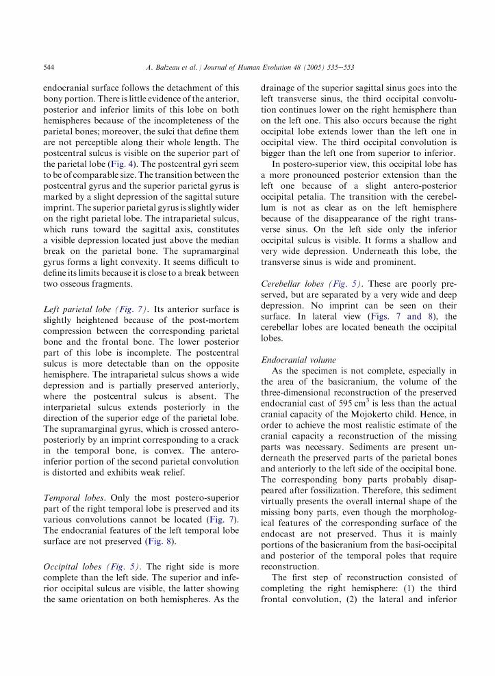
Fig. 1. (a) Transverse, and (b) midsagittal CT scans, and (c) a volume-rendered 3D reconstruction of Mojokerto, showing the
stratification of the sediments inside the Mojokerto calvaria. In ‘b’ the white line indicates the position of ‘a’. White arrows indicate
coarser sedimentation and black arrows finer one.
Material and methods
For the purposes of the present study, thecalvaria was CT scanned with a General ElectricHigh Speed HAS scanner at the ‘‘CHNO desQuinze-vingts’’ in Paris (120 kV, 80 mAs, scantime 3 s). The full three-dimensional image stackconsists of 112 contiguous, 1 mm thick slices in thecoronal plane. The images have a pixel matrix of512! 512, and with a field of view (FOV) of23 cm the pixel size is 0.45 mm. The CT scans donot show any noticeable artifacts such as beamhardening or scattering even though the calvaria isheavily mineralized.
The use of CT in anthropological researchbegan nearly twenty years ago. In spite of thetechnical limits of the earlier equipment, pioneerresearchers (e.g. Tate and Cann, 1982; Wind, 1984;Vannier et al., 1985; Zonneveld and Wind, 1985;Ruff and Leo, 1986) were attracted by theopportunity to get access to the hidden structuresof fossils. Since then, this remarkable technolog-ical enhancement produced numerous and diverseapplications (e.g. Hublin et al., 1996; Spoor et al.,2000, 2003; Ponce de Leon and Zollikofer, 2001;Baba et al., 2003; Bookstein et al., 2003; Silcox,
2003; Brauer et al., 2004; Rook et al., 2004),including attempts to estimate the endocranialvolume (e.g. Conroy and Vannier, 1985; Conroyet al., 1990, 1998; Falk, 1998; Recheis et al., 1999;Tobias, 2001; Prossinger et al., 2003).
The CT data set was visualized and analysedusing eFilm 1.8.1 and Mimics 7.1 software. Fol-lowing previous work (Balzeau et al., 2002a,b,2003a,b), we used the data to extract informationconcerning both the endocranial cavity, and thedetailed morphology of the internal structuresof the Mojokerto calvaria (Figs. 1 and 2).The method of using global thresholds of CT(Hounsfield) values to separate bone, sedimentsand air cannot be applied reliably to the Mojo-kerto CT scans, because their density rangesoverlap, and part of the sediments would beincluded as bone, and part of the bone assediments. We therefore developed a specific pro-tocol to define the exact position of the differentinterfaces in order to obtain the most accurateresults. On each CT slice, the boundary betweenthe fossil and the filling sediment was identified bymanual segmentation. This procedure consists ofmeasuring the median value (or Half MaximumHeight, HMH) from the CT value of the two

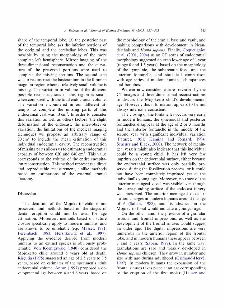
538 A. Balzeau et al. / Journal of Human Evolution 48 (2005) 535e553
Fig. 2. Morphology of the anterior fontanelle of Mojokerto. The position of the three oblique CT sections (left posterior to right
anterior) are indicated on the three-dimensional image. A anterior fontanelle, B osseous fragment, C fracture, D right coronal suture,
rp right parietal, rf right frontal, lf left frontal.
elements (the fossil and the sediment) of which theinterface should be defined (Spoor et al., 1993;Schwartz et al., 1998; Balzeau et al., 2002c). As theHounsfield numbers of the matrix are lower thanthose of the fossilized bone, this value is used asthe maximal one of the mask characterizing theendocast. This adjustment has to be made eachtime the attenuation coefficient of one of theelements varies all along the interface and on eachCT slice. We named this protocol SMM (SeuillageManuel Multiple for Multiple Manual Thresh-olding). It is time consuming, but it allows foraccurate identification of the interface betweentwo structures, despite local fluxtuation in CTnumbers. Finally, the boundaries obtained by theSMM protocol were used to calculate three-dimensional reconstructions of the endocast andthe cranium.
The accuracy of the surface reconstructions aremainly determined by the spatial resolution of theCT data set, which is adequate to visualizevascular, sulcal and gyral impressions of theMojokerto endocast (Fig. 3). Thus, the slicethickness and pixel size used here are a goodcompromise between obtaining sufficient spatialresolution, and the time-consuming process ofisolating the virtual endocast.
We analysed the vascular, gyral and sulcalimpressions of the Mojokerto child’s endocast andcompared its morphology with that observed onendocasts of fossil hominids and modern humans.The fossil sample includes H. erectus from Asia(NZ 22 from Trinil, Sangiran, Zhoukoudian,Ngandong and Sambungmacan), Neanderthals(NZ 10), and fossil H. sapiens (NZ 9) (Gri-maud-Herve, 1997; Balzeau et al., 2002a,b). The

539A. Balzeau et al. / Journal of Human Evolution 48 (2005) 535e553
modern human sample consists of 20 virtualendocasts obtained from CT data of 1.5 to 13-year old children (Bosma collection; Shapiro andRichtsmeier, 1997) and 30 adult physical endocastsall of unknown geographic origin (coll. Musee del’Homme; DGH personal data).
Previous studies have shown that metric anal-yses of endocasts provide little useful informationregarding the differences between fossil hominidsand modern humans (Holloway, 1981; Grimaud-Herve, 1994, 1997). Moreover, direct absolutemetric comparison of the Mojokerto individualwith H. erectus adult specimens and modernhumans were not conducted because of its young
Fig. 3. Supero-lateral and postero-lateral three-dimensional
visualization of the Mojokerto endocast. FG granular foveola,
RA ascending ramus, B Broca’s area number 44 and 45, Sg
superior sagittal sinus, H confluence of sinuses, St left trans-
verse sinus; the maximum length of the endocast is 128 mm.
age at death. Geometric morphometric analysesare still in progress.
Results
Preservation
The CT images and the 3D reconstructionsshow detailed morphological features of theendocranial surface, such as some parts of thepattern of meningeal vessels and the gyral andsulcal imprints. This proves that the inner surfaceof the Mojokerto calvaria is well preserved.Interestingly, some bony fractures visible on theCT images extend also into the sediment. Conse-quently, it appears that the sediment filled thecranium before the specimen underwent substan-tial structural changes because of postdepositionaltaphonomic dynamics. Where parts of the vaultbones are missing the endocranial surface has keptits original shape, suggesting that these part werelost after fossilisation, and perhaps even duringcollection/preparation of the specimen (Huffmanand Zaim, 2003).
The CT data shows a heterogeneous sedimentinside the cranium (Fig. 1). The deposit showsa stratification of two layers: a coarser one, locatedantero-superiorly, and a finer one. Some elementscontained in the sediment are spherical witha diameter close to 2 cm, substantial comparedto the dimensions of the specimen, and probablycorrespond to pumice balls. No element of thesediment seems to represent bone from thecranium itself. The specimen seems to come froma high-energy fluvial sediment (Huffman andZaim, 2003). However, the action of the waterdid not disarticulate the still unfused bones of thevault (Fig. 1). Also, the relatively sharp morphol-ogy of some free bony margins (i.e., the lateral partof the supraorbital torus, the preserved regions ofthe basis) shows that any transport was short andthat burial was fast. Available evidence suggeststhat the sediment penetrated the cranium when ithad just lost its base, and that the specimen wasembedded quickly before being dislocated. Eventhough this indicates that the endocranial matrix

540 A. Balzeau et al. / Journal of Human Evolution 48 (2005) 535e553
and the fossil are contemporaneous, this may notrepresent the age of volcanic material itself.
The specimen is slightly deformed: the leftparietal bone is raised in its median part. Afragment of the right parietal is isolated from themain part by an area of missing bone. Thisfragment is rotated to a more vertical orientation,and the calvaria probably suffered slight pressureon its right side postmortem. In addition, the leftparietal bone is anteriorly compressed onto thefrontal bone, and both the right parietal and righthalf of the occipital are shifted slightly posteriorly.The preservation of the internal surfaces will beconsidered together with the endocast.
Only the left frontal sinus is preserved. It is longand flat due to a lateral extension and its volume issmall (greatest width of 34 mm, height less than13 mm). The temporal bones are incomplete. Thepetrous parts are partially preserved, as is theanteriormost part of the left squamous part.The CT images show that the bony labyrinth, thevestibule and the cochlea are entirely preserved onboth sides, whereas both the external and internalacoustic meati are absent. The external andinternal bony laminae, as well as the diploe, arewell-differentiated in all vault bones. Similardiploic differentiation in all vault bones wasobserved in modern humans from our compara-tive sample from the age of 3.
Suture synostosis on the internal surface
The state of suture synostosis on the internalsurface was examined using cross-sectional imagesextracted from the original CT dataset (Fig. 2). Assome sediments partially filled the still unfusedsutures and the inner cracks of the originalspecimen, these features appear on the 3D re-construction of the endocast.
The coronal, sagittal and lambdoid sutures areclearly detectable, as is the anterior fontanelle. Theregion corresponding to the mastoid fontanelle isnot preserved on either side. A fine marking alongthe mid-internal surface of the frontal bone couldcorrespond to the metopic suture, but this imprintappears to be a bony surface crack (Fig. 2, C).Thus, this suture is fused on both the exocranial(Anton, 1997) and the endocranial parts of the
Mojokerto frontal bone. The open coronal sutureis about 6 mm wide. The sagittal suture is visiblealong its extension, while the lambdoid suture hasthe least marked inner suture.
Fig. 2 illustrates the vault morphology of thebregma region. The posteriormost slice showsa separation between the posteromedial part ofthe frontal bone, and the anteromedial corner ofthe right parietal bone (Fig. 2, A). At the marginsof both parietal bones the diploe is covered withcortical bone. This morphology is typical of in vivoapposition of bone matrix during normal perios-teal growth in a suture. It appears then that thespace in this region is not caused by damage, butrepresents a small anterior fontanelle. Bothmargins of the frontal bone show a broken edgein this region, but they conserved their originalconformation at the coronal suture. On theintermediate slice, an isolated bony fragment islocated above the region of the fontanelles andcorresponds to localized post-mortem damagerepresenting an isolated element of the mostposterior tip of the right frontal bone (Fig. 2, B).On the anterior slice, a fracture extends betweentwo frontal halves (Fig. 2, C), whereas the coronalsuture is very clear (Fig. 2, D). This coronal sutureis a narrow sutural space between the right parietaland the frontal. Surface detail of the original fossilshows the same features, with fragments of thefrontal joined with the sediment by preservativearound the anterior fontanelle.
Morphological description of the endocranial cast
Overall morphologyAs mentioned above the calvaria shows some
taphonomic deformation. However, the overallshape of the endocranial cavity is well preservedbecause the vault does not show major breakage,and unfused bones stayed in articulation.
In superior view, the groove of the superiorsagittal sinus is clearly discernible, but partiallycovered by an imprint related to the sagittal suture(Fig. 4). In fact, the sediment went into this suture,thus forming a fine ridge. In the same view it isdifficult to identify precisely the position of themaximal width of the endocast because thecorresponding region is not entirely preserved on

541A. Balzeau et al. / Journal of Human Evolution 48 (2005) 535e553
the left side. Nevertheless, it seems located in theposterior half of the endocast. The preserved partsof the frontal lobes show a similar extension (nofrontal petalia), while the right occipital lobeslightly extends posteriorly to the left one (occip-ital petalia) (Fig. 4). This last may be due to thetaphonomic posterior slight shift of the occipitalbone.
In lateral view (Figs. 7 and 8), the outline of theendocast is regularly curved as far posteriorly asmidway between bregma and lambda, with a moreflattened area further posteriorly, which corre-sponds to the marked supralambdoid flattening onthe outer surface. The endocast maximal height islocated in its median part. The curvature of theoccipital lobes is unchanged.
In frontal and occipital view (Figs. 5 and 6), thetwo parietal lobes are similar in their ratherstraight outline superiorly, which makes an anglewith the straight, and a sub-vertical outline
Fig. 4. Vertical view of the endocast of Mojokerto. ss sagittal
sulcus, gfs superior frontal gyrus, gfm medial frontal gyrus, gfi
inferior frontal gyrus, sc central sulcus, spc post central sulcus,
gps superior parietal gyrus, si intraparietal sulcus, gs supra-
marginal gyrus, sp parietoocipital sulcus.
more inferiorly. The base of the endocast isbetter preserved on the left hemisphere. Themaximum width of the endocast is positionedinferiorly.
Vascular impressions
Venous sinuses. The groove of the superior sagittalsinus cannot be observed between the frontal lobes,but is visible in the posterior region of the parietallobes beneath the imprint of the sagittal suture thatcovers it. It is visible again posteriorly between theendolambda and the endinion (Fig. 3, SS), whereits width lessens (less than 8 mm) with respect tothe anterior tract (average width of 10 mm). Thesinus deviates laterally to the left into the lefttransverse sinus (Fig. 3, SL). No evidence of atransverse sinus is visible on the right hemisphere(Fig. 5). The region between this confluence ofsinuses and the most basal available limit of thebasi-occipital is well preserved, yet there is noindication of the presence of an occipital ora marginal sinus. The bilateral areas, where thepretrosquamosal sini should be found, are badlypreserved and there is no trace of the sinus. Onboth side, the sphenoparietal sini seem to beabsent, but the morphology of the corresponding
Fig. 5. Occipital view of the Mojokerto endocast. gps superior
parietal gyrus, si intraparietal sulcus, sp parietoocipital sulcus,
Sg superior sagittal sinus, St left transverse sinus, sos superior
occipital sulcus, soi inferior occipital sulcus.
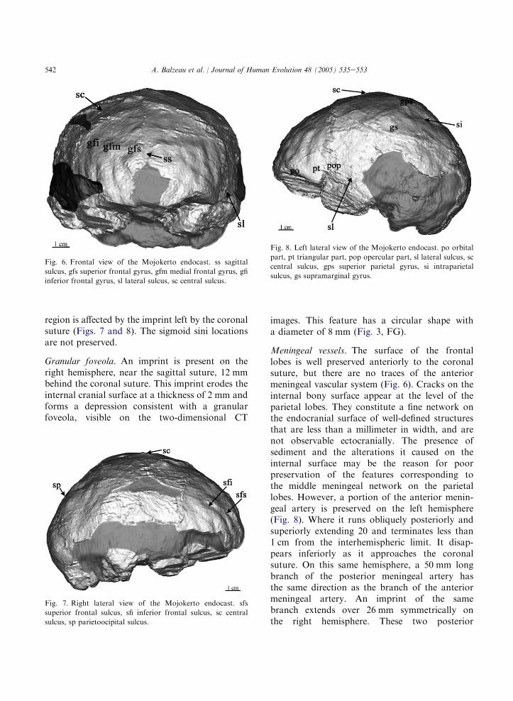
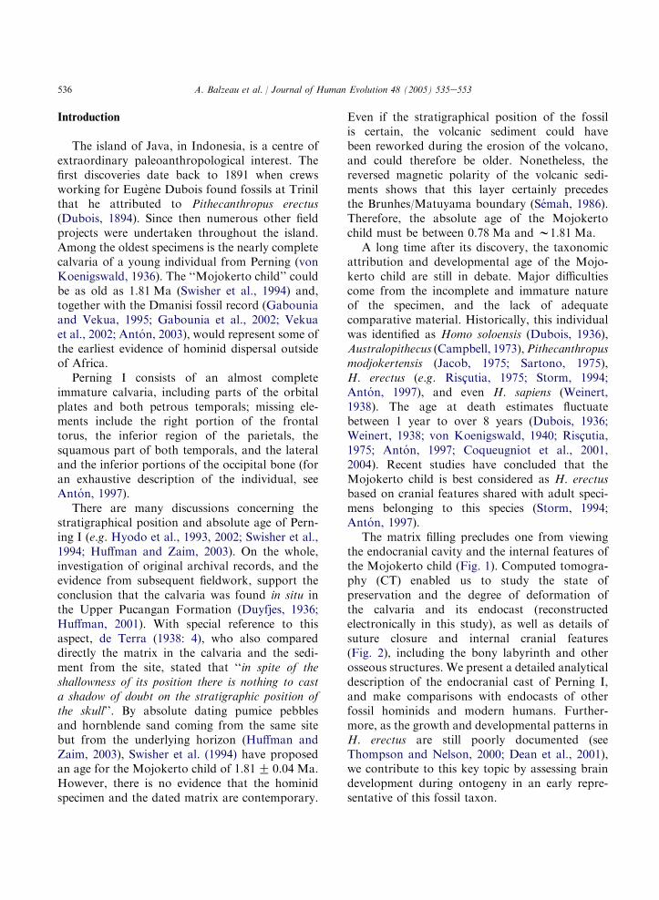
542 A. Balzeau et al. / Journal of Human Evolution 48 (2005) 535e553
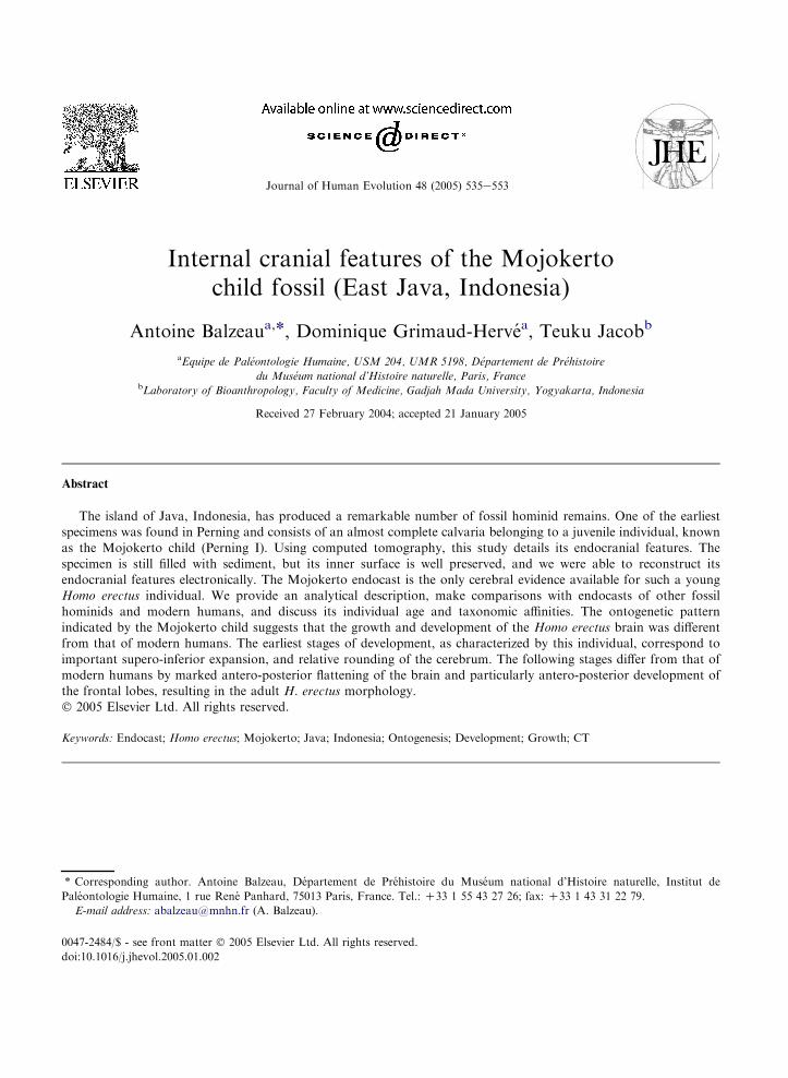
region is affected by the imprint left by the coronalsuture (Figs. 7 and 8). The sigmoid sini locationsare not preserved.
Granular foveola. An imprint is present on theright hemisphere, near the sagittal suture, 12 mmbehind the coronal suture. This imprint erodes theinternal cranial surface at a thickness of 2 mm andforms a depression consistent with a granularfoveola, visible on the two-dimensional CT
Fig. 6. Frontal view of the Mojokerto endocast. ss sagittal
sulcus, gfs superior frontal gyrus, gfm medial frontal gyrus, gfi
inferior frontal gyrus, sl lateral sulcus, sc central sulcus.
Fig. 7. Right lateral view of the Mojokerto endocast. sfs
superior frontal sulcus, sfi inferior frontal sulcus, sc central
sulcus, sp parietoocipital sulcus.
images. This feature has a circular shape witha diameter of 8 mm (Fig. 3, FG).
Meningeal vessels. The surface of the frontallobes is well preserved anteriorly to the coronalsuture, but there are no traces of the anteriormeningeal vascular system (Fig. 6). Cracks on theinternal bony surface appear at the level of theparietal lobes. They constitute a fine network onthe endocranial surface of well-defined structuresthat are less than a millimeter in width, and arenot observable ectocranially. The presence ofsediment and the alterations it caused on theinternal surface may be the reason for poorpreservation of the features corresponding tothe middle meningeal network on the parietallobes. However, a portion of the anterior menin-geal artery is preserved on the left hemisphere(Fig. 8). Where it runs obliquely posteriorly andsuperiorly extending 20 and terminates less than1 cm from the interhemispheric limit. It disap-pears inferiorly as it approaches the coronalsuture. On this same hemisphere, a 50 mm longbranch of the posterior meningeal artery hasthe same direction as the branch of the anteriormeningeal artery. An imprint of the samebranch extends over 26 mm symmetrically onthe right hemisphere. These two posterior
Fig. 8. Left lateral view of the Mojokerto endocast. po orbital
part, pt triangular part, pop opercular part, sl lateral sulcus, sc
central sulcus, gps superior parietal gyrus, si intraparietal
sulcus, gs supramarginal gyrus.

543A. Balzeau et al. / Journal of Human Evolution 48 (2005) 535e553
meningeal branches extend almost as far as theinterhemispheric limit (Fig. 4).
Gyral impressionsThe sagittal sulcus forms a slight depression
posterior to the endoglabella and extends verti-cally as far as the endobregma (Fig. 6). The centralsulci are visible, especially near the sagittal region.In their connection, the imprint left by the sagittalsuture stands out in a slight depression (Fig. 4). Onthe right hemisphere, the central sulcus extendsjust posteriorly to the granular foveola (Figs. 4and 7). As the fontanelle is open, it is difficult tolocalize precisely the position of the endobregma.The endolambda is also not easy to localizebecause the sagittal and lambdoid sutures arewide at their junction. Thus, the connection of thecentral sulci is located approximately 29 mm fromendobregma and 62 mm from endolambda.
The frontal and temporal lobes are separated atthe lateral sulcus starting point by a wide and deepdepression that goes on posteriorly by a shortoblique depression on the left hemisphere (Fig. 8).The corresponding region is not preserved on theright hemisphere (Fig. 7). On the two hemispheres,the ascending ramus is visible (Fig. 3).
On the right hemisphere, the parietoocipitalsulcus forms a brief and light depression anteriorlyto the lambdoid suture (Fig. 7). The correspondingregion on the opposite hemisphere, which does notpresent any track of this sulcus, is distortedbecause of the upthrust of the parietal boneanteriorly to the lambdoid suture.
Sulcal impressions
Right frontal lobe (Fig. 7). On the two-dimen-sional CT images it appears that the ethmoid andthe sphenoid bones are damaged, and the basalparts of the frontal lobes are not well-preserved.The most anterior region of the lobe is absent andthe inferior surfaces of the frontal lobes aredamaged. The bulge formed by the coronal suturein relief and the raised parietal bone make theprecentral sulci barely recognizable on both sides(Fig. 4). As a result, the width of the precentralgyri cannot be assessed accurately. The superiorfrontal sulcus is slightly marked and regularly
curved from the most anterior preserved part ofthe frontal lobe. It goes as far as the coronal sutureby moving away from the interhemispheric limit.The superior frontal gyri have a comparable widthon both hemispheres. The inferior frontal sulcus isvery marked and slightly sinuous and is preservedto the coronal suture. The middle frontal gyri arewide on both hemispheres. The external part of theinferior frontal gyrus is rather complete missingonly the orbital portion and the inferior region ofthe triangular portion. The preserved portion ofthe ascending ramus is tilted posteriorly anddisappears at the level of the coronal suture. Theopercular portion is not preserved.
Left frontal lobe (Fig. 8). The superior frontalsulcus and the inferior frontal sulcus are moresinuous than on the opposite lobe and delimitclearly the three frontal convolutions (Fig. 6). Theascending ramus and the anterior ramus arevisible. The former is slightly tilted anteriorlyand extends over 15 mm (Fig. 3, RA), while thelatter curves upward. The cap incisure formsa rather wide and shallow depression. The so-delimited triangular part constitutes a well de-veloped relief. This bulge corresponds to area 45situated on the left inferior frontal gyrus andanteriorly to the ascending ramus (Fig. 3, B). It islocalized anteriorly to the temporal pole fromwhich it is separated by a wide and deep de-pression. The orbital part is complete anteriorlyand extends antero-posteriorly to about 30 mm.Behind it, the surface corresponding to theopercular part obscured by the coronal suture.The inferior surface of the frontal lobe is completebut poorly preserved. A depression can beobserved on its external part that might representthe external orbital gyrus.
Right parietal lobe (Fig. 7). This lobe is incompletein its inferior part due to the partial preservation ofthe right parietal bone. A 44 mm triangular-shapedzone is missing along its median part, antero-posteriorly of the coronal suture. A crack runsfrom the superior limit of this break to join thesagittal suture. Two other cracks run inferiorly andisolate a bone fragment posterior to the coronalsuture. The orientation of the corresponding
544 A. Balzeau et al. / Journal of Human Evolution 48 (2005) 535e553
endocranial surface follows the detachment of thisbony portion. There is little evidence of the anterior,posterior and inferior limits of this lobe on bothhemispheres because of the incompleteness of theparietal bones; moreover, the sulci that define themare not perceptible along their whole length. Thepostcentral sulcus is visible on the superior part ofthe parietal lobe (Fig. 4). The postcentral gyri seemto be of comparable size. The transition between thepostcentral gyrus and the superior parietal gyrus ismarked by a slight depression of the sagittal sutureimprint. The superior parietal gyrus is slightly wideron the right parietal lobe. The intraparietal sulcus,which runs toward the sagittal axis, constitutesa visible depression located just above the medianbreak on the parietal bone. The supramarginalgyrus forms a light convexity. It seems difficult todefine its limits because it is close to a break betweentwo osseous fragments.
Left parietal lobe (Fig. 7). Its anterior surface isslightly heightened because of the post-mortemcompression between the corresponding parietalbone and the frontal bone. The lower posteriorpart of this lobe is incomplete. The postcentralsulcus is more detectable than on the oppositehemisphere. The intraparietal sulcus shows a widedepression and is partially preserved anteriorly,where the postcentral sulcus is absent. Theinterparietal sulcus extends posteriorly in thedirection of the superior edge of the parietal lobe.The supramarginal gyrus, which is crossed antero-posteriorly by an imprint corresponding to a crackin the temporal bone, is convex. The antero-inferior portion of the second parietal convolutionis distorted and exhibits weak relief.
Temporal lobes. Only the most postero-superiorpart of the right temporal lobe is preserved and itsvarious convolutions cannot be located (Fig. 7).The endocranial features of the left temporal lobesurface are not preserved (Fig. 8).
Occipital lobes (Fig. 5). The right side is morecomplete than the left side. The superior and infe-rior occipital sulcus are visible, the latter showingthe same orientation on both hemispheres. As the
drainage of the superior sagittal sinus goes into theleft transverse sinus, the third occipital convolu-tion continues lower on the right hemisphere thanon the left one. This also occurs because the rightoccipital lobe extends lower than the left one inoccipital view. The third occipital convolution isbigger than the left one from superior to inferior.
In postero-superior view, this occipital lobe hasa more pronounced posterior extension than theleft one because of a slight antero-posterioroccipital petalia. The transition with the cerebel-lum is not as clear as on the left hemispherebecause of the disappearance of the right trans-verse sinus. On the left side only the inferioroccipital sulcus is visible. It forms a shallow andvery wide depression. Underneath this lobe, thetransverse sinus is wide and prominent.
Cerebellar lobes (Fig. 5). These are poorly pre-served, but are separated by a very wide and deepdepression. No imprint can be seen on theirsurface. In lateral view (Figs. 7 and 8), thecerebellar lobes are located beneath the occipitallobes.
Endocranial volumeAs the specimen is not complete, especially in
the area of the basicranium, the volume of thethree-dimensional reconstruction of the preservedendocranial cast of 595 cm3 is less than the actualcranial capacity of the Mojokerto child. Hence, inorder to achieve the most realistic estimate of thecranial capacity a reconstruction of the missingparts was necessary. Sediments are present un-derneath the preserved parts of the parietal bonesand anteriorly to the left side of the occipital bone.The corresponding bony parts probably disap-peared after fossilization. Therefore, this sedimentvirtually presents the overall internal shape of themissing bony parts, even though the morpholog-ical features of the corresponding surface of theendocast are not preserved. Thus it is mainlyportions of the basicranium from the basi-occipitaland posterior of the temporal poles that requirereconstruction.
The first step of reconstruction consisted ofcompleting the right hemisphere: (1) the thirdfrontal convolution, (2) the lateral and inferior
545A. Balzeau et al. / Journal of Human Evolution 48 (2005) 535e553
shape of the temporal lobe, (3) the posterior partof the temporal lobe, (4) the inferior portions ofthe occipital and the cerebellar lobes. This waspossible by using the morphology of the morecomplete left hemisphere. Mirror imaging of thethree-dimensional reconstruction and the curva-ture of the preserved portions were used tocomplete the missing sections. The second stepwas to reconstruct the basicranium in the foramenmagnum region where a relatively small volume ismissing. The variation in volume of the differentpossible reconstructions of this region is small,when compared with the total endocranial volume.The variation encountered in our different at-tempts to complete the missing parts of thisendocranial cast was 13 cm3. In order to considerthis variation as well as others factors (the slightdeformation of the endocast, the inter-observervariation, the limitations of the medical imagingtechnique) we propose an arbitrary range of20 cm3 to include the mean estimation of thisindividual endocranial cavity. The reconstructionof missing parts allows us to estimate a endocranialcapacity of between 620 and 640 cm3. This valuecorresponds to the volume of the entire encepha-lon reconstruction. This method represents a directand reproducible measurement, unlike methodsbased on estimations of the external cranialanatomy.
Discussion
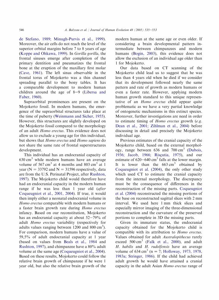
The dentition of the Mojokerto child is notpreserved, and methods based on the stages ofdental eruption could not be used for ageestimation. Moreover, methods based on sutureclosure specifically apply to modern humans, andare known to be unreliable (e.g. Masset, 1971;Ferembach, 1983; Hershkovitz et al., 1997).Applying the evidence derived from modernhumans to an extinct species is obviously prob-lematic. Von Koenigswald (1940) considered theMojokerto child around 5 years old at death.Riscutia (1975) suggested an age of 2.5 years to 3.5years, based on estimates of the specimen’s adultendocranial volume. Anton (1997) proposed a de-velopmental age between 4 and 6 years, based on
the morphology of the cranial base and vault, andmaking comparisons with development in Nean-derthals and Homo sapiens. Finally, Coqueugniotet al. (2001, 2004) using CT scans of endocranialmorphology suggested an even lower age of 1 year(range 0 and 1.5 years), based on the morphologyof the tympanic, the subarcuate fossa and theanterior fontanelle, and statistical comparisonwith age series of modern humans, chimpanzeesand bonobos.
We can now consider features revealed by theCT images and three-dimensional reconstructionsto discuss the Mojokerto child’s developmentalage. However, this information appears to be notalways internally consistent.
The closing of the fontanelles occurs very earlyin modern humans: the sphenoidal and posteriorfontanelles disappear at the age of 2 or 3 monthsand the anterior fontanelle in the middle of thesecond year with significant individual variation(Paturet, 1951; Kamina and Renard, 1994;Scheuer and Black, 2000). The network of menin-geal vessels might also indicate that this individualcould be a young child. It has left very fewimprints on the endocranial surface, either becausethe endocranial surface was only partially pre-served during the fossilization process, or it couldnot have been completely imprinted yet at theindividual’s young age. Moreover, no trace of theanterior meningeal vessel was visible even thoughthe corresponding surface of the endocast is verywell preserved. The anterior meningeal vascular-isation emerges in modern humans around the ageof 6 (Saban, 1988), and its absence on theMojokerto fossil would indicate a younger age.
On the other hand, the presence of a granularfoveola and frontal impressions, as well as thedevelopment of the frontal sinuses would suggestan older age. The digital impressions are verynumerous in the anterior region of the frontallobe, and in modern humans these appear between3 and 5 years (Saban, 1988). In the same way,granulations are rare and weakly developed inHomo sapiens children. They grow in number andsize with age during adulthood (Grimaud-Herve,1997). In modern humans the expansion of thefrontal sinuses takes place at an age correspondingto the eruption of the first molar (Hauser and
546 A. Balzeau et al. / Journal of Human Evolution 48 (2005) 535e553
de Stefano, 1989; Minugh-Purvis et al., 1999).Moreover, the air cells do not reach the level of thesuperior orbital margins before 7 to 8 years of age(Koppe and Ohkawa, 1999). In Gorilla gorilla, thefrontal sinuses emerge after completion of theprimary dentition and pneumatizes the frontalbone at the eruption of the maxillary first molar(Cave, 1961). The left sinus observable in thefrontal torus of Mojokerto was a thin channelspreading parallel to the bony tables. It hasa comparable development to modern humanchildren around the age of 8e9 (Libersa andFaber, 1960).
Supraorbital prominences are present on theMojokerto fossil. In modern humans, the emer-gence of the supraorbital structures take place atthe time of puberty (Weinmann and Sicher, 1955).However, this structures are slightly developed onthe Mojokerto fossil compared to the morphologyof an adult Homo erectus. This evidence does notallow us to exclude a young age for this individual,but shows that Homo erectus and Homo sapiens donot share the same rate of frontal superstructuresdevelopment.
This individual has an endocranial capacity of630 cm3 while modern humans have an averagevolume of 567 cm3 at 4 months and 803 cm3 at 1year (NZ 33792 and NZ 31596 respectively, dataare from the U.S. Perinatal Project, after Rushton,1997). The Mojokerto child would therefore havehad an endocranial capacity in the modern humanrange if he was less than 1 year old (afterCoqueugniot et al., 2001, 2004). If true, it wouldthen imply either a neonatal endocranial volume inHomo erectus comparable with modern humans ora faster brain growth rate during Homo erectusinfancy. Based on our reconstitution, Mojokertohas an endocranial capacity at about 52e79% ofadult Homo erectus variability (respectively foradults values ranging between 1200 and 800 cm3).For comparison, modern humans have a value of59,5% of adult endocranial capacity at 1 year(based on values from Beals et al., 1984 andRushton, 1997), and chimpanzee have a 80% adultvolume at the same age (Coqueugniot et al., 2004).Based on these results, Mojokerto could follow therelative brain growth of chimpanzee if he were 1year old, but also the relative brain growth of the
modern human at the same age or even older. Ifconsidering a brain developmental pattern in-termediate between chimpanzees and modernhumans (Bogin, 2003), this evidence does notallow the exclusion of an individual age older than1 for Mojokerto.
Our data based on CT scanning of theMojokerto child lead us to suggest that he wasless than 4 years old when he died if we considerthat its development followed nearly the samepattern and rate of growth as modern humans oreven a faster rate. However, applying modernhuman growth standard to this unique represen-tative of an Homo erectus child appear quiteproblematic as we have a very partial knowledgeof ontogenetic mechanisms in this extinct species.Moreover, further investigations are need in orderto estimate timing of Homo erectus growth (e.g.Dean et al., 2001; Zihlman et al., 2004) beforediscussing in detail and precisely the Mojokertoindividual age.
Previous estimates of the cranial capacity of theMojokerto child, based on the external morphol-ogy, range between 636 and 700 cm3 (Dubois,1936; Jacob, 1966; Riscutia, 1975), and ourestimate of 620e640 cm3 falls at the lower margin.It is lower than the 663 cm3 obtained byCoqueugniot et al. (2004), the only other studywhich used CT to estimate the cranial capacityfrom the internal morphology. The discrepancymust be the consequence of differences in thereconstruction of the missing parts. Coqueugniotet al. (2004) reconstructed the missing portions ofthe base on reconstructed sagittal slices with 2 mminterval. We used here 1 mm thick slices andespecially mirror imaging of the three-dimensionalreconstruction and the curvature of the preservedportions to complete in 3D the missing parts.
Given its immature status, the endocranialcapacity obtained for the Mojokerto child iscompatible with its attribution to Homo erectus.Values obtained for adult Australopithecus rarelyexceed 500 cm3 (Falk et al., 2000), and adultH. habilis and H. rudolfensis have an averagevolume of 634 cm3 (nZ 7; Holloway, 1973, 1978,1983a; Stringer, 1986). If the child had achievedadult growth he would have attained a cranialcapacity in the adult Asian Homo erectus range of
547A. Balzeau et al. / Journal of Human Evolution 48 (2005) 535e553
variability, between 840 and 1245 cm3 for thefossils of Trinil, Sangiran and the Sinanthropus(nZ 10; Grimaud-Herve, 1997).
The superior sagittal sinus is continuous withthe right transverse sinus in modern humans andmost fossils assigned to the genus Homo (re-spectively 80 and 85%, Grimaud-Herve, 1997). Inthe Mojokerto child it continues into the lefttransverse sinus (Fig. 3), as is the case in 20%of modern humans (with some variations e.g.Grimaud-Herve, 1997; Bruner et al., 2003). Thepredominant presence of an enlarged occipital-marginal sinus in Paranthropus robustus, P. boisei,and Australopithecus afarensis is established(e.g.Tobias, 1987; Tobias and Falk, 1988; Saban,1993; Falk et al., 1995). In adult Homo erectus, thesuperior sagittal sinus is continuous with the righttransverse sinus in Trinil 2, Sangiran 12 and 17,Ckn.E 1.PA.16 (III), Ckn.L 1.PA.98 (X), Ckn.L2.PA.99 (XI), Ckn.L 3.PA.100 (XII), Ngandong 6,7 and 12, and with the left in Sangiran 2 and 10.Only Sambungmacan 3 shows an occipital-mar-ginal sinus (Broadfield et al., 2001; Balzeau et al.,2002b). The presence of this feature on Trinil 2 isnot confirmed (contra Falk, 1986). This endocastshows a preponderant right transerve sinus wherethe superior sagittal sinus continues in majority.The left transerve sinus is less printed. It is situatedjust above the preserved limit of the endocast andno relief in this region can be interpreted as anoccipito-marginal sinus. This system is absent inMojokerto, as is the most frequent condition in thegenus Homo.
The asymmetric pattern of the transversesinuses and the confluence of sinuses may beassociated with the asymmetric development ofone of the cerebral hemispheres (Delmas andChifflet, 1950). In spite of the strong developmentof the left transverse sinus (Fig. 3, St), the endocastof the Mojokerto individual does not show a clearpredominance of one of the hemispheres. The rightone is larger and the occipital pole extendposteriorly to the left one in superior view, butthis extension was certainly slightly amplified bythe diagenetic processes that this fossil underwent.
The sphenoparietal sinus seem to be absent onMojokerto, even if the corresponding areas areaffected by the presence of the coronal suture. This
feature is variably present and slightly developedon the Trinil, Sangiran and Zhoukoudian endo-casts. It is always absent on the Ngandong andSambungmacan hominids and the actual modernhumans sample (Grimaud-Herve, 1997; Balzeauet al., 2002b).
The meningeal network left very few prints onthe endocast. No trace of the anterior meningealvessel was visible whereas the correspondingsurface is very well preserved. Meanwhile, a gran-ular foveola is visible in the posterior part of theright frontal lobe, close to the sagittal sulcus. Inmodern humans these granulations are absent infoetuses, rare and less developed in children. Theyincrease in number while the cerebral volumeincreases with age. Therefore, the importantdevelopment of this feature on Mojokerto mightindicate a faster vascular development of the brainthan in modern humans. In adults Homo erectus,granular foveolae are very frequent on theposterior part of the frontal lobes in the Trinil,Sangiran and Zhoukoudian fossils and less in theNgandong and Sambungmacan hominids. Thesefeatures are more numerous and developed inthese adults than on the Mojokerto endocast.
In modern humans, Broca’s motor and speecharea is composed of areas number 44 and 45(Bourret and Louis, 1974; Grimaud-Herve, 1997).On the Mojokerto endocast, there is a bulgecorresponding to area 45 situated on the leftinferior frontal gyrus and anteriorly to theascending ramus (Fig. 3, B). The presence of thisfeature is variable in australopithecines (Tobias,1975), and appears more consistently in H. habilisand H. erectus (Holloway, 1983b; Tobias, 1983;Broadfield et al., 2001). Its relevance as evidencefor the existence of some hypothetical languagefacilities is dubious (Maitrerobert, 2002). How-ever, this relief has a similar development and thesame localization relatively to the temporal lobe inMojokerto than in Asian Homo erectus. Similarly,they all present a wide and deep depression at theorigin of the lateral sulcus.
The overall shape of the frontal lobes inanterior view is characteristic of various groupsof hominids (Grimaud-Herve, 1997). Outlineswhich correspond to the orientation of the inferiorsurface of the frontal lobes and the encephalic

548 A. Balzeau et al. / Journal of Human Evolution 48 (2005) 535e553
rostrum show a regular curvature in the case of theendocasts of the Trinil, Sangiran and Zhoukou-dian hominids. Ngandong and Sambungmacanendocasts show a clear discontinuity between thesetwo planes, as it becomes more oblique in laterrepresentatives of the genus Homo. In Mojokertothe encephalic rostrum region is not preserved, butthe left frontal lobe is similar to that of theZhoukoudian, the Trinil and the Sangiran speci-mens (Fig. 6). This morphology indicates a relativelow development of the prefrontal cortex incomparison with that observed in Homo sapiens.Indeed, the inferior part of the inferior frontalgyrus in the Mojokerto child is similar in shapeand development to all the Asian Homo erectusexcept the Ngandong and Sambungmacan indi-viduals (Grimaud-Herve, 1997; Balzeau et al.,2002a,b).
The Mojokerto specimen is similar to Homoerectus from Trinil, Sangiran and Zhoukoudian inthat the anterior-inferior part of the secondparietal convolution shows weak relief, the supra-marginal gyri are not prominent, the occipitallobes are separated by a wide inter-hemisphericspace, and are located behind the extension of theparietal and temporal lobes, and the cerebellarlobes are prominent, separated by a large grooveand located underneath the occipital lobes. Allthese features show a continuous shift in directionof modern human morphology during hominidevolution (see Grimaud-Herve, 1997 for an ex-haustive description of these transformations).The Ngandong and Sambungmacan endocastshave a particular morphology for these features(Grimaud-Herve, 1997; Balzeau et al., 2002a,b),not shared by others Asian Homo erectus nor theMojokerto individual.
Consequently, all these vascular and gyralfeatures suggest closer affinities for the Mojokertochild with the Trinil, Sangiran and ZhoukoudianHomo erectus while the Ngandong and Sambung-macan fossils present a relatively more evolvedmorphology (Grimaud-Herve, 1986, 1997; Balzeauet al., 2002a,b).
The orientation of the ascending ramus, com-pared to the posterior ramus, varies amonghominids. It is quite vertical on the Asian Homoerectus endocasts but goes forward in modern
humans; a change correlated with the anterior shiftof the frontal lobes. The ascending ramus isslightly tilted forwards on the Mojokerto lefthemisphere (Fig. 3, RA), showing some similaritywith modern humans. This is due to the relativeheight and convexity of the endocast in the lateralview (Figs. 7 and 8) and particularly to the frontallobes anterior rounding. Indeed, the correspond-ing endocranial outline is more globular than seenin any adult Homo erectus individuals. On theMojokerto endocranial cast, the connection of thecentral sulci is located in the anterior third ofthe endobregma-endolambda axis. This position issimilar to that in modern humans while theSangiran, Trinil, and Zhoukoudian endocasts havea connection situated at the 2/5 of this axis(Grimaud-Herve, 1997). Thus the parietal lobesof Mojokerto are relatively more developed thanthose of its contemporaries and the frontal lobesshow a relatively less antero-posterior extension.This individual differs from adult Homo erectus inthe position of the endocranial vertex because it islocated near the central sulci intersection. TheMojokerto cerebral shape would have got longerand flatter during growth making it more similarto adult individuals. These modifications areparticularly characterized by an antero-posteriordevelopment of the frontal lobes and associatedcompression of the parietal lobes. Alternatively,the cerebrum increases in height between modernhuman children and adults resulting in cerebralrounding. This suggests that Homo erectus had anontogenetic pattern that differed from the modernhuman pattern.
Studies of young modern human endocastsshow that the digital impressions appear on thefrontal lobes between 3 and 5 years (Saban, 1988).The digital impressions are numerous on theanterior region of Mojokerto’s frontal lobes,whereas the anterior fontanelle and sutures aredistinctly unfused. The imprints on this endocastare more developed than in most adult Homoerectus specimens and than those in chimpanzeesregardless of their developmental stage. They aresimilar to those observed for 6-year-old modernhumans in our comparison sample (Fig. 9). Thisshould not be considered as a diagnostic featurefor modern humans, but rather as the consequence
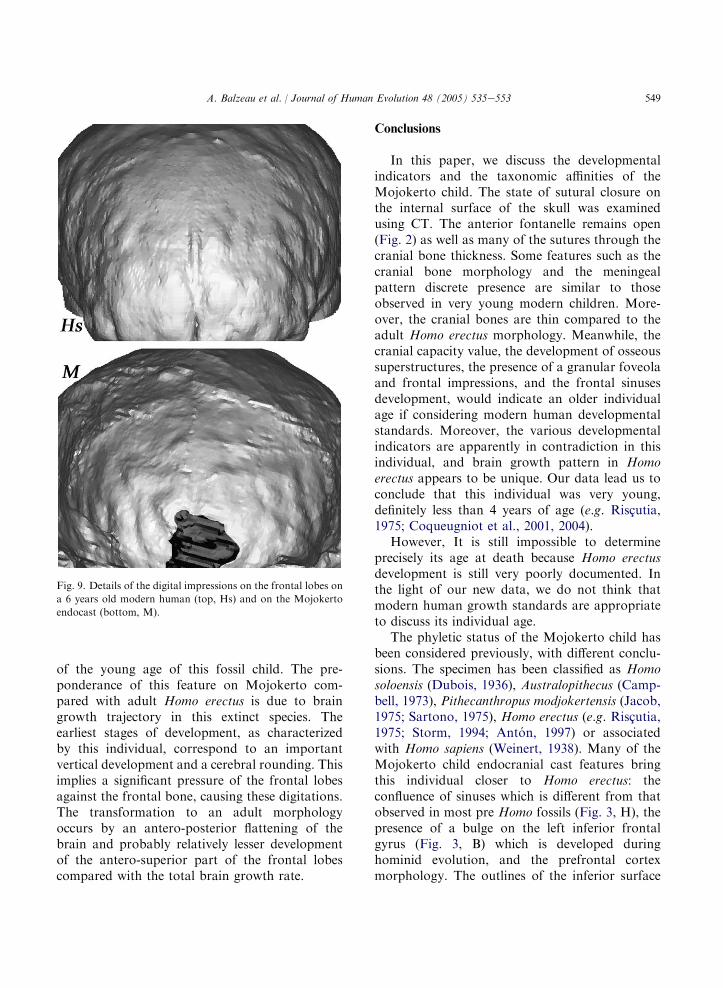
549A. Balzeau et al. / Journal of Human Evolution 48 (2005) 535e553
of the young age of this fossil child. The pre-ponderance of this feature on Mojokerto com-pared with adult Homo erectus is due to braingrowth trajectory in this extinct species. Theearliest stages of development, as characterizedby this individual, correspond to an importantvertical development and a cerebral rounding. Thisimplies a significant pressure of the frontal lobesagainst the frontal bone, causing these digitations.The transformation to an adult morphologyoccurs by an antero-posterior flattening of thebrain and probably relatively lesser developmentof the antero-superior part of the frontal lobescompared with the total brain growth rate.
Fig. 9. Details of the digital impressions on the frontal lobes on
a 6 years old modern human (top, Hs) and on the Mojokerto
endocast (bottom, M).
Conclusions
In this paper, we discuss the developmentalindicators and the taxonomic affinities of theMojokerto child. The state of sutural closure onthe internal surface of the skull was examinedusing CT. The anterior fontanelle remains open(Fig. 2) as well as many of the sutures through thecranial bone thickness. Some features such as thecranial bone morphology and the meningealpattern discrete presence are similar to thoseobserved in very young modern children. More-over, the cranial bones are thin compared to theadult Homo erectus morphology. Meanwhile, thecranial capacity value, the development of osseoussuperstructures, the presence of a granular foveolaand frontal impressions, and the frontal sinusesdevelopment, would indicate an older individualage if considering modern human developmentalstandards. Moreover, the various developmentalindicators are apparently in contradiction in thisindividual, and brain growth pattern in Homoerectus appears to be unique. Our data lead us toconclude that this individual was very young,definitely less than 4 years of age (e.g. Riscutia,1975; Coqueugniot et al., 2001, 2004).
However, It is still impossible to determineprecisely its age at death because Homo erectusdevelopment is still very poorly documented. Inthe light of our new data, we do not think thatmodern human growth standards are appropriateto discuss its individual age.
The phyletic status of the Mojokerto child hasbeen considered previously, with different conclu-sions. The specimen has been classified as Homosoloensis (Dubois, 1936), Australopithecus (Camp-bell, 1973), Pithecanthropus modjokertensis (Jacob,1975; Sartono, 1975), Homo erectus (e.g. Riscutia,1975; Storm, 1994; Anton, 1997) or associatedwith Homo sapiens (Weinert, 1938). Many of theMojokerto child endocranial cast features bringthis individual closer to Homo erectus: theconfluence of sinuses which is different from thatobserved in most pre Homo fossils (Fig. 3, H), thepresence of a bulge on the left inferior frontalgyrus (Fig. 3, B) which is developed duringhominid evolution, and the prefrontal cortexmorphology. The outlines of the inferior surface
550 A. Balzeau et al. / Journal of Human Evolution 48 (2005) 535e553
of the frontal lobe and of the encephalic rostrumlateral part show a regular curvature. The occipitallobes are located behind the extension of theparietal and temporal lobes. The cerebellar lobesare prominent, separated by a large groove, andlocated underneath the occipital lobes. Thesemorphological features suggest closest affinitieswith the Trinil, Sangiran and Zhoukoudian Homoerectus. Moreover, because of its endocranialmorphology, Mojokerto is more distinct from theNgandong and Sambungmacan specimens, whichare now considered as a particular group ofevolved Homo erectus (Grimaud-Herve, 1986,1997; Balzeau et al., 2002a,b). The informationobtained from the endocranial cavity allows us tosay that this fossil belongs to Homo erectus.Within Javanese Homo erectus, precise phyleticor chronological positions are not easily identifi-able because of the archaic features of theMojokerto endocast. Moreover, its young individ-ual age complicates morphological comparisons.The lack of comparative material (the absence ofendocranial data for the earlier Javanese hominids,such as Sangiran 27 and 31, or the absence of fossiljuvenile individuals) make it impossible to bringthe Mojokerto child closer to a particular group ofAsian Homo erectus if basing arguments on sharedarchaic features.
However, some features observed on thisendocast are similar to those of modern humans.The ascending ramus is slightly tilted forward onthe left hemisphere (Fig. 3 RA), the central sulciintersection is anteriorly situated, the endocranialvertex is close to this intersection, so the parietallobes propagate themselves anteriorly, and theendocast outlines are rounded. On one hand, itappears that this very young Homo erectuspresents some similarities in its internal characterswith adult Homo sapiens, and a large brain despiteits young age. On the other hand, the presence ofa granular foveola (Fig. 3, FG) and numerousdigital impressions on the anterior region of thefrontal lobes is quite unusual in very youngmodern humans.
All these evidence concerning the Mojokertoendocranial morphology suggests that Homoerectus followed a unique brain growth trajectory.The earliest stages of development, as characterized
by this individual, correspond to an importantvertical development and a relative cerebral round-ing. Thus the parietal lobes of Mojokerto arerelatively more developed than those of adultindividuals and the frontal lobes are anteriorlyrounded. The ontogeny of Homo sapiens presentsneotenic retention (Gould, 1977; Deacon, 1990;Begun and Walker, 1993) of ancestral features ofits genus, as shown by the shared relative de-velopment of the cerebral lobes in Mojokerto andmodern humans adults.
The following stages in Homo erectus ontogenycorrespond to a different growth and developmentpatterns than in Homo sapiens (Thompson andNelson, 2000; Dean et al., 2001). Indeed, theendocranial transformation to an adult morphol-ogy occurs by an antero-posterior flattening of thebrain. These modifications are particularly char-acterized by an antero-posterior development ofthe frontal lobes while the parietal lobes arerelatively compressed by the posterior shift of thefrontal lobes.
Acknowledgments
We would like to thank Professor E.A.Cabanis e CHNO des Quinze-Vingts, Paris- forthe scanning of the fossils, J.T. Richtsmeier forproviding the Bosma collection CT data. Wethank four anonymous reviewers and the associateeditor for their helpful comments. C. Falgueres, J.Badawi-Fayad provided invaluable discussions,B.M. Parisi, F. Semah and R. Macchiarelli helpfulcomments on earlier versions of this paper. Thefirst author thanks F. Zonneveld for his preciousadvice. To all of them we are grateful.
References
Anton, S.C., 1997. Developmental age and taxonomic affinity
of the Mojokerto child, Java, Indonesia. Am. J. Phys.
Anthropol. 102, 497e514.Anton, S.C., 2003. Natural history of Homo erectus. Yearb.
Phys. Anthropol. 46, 126e170.
Baba, H., Aziz, F., Kaifu, Y., Suwa, G., Kono, R.T., Jacob, T.,
2003. Homo erectus calvarium from the Pleistocene of Java.
Science 299, 1384e1388.

551A. Balzeau et al. / Journal of Human Evolution 48 (2005) 535e553
Balzeau, A., Jacob, T., Indriati, E., 2002a. Internal cranial
features of the Sambungmacan 1 Homo erectus (Java,
Indonesia). C.R. Palevol 1, 305e310.Balzeau, A., Grimaud-Herve, D., Indriati, E., Jacob, T.,
Semah, F., 2002b. Etude comparative de l’endocrane de
l’Homo erectus Sambungmacan 3 (Java, Indonesie). Bull.
Mem. Soc. Anthrop. Paris 14, 154e155.Balzeau, A., Grimaud-Herve, D., Jacob, T., Semah, F.,
Cabanis, E., 2002c. Virtual anthropology: the internal
characters of the ‘‘Mojokerto child’’. Coll. Antropol. 26
(Suppl.), 13.
Balzeau, A., Indriati, E., Grimaud-Herve, D., Jacob, T., 2003a.
Computer tomography scanning of Homo erectus crania
Ngandong 7 from Java: internal structure, paleopathology
and post-mortem history. B.I. Ked. (J. Med. Sci.) 35, 133e
140.
Balzeau, A., Grimaud-Herve, D., Indriati, E., Semah, F.,
Jacob, T., 2003b. Caracteres morphologiques et paleopa-
thologiques craniens et deformations taphonomiques chez
les Homo erectus Ngandong 7 et Sangiran 31. Bull. Mem.
Soc. Anthrop. Paris 15 (n.s.), 6.
Beals, K.L., Smith, C.L., Dodd, S.M., 1984. Brain size, cranial
morphology, climate, and time machines. Curr. Anthropol.
25, 301e330.
Begun, D., Walker, A., 1993. The endocast. In: Walker, A.,
Leakey, R. (Eds.), The NariokotomeHomo erectus Skeleton.
Springer-Verlac, pp. 326e358.
Bogin, B., 2003. The human pattern of growth and de-
velopment in paleontological perspective. In:
Thompson, J.L., Krovitz, G.E., Nelson, A.J. (Eds.),
Patterns of Growth an Development in the Genus Homo.
Cambridge University Press, New York, pp. 15e44.
Bookstein, F.L., Gunz, P., Mitterœcker, P., Prossinger, H.,
Schæfer, K., Seidler, H., 2003. Cranial integration in Homo:
singular warps analysis of the midsagittal plane in ontogeny
and evolution. J. Hum. Evol. 44, 167e187.Bourret, P., Louis, R., 1974. Anatomie du systeme nerveux
central. L’expansion scientifique francaise, Paris.
Brauer, G., Groden, C., Groning, F., Kroll, A., Kupczik, K.,
Mbua, E., Pommert, A., Schiemann, T., 2004. Virtual study
of the endocranial morphology of the matrix-filled cranium
from Eliye Springs, Kenya. Anat. Rec. 276A, 111e133.
Broadfield, D.C., Holloway, R.L., Mowbray, K., Silvers, A.,
Yuan, M.S., Marquez, S., 2001. Endocast of Sambungma-
can 3 (Sm 3): a new Homo erectus from Indonesia. Anat.
Rec. 262, 369e379.
Bruner, E., Averini, M., Manzi, G., 2003. Endocranial traits.
Prevalence and distribution in a recent human population.
Eur. J. Anat. 7, 23e33.
Campbell, B.G., 1973. A new taxonomy of fossil man. Yearb.
Phys. Anthropol. 17, 194e201.Cave, A.J.E., 1961. The frontal sinus of gorilla. Proc. Zool. Soc.
Lond. 136, 359e373.
Conroy, G.C., Vannier, M.W., 1985. Endocranial volume
determination of matrix-filled fossil skulls using high-
resolution CT. Hominid Revolution: Past, Present and
Future. Alan R Liss. pp. 419e426.
Conroy, G.C., Vannier, M.W., Tobias, P., 1990. Endocranial
features of Australopithecus africanus revealed by 2 and 3D
computed tomography. Science 247, 838e841.Conroy, G.C., Weber, G.W., Seidler, H., Tobias, P.V.,
Kane, A., Brunsden, B., 1998. Endocranial capacity in an
early hominid from Sterkfontein, South Africa. Science 280,
1730e1731.Coqueugniot, H., Hublin, J.-J., Jacob, T., 2001. Revision de
l’age individuel de l’enfant de Modjokerto (Java, Indonesie).
XIVe Congres de l’UISPP, Liege, Belgique.
Coqueugniot, H., Hublin, J.-J., Veillon, F., Houet, F.,
Jacob, T., 2004. Early brain growth in Homo erectus and
implications for cognitive ability. Nature 431, 299e302.
Deacon, T.W., 1990. Problems of ontogeny and phylogeny in
brain size evolution. Int. J. Primatol. 11, 237e282.
Dean, C., Leakey, M.G., Reid, D., Schrenk, F., Schwartz, G.T.,
Stringer, C., Walker, A., 2001. Growth processes in teeth
distinguish modern humans from Homo erectus and earlier
hominins. Nature 414, 628e631.
Delmas, A., Chifflet, J., 1950. Le pressoir d’Herophyle. C.R.
Assoc. Anat. 123e131. 37eme reunion, Louvain.
Dubois, E., 1894. Pithecanthropus erectus, eine menschenahn-
liche uebergangsform aus Java. Landesdruckerei, Batavia.
Dubois, E., 1936. Racial identity of Homo soloensis Oppe-
noorth (including Homo modjokertensis von Koenigswald)
and Sinanthropus pekinensis Davidson Black. Proc. Kon.
Akd. Wet. Amsterdam 34, 1180e1185.
Duyfjes, J., 1936. Zur geologie und stratigraphie des Kendeng-
gebietes zwischen Trinil und Soerabaja (Java). De Ingenieur
in Ned.-Indie. 4, 136e149.
Falk, D., 1986. Evolution of cranial blood drainage in
hominids: enlarged occipital/marginal sinuses and emissary
foramina. Am. J. Phys. Anthropol. 70, 311e324.
Falk, D., Gage, T.B., Dudek, B., Olson, T.R., 1995. Did more
than one species of hominid coexist before 3.0 Ma? Evidence
from blood and teeth. J. Hum. Evol. 29, 591e600.Falk, D., 1998. Hominid brain evolution: looks can be
deceiving. Science 280, 1714.
Falk, D., Redmond Jr., J.C., Guyer, J., 2000. Early hominid
brain evolution: a new look at old endocasts. J. Hum. Evol.
38, 695e717.
Ferembach, D., 1983. Bilan sur la fiabilite des techniques de
determination de l’age a partir du squelette. Bull. Mem. Soc.
Anthrop. Paris 10 (serie XIII), 435e440.Gabounia, L., Vekua, A., 1995. A Plio-Pleistocene hominid from
Dmanisi, East Georgia, Caucasus. Nature 373, 509e512.
Gabounia, L., Lumley de, M.-A., Vekua, A.,
Lordkipanidze, D., Lumley de, H., 2002. Decouverte d’un
nouvel hominide a Dmanissi (Transcaucasie, Georgie). C.R.
Palevol 1, 243e253.
Gould, S.J., 1977. Ontogeny and phylogeny. Harvard Univer-
sity Press, Cambridge.
Grimaud-Herve, D., 1986. The parietal bone of Indonesian
Homo erectus. Hum. Evol. 1 (2), 167e182.
Grimaud-Herve, D., 1994. Evolution of the Javanese fossil
hominid brain. Cour. Forsch.-Inst. Senckenberg 171,
61e68.
552 A. Balzeau et al. / Journal of Human Evolution 48 (2005) 535e553
Grimaud-Herve, D., 1997. L’evolution de l’encephale chez
l’Homo erectus et l’Homo sapiens: exemples de l’Asie et de
l’Europe. Cahiers de paleoanthropologie. CNRS, Paris.
Hauser, G., de Stefano, G.F., 1989. Epigenic variants of the
human skull. E. Schweizerbart’sche Verlagsbuchhandlung,
Stuttgart.
Hershkovitz, I., Latimer, B., Dutour, O., Jellema, L.M., Wish-
Baratz, S., Rothschild, Rothschild, B.M., 1997. Why do we
fail in aging the skull from the sagittal suture? Am. J. Phys.
Anthropol. 103, 393e399.
Holloway, R.L., 1973. New endocranial values for the East
African early hominids. Nature 243, 97e99.
Holloway, R.L., 1978. Problems of brain endocast interpreta-
tion and African hominid evolution. In: Jolly, C.J. (Ed.),
Early Hominids of Africa. St Martin’s Press, New York, pp.
379e401.
Holloway, R.L., 1981. The Indonesian Homo erectus brain
endocast revisited. Am. J. Phys. Anthropol. 55, 503e521.Holloway, R.L., 1983a. Human brain evolution: a search for
units, models and synthesis. Can. J. Anthropol. 3, 215e230.
Holloway, R.L., 1983b. Human paleontological evidence rele-
vant to language behavior. Hum. Neurobiol. 2, 105e114.Hublin, J.J., Spoor, F., Braun, M., Zonneveld, F., Condemi, S.,
1996. A late Neanderthal with upper Palaeolithic artefacts.
Nature 381, 224e226.Huffman, O.F., 2001. Geologic context and age of the Perning/
MojokertoHomo erectus, East Java. J. Hum. Evol. 40, 353e
362.
Huffman, O.F., Zaim, Y., 2003. Mojokerto Delta, East Jawa:
Paleoenvironment of Homo modjokertensis, first results. J.
Miner. Technol. 10 (2) The Faculty of Earth Sciences and
Mineral Technology, Institute Technology, Bandung.
Hyodo, M., Watanabe, N., Sunata, W., Susanto, E.E.,
Wahyono, H., 1993. Magnetostratigraphy of hominid fossil
bearing formations in Sangiran and Mojokerto, Java.
Anthropol. Sci. 101 (2), 157e183.
Hyodo, M., Nakaya, H., Urabe, A., Saegusa, H., Shunrong, X.,
Jiyun, Y., Xuepin, J., 2002. Paleomagnetic dates of hominid
remains from Yuanmou, China, and other Asian sites. J.
Hum. Evol. 43, 27e41.
Jacob, T., 1966. The sixth skull cap of Pithecanthropus erectus.
Am. J. Phys. Anthropol. 25, 243e260.
Jacob, T., 1975. Morphology and paleoecology of early Man in
Java. In: Tuttle, R.H. (Ed.), Paleoanthropology, Morphol-
ogy and Paleoecology. Mouton, The Hague, pp. 321e325.
Kamina, P., Renard, M., 1994. Tete osseuse, articulation
temporo-madibulaire et dents. Maloine.
von Koenigswald, G.H.R., 1936. Ein fossiler hominide aus dem
Altpleistocan Ostjavas. De Ingenieur in Ned.-Indie. 8, 149e
158.
von Koenigswald, G.H.R., 1940. neue Pithecanthropus funde.
Landsdrukkerij, Batavia.
Koppe, T., Ohkawa, Y., 1999. Pneumatization of the facial
skeleton in Catarrhine primates. In: Koppe, T., Nagai, H.,
Alt, K.W. (Eds.), The Paranasal Sinuses of Higher Primates;
Development, Function and Evolution. Quintessence, Chi-
cago, pp. 77e120.
Libersa, C., Faber, M., 1960. Pneumatisation de la tete du
cornet moyen. J. Otorhinolaryngol. 6, 723e740.
Maitrerobert, A., 2002. L’origine du langage articule, le tractus
vocal et ses relations avec la base du crane et de la
mandibule. These de doctorat, Museum national d’Histoire
naturelle.
Masset, C., 1971. Erreurs systematiques dans la determination
de l’age par les sutures craniennes. Bull. Mem. Soc.
Anthropol. Paris 7 (serie XII), 85e105.
Minugh-Purvis, N., Radovcic, J., Smith, F.H., 1999. Krapina 1:
a juvenile Neandertal from the early late Pleistocene of
Croatia. Am. J. Phys. Anthropol. 111, 393e424.
Paturet, G., 1951. Traite d’anatomie humaine, osteologie,
anthropologie-myologie. Tome I. Masson., Paris.
Ponce de Leon, M.S., Zollikofer, C.P.E., 2001. Neandertal
cranial ontogeny and its implications for late hominid
diversity. Nature 412, 534e538.
Prossinger, H., Seidler, H., Wicke, L., Weaver, D., Recheis, W.,
Stringer, C., Muller, G., 2003. Electronic removal of
encrustrations inside the Steinheim cranium reveals para-
nasal sinus features and deformations, and provides
a revised endocranial volume estimate. Anat. Rec. 273B,
132e142.
Recheis, W., Macchiarelli, R., Seidler, H., Weaver, D.,
Schafer, K., Bondioli, L., Weber, G.W., zur Nedden, D.,
1999. Re-evaluation of the endocranial volume of the
Guattari 1 Neandertal specimen. Coll. Antropol. 23 (2),
397e405.
Riscutia, C., 1975. A study on the Modjokerto infant
calvarium. In: Tuttle, R.H. (Ed.), Paleoanthropology,
Morphology and Paleoecology. Mouton, The Hague, pp.
373e375.
Rook, L., Bondioli, L., Casali, F., Rossi, M., Kohler, M., Moya
Sola, S., Macchiarelli, R., 2004. The bony labyrinth of
Oreopithecus bambolii. J. Hum. Evol. 46, 347e354.
Ruff, C.B., Leo, F.P., 1986. Use of computed tomography in
skeletal structure research. Am. J. Phys. Anthropol. 29,
181e196.
Rushton, J.P., 1997. Cranial size and IQ in Asian Americans
from birth to age seven. Intelligence 25, 7e20.Saban, R., 1988. Le reseau meninge dans les cranes deformes:
deformation toulousaine et deformation peruvienne. Soc.
d’etudes rech. prehist., Les Eyzies 37, 99e120.
Saban, R., 1993. Aux sources du language. Masson, Paris.
collection Prehistoire.
Sartono, S., 1975. Implications arising from Pithecanthropus
VIII. In: Tuttle, R.H. (Ed.), Paleoanthropology, Morphol-
ogy and Paleoecology. Mouton, The Hague, pp. 327e360.Scheuer, L., Black, S., 2000. Developmental juvenile osteology.
Academic Press, San Diego.
Schwartz, G.T., Thackeray, J.F., Reid, C., Van Reenan, J.F.,
1998. Enamel thickness and the topography of the enamel-
dentine junction in South Africa Plio-Pleistocene hominids
with special reference to the Carabelli trait. J. Hum. Evol.
35, 523e542.
Semah, F., 1986. Le peuplement ancien de Java. Chronologie.
L’anthropologie 90, 359e400.
553A. Balzeau et al. / Journal of Human Evolution 48 (2005) 535e553
Shapiro, D., Richtsmeier, J.T., 1997. Brief communication:
a sample of pediatric skulls available for study. Am. J. Phys.
Anthropol. 103, 415e416.Silcox, M.T., 2003. New discoveries on the middle ear anatomy
of Ignacius graybullianus (Paromomyidae, Primates) from
ultra high resolution X-ray computed tomography. J. Hum.
Evol. 44, 73e86.Spoor, F., Zonneveld, F., Macho, G.A., 1993. Linear measure-
ments of cortical bone and dental enamel by CT:
applications and problems. Am. J. Phys. Anthropol. 91,
469e484.Spoor, F., Jeffery, N., Zonneveld, F., 2000. Imaging skeletal
growth and evolution. In: O’Higgins, P., Cohn, M. (Eds.),
Development, Growth and Evolution: Implications for the
Study of the Hominid Skeleton. Academic press, London,
pp. 123e161.
Spoor, F., Hublin, J.J., Braun, M., Zonneveld, F., 2003. The
bony labyrinths ofNeanderthals. J. Hum. Evol. 44, 141e165.
Storm, P., 1994. De morfologie van Homo modjokertensis.
Cranium 11, 97e102.
Stringer, C.B., 1986. The credibility of Homo habilis. In:
Wood, B., Martin, L., Andrews, P. (Eds.), Major Topics in
Primate and Human Evolution. Cambridge University
Press, Cambridge, pp. 266e294.
Swisher III, C.C., Curtis, G.H., Jacob, T., Getty, A.G.,
Suprijo, A., Widiasmoro, 1994. Age of the earliest known
hominids in Java, Indonesia. Science 263, 1118e1121.
Tate, J.R., Cann, C.E., 1982. High-resolution computed
tomography for the comparative study of fossil and extand
bone. Am. J. Phys. Anthropol. 58, 67e73.
Terra, H.de, 1938. Fourth scientific field report of the American
southeast Asiatic expedition for cenozoic geology and early
man, Academy of Natural Sciences of Philadelphia, the
Peabody museum of Harvard University, and Carnegie
Institution of Washington. Report on file (9 pages; col-
lection 113 IV No. 6), Carnegie Institution of Washington,
Washington D.C.
Thompson, J.L., Nelson, A.J., 2000. The place of Neandertals
in the evolution of hominid patterns of growth and
development. J. Hum. Evol. 38, 475e495.
Tobias, P.V., 1975. Brain evolution in the Hominoidea. In:
Tuttle, R.H. (Ed.), Primate Functional Morphology and
Evolution. Mouton, The Hague, pp. 353e392.Tobias, P.V., 1983. Recent advances in the evolution of the
hominids with especial reference to brain and speech. In:
Chagas, C. (Ed.), Recent Advances in the Evolution of
Primates. Pontifica Academia Scientiarum, Citta del Vat-
icano, pp. 85e140.
Tobias, P.V., 1987. The brain of Homo habilis: a new level
of organization in cerebral evolution. J. Hum. Evol. 16,
741e761.Tobias, P.V., Falk, D., 1988. Evidence for a dual pattern of
cranial venous sinuses on the endocranial cast of Taung
(Australopithecus africanus). Am. J. Phys. Anthropol. 76,
309e312.
Tobias, P.V., 2001. Re-creating ancient hominid virtual
endocasts by CT-scanning. Clin. Anat. 14, 134e141.
Vannier, M.W., Conroy, G.C., Marsh, J.L., Knapp, R.H.,
1985. Three-dimensional cranial surface reconstructions
using high-resolution computed tomography. Am. J. Phys.
Anthropol. 67, 299e311.
Vekua, A., Lordkipanidze, D., Rightmire, G.P., Agusti, J.,
Ferring, R., Maisuradze, G., Mouskhelishvili, A.,
Nioradze, M., Ponce de Leon, M., Tappen, M.,
Tvalchrelidze, M., Zollikofer, C., 2002. A new skull of
early Homo from Dmanissi, Georgia. Science 297, 85e89.
Weinert, H., 1938. Entstehung Der Menschrassen. Ferdinand
Enke Verlag, Stuttgart.
Weinmann, J.P., Sicher, H., 1955. Bone and bones: fundamen-
tals of bone biology, second ed. Kimpton, London.
Wind, J., 1984. Computerized X-Ray tomography of fossil
hominid skulls. Am. J. Phys. Anthropol. 63, 265e282.
Zihlman, A., Bolter, D., Boesch, C., 2004. Wild chimpanzee
dentition and ist implications for assessing life history in
immature hominin fossils. Proc. Nat. Acad. Sci. 101,
10541e10543.Zonneveld, F., Wind, J., 1985. High-resolution CT of fossil
hominid skulls: a new method and some results. Hominid
Revolution: Past, Present and Future. Alan R Liss. pp.
427e436.