Embed Size (px)
Citation preview

ILS Pre reading and Materials Page 1
Intermediate Life Support
(ILS)
For Stage 3 Medical Students
Reading Materials and Resources
Nathan Moore and Andrew Coggins (SILECT Instructors)

ILS Pre reading and Materials Page 2
Introduction
During the Stage 3 of the University of Sydney Graduate Medical Program there has been student feedback that they
would like further teaching of Advance Life Support (ALS) Skills. While an ALS ‘qualification’ requires the
participation in an accredited 2 day course (and on ongoing training) we have been presented with an ideal
opportunity to cover some of the basic concepts that underpin the teachings of ALS in a 4 hour program at Westmead
Hospital. This Intermediate Life Support (ILS) course aims to deliver short clinically relevant training sessions for
Stage 3 medical students to gain further ALS skills. In the program we will use short didactic lectures and tutorials to
teach the concepts from the 2010 ALS guidelines. Further learning will be provided by the participation in simulated
clinical scenarios using a high fidelity training environment. In these scenarios we will cover common ALS scenarios
encountered on the ward. Participants are expected to read though some pre-reading materials to get the most out of
the 4 hour session. Participants will be given laminated cards and encouraged to use check-lists and cognitive aids
in the simulations and on the ward. Good communication and teamwork will be encouraged during in the session.
Learning Objectives
Basic Life Support (Review) and Advanced Life Support (Introduction)
Teamwork
Communication and the importance of Clinical Handover (ISBAR).
When to call for help and Criteria for Escalation (e.g. PACE, MET and ALS calls)
How to call for help
The Importance of Local Guidelines (e.g. Sepsis)
ECG and Arrhythmia Interpretation
Arterial Blood Gas analysis
Introduction to the approach to the deteriorating patient on the ward

ILS Pre reading and Materials Page 3
Target group
These sessions are targeted at STAGE 3 University of Sydney Medical Students.
Overview
Time Allowance
This program will take approximatley 4 hours to complete.
There will be groups of 5-10 students.
Other medical students can observe but not participate in the scenarios.
The second 2 hours of the program will be combined with Junior Medical Officer Teaching where possible

ILS Pre reading and Materials Page 4
Program Aims and Delivery
The main aims of the program are:
To provide training and education for future medical staff
To provide a ‘safe’ simulation environment for participant questions and practice of clinical scenarios
To increase the student’s confidence in knowledge and application of basic ALS concepts
To provide information on the Ministry of Health’s policies and educational resources associated with the
patient care including Sepsis Kills, Deteriorating Patients (Between the Flags), ACS and ISBAR.
Delivery of the ILS program:
The program has been designed to be delivered in a combination of both workshops and simulation based modalities.
SILECT will aim to provide accommodating faculty and a comfortable venue.
Face to face workshops
All workshops are designed to be delivered as interactive
Simulation sessions
All simulation sessions will be run with an approximately 5 minute introduction discussion related to the topic followed
by a 25 minute simulation session. The simulation component will be conducted as per SILECT debriefing practice
aiming to spend no less than twice the length of scenario time spent in debriefing. ‘Good judgement’ in debriefing
should be undertaken and there must be a SILECT accredited instructor in attendance for the debriefing. Appropriate
methods of debriefing used include advocacy/inquiry and plus delta techniques.
Evaluation
Course participants are encouraged to evaluate the program and offer suggestions for future programs
Presenters and facilitators will evaluate the sessions and offer suggestions for future programs

ILS Pre reading and Materials Page 5
(1) Suggested Approach to the Deteriorating Patient
Detect Manual (2010)

ILS Pre reading and Materials Page 6
(2) Basic Life Support (BLS)

ILS Pre reading and Materials Page 7
(3) The Advanced Life Support (ALS) Algorithm

ILS Pre reading and Materials Page 8
Key Steps in the Chain of Survival
See - http://emergencypedia.com/2014/01/17/push-my-buttons-mechanical-cpr
In Hospital ALS Team Actions

ILS Pre reading and Materials Page 9
MET (PACE) Call Criteria
When to Worry

ILS Pre reading and Materials Page 10
The Sepsis Pathway
Key Concept - Sepsis is a leading cause of admission to ICU and has a high mortality.

ILS Pre reading and Materials Page 11

ILS Pre reading and Materials Page 12
The ABG Framework
Adapted from Adult Life Support Workbook 2013
Key Concept - Respiratory Failure is a leading cause of admission to the ICU and a leading cause of mortality.
Early recognition using blood gases and appropriate actions following their interpretation are important.
(For more on ABGs go to - http://emergencypedia.com/2013/05/23/the-ed-arterial-blood-gas-abg)
1. How is the patient?
All ABG’s should be interpreted in the context of the patients’ current condition and past medical history
2. Is the patient hypoxic?
Major principles when managing hypoxia:
All critically ill patients need oxygen. “Failure to correct hypoxia for fear of causing CO₂
retention is unacceptable clinical practice.” Bateman & Leach, BMJ 2001.
If the patient requires ≥50% oxygen to keep SpO₂ ≥90%, an ALS call must be made, and the
patient should be managed on HDU or ICU.
PaO₂ should be 3.5 – 5 times the FiO₂.
Elevated PaCO₂ in the context of Acute Respiratory Failure indicates the patient is tiring until
proven otherwise. Any patient with elevated PaCO₂ and low pH has an acute respiratory
acidosis and may need ventilatory support. These patients need an ALS call.
Don’t take off the mask to take an ABG on room air.
Rapid removal of supplemental O₂ in the context of high PaCO₂ leads to rapid and dangerous
hypoxia.
3. Is the patient acidaemic or alkalaemic?
The pH of the body is maintained within a narrow range (7.35 – 7.45) by 3 main homeostatic mechanisms:
1. Lungs/Respiratory centre
2. Kidneys & Liver handling of HCO₃⁻
3. Buffer systems
The most important buffer system by far is the Bicarbonate-Carbonic Acid system, which links the Respiratory and
HCO₃⁻ systems and is represented by the equation:
CO₂ + H₂O ↔ H₂CO₃ ↔ H⁺ + HCO₃⁻
4. What has happened to the CO2?
Is the abnormality due wholly or partially to a defect in the respiratory system?
If the pH < 7.35 (acidaemia):
Is the PaCO₂ increased (>45mmHg)? If so, there is a respiratory acidosis that may account
for all or part of the derangement. This is most common in patients with COPD, sedated
patients, and in patients with Acute Respiratory Failure who are tiring.

ILS Pre reading and Materials Page 13
This causes a compensatory metabolic alkalosis – in the acute stage, the HCO₃⁻ increases a
small amount by a shift in the balance of the Bicarbonate-Carbonic Acid relationship to the
right. Over several days, the kidneys/liver retain HCO₃⁻ to provide compensation.
If the pH is > 7.45 (alkalaemia):
Is the PaCO₂ reduced (< 35mmHg)? If so, there is a respiratory alkalosis that may account
for all or part of the derangement. This is most common in patients with Acute Respiratory
Failure with tachypnoea.
This causes a compensatory metabolic acidosis – in the acute stage the HCO₃⁻ decreases a
small amount by a shift in the balance of the Bicarbonate-Carbonic Acid relationship to the left.
Over several days, the kidneys/liver excrete HCO₃⁻.
5. What has happened to the base excess and bicarbonate?
Is the abnormality due wholly or partially to a defect in the metabolic system?
If the pH < 7.35 (acidaemia):
Is the HCO₃⁻ reduced (< 22 mmol/L) or Base Excess reduced (more negative than -2)? If so,
there is a metabolic acidosis accounting for all or part of the derangement.
Compensatory respiratory alkalosis: Respiratory centre is stimulated and minute ventilation
increases, with a fall in PaCO₂.
If the pH is > 7.45 (alkalaemia):
Is the HCO₃⁻ increased (> 26mmol/L) or Base Excess increased (> +2mmol/L)? If so, there is
a metabolic alkalosis accounting for all or part of the derangement.
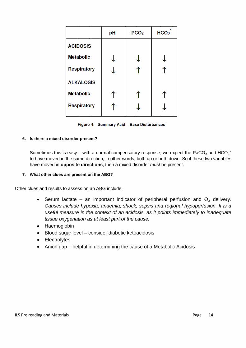
ILS Pre reading and Materials Page 14
6. Is there a mixed disorder present?
Sometimes this is easy – with a normal compensatory response, we expect the PaCO₂ and HCO₃⁻
to have moved in the same direction, in other words, both up or both down. So if these two variables
have moved in opposite directions, then a mixed disorder must be present.
7. What other clues are present on the ABG?
Other clues and results to assess on an ABG include:
Serum lactate – an important indicator of peripheral perfusion and O₂ delivery.
Causes include hypoxia, anaemia, shock, sepsis and regional hypoperfusion. It is a
useful measure in the context of an acidosis, as it points immediately to inadequate
tissue oxygenation as at least part of the cause.
Haemoglobin
Blood sugar level – consider diabetic ketoacidosis
Electrolytes
Anion gap – helpful in determining the cause of a Metabolic Acidosis

ILS Pre reading and Materials Page 15
Example ABG 1
On 28% Oxygen

ILS Pre reading and Materials Page 16
Example ABG 2
On 50% Oxygen
Example ABG 3

ILS Pre reading and Materials Page 17
On Oxygen – Non Re-breather Mask (15 Litres/min)

ILS Pre reading and Materials Page 18
Example ABG 4
On Room Air

ILS Pre reading and Materials Page 19
Management of Acute Hypoxia

ILS Pre reading and Materials Page 20
Oxygen Therapy
- All Critically Unwell and Deteriorating Patients require Oxygen Therapy and Optimum Positioning
- “Failure to correct hypoxia for fear of causing hypoventilation and CO₂ retention, is unacceptable clinical practice.”
(Bateman & Leach, BMJ)
- Patients with COPD are at risk of oxygen induced hypoventilation.
- However, it must be remembered hypoxia kills in minutes hypercarbia kills in hours
- In COPD patients O2 should never be suddenly removed as this may precipitate a rapid fall in SaO2
- When ‘unwell’ these patients should receive titrated oxygen aiming for a saturation of 88-92%
Relative Hypoxia and the Alveolar Gas Equation
How sick is your patient? - Think about the concept of ‘relative hypoxia’ when looking at ABG:
- In a healthy individual breathing air normal PaO₂ is over 75 mmHg
- In other words - 3.5 to 5 times the inspired oxygen concentration
- For Example: at 40% FiO₂ your PaO₂ should be 140-200 mmHg
Initial Actions in the patient with Respiratory Distress
• ABCDEFG
• Give Oxygen
• Sit the patient up
• Call for Help
• Reassure and Commutate with Patient
• Consider bronchodilators
• Consider reversing opiates
• Consider need to call for help for ventilation support

ILS Pre reading and Materials Page 21
ECG Rhythm Interpretation
Tip for Managing Arrhythmias
When Dealing with Bradycardia always think of “DIE”
Drugs (therapeutic and overdose)
Ischaemia
Electrolytes (especially postassium)
Don’t worry about the Rhythm – focus on the patient
Ensure the patient has “IV O2 MONITOR”

ILS Pre reading and Materials Page 22
ECG Interpretation
For further reading see - http://emergencypedia.com/ecg/
Use of a System and Checklists
If you are new to ECG interpretation, using a methodical approach at first helps develop the pattern recognition
abilities that are common in experienced doctors and nurses... (i.e. a good example of pattern recognition in clinical
practice would be a nurse's "endofthebedogram" or "gestalt" suggesting that the patient is in hypovolaemic shock -
she knew this was likely to be the problem because she had seen it many times before in her career).
There are lots of examples of checklists in medicine (e.g. Intensive Care, Theatre and Intubation) and the experienced
clinician can also make use of checklists and templates.
It is often said that the most commonly missed injury is said to be the "second injury". This second injury or problem
often goes unnoticed due to a sense of 'relief' or 'satisfaction' which comes from the practitioner discovering the first
abnormality... Human errors (that we are all at risk of making) are common and often predictable. Some of the
common error producing pitfalls have been described as 'Search Satisfaction', 'Confirmation Bias', 'Anchoring'
and 'Premature Closure'. These pitfalls are summarised in the following link and picture:
Dr Chris Nickson's Cognitive Pitfalls

ILS Pre reading and Materials Page 23
How is all this relevant to ECG interpretation and this course?
We believe that the error producing pitfalls can occur in simple tasks like ECG interpretation as well as complex team
tasks such as Managing ALS Emergencies on the Ward.
In ECG interpretation jumping to (and fixing early) on a diagnosis without a checklist or systematic approach could be
perilous as significant pathology could be missed.
Look at the following ECG as an example:
You might notice that on the ECG Shown that the patient is in 'Rapid Atrial Fibrillation' just by a cursory glance.
However, it would be easy to miss the 'Inferior Myocardial Infarction' if you were distracted by the first (most obvious)
abnormality... If you saw the Myocardial Infarction did you notice the Rapid Atrial Fibrillation?
This is a good example of how even is something as simple and focussed as an ECG interpretation it is very easy to
miss things. In a complex ALS call with challenges in communication as well as clinical skills and medical decision
making problems are enviable…
The PRINT Term ACTS course will discuss the concept of Human Factors and Crew resource management and we
will discuss the Pros and Cons of Checklists and Guidelines.

ILS Pre reading and Materials Page 24
The ECG Checklist
An ECG checklist or template, not only serves as a good learning tool for the novice ECG reader, but also should be
useful for more experienced clinicians who are aware of 'human factors' and don't want to miss significant
abnormalities on the ECG. We will be giving you other laminated checklists for you in your work as an Intern…

ILS Pre reading and Materials Page 25
ECG Basics An Interpretation System
We found the process of learning ECGs frustrating at times.
Despite many books and apps reassuringly titled along the lines of 'the ECGs made Easy' there is, in fact, often a
significant degree of difficulty when starting learning about ECGs.
Rest assured, time and patience will get you to your goals eventually...
We suggest you practice as many ECGs as you can both from books and on the wards.
An ECG System
Identify Demographics (name of patient, time take, old ECG for comparison)
Identify as a Complete 12 lead ECG (e.g. not a derived ECG from a monitoring system)
Check Paper speed (25mm/s)
Standard Calibration is 5mm by 10mm
On the ECG look at the bottom centre (paper speed) and bottom right (calibration 'vertical block' of 10mm) for
these details
We have circled the key areas to check in the following ECG portion:
o At a paper speed of 25mm/s:
A 'BIG' square is 0.2 seconds in duration and a 'SMALL' square is 0.04 seconds
Click Here for more on ECG Basics
RATE, RHYTHM and AXIS
o Rate (Big Squares are 0.2 seconds at a paper speed of 25mm/s) = 300/R-R interval squares
o Rhythm (Card Method)
o Axis
Lies at 90 degrees to Isoelectric Lead
This AXIS is Normal if I and II have a positive deflection and AVR is negative

ILS Pre reading and Materials Page 26
Define the Baseline
o Look at the 'TP' Segment – use this to define ‘ST segment elevation’ and ‘PR depression’
The importance of this is described by Brady and Mattu in the following figure:
P waves
o P waves correlate with Atrial electrical activity arising in the Sino-atrial (SA) Node
o The P wave is marked with the Red Arrow:
P wave marked with Red Arrow

ILS Pre reading and Materials Page 27
o Ask yourself:
Does each QRS complex have an associated P wave?
Is the rhythm Nodal in origin (Narrow Complex, No P Waves) or Ventricular in origin
(wide complex and No P wave)
o Types of P wave:
A Bifid P wave - May suggest Left Atrial Enlargement
Peaked P wave = P-pulmonale
This can represent Right Atrial Hypertrophy or represent ‘a pseudo peak’ (Hypokalaemia)
Types of P Wave
Absent P Waves - Think of AF (most common), sinoatrial blocks, junctional and ventricular rhythms
PR interval
o What is the PR interval?
It should be 0.12 to 0.2 seconds
It is measured from the start of the P wave to the start of the QRS complex
o Is the interval constant?
o Is there a QRS complex for every P wave?
Specific PR Interval Problems
Is there a 1st degree heart block?

ILS Pre reading and Materials Page 28
Wolf-Parkinson- White Syndrome (WPW) type I is Upright Delta Wave in V1
WPW type II is Down-going Delta Wave in V1
WPW Syndrome
If increasing PR interval – Consider Wenkebach’s Phenomenon (AKA Mobitz I)
Wenckebach Phenomenon (Mobitz Type I)
If constant but a QRS is dropped regularly – Consider Mobitz Type 2 Heart Block
Mobitz 2
P waves seen but NO association – this may be 3rd
degree (complete) Heart Block
Complete Heart Block

ILS Pre reading and Materials Page 29
PR Segments
o Depression in most leads or elevation of the PR segment in AVR is suggestive of Pericarditis when
associated with concave up ST elevations globally
Q waves
o Non pathological waves are common
These represent normal L-R depolarization in the septum
o Pathological is likely with the following:
- Q waves more than 40ms (1mm) wide
- Q waves more than 25% of R wave or Q waves more than 2mm deep
QRS Complex
o The QRS Complex (marked with the Red Arrow) should be less than 0.12 seconds or 3 small boxes
QRS complex - marked with Red Arrow
o When prolonged suggests a conduction delay as depolarization occurs across the Myocardium
(There is the bundle of His which becomes a right bundle, a left bundle with 2 fascicles (anterior and
posterior) – these can become ‘blocked’ as a result of conduction delay due to Myocardial
Ischaemia, Drugs or Electrolyte Abnormalities
o Look at the Chest leads V1 – V6
If shaped as an ‘M’ in V1 (that is mostly positive RSR pattern in lead V1) and ‘W’ in lead V6
think MARROW – Right Bundle Branch Block
If shaped as W in V1 and M in V6 thing WILLIAM – Left Bundle Branch Block
o You can have Unifascicular Block (that is an isolated BBB), Bifascicular Block (classically RBBB with
Left Axis Deviation) and Trifasicular (RBBB, LAD, Heart block)
ST segments (depression or elevation)
o Elevation or depression of the ST segments classically represents Ischaemic Heart Disease and/or
Myocardial Ischaemia. The elevation or depression is measured from the J point
o J Point (the junction of the QRS complex and beginning of the ST segment)

ILS Pre reading and Materials Page 30
o How much is too much? This depends on the lead – in the Chest lead 2mm of elevation in 2 leads if
significant, in the limb leads (further away) 1mm is significant
QT interval
o Represents Potassium channels – i.e. a marker of repolarisation
Drugs or disease that cause QT interval prolongation can lead to sudden syncope or sudden
death due to an arrhythmia called Torsades de Pointes (Twisting around the point). This is
also known as Polymorphic Ventricular Tachycardia.
While rare QT prolongation is a concern. Treatment includes Magnesium and Potassium
infusions.
o Consider using the QT nomogram to estimate risk of Torsades:

ILS Pre reading and Materials Page 31
T waves
o If peaked think about Hyperkalaemia. If Inverted (outside of III and AVR) think about Paediatrics (T
wave inversions are normal in Kids) and young females (also normal variant). Pathological Causes
of T wave inversion include Myocardial Ischaemia, Pulmonary Embolism, Cardiomyopathy,
Electrolytes Disturbances and Stroke (especially Haemorrhagic)
U waves (and T-U fusion)
o Can be seen when the K is low – an ‘extra’ wave ‘blip’ after the T wave
J waves (Osborn)
o When seen are suggestive of Hypothermia
Epsilon Wave
o Rare finding that suggests Arrythmogenic Right Ventricular Dysplasia
Check for Pacemaker
o Various problems and presence of a pacemaker are detected on the ECG
ECG interpretation is about pattern recognition. However, so as not to miss subtle abnormalities, and to gradually
build up to an adequate level of expertise we need to practice.
In doing this it is best to use a systematic approach such as the approach outlined above. The best way to learn is to
go through lots of sample ECGs (the textbooks below are good resources for this) and take a problem based
approach to learning the theory...

ILS Pre reading and Materials Page 32
ECG 1 – Anterior STEMI

ILS Pre reading and Materials Page 33
ECG 2 – Inferior STEMI with RV Infarction

ILS Pre reading and Materials Page 34
ECG 3 – Hyperkalaemia 1

ILS Pre reading and Materials Page 35
ECG 4 – Hyperkalaemia 2

ILS Pre reading and Materials Page 36
ECG 5 – Bradycardia (Mobitz II)

ILS Pre reading and Materials Page 37
ECG 6 – Narrow Complex Tachycardia

ILS Pre reading and Materials Page 38
ECG 7 – Wide Complex Tachycardia

ILS Pre reading and Materials Page 39
General Tips Making a Referral to a Senior Colleague
• Prepare your information and resources before you call
• Anticipate information you’ll need for the call (e.g. results)
• Consultation when you Need Something or have a Specific Clinical Question
• Be nice (kill them with kindness)
• Don’t respond in a passive or rude manner (even if the consultant you are calling is coming across as rude – you may have interrupted their dinner and they are a human being so give them the benefit of the doubt).
• Listen Carefully to advice from the Specialist
• This involves you being brief with your explanation
• Use The I S B A R Framework
• Don’t talk for too long – it’s not a long case presentation…

ILS Pre reading and Materials Page 40
• Use Negotiation Skills (see below) • “Credibility, authority, and being LIKED are powerful persuasion tools” Cliff Reid (2013)
• Show a genuine respect for the colleagues opinion – show respect for their point of view even if you
don’t agree with them…
• Compromise
• Be specific about your concerns and questions
• It may take a few calls to get what you need
• “If I send that D-dimer off straight away are you happy to have a look at the patient for me? I think
they’d benefit from you having a look.”
• Close the loop – repeat back what has been discussed – cross-check what will happen with the
patient
• Ask a helpful senior colleague for help early if things are not going as planned…
Negotiation Strategies
• Authority Individuals are more likely to comply with experts/authority – you may not have this
as an intern but you may be able to call on the help of someone who has…
• Reciprocity (“Do us a favour”) If you give something to people, they feel compelled to return the favour. - e.g. It sounds like the d-dimer sounds like an important test for you to have – I’ll
make sure that gets done right now if you wouldn’t mind seeing the patient in the next 20 mins or so?
• Scarcity
This is less applicable to medicine – i.e. rare items are more valuable to people
• Liking We are more inclined to follow the lead of someone who is similar to us rather than
someone who is dissimilar
• Consistency • Commitment • Social Proof We view a behaviour as correct if others are performing in a similar manner.

ILS Pre reading and Materials Page 41
Essential Crisis Management Skills
• Know your environment • Anticipate and plan
• Effective team leadership • Active team membership • Effective communication
• Be situational aware • Manage your resources
• Avoid and manage conflicts • Be aware of potential errors
The Concept of Human Factors
We suggest you think about management of the TEAM, YOURSELF and the ENVIRONMENT in order to remain in control when managing difficult situations:
• YOU – Are you H.A.L.T? Hungry, Angry, Late or Tired – ‘stop yourself’ making a mistake if you are
• ENVIRONMENT – Are you familiar? Noise Levels (patient, team)? Distractions? Use Checklists to control the Emergency and free up your brain to think clearly
• The highly functioning TEAM requires good leadership and followership to move forward effectively
and efficiently towards shared common goals. As the team leader it is important to continuously allow for feedback, share your thought processes and summarise at regular intervals. As a team member it is important to provide feedback and support for the team dynamics to work well…
Elements of Good Teamwork
• A Clear Team Structure • Adequate plan and preparation • Skilled Members – (recognition of extra needs is part of good team work – call for help early) • Effective Team Leadership • Good Team Communication • Full utilization of resources • Wise management of people • Share Common Goals • Collaboration • Regular Education and Training

ILS Pre reading and Materials Page 42
Other Tips for Communication Skills
When managing an Emergency Share your goals with the rest of the team (rather than a list of tasks).
Communicate using names and closed loop communication.
Debrief after Difficult cases and Emergencies and reflect on what went well and what could go better.
Try to share your thoughts and learning points with seniors as well as other Team Members (e.g. Nurses).
You should never feel completely isolated at a big hospital – there’s support available so pick up the phone!
Graded Assertiveness
Having and bringing up a concern about a Senior’s actions in an Emergency can be very difficult and can feel like an impossible task.
One suggested method of bringing to attention a potential error is Graded Assertiveness – we suggest you gently “Cuss your Consultant”:
• C – CONCERN - I am concerned that we haven’t checked for allergies • U – UNSURE - I am uncertain that this Augmentin duo forte medicine can be given to someone with a
possible penicillin allergy • S – SAFETY – I am really worried it is UNSAFE to give this patient a penicillin like drug given his known
allergy. I think this is a patient safety issue…
• S – STOP – Please stop – we need to take a timeout and discuss this further…

ILS Pre reading and Materials Page 43
General Resources
Australian Resuscitation Council (2011) ARC Advanced Life Support Level 2 Manual. Australian
Resuscitation Council.
o http://www.resus.org.au
Westmead Postgraduate Medical Education Centre (2013) Advanced Life Support Workbook.
Westmead Hospital
o A copy can be borrowed by attending the Post Graduate Education Centre (PMEC) during
office hours Monday to Friday or emailing [email protected]
Surviving as an Intern - http://emergencypedia.com/2013/09/01/tips-for-the-junior-doctor-of-2014/
Specific Resources
The use of a systematic approach in assessment of the deteriorating patient
o Jacques T et al (2010). DETECT Manual 2nd Edition. NSW- Clinical Excellence Commission.
o http://nswhealth.moodle.com.au/DOH/DETECT/content/
Basic Life Support (Review) and Advanced Life Support (Introduction)
o ARC Website (See above)
o Westmead ALS Team video - http://www.youtube.com/watch?v=KzA-7o6IdyM
o Crisis Code Videos - http://www.crisiscode.org/course-contents/
Teamwork and an Introduction to the Concept of Human Factors
o Westmead Team Training - http://emergencypedia.com/2013/05/02/human-factors-and-team-training/
Communication and an Introduction to the Concept of Crew Resource Management (CRM)
o Human Factors in Medicine: Lessons from Commercial Aviation
http://www.youtube.com/watch?v=OevZsR6mwZc

ILS Pre reading and Materials Page 44
Handover (ISBAR)
o http://nswhealth.moodle.com.au/DOH/DETECT/content/00_worry/when_to_worry_06.htm
o http://www.sahealth.sa.gov.au/wps/wcm/connect/public+content/sa+health+internet/clinical+resources
/safety+and+quality/clinical+handover/isbar
Criteria for Escalation at Westmead Hospital (PACE and ALS calls)
o This will be discussed in our introductory facilitated discussions
o http://www.health.vic.gov.au/sssl/downloads/bib_merit.pdf
Local Guidelines
o Myocardial Infarction - http://www0.health.nsw.gov.au/policies/pd/2011/pdf/pd2011_037.pdf
o Sepsis – “Recognition, Resuscitate and Refer” -
http://www.cec.health.nsw.gov.au/__documents/programs/sepsis/sepsis_pathway_final_3_may_2011
_adult.pdf
ECG and Arrhythmia Interpretation
o http://www.resus.org.uk/pages/alsabgGd.pdf
Arterial Blood Gas analysis
o http://emergencypedia.com/2013/05/23/the-ed-arterial-blood-gas-abg/
Management of the Patient with Acute Respiratory Distress

ILS Pre reading and Materials Page 45
Appendix
Changes to the 2010 ALS Guidelines
Adapted from http://lifeinthefastlane.com/education/ccc/ilcor-guideline-changes-2010
(1) An Emphasis on Good Compressions
• Make sure good BLS is occurring • CAB rather than ABC • Minimise interruptions • Allow complete chest recoil • At least 100/min rather than approximately 100/min • Compression depth: at least 5cm in adults and 4cm in infants • No change in ratios: 30:2 adults and children (with children can go to 15:2) • Avoid excessive ventilation (2) Basic Life Support
• Immediate recognition of unresponsiveness and activation of emergency response services • Initiation of CPR if unresponsive or not breathing normally • Look-listen-feel removed • Good quality CPR (push hard, push fast)
(3) Advanced Life Support
• Emphasis in minimising duration of pre & post shock pauses (we teach COACH at Westmead) • CPR should not stop > 5 seconds • Early defibrillation (1 better than 3 stacked shocks) • AED’s in hospitals as well as for children and infants • If initial shock is unsuccessful -> give same or greater energy • No CPR device has been shown to be superior to manual CPR • Precordial thump only for witnessed arrest - should not delay defibrillation or CPR • Early intubation with minimal disruption to CPR (then continuous CPR with breaths 10/min) • Capnography recommended: ET tube placement, quality of CPR and detection of ROSC ALS Drug Changes: • Atropine not recommended for PEA/asystole • Adrenaline 1mg every 4 minutes during CPR • Amiodarone 300mg after 3rd shock
• Don’t use ETT for drugs (IV or IO)

ILS Pre reading and Materials Page 46
• Adenosine used for diagnosis and treatment of unstable, undifferentiated, regular, monomorphic wide-complex tachycardia (don’t use in irregular wide-complex tachycardia)
Cardioversion:
• AF: 120-200J (biphasic), 200J (monophasic) • Atrial flutter: 50-100J (bi or monophasic) • Stable VT: 100J (bi or monophasic) (4) Post Cardiac Arrest Care
• Optimise cardiopulmonary function and perfusion • Transport to appropriate hospital (cath lab, neurological care, goal directed critical care,
hypothermia) • Identify and treat precipitating causes • Avoid hyperoxia (SpO2 >94% but not 100%) • Primary PCI is appropriate in the comatosed with ROSC • Glucose < 10mmol/L • Therapeutic hypothermia • Traditional measures of prognosis @ 72 hrs are now not as predictive given benefits seen of
therapeutic hypothermia. • Monitor for and treat seizures
(5) Acute Coronary Syndromes
• Out of hospital 12 lead ECG: reduce time to thrombolysis and PCI • Cardiac arrest and ACS if ischaemic in origin -> requires PCI (coma is not a contraindication) • If saturations > 94% don’t require O2 • Chest pain: use nitrates, use morphine cautiously (associated with increased mortality)