Embed Size (px)
Citation preview

The 78,000-Mr Intermediate Chain of Chlamydomonas Outer Arm Dynein Is a Microtubule-binding Protein Stephen M. King,** Ramila S. Patel-King,* Curtis G. Wilkerson,* and George B. Witman* *Department of Biochemistry, The University of Connecticut Health Center, Farmington, Connecticut 06032; and*Cell Biology Group, Worcester Foundation for Biomedical Research, Shrewsbury, Massachusetts 01545
Abstract. A previous study (King et al., 1991. J. Biol. Chem. 266:8401-8407) showed that the 78,000-Mr inter- mediate chain (IC78) from the Chlamydomonas outer arm dynein is in direct contact with e~-tubulin in situ, suggesting that this protein may be involved in binding the dynein to the doublet microtubules. Molecular ge- netic analysis of this chain recently demonstrated that it is a WD repeat protein essential for outer arm assembly (Wilkerson et al., 1995. J. Cell Biol. 129:169-178). We have now transcribed and translated IC78 in vitro, and demonstrate that this molecule binds axonemes and mi- crotubules, whereas a homologous protein (the 69,000- M r intermediate chain [IC69] of Chlamydomonas outer arm dynein) does not. Thus, IC78 is a bona fide micro- tubule-binding protein. Taken together with the previ- ous results, these findings indicate that IC78 is likely to
provide at least some of the adhesive force that holds the dynein to the doublet microtubule, and support the general hypothesis that the dynein intermediate chains are involved in targeting different dyneins to the spe- cific cell organelles with which they associate. Analysis of the binding activities of various IC78 deletion con- structs translated in vitro identified discrete regions of IC78 that affected the binding to microtubules; two of these regions are specifically missing in IC69. Previous studies also showed that IC78 is in direct contact with IC69; the current work indicates that the region of IC78 that mediates this interaction is coincident with two of IC78's WD repeats. This supports the hypothesis that these repeats are involved in protein-protein interac- tions within the dynein complex.
YNEINS are molecular motors responsible for ciliary and flagellar movement, the directed movement of membrane-bound organelles, positioning of the
Golgi apparatus, nuclear movements, spindle formation and orientation, and possibly some types of chromosomal movement. Dyneins occur as several distinct isoforms, each of which consists of two or more heavy chains (DHCs) 1 associated with a variety of accessory proteins (for reviews see Holzbauer et al., 1994 and Witman et al., 1994). Recent molecular studies have indicated that at least some of the accessory proteins, namely, the interme- diate chains (ICs), found in cytoplasmic and flagellar outer arm dynein derive from a common ancestor (Mitchell and Kang, 1991; Paschal et al., 1992; Wilkerson et al., 1995). This observation has raised the possibility that the ICs of flagellar outer arm dynein and cytoplasmic dynein per-
Address all correspondence to Stephen M. King, Department of Bio- chemisty, The University of Connecticut Health Center, 263 Farmington Avenue, Farmington, CT 06032. Tel.: (203) 679-3347. Fax: (203) 679-3408. E-mail: [email protected]
1. Abbreviations used in this paper. DHC, dynein heavy chain; EDC, 1-ethyl- 3-(3-dimethylaminopropyl) carbodiimide; IC, dynein intermediate chain; IC/LC, intermediate chain/light chain complex; LC, dynein light chain; MARP, microtubule-associated repetitive protein.
form related functions. It has been suggested that one function of the ICs is to bind the dynein to its cargo (mi- crotubules in the case of flagellar dynein; membranous vesicles and other cell organelles in the case of cytoplasmic dynein) (King and Witman, 1990; King et al., 1991; Paschal et al., 1992). However, to date, there has been little experi- mental evidence (see below) to support this proposal.
We have been using the outer arm dynein from Chlamy- domonas as a model system to learn more about the func- tions of specific dynein subunits. This dynein (Fig. 1 A) consists of 3 DHCs (et, 13, and -/; each >500 kD [Mitchell and Brown, 1994; Wilkerson et al., 1994]), 2 ICs (IC78 and IC69 [Also referred to as IC70; Mitchell and Kang, 1991]; 76.5 and 63.4 kD, respectively [Mitchell and Kang, 1991; Wilkerson et al., 1995]), and 10 light chains (LCs; 8-22 kD [King and Patel-King, 1995; Pfister et al., 1982; Piperno and Luck, 1979]). The purified dynein retains both ATP- sensitive (motor) and -insensitive (structural) microtubule- binding domains (Haimo et al., 1979; Haimo and Fenton, 1988). Structurally, each DHC consists of a globular head and a flexible stem. The head, formed from the COOH- terminal ~350 kD, contains the ATPase and motor do- mains, and is presumed to bind transiently to the B tubule of the opposing doublet microtubule during force genera- tion. The stem, formed from the NHe-terminal part of the
© The Rockefeller University Press, 0021-9525/95/10/399/11 $2.00 The Journal of Cell Biology, Volume 131, Number 2, October 1995 399-409 399
on April 10, 2019jcb.rupress.org Downloaded from http://doi.org/10.1083/jcb.131.2.399Published Online: 15 October, 1995 | Supp Info:

Figure 1. (A) Model for the outer arm dynein of Chlamydomo- nas and its association with the outer doublet microtubule (shaded). Intermediate chains IC78 and IC69 are closely associ- ated with each other and with the 8-, 11-, 14-, and 20-kD LCs in an IC/LC complex (Mitchell and Rosenbaum, 1986; King et al., 1991) located at the base of the stems of the et, 13, and y DHCs (King and Witman, 1990). The 19-kD LC is associated with the NH2-terminal one-third of the [3 DHC (which presumably corre- sponds to that chain's stem) (Sakakibara et al., 1993), but has not been shown to be a part of the IC/LC complex. The 16-kD LC and 18- and 22-kD LCs are associated with the a and y DHCs, re- spectively (Pfister and Witman, 1984), although their location in the heads of these chains is speculative. (B) Predicted domain structure of IC78 showing the basic NH2-terminus (diagonally hatched box), the glutamine-proline-rich region that is expected to form a polyproline II helix (filled box), and WD repeats A-F (horizontally hatched boxes) (Wilkerson et al., 1995).
chain, extends to the base of the dynein and appears to in- teract with the stems of the other DHCs to hold the dynein together (Johnson and Wall, 1983; Sakakibara et al., 1993; Witman et al., 1983; Goodenough and Heuser, 1984; Sale et al., 1985; for review see Witman et al., 1994). The ICs in- teract with each other and with several of the LCs to form an IC/LC complex (Mitchell and Rosenbaum, 1986; King et al., 1991) that is located at the base of the soluble dynein particle (King and Witman, 1990). Studies using the zero- length cross-linker bethyl-3-(3-dimethylaminopropyl) car- bodiimide (EDC) revealed that IC78 is in direct contact with et-tubulin in situ, suggesting a role for this component in the ATP-insensitive (or structural) binding of the outer arm to the flagellar doublet microtubule (King et al., 1991).
To date, this latter observation provides the sole experi- mental basis for the hypothesis that ICs are involved in at- tachment of the dynein motor to its cargo.
Recently, we described the cloning and sequencing of a full-length cDNA encoding IC78 (Wilkerson et al., 1995). The predicted sequence indicates that the NH2-terminal 22 residues constitute a highly charged, lysine-rich region (Fig. 1 B). This basic region is separated from the rest of the protein by a short glutamine-proline-rich region pre- dicted to form a polyproline II helix. Polyproline II helices generally form flexible linkers between domains of a pro- tein (Adzhubei and Sternberg, 1994), suggesting that the highly charged NH2-terminal region is a discrete structural domain and probably a discrete functional domain as well. The COOH-terminal half of the protein contains a series of six repeated elements known as WD repeats; such re- peats are believed to be involved in subunit-subunit inter- actions within multisubunit complexes (Neer et al., 1994). Although not previously reported, the WD repeats also are present in Chlamydomonas IC69 and in the 74,000-Mr IC (IC74) of cytoplasmic dynein, which are homologous to IC78 (Wilkerson et al., 1995). The fact that the WD re- peats are conserved suggests that they may perform simi- lar functions in the different chains. Using the IC78 cDNA as a probe, we identified seven independently isolated strains, generated by insertional mutagenesis and selected on the basis of a slow swimming phenotype, in which the IC78 gene was completely removed from the genome, and one strain in which there was a large insertion within this gene (Wilkerson et al., 1995). Electron microscopic analy- sis of the disruption mutant and of several of the deletion mutants revealed that in every case the outer arm was completely and specifically lost, indicating that IC78 is es- sential for outer arm assembly.
To clarify the role of IC78 in outer arm assembly, we have now investigated the ability of in vitro-translated IC78 to bind to microtubules and axonemes. The results demonstrate that IC78 is a bona fide microtubule-binding protein, suggesting that IC78 is essential for outer arm as- sembly because it provides at least some of the adhesive force for binding the arm to the flagellar microtubules. This finding provides additional support for the general hypothesis that dynein ICs are involved in binding dyneins to the organelles with which they associate. We also have investigated the relationship between IC78 functional do- mains and the predicted structural domains of the polypep- tide. We have identified two, and possibly three distinct regions within IC78 that affect microtubule-binding activ- ity; one of these is contained within the highly charged NH2-terminal region, the other is located in the center of the molecule and includes another highly basic sequence. The homologous protein IC69 does not bind microtubules in vitro; although IC69 also contains basic regions, the spe- cific sequences that would have been homologous to the regions important for microtubule-binding in IC78 are not present in IC69. Finally, we have identified the region of IC78 that is involved in binding to IC69. This activity is lo- calized to two of the WD repeats in IC78, supporting the hypothesis that these repeats may be responsible for sub- unit-subunit interactions within the dynein complex. It is likely that at least some of the WD repeats in IC69 of outer arm dynein and IC74 of cytoplasmic dynein have a
The Journal of Cell Biology, Volume 131, 1995 400
similar function, as such subuni t -subuni t interact ions are common to all dynein ICs.
Materials and Methods
Plasmids and Constructions Two plasmids were used in this study: pc78k3, which consists of a 2.65-kb insert containing the entire coding sequence for IC78 cloned between the EcoRI and XhoI sites of pBluescript II S K - (Wilkerson et al., 1995), and pBc70-16, which contains the cDNA for IC69 cloned into pBluescript II KS+ (Mitchell and Kang, 1991). pBc70-16 was the generous gift of Dr. D. Mitchell (SUNY Health Science Center, Syracuse, NY). NHE-terminal de- letions were constructed in one of two ways. In the first, pc78k3 was di- gested with Sinai, Pstl, and BamHI (each enzyme cuts once within the coding sequence and once within the 5'-polylinker) and religated. The re- sulting constructs (78ASmaI, 78APstI, and 78ABamHI) rely on internal codons for translation initiation and are expected to produce fragments corresponding to M324-H683, M453-H6s3, and M480-H683, respectively. To construct the NH2-terminal deletion series 78A213N-78A819N 2, a linker containing part of the 5'-untranslated region of IC78, a translation initia- tion signal abutted to an EcoRI site, and an in-frame stop codon flanked by an upstream ApaI site and a downstream SalI site was cloned between the XbaI and EcoRI sites of pc78k3. This plasmid then was digested with ApaI to protect the vector and with SalI to provide a substrate for the exo- nuclease ExoIIl. The digestion was stopped at various times, the plasmid cut with EcoRI, blunt ended with $1 nuclease, and the ends repaired with T7 DNA polymerase. The 5' ends of individual clones then were se- quenced to determine the extent of the deletion; only those that retained the correct reading frame were used.
For COOH-terminal deletions of IC78 (the series 78A679C-78A1951C) and 78ASmaI (78ASmaIAC), a linker containing KpnI and NruI sites was inserted between the XhoI and KpnI sites (the latter was not preserved upon religation) in the 3'-polylinker of both plasmids. Plasmids then were cut with XhoI and KpnI to provide appropriate suhstrates for exonuclease digestion. Individual .clones were sequenced to determine the precise point of truncation. Insertion of the linker, which provides a unique site with which to linearize the plasmid for m R N A runoff, was made necessary by our observation that vector protected from digestion by the 3' over- hang of the ApaI site (located between XhoI and KpnI in the original polylinker) apparently could not be digested with $1 nuclease and there- fore could not be religated.
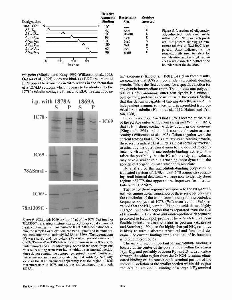
Oligonucleotide-directed deletions within 78A1309C were made essen- tially as described by Kunkel et al. (1987). For selection purposes, each oligonucleotide incorporated a restriction site determined by the terminal three bases 5' to the desired deletion. Therefore, for each construct a sin- gle residue encoded by the other half of the restriction site is inserted within the boundaries of the deletion. The restriction sites used and the residue inserted are tabulated in Fig. 8.
In Vitro Transcription and Translation Plasmid constructs were linearized and transcribed in vitro using T3 RNA polymerase; pbc70-16 was transcribed with T7 RNA polymerase. Individ- ual RNAs were translated using the rabbit reticulocyte lysate system (Promega Corp., Madison, WI) containing [35S]methionine (New England Nuclear, Boston, MA). Before use for axoneme/microtubule-binding ex- periments, the lysates were spun at ~180,000 g for 5 min in an airfuge (Beckman Instruments, Inc., Palo Alto, CA) to pellet any nascent polypep- tide chains still attached to polysomes.
The specific activity of the [35S]methionine pool in the lysate was deter- mined as described by Butner and Kirschner (1991). From this value and the number of methionines in IC78, the specific activity of the in vitro
2. Nomenclature of the IC78 deletion constructs is based on the number- ing of the cDNA clone 78k3 (Wilkerson et al., 1995). For NH2- and COOH-terminal deletions, the number shown indicates the first or last base in the construct, respectively. For example, the designation 78A679C indicates a COOH-terminal deletion that has removed all but the first 679 bases of the clone 78k3. The three constructions made using internal re- striction sites to remove NH2-terminal regions are indicated with the name of the endonuclease used, e.g., 78ABamHI. The residues of IC78 en- coded by each construct are tabulated in Fig. 3.
translated polypeptide was calculated. The amount of IC78 translated in vitro was then determined by cutting IC78 bands (identified by autora- diography) out of gels and counting the amount of radioactivity in the bands.
Preparation of Axonemes and Microtubules Chlamydomonas flagella were isolated from the wild-type strain l132D- and from the outer armless mutant odal by standard methods (Witman, 1986). Flagella were demembranated with HMEK buffer (1% NP-40, washed with 10 mM Hepes, pH 7.5, 5 mM MgSO4, 0.5 mM EDTA, 25 mM KC1), and either used immediately or stored in 50% glycerol at -20°C. Stored axonemes were washed twice with HMEK before use.
DEAE-purified bovine brain tubulin was prepared by the procedure of Vallee (1986) and was kindly provided by Dr. Anthony Moss (Auburn University, Auburn, AL). Polymerized microtubules were stabilized with taxol before use in the sedimentation assays.
Sedimentation Assays To assess the axoneme/microtubule-binding activity of in vitro translation products, 10 ~tl of the reticulocyte lysate was added to a microfuge tube containing 10 ~l HMEK buffer and 10 txl axonemes (6 mg/ml) or microtu- bules (18 mg/ml). The same amounts were used in all experiments. Con- trol tubes contained reticulocyte lysate and 20 p,1 HMEK, but no axo- nemes or microtubules. Samples were incubated at room temperature for 30 min and then sedimented for 1 min in a microfuge or, when microtu- bules were used, in an airfuge. Supernatants were removed and the pellets washed in 500 izl HMEK before preparation for gel electrophoresis (King et al., 1986). It is important to note that all binding experiments took place in the presence of >50 mg/ml cellular protein derived from the reticulo- cyte lysate. Quantitation of autoradiographs was performed using the pro- gram Quantity One (pdi, Huntington Station, NY) running on a Sparc workstation (Sun Microsystems Inc., Mountain View, CA). The amount of binding observed for any one construct was quite reproducible, e.g., in four different experiments performed on different days, values of 67, 70, 72, and 77% were obtained for the amount of 78A1309C bound to wild- type axonemes.
Cross-linking experiments were performed as described in King et al, (1991). Briefly, axonemes with bound in vitro translation products were resuspended in HMEK buffer and treated with 0 or 1 mM EDC (Pierce Chemical Co., Rockford, IL) for 60 min at room temperature. The reac- tion was terminated by the addition of a fivefold molar excess of I~-mer- captoethanol before preparation for electrophoresis. Samples were separated in 8 or 10% acrylamide minigels and then subjected to autora- diography. 14C-labeled molecular weight standards were obtained from Amersham Corp. (Arlington Heights, IL).
Immunoprecipitation mAbs 1878A and 1869A (King et al., 1985; 1986) were purified from as- cites fluid and bound to protein A-Affigel beads (Bio-Rad Laboratories, Hercules, CA). The antibody-protein A interaction was stabilized by cross-linking with dimethylpimelimidate (Pierce Chemical Co.) by the procedure of Schneider et al. (1982). For immunoprecipitation experi- ments, the beads were incubated for 30-60 min with reticulocyte lysates containing in vitro translation products and washed several times with 0.05% Tween 20 in TBS, before resuspension in electrophoresis sample buffer.
Computational Methods The program PHD was used to predict the secondary structure of IC78 (Rost and Sander, 1993). Hydrophobicity and surface probability were de- termined using the GCG suite of programs (Devereux et al., 1984).
Results
IC78 Is a Bona Fide Microtubule-binding Protein
Previously, we used the zero-length cross-linking reagent EDC to demonstrate that, within the axoneme, IC78 inter-
King et al. lC78 of Outer Arm Dynein Is a Microtubule-binding Protein 401
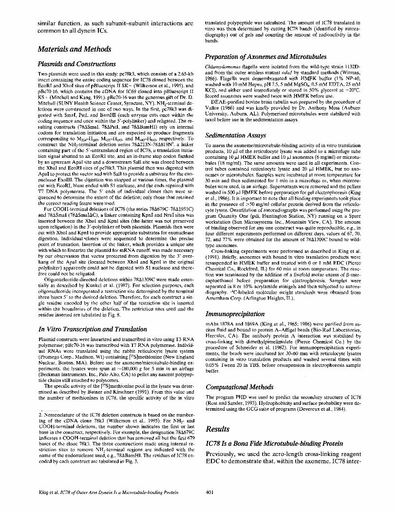
acts directly with a-tubulin (King et al., 1991). However, those experiments could not determine whether IC78 merely is in contact with tubulin or is a microtubule-bind- ing protein per se. To address this important question, we investigated the ability of in vitro-translated IC78 to bind to microtubules. The plasmid pc78k3 (Wilkerson et al., 1995), which contains the full-length c D N A encoding IC78, was transcribed in vitro, and the resulting m R N A translated in a rabbit reticulocyte lysate system. Quantita- tion of radioactivity incorporated into IC78 revealed that ~1 fmol (~80 pg) protein was synthesized per microliter reticulocyte lysate. Subsequently, the ability of the trans- lation products to bind axonemes and taxol-stabilized bovine brain microtubules was assessed in a simple sedi- mentation assay. In the absence of axonemes, in vi tro- translated IC78 remained in the supernatant. In the pres- ence of axonemes (derived either from wild-type or from the outer armless strain odal), significant amounts (up to 60%) of the IC78 translation product were found in the pellet (Fig. 2 A, top panel). Similar results were obtained when microtubules were substituted for axonemes in the assay (Fig. 2 B), indicating that the binding was due to di- rect interaction between IC78 and tubulin. Binding of IC78 to axonemes was sensitive to the addition of 0.6 M NaC1 (not shown), which is the salt concentration required to release >90% of outer arm dynein from the axoneme. Approximately 86% of bound IC78 was released by the salt treatment, indicating that binding is mediated largely by ionic interactions similar to those which attach the arm to the doublet microtubule in vivo. Further evidence that this interaction is similar to that which occurs in vivo was obtained from protein cross-linking experiments described below.
Assuming that tubulin accounts for ~ 5 0 % of total axo- nemal protein, each assay contained ~1 nmol tubulin; when brain microtubules were used, the concentration of tubulin was even higher. Inasmuch as the assay mixture contained only ~10 fmol of IC78, these experiments were performed under conditions where a large excess (~105- fold in molar terms) of potential IC78 binding sites were present. Because only extremely small amounts of in vitro translated product were available, it was not possible to saturate the available binding sites in the sedimentation assays. However, the fact that much of the IC78 bound to axonemes or microtubules, even though IC78 was present at a concentration of only 80 ng/ml in a background of >50 mg/ml cellular protein from the lysate, strongly suggested that the IC78-microtubule interaction was specific.
As an alternative test of whether the interaction of IC78 with the microtubules was specific, we examined the bind- ing properties of IC69. IC69 is homologous to IC78, has a similar isoelectric point (predicted 5.4 vs. 6.6 for IC78), has similar WD repeats, and also has a basic NH2-terminus (see Fig. 10) (Wilkerson et al., 1995), but it is not cross- linked to tubulin by treatment of the intact axoneme with E D C (King et al., 1991). IC69 was produced by in vitro transcription and translation of the plasmid pBc70-16 (Mitchell and Kang, 1991). Under the same assay condi- tions as were used for IC78, IC69 remained exclusively in the supernatant and showed no detectable axoneme-bind- ing activity (Fig. 2 A, bottom panel). These results indicate that the interaction of IC78 with axonemes and microtu-
Figure 2. IC78 binds axonemes in vitro. (A) IC78 and IC69 were transcribed and translated in vitro. Translation mixes (10 ixl) were added to HMEK buffer (20 ~1) alone or to buffer contain- ing 3 mg/ml wild-type axonemes. The samples were incubated at room temperature for 30 min, sedimented in a microfuge, and the pellets washed with 500 p.1 HMEK. Samples were separated in an 8% acrylamide minigel and autoradiographed. In the absence of axonemes (-Axo), both IC78 (top panel) and IC69 (bottom panel) were found in the supernatant (S). After axoneme addi- tion (+Axo), ~40% of IC78 was found associated with the pellet (P), whereas IC69 remained in the supernatant. The minor short fragments visible in these and subsequent autoradiographs derive from translation initiation within the IC78 and IC69 coding re- gions, presumably at internal methionines. (B) In vitro translated IC78 was incubated in the presence or absence of taxol-stabilized brain microtubules. Samples were pelleted in the airfuge and treated as described in A.
bules is specific. We conclude that IC78 is a bona fide mi- crotubule-binding protein. The results also indicate that, in contrast to IC78, IC69 is not a microtubule-binding pro- tein.
Identification oflC78 Regions that Affect Microtubule Binding
To determine which regions of IC78 are important for binding to microtubules, we generated a series of con- structs designed to produce truncated versions of IC78 in which various amounts of the molecule were deleted from the NH2-terminus, the COOH-terminus, or both (Fig. 3).
The Journal of Cell Biology, Volume 131, 1995 402

These constructs were then transcribed and translated in vitro, and assayed as above to determine if they were capa- ble of binding to axonemes or brain microtubules.
As reported above, the intact IC78 bound to and cosedi- mented with axonemes or brain microtubules. However, truncation of the NHe-terminal 20 residues of IC78 (con- struct 78A213N) resulted in loss of binding activity (Fig. 4); the remaining portion of the chain (G21-H683, preceded by a methionine inserted for initiation) was unable to bind to the axonemal or brain microtubules. Fragments in which more extensive portions of the NH2-terminal region were deleted (up to K223; constructs 78A414N, 78A522N, 78A735N, and 78A819N) similarly were unable to bind to microtubules. These results indicate that at least the first 20 residues of IC78 are important for microtubule binding.
Truncated versions of IC78 in which the COOH-ter- minus was deleted down to L386 (constructs 78A1951C- 78A1309C) bound readily to axonemal or brain microtu- bules. For example, 70% of 78A1309C (M1-L386) bound to and cosedimented with the microtubules (Fig. 5, top panel). However, further truncation towards the NH2-ter- minus (down to L263 or beyond; constructs 78A941C and 78A679C) completely abolished binding (Fig. 5, middle and bottom panels). Therefore, the central region of IC78 (D264-L386) contains a second domain essential for micro- tubule binding.
Surprisingly, if IC78 was truncated from the NH2-termi- nus partially or completely through this second domain (to M324 and beyond), then axonemal and brain microtubule- binding activity was restored to the resulting COOH- terminal fragments (constructs 78ASmaI, 78APstl, and 78ABamHI) (Fig. 6). These results indicate that removal of the entire NH2-terminal half of IC78 unmasks microtu- bule-binding activity in the COOH-terminal half of the molecule. COOH-terminal fragments as short as 203 amino acids (residues M480-H683; construct 78ABamHI) were ca- pable of binding to axonemal microtubules (Fig. 6, bottom panel). A fragment (M324-5604; construct 78ASmalAC) in which the COOH-terminal 79 residues were deleted also
bound to microtubules (Fig. 6, bottom panel). Thus, at least some of the COOH-terminal microtubule-binding activity is likely to be contained in the region of overlap between the latter two fragments (residues M480-$604). This does not rule out the possibility that there are addi- tional microtubule-binding sites COOH- or NH2-terminal to this region. The interaction between 78ASmaI and ax- onemes was partially sensitive to increasing ionic strength; 58% of the bound translation product was released by washing with HMEK buffer containing 0.6 M NaCI, and additional amounts were released by increasing the salt concentration to 1 M (not shown).
NH2-terminal Hal f o f lC78 Is Cross-linked to a-tubulin by E D C
It was of considerable interest to determine which of the regions identified above were binding to microtubules in a physiologically relevant manner. Inasmuch as IC78 in dynein in situ can be readily cross-linked to axonemal ct-tubulin by low concentrations of EDC (King et al., 1991), we investigated whether the intact chain, and frag- ments representing the NH2- and the COOH-terminal half of the chain, could be similarly cross-linked to a-tubulin after their binding to odal axonemes in vitro.
IC78 was translated in vitro and bound to odal ax- onemes. An aliquot of the axonemes was then treated with 1 mM EDC; an identical aliquot was left untreated. After analysis of the aliquots by SDS-PAGE (Fig. 7, top panels), a single additional 127-kD band was observed in the EDC- treated sample. This is precisely the size of the IC78/et- tubulin conjugate identified previously after treatment of intact, native axonemes with EDC (King et al., 1991). Be- cause EDC cross-links only proteins in which aspartate or glutamate residues on one protein are in intimate contact with lysine residues on the other protein (Hoare and Koshland, 1966), the IC78/tubulin interaction in vitro is likely to be very similar to that which occurs in vivo. This provides additional evidence that in vitro translated IC78
Figure 3. Properties of IC78 and NH2- and COOH-terminally de- leted constructs. This diagram indicates the regions of IC78 covered by the various con- structs described in Results, and whether that product was com- petent to bind axonemes/micro- tubules and IC69. Predicted structural features of IC78 are as in Fig. 1 B. Regions important for microtubule binding (solid bars 1, 2, and 3) and IC69 bind- ing (vertically hatched bar) are indicated at the top of the figure; solid bar 2 corresponds to resi- dues D264-D323 (see Results). n.d., not determined.
King et al. 1C78 of Outer Arm Dynein Is a Microtubule-binding Protein 403

Figure 4. The NH2-terminus of IC78 is essential for microtubule binding. The fragment 78A213N, which lacks only the first 20 amino acids of IC78, was expressed in vitro and assayed for mi- crotubule binding activity as in Fig. 2. All of the product re- mained in the supernatant in both the presence and absence of axonemes.
binds to axonemal microtubules in a specific and physio- logically re levant manner .
A similar exper iment was carried out with the f ragment 78A1309C (M1-L386) , which contains both the NHE-termi- nal and the central regions that were observed to be im-
Figure 6. Microtubule-binding activity in the COOH-terminal re- gion of IC78. Three NH2-terminal deletion constructs (78ASmaI, 78ABamHI, and 78ASmaIAC), encoding overlapping sections of the COOH-terminal region of IC78, were transcribed and trans- lated in vitro. The ability of each translated product to bind odal axonemes then was assessed in the sedimentation assay. Samples were separated in an 8% minigel and autoradiographed. All three products bound avidly, indicating that IC78 contains a mi- crotubule-binding domain within the region of overlap (shaded box in diagram). Note that for 78ABamHI, the region containing the centrally located site that is important for microtubule bind- ing (see Fig. 5) has been completely removed. The multiple bands are due to translation initiation at several internal residues.
Figure 5. The central region of IC78 is important for microtubule binding. The axoneme-binding behavior of three COOH-termi- nally truncated versions of IC78 are shown. Constructs 78A1309C, 78A941C, and 78A679C represent IC78 residues M1-L386, Ml-L263 ' and MI-Q175, respectively. After the sedimentation assay, sam- ples were separated in a 10% minigel and autoradiographed. In the absence of axonemes, all three products remained in the su- pernatant. However, in the presence of odal axonemes, >70% of 78A1309C was found in the pellet. Neither 78A941C nor 78A679C showed detectable axoneme-binding activity. The lower panel di- agramatically illustrates the regions of IC78 covered by these constructs; the shaded box indicates the region important for mi- crotubule binding.
por tant for microtubule binding in vitro. React ion of ax- oneme-bound 78A1309C with E D C genera ted a single addi t ional band with a relat ive molecular mass of 92,000 (Fig. 7, bottom panels). The mass of this conjugate is con- sistent with the cross-linking of the 43-kD translat ion product to tubulin. In contrast, when the same exper iment was carried out with bound 78ASmaI (M324-H683) , which contains the COOH- te rmina l microtubule-binding activ- ity, no cross-l inked products were observed, even at higher concentrat ions of E D C (Fig. 7, middle panels). Therefore , it appears that the sites responsible for cross-linking of IC78 to a- tubul in reside exclusively in the NH2-terminal half of the molecule.
Further Definition of Regions in the NH2-terminal Half of lC78 Important for Microtubule Binding
Because microtubule binding was observed only for those large NHE-terminal fragments which contained both the ext reme NH2-terminal domain (Ma-Te0) and the region from D264-L386, it was not possible to use simple trunca- tions to assess the impor tance to microtubule binding of parts of the molecule located be tween these two regions. To investigate the contr ibut ion of these o ther parts to this activity, we produced a series of constructs in which vari- ous small internal regions were de le ted from the fragment
The Journal of Cell Biology, Volume 131, 1995 404

Figure 7. EDC cross-linking of in vitro--translated IC78 and frag- ments. In vitro-translated IC78, 78ASmaI, and 78A1309C were bound to axonemes, washed with HMEK buffer, and then treated with 1 mM EDC. The samples were separated in an 8% minigel and autoradiographed. For IC78, reaction with EDC generated a cross-linked product with a relative molecular mass of 127,000 that appeared to be identical to the product previously shown to derive from the cross-linking of IC78 to ct-tubulin (King et al., 1991). No detectable difference between EDC-treated and un- treated samples was observed for 78ASmaI. However, after treat- ment with EDC, an extra product with a relative molecular mass of 92,000 was obtained with 78A1309C. The apparent mass of this product is consistent with the cross-linking of 78A1309C (43 kD) to tubulin. The diagram indicates the regions of IC78 represented by the constructs.
78A1309C (Fig. 8). These fragments were then assayed for the ability to bind to and cosediment with axonemes. The deletion of the region from $5-D29 reduced by 58% the amount of fragment cosedimenting with axonemes, con- firming the importance of this region for microtubule binding. The deletion P298-D323 reduced binding by 35%. In contrast, deletion of the adjacent regions T272-E293 or A326-5339 had little or no effect on binding. This confirms that most if not all of the microtubule-binding activity in this central part of the molecule is contained within the re- gion extending from D264-D323 (see above), and probably within the smaller region from P298-D323. None of the
other small deletions (ASll6-D140, AK181-E204, AA236-5253) had a noticeable effect on the binding activity of the frag- ment. Therefore, within the NHz-terminal half of IC78, the highly charged NH2-terminal domain and the region from D264-D323 appear to be uniquely important for microtu- bule binding.
IC78 Region that Binds IC69 Is Coincident with Two WD Repeats
IC78 interacts with IC69 in situ (Mitchell and Rosenbaum, 1986; King et al., 1991). To determine the region of IC78 responsible for this interaction, IC69 was translated in vitro and allowed to interact for 30 min with various IC78 constructs that likewise had been translated in vitro. The samples were then immunoprecipitated with either anti- body 1878A or 1869A (both antibodies are highly specific for their respective IC; see King et al., 1985; 1991). The im- munoprecipitates were then examined by SDS-PAGE to determine if the nonimmunoreactive intermediate chain or fragment was coprecipitated.
When intact IC78, 78ASmaI, and 78A1309C were immu- noprecipitated with 1878A, significant amounts of IC69 also were found in the pellets (Fig. 9). These data suggest that the region of IC78 involved in binding IC69 is located in residues M324-L386 (i.e., the overlap region between 78ASmaI and 78A1309C). The 1878A epitope is correlated with the sequence extending from A326 tO F334 (King, S. M., and G. B. Witman, unpublished results); hence, fragments truncated further towards the NH2- or COOH-terminus (e.g., 78A679C or 78ABamHI) lack the 1878A epitope and could not be used here.
In the converse experiment, the antibody 1869A immu- noprecipitated IC69 together with minor amounts of 1C78, 78ASmaI, and 78A1309C (Fig. 9), but did not coprecipitate 78A679C or 78ABamHI along with IC69 (not shown). We previously observed that binding of the 1869A antibody to native outer arm dynein causes significant disruption of the complex (see King and Witman, 1990) and this likely accounts for the small amounts of 1C78 and constructs co- precipitated with IC69 by this antibody. In any case, the data obtained with antibody 1869A support the assign- ment of residues M324-L386 of IC78 as containing the site of interaction with IC69. This section of 1C78 coincides closely with WD repeats A and B (5307--5339 and H358-V390, respectively) (see Fig. 1 B and Wilkerson et al., 1995).
Discussion
In the studies reported here, we found that the 78,000-Mr intermediate chain (IC78) of Chlamydomonas outer arm dynein, produced by translation in a rabbit reticulocyte ly- sate, is able to bind to axonemes and bovine brain micro- tubules in vitro. The interaction appears to be specific and similar to that which occurs in vivo based on the following observations: (a) Binding occurs when IC78 is present at a concentration of only ~80 ng/ml in a background of >50 mg/ml cellular protein in the reticulocyte lysate. (b) The bound protein is released by 0.6 M NaC1, which is the con- centration that releases >90% of outer arm dynein from the axoneme. (c) Binding occurs under conditions where 1C69, a homologous protein with similar WD repeats, a similar basic NH2-terminus, and a similar size and isoelec-
King et al. IC78 of Outer Arm Dynein Is a Microtubule-binding Protein 405
Relative Axoneme Restriction
Designation Binding Site 78A1309C N C 100 .....
ASs-D29 .A 42 XhoI AS.6-D~4o A 100 HindIII ALlsl_E2o4 A 99 SaclI AA23 _S253 A 100 PvuI AY272_E293 /N 100 NsiI /~D29s-D323 ~ 65 Pstl ~t_326- S 339 A 90 BstBI
I I I 3100 0 100 200 Residue
Residue Inserted
E L R S H Q E
Figure 8. Location of oligonucle- otide-directed deletions made within 78A1309C. For each prod- uct, the percent binding to axo- nemes relative to 78A1309C is re- ported. Also indicated is the restriction site used to select for each deletion and the single amino acid residue inserted between the boundaries of the deletion.
tric point (Mitchell and Kang, 1991; Wilkerson et al., 1995; Ogawa et al., 1995), does not bind. (d) EDC treatment of IC78 bound to axonemes in vitro results in the formation of a 127-kD complex which appears to be identical to the IC78/o~-tubulin conjugate formed by EDC treatment of in-
Figure 9. IC78 binds IC69 in vitro. 10 i~1 of the IC78, 78ASmaI, or 78A1309C translation mixture was added to an equal volume of lysate containing in vitro-translated IC69. After incubation for 30 min, the samples were divided into two aliquots and immunopre- cipitated either with antibody 1878A or 1869A. The supernatants (S) were saved and the pellets (P) washed several times with 0.05% Tween 20 in TBS before electrophoresis in an 8% acryla- mide minigel and autoradiography. Some of the short fragments of IC69 resulting from translation initiation at internal methio- nines do not contain the epitope recognized by mAb 1869A and hence are not immunoprecipitated by that antibody. Similarly, some of the IC69 fragments apparently lack the region of IC69 that interacts with IC78 and are not coprecipitated by antibody 1878A.
tact axonemes (King et al., 1991). Based on these results, we conclude that IC78 is a bona fide microtubule-binding protein. This is the first evidence for a specific function for any dynein intermediate chain. That at least one polypep- tide of Chlamydomonas outer arm dynein is a microtu- bule-binding protein is consistent with the earlier finding that this dynein is capable of binding directly, in an ATP- independent manner, to microtubules assembled from pu- rified brain tubulin (Haimo et al., 1979; Haimo and Fen- ton, 1988).
Previous results showed that IC78 is located at the base of the soluble outer arm dynein (King and Witman, 1990), that it is in direct contact with tx-tubulin in the axoneme (King et al., 1991), and that it is essential for outer arm as- sembly (Wilkerson et al., 1995). Taken together with the current finding that IC78 is a microtubule-binding protein, these results indicate that IC78 is almost certainly involved in attaching the outer arm dynein to the doublet microtu- bule by virtue of its microtubule-binding activity. This raises the possibility that the ICs of other dynein isoforms may have a similar role in attaching those dyneins to the specific cell organelles with which they associate.
By analysis of the microtubule-binding properties of truncated versions of IC78, and of IC78 fragments contain- ing small internal deletions, we were able to identify three regions of IC78 that appear to be important for microtu- bule binding in vitro.
The first of these regions corresponds to the NH2-termi- nal ~20 amino acids; truncation of these residues prevents the remainder of the chain from binding to microtubules. Sequence analysis of IC78 (Wilkerson et al., 1995) re- vealed that the NH2-terminal 24 amino acids form a highly charged, lysine-rich region that is separated from the rest of the molecule by a short glutamine-proline-rich segment predicted to form a polyproline II helix. Such helices form flexible linkers between domains in proteins (Adzhubei and Sternberg, 1994), so the highly charged NH2-terminus is likely to form a discrete structural and functional do- main. The current findings imply that one of its functions is to bind microtubules.
The second region important for microtubule binding is located in the center of the polypeptide, within the region D264-D323 and probably between P29S and D323. Truncation through the wider region from the COOH-terminus elimi- nated binding of the remaining N-terminal portion of the molecule; deletion of the smaller section within this region reduced the amount of binding of a large NH2-terminal
The Journal of Cell Biology, Volume 131, 1995 406
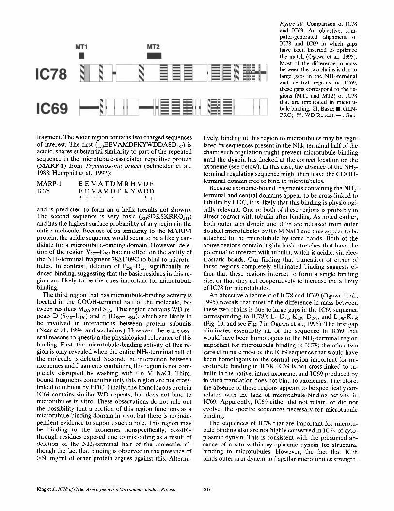
Figure 10. Comparison of IC78 and IC69. An objective, com- puter-generated alignment of IC78 and IC69 in which gaps have been inserted to optimize the match (Ogawa et al., 1995). Most of the difference in mass between the two chains is due to large gaps in the NH2-terminal and central regions of IC69; these gaps correspond to the re- gions (MT1 and MT2) of IC78 that are implicated in microtu- bule binding. [], Basic; II, GLN- PRO; [~, WD Repeat; B , Gap.
fragment. The wider region contains two charged sequences of interest. The first (273EEVAMDFKYWDDASD287) is acidic, shares substantial similarity to part of the repeated sequence in the microtubule-associated repetitive protein (MARP-1) from Trypanosoma brucei (Schneider et al., 1988; Hemphill et al., 1992):
MARP-1 E E V A T D M R H V D E IC78 E E V A M D F K Y W D D
* * * * , + * +
and is predicted to form an a helix (results not shown). The second sequence is very basic (304SDKSKRRQ3v) and has the highest surface probability of any region in the entire molecule. Because of its similarity to the MARP-1 protein, the acidic sequence would seem to be a likely can- didate for a microtubule-binding domain. However, dele- tion of the region Y272-E293 had no effect on the ability of the NH2-terminal fragment 78A1309C to bind to microtu- bules. In contrast, deletion of P298-D323 significantly re- duced binding, suggesting that the basic residues in this re- gion are likely to be the ones important for microtubule binding.
The third region that has microtubule-binding activity is located in the COOH-terminal half of the molecule, be- tween residues M480 and 5604. This region contains WD re- peats D ($518-L550) and E (D567-L594) , which are likely to be involved in interactions between protein subunits (Neer et al., 1994, and see below). However, there are sev- eral reasons to question the physiological relevance of this binding. First, the microtubule-binding activity of this re- gion is only revealed when the entire NH2-terminal half of the molecule is deleted. Second, the interaction between axonemes and fragments containing this region is not com- pletely disrupted by washing with 0.6 M NaCI. Third, bound fragments containing only this region are not cross- linked to tubulin by EDC. Finally, the homologous protein IC69 contains similar WD repeats, but does not bind to microtubules in vitro. These observations do not rule out the possibility that a portion of this region functions as a microtubule-binding domain in vivo, but there is no inde- pendent evidence to support such a role. This region may be binding to the axonemes nonspecifically, possibly through residues exposed due to misfolding as a result of deletion of the NH2-terminal half of the molecule, al- though the fact that binding is observed in the presence of >50 mg/ml of other protein argues against this. Alterna-
tively, binding of this region to microtubules may be regu- lated by sequences present in the NH2-terminal half of the chain; such regulation might prevent microtubule binding until the dynein has docked at the correct location on the axoneme (see below). In this case, the absence of the NH2- terminal regulating sequence might then leave the COOH- terminal domain free to bind to microtubules.
Because axoneme-bound fragments containing the NH2- terminal and central domains appear to be cross-linked to tubulin by EDC, it is likely that this binding is physiologi- cally relevant. One or both of these regions is probably in direct contact with tubulin after binding. As noted earlier, both outer arm dynein and IC78 are released from outer doublet microtubules by 0.6 M NaC1 and thus appear to be attached to the microtubule by ionic bonds. Both of the above regions contain highly basic stretches that have the potential to interact with tubulin, which is acidic, via elec- trostatic bonds. Our finding that truncation of either of these regions completely eliminated binding suggests ei- ther that these regions interact to form a single binding site, or that they act cooperatively to increase the affinity of IC78 for microtubules.
An objective alignment of IC78 and IC69 (Ogawa et al., 1995) reveals that most of the difference in mass between these two chains is due to large gaps in the IC69 sequence corresponding to IC78's L4-Ds2 , K229-D287, and L296-K308 (Fig. 10, and see Fig. 7 in Ogawa et al., 1995). The first gap eliminates essentially all of the sequence in IC69 that would have been homologous to the NH2-terminal region important for microtubule binding in IC78; the other two gaps eliminate most of the IC69 sequence that would have been homologous to the central region important for mi- crotubule binding in IC78. IC69 is not cross-linked to tu- bulin in the native, intact axoneme, and IC69 produced by in vitro translation does not bind to axonemes. Therefore, the absence of these regions appears to be specifically cor- related with the lack of microtubule-binding activity in IC69. Apparently, IC69 either did not retain, or did not evolve, the specific sequences necessary for microtubule binding.
The sequences of IC78 that are important for microtu- bule binding also are not highly conserved in IC74 of cyto- plasmic dynein. This is consistent with the presumed ab- sence of a site within cytoplasmic dynein for structural binding to microtubules. However, the fact that IC78 binds outer arm dynein to flagellar microtubules strength-
King et al. 1C78 of Outer Arrn Dynein Is a Microtubule-binding Protein 407

ens the argument that the homologous IC74 may be in- volved similarly in targeting cytoplasmic dynein to the or- ganelles with which it associates. Multiple forms of IC74 generated by phosphorylation (Dillman and Pfister, 1994) and alternative splicing (Paschal et al., 1992) have been described, and these variants may well play a role in the at- tachment of cytoplasmic dynein to different cellular struc- tures.
Multiple microtuble-binding sites have been identified within other MAPs, including tau (Butner and Kirschner, 1991), MAP-2 (Lewis et al., 1988) and MAP-4 (Chapin and Bulinski, 1991); in these cases at least some of the con- tact sites are arranged as a series of imperfect repeats end- ing in the glycine-rich sequence PGGG. In MAP-4, these repeats are adjacent to a basic proline-rich region; in vitro studies indicate that both regions bind to microtubules, al- though the proline-rich region appears to bind most tightly (Aizawa et al., 1991). This interaction may thus be analo- gous to that of IC78 and microtubules. However, with the possible exception of MARP-1 (which also exhibits a mul- tiple repeat structure [Schneider et al., 1988; Hemphill et al., 1992]), IC78 shows no overt similarity with other MAPs, indicating that it contains at least one and perhaps two novel microtubule:binding motifs.
After high salt extraction from the axoneme, the Chla- mydomonas outer arm routinely is obtained as two dis- crete particles: a ~ subunit containing the "y DHC and 2 LCs, and an a/13 dimer consisting of the et and 13 DHCs, IC78 and IC69, and the remaining LCs (Pfister et al., 1982). Efficient rebinding of either particle to dynein- depleted axonemes requires the presence of the other par- ticle (Fay, R. B., and G. B. Witman. 1977. The localization of flagellar ATPases in Chlamydomonas reinhardtii. J. Cell Biol. 75:286a; Takada and Kamiya, 1994; King, S. M., and G. B. Witman, unpublished results), suggesting that the a/ 13 dimer must associate with the ",/ subunit before strong binding can occur. Thus, although we demonstrate here that IC78 is a microtubule-binding protein, this compo- nent apparently is not sufficient for strong binding of the outer arm to the doublet microtubule. It is possible that other dynein polypeptides also contribute to the ATP- insensitive microtubule-binding of the outer arm to the doublet microtubules; for example, the c~ DHC from sea urchin sperm flagella has been suggested to participate in this interaction (Bell et al., 1982; Moss et al., 1992). Alter- natively, the interaction of the "y subunit with the a/13 dimer may cause a conformational change in IC78 that al- lows binding to occur; this would ensure that outer arm binding sites were not occupied by the e~/13 dimer alone.
It also is likely that the outer arm interacts with other axonemal proteins in addition to tubulin. Flagellar outer arm dynein decorates the entire surface of microtubules assembled from purified brain tubulin (Haimo et al., 1979; Haimo and Fenton, 1988), whereas outer arm dynein binds to a specific site on outer doublet microtubules both in vitro (Fay, R. B., and G. B. Witman. 1977. The localiza- tion of flagellar ATPases in Chlamydomonas reinhardtii. J. Cell Biol. 75:286a; Takada and Kamiya, 1994) and in vivo. Thus, the outer doublet microtubules probably contain some protein which specifies the outer arm binding site. Recently, Takada and Kamiya (1994) reported that a 7S factor was necessary for efficient assembly of outer arms
onto outer doublet microtubules. This factor was corre- lated with an ,'o70-kD polypeptide, and with a small pointed structure at the site where the outer arm normally attaches to the outer doublet. Thus, the 70-kD polypeptide and this structure may specify the outer arm binding site. Further experiments will be necessary to determine if this factor interacts with IC78 or with some other dynein polypeptide. In any case, it should be noted that some of the experiments reported here used axonemes from the mutant odal, which lacks the 7S factor; thus, the binding we observed using odal axonemes, as well as that ob- served with brain microtubules, could not have been medi- ated by the 7S factor.
Previous studies have used detergent-induced dissocia- tion (Mitchell and Rosenbaum, 1986) and direct chemical cross-linking (King et al., 1991) to determine that IC78 and IC69 interact directly with each other and with several of the dynein LCs. Such IC/LC complexes also have been found in dyneins from sea urchin (Tang et al., 1982; Moss et al., 1992) and trout (King et al., 1990) sperm flagella and probably are a general feature of outer arm design (for re- view see Witman et al., 1991). In the present study, we found that the site in IC78 that interacts with IC69 appar- ently is localized to the central region (M324-L386) of IC78. This region correlates very closely with IC78's WD repeats A (5307--5339) and B (H358-V390) (Wilkerson et al., 1995). It has been proposed that WD repeats are involved in sub- unit-subunit interactions within multisubunit complexes, and that they may act in pairs to form a site for subunit binding (Neer et al., 1994). Our findings are in good agree- ment with this proposal. It will be of considerable interest to determine if IC78's other WD repeats are similarly in- volved in binding the dynein LCs and perhaps the 13 DHC. Inasmuch as subunit-subunit interactions appear to be a feature common to all dynein ICs, it is likely that the WD repeats in IC69 and IC74 have a similar role.
The present study has identified two, and possibly three, regions of IC78 that appear to be important for binding the outer arm to the outer doublet microtubule, as well as one region that appears to be important for binding IC78 to IC69 within the dynein complex. Recently, several in- sertional mutants of Chlamydomonas were identified in which the IC78 gene has been completely deleted (Wilker- son et al., 1995). Thus, it should now be possible to use site-directed mutagenesis to alter those regions of the IC78 cDNA or gene that encode the presumptive func- tional domains identified in this study, and then transform the altered DNA into the null mutants to test the roles of these domains, and of specific residues within these do- mains, in vivo.
We are grateful to Dr. David Mitchell for providing the cDNA clone for IC69 and to Dr. Anthony Moss for bovine brain tubulin.
This study was supported by grants GM30626 and CA12708 awarded by the National Institutes of Health, and by a grant from the Patrick and Catherine Weldon Donaghue Medical Research Foundation.
Received for publication 4 October 1994 and in revised form 8 June 1995.
References
Adzhubei, A. A., and M. J. E. Sternberg. 1994. Conservation of polyproline II helices in homologous proteins: implications for structure prediction by model building. Protein Sci. 3:2395-2410.
Aizawa, H., Y. Emori, A. Mori, H. Murofushi, H. Sakai, and K. Suzuki. 1991.
The Journal of Cell Biology, Volume 131, 1995 408

Functional analyses of the domain structure of microtubule-associated pro- tein-4 (MAP-U). J. BioL Chem. 266:9841-9846.
Bell, C. W., and I. R. Gibbons. 1982. Structure of the dynein-1 outer arm in sea urchin sperm flagella. II. Analysis by proteolytic cleavage. J. Biol. Chem. 257:516--522.
Butner, K. A., and M. W. Kirschner. 1991. Tau protein binds to microtubules through a flexible array of distributed weak sites. Z Cell Biol. 115:717-730.
Chapin, S. J., and J. C. Bulinski. 1991. Non-neuronal 210 × 103 Mr microtubule- associated protein (MAP4) contains a domain homologous to the microtu- bule-binding domains of MAP2 and tau. J. Cell Sci. 98:27-36.
Devereux, J., P. Haeberli, and O. Smithies. 1984. A comprehensive set of se- quence analysis programs for the VAX. Nucleic Acids Res. 12:387-395.
Dillman, J. F., III, and K. K. Pfister. 1994. Differential phosphorylation in vivo of cytoplasmic dynein associated with anterogradely moving organelles. J. Cell Biol. 127:1671-1681.
Goodenough, U. W., and J. E. Heuser. 1984. Structural comparison of purified dynein proteins with in situ dynein arms. Z Mol, Biol. 180:1083-1118.
Haimo, L. T., and R. D. Fenton. 1988. Interaction of Chlamydomonas dynein with tubulin. Cell Motif, Cytoskeleton. 9:129-139.
Haimo, L. T., B. R. Telzer, and J. L. Rosenbaum. 1979. Dynein binds to and crossbridges cytoplasmic microtubules. Proc. Natl. Acad. Sci. USA. 76:5759- 5763.
Hoare, D. G., and D. E. Koshland. 1966. A procedure for the selective modifi- cation of carboxyl groups in proteins. J. Am. Chem. Soc. 88:2057-2058.
Hemphill, A., M. Affolter, and T. Seebeck. 1992. A novel microtubule-binding motif identified in a high molecular weight microtubule-associated protein from Trypanosoma brucei. J. Cell Biol. 117:95-103.
Holzbauer, E. L. F., A. Mikami, B. M. Paschal, and R. B. Vallee. 1994. Molecu- lar characterization of cytoplasmic dynein. In Microtubules. J. S. Hyams, and C. W. Lloyd, editors. Wiley-Liss, Inc., New York. 251-267.
Johnson, K. A., and J. S. Wall. 1983. Structure and molecular weight of the dy- nein ATPase. J. Cell Biol. 96:669-678.
King, S. M., and R. S. Patel-King. 1995. The Mr8,000 and 11,000 outer arm dy- nein light chains from Chlamydomonas flagella have cytoplasmic homo- logues. J. Biol, Chem. 270:11445-11452.
King, S. M., and G. B. Witman. 1990. Localization of an intermediate chain of outer arm dynein by immunoelectron microscopy. J. Biol. Chem. 265:19807- 19811.
King, S. M., T. Otter, and G. B. Witman. 1985. Characterization of monoclonal antibodies against Chlamydomonas flagellar dyneins by high resolution pro- tein blotting. Proc. Natl. Acad. Sci. USA. 82:4717--4721.
King, S. M., T. Otter, and G. B. Witman. 1986. Purification and characterization of Chlamydomonas flagellar dyneins. Methods Enzymol, 134:291-306.
King, S. M., J.-L. Gatti, A. G. Moss, and G. B. Witman. 1990. Outer arm dynein from trout spermatozoa: substructural organization. Cell Motif, Cytoskele- ton. 16:266-278.
King, S. M., C. G. Wilkerson, and G. B. Witman. 1991. The Mr78,000 intermedi- ate chain of Chlamydomonas outer arm dynein interacts with a-tubulin in situ. J. BioL Chem. 266:8401-8407.
Kunkel, T. A., J. D. Roberts, and R. A. Zakour. 1987. Rapid and efficient site- specific mutagenesis without phenotypic selection. Methods Enzymol. 154: 367-382.
Lewis, S. A., D. Wang, and N. J. Cowan. 1988. Microtubule-associated protein 2 (MAP2) shares a microtubule-binding motif with tau protein. Science (Wash. DC.). 242:936-939.
Mitchell, D. R., and K. Brown. 1994. Sequence analysis of the Chlamydomonas a and 13 dynein heavy chain genes. J. Cell Sci. 107:635-644.
Mitchell, D. R., and Y. Kang. 1991. Identification of oda6 as a Chlamydomonas dynein mutant by rescue with the wild-type gene. J. Cell BioL 113:835-842.
Mitchell, D. R., and J. L. Rosenbaum. 1986. Protein-protein interactions in the 18S ATPase of Chlamydomonas outer dynein arms. Cell Motil. Cytoskele- ton. 6:510-520.
Moss, A. G., W. S. Sale, L. A. Fox, and G. B. Witman. 1992. The a subunit of
sea urchin sperm outer arm dynein mediates structural and rigor binding to microtubules. J. Cell Biol. 118:118%1200.
Neer, E. J., C. J. Schmidt, R. Nambudripad, and T. F. Smith. 1994. The ancient regulatory-protein family of WD-repeat proteins. Nature (Wash. DC). 371: 297-300.
Ogawa, K., R. Kamiya, C. G. Wilkerson, and G. B. Witman. 1995. Interspecies conservation of outer arm dynein intermediate chain sequences defines two intermediate chain subclasses. Mol. Biol. Cell, 6:685-696.
Paschal, B. M., A. Mikami, K. K. Pfister, and R. B. Vallee. 1992. Homology of the 74-kD cytoplasmic dynein subunit with a flagellar dynein polypeptide suggests an intracellular targeting function. J. Cell Biol. 118:1133-1143.
Pfister, K. K., and G. B. Witman. 1984. Subfractionation of Chlamydomonas 18 S dynein into two unique subunits containing ATPase activity. J. BioL Chem. 259:12072-12080.
Pfister, K. K., R. B. Fay, and G. B. Witman. 1982. Purification and polypeptide composition of dynein ATPases from Chlamydomonas flagella. Cell Motil. 2: 525-547.
Piperno, G., and D. J. L. Luck. 1979. Axonemal adenosine triphosphatases from flagella of Chlamydomonas reinhardtii: purification of two dyneins. J. Biol. Chem. 254:3084--3090.
Rost, B., and C. Sander. 1993. Prediction of protein structure at better than 70% accuracy. J. Mol. Biol. 232:584-599.
Sakakibara, H., S. Takada, S. M. King, G. B. Witman, and R. Kamiya. 1993. A Chlamydomonas outer arm dynein mutant with a truncated 13 heavy chain. J. Cell Biol. 122:653-6-661.
Sale, W. S., U. W. Goodenough, and J. E. Heuser. 1985. The substructure of isolated and in situ outer dynein arms of sea urchin sperm flagella. J. Cell Biol. 101:1400-1412.
Schneider, C., R. A. Newman, D. R. Sutherland, U. Asser, and M. F. Greaves. 1982. A one-step purification of membrane proteins using a high affinity im- munomatrix. J. Biol. Chem. 257:10766-10769.
Schneider, A., A. Hemphill, T. Wyler, and T. Seebeck. 1988. The large microtu- bule-associated protein of Trypanosoma brucei has tandemly repeated, near identical sequences. Science (Wash. DC.). 241:459--462.
Takada, S., and R. Kamiya. 1994. Functional reconstitution of Chlamydomonas outer dynein arms from ~-13 and ~, subunits: requirement of a third factor. J. Cell Biol. 126:737-745.
Tang, W.-J. Y., C. W. Bell, W. S. Sale, and I. R. Gibbons. 1982. Structure of the dynein-1 outer arm in sea urchin sperm flagella. I. Analysis by separation of subunits. J. BioL Chem. 257:508-515.
VaUee, R. B. 1986. Reversible assembly purification of microtubules without assembly-promoting agents and further purification of tubulin, microtubule- associated proteins, and MAP fragments. Methods Enzymol, 134:89-104.
Wilkerson, C. G., S. M. King, and G. B. Witman. 1994. Molecular analysis of the ~/heavy chain of Chlamydomonas flagellar outer-arm dynein. J. Cell Sci. 107:497-506.
Wilkerson, C. G., S. M. King, A. Koutoulis, G. J. Pazour, and G. B. Witman. 1995. The 78,000 Mr intermediate chain of Chlamydomonas outer arm dy- nein is a WD-repeat protein required for arm assembly. Z Cell Biol. 129:169- 178.
Witman, G. B. 1986. Isolation of Chlamydomonas flagella and axonemes. Methods Enzymol. 134:280-290.
Witman, G. B., K. A. Johnson, K. K. Pfister, and J. S. Wall. 1983. Fine structure and molecular weight of the outer arm dyneins of Chlamydomonas. J. Sub- microsc. CytoL 15:193-197.
Witman, G. B., S. M. King, A. G. Moss, and C. G. Wilkerson. 1991. The inter- mediate chain/light chain complex: an important structural entity of outer arm dynein. In Comparative Spermatology, 20 Years After. B. Baccetti, edi- tor. Raven Press, New York. 439-443.
Witman, G. B., C. G. Wilkerson, and S. M. King. 1994. The biochemistry, genet- ics and molecular biology of flagellar dynein. In Microtubules J. S. Hyams, and C. W. Lloyd, editors. Wiley-Liss, Inc., New York. 229-249.
King et al. 1C78 of Outer Arm Dynein Is a Microtubule-binding Protein 409