Embed Size (px)
Citation preview

Kidney International, Vol. 45 (1994), pp. 150—158
Interleukin-1-induced cyclooxygenase 2 expression issuppressed by cyclosporin A in rat mesangial cells
MICHAEL MARTIN, DETLEF NEUMANN, TORSTEN HOFF, KLAUS RESCH, DAVID L. DEWITT,and MARGARETE GOPPELT-STRUEBE
Institute of Molecular Pharmacology, Medical School Hannover, Hannover, Germany; Department of Biochemistry, Michigan StateUniversity, East Lansing, Michigan, USA; and Department of Medicine IV, Nephrology Laboratory, University of Erlangen-Nuernberg,
Erlangen, Germany
Interleukin-1-induced cyclooxygenase 2 expression Is suppressed bycyclosporin A In rat mesangial cells. The immunosuppressive drugcyclosporin A (CsA) has considerable nephrotoxic side effects whichseem to be related to its interference with the synthesis of vasoactiveprostanoids. Therefore, the molecular mechanism of the effect of CsAon the synthesis of prostaglandin E2 (PGE2) was investigated in rat renalmesangial cells (RMC). CsA effectively inhibited the PGE2 synthesisinduced by inflammatory cytokines such as interleukin I (IL-I) or tumornecrosis a (TNFa). The induction by IL-i and the inhibition by CsAwere reflected in the enzyme activity of the cyclooxygenase. Thechanges in activity could be correlated with the expression of theinducible cyclooxygenase isoform (COX2), which is characterized byits 4.4kb mRNA: the expression of this enzyme was enhanced by IL-iand suppressed by CsA on the mRNA and protein level as determinedby Northern and Western blot analyses. Suppression of COX2 mRNAwas also observed when the message was induced by LPS or ionophoreA23187. The expression of the basal cyclooxygenase isoform (COXI),which was constitutively expressed in proliferating mesangial cells, wasnot affected by IL-i or CsA. Interferon y, which did not induceprostaglandin synthesis or influence COX mRNA expression, aug-mented the expression of MHC antigens in RMC. This induction wasinsensitive to CsA treatment. We could thus show that the induciblecyclooxygenase isoform in mesangial cells is a molecular target for CsAproviding a possible mechanism for the interference of the drug with thebalance of vasoactive prostanoids.
The prolonged use of cyclosporin A (CsA), necessary toachieve adequate immunosuppression, is limited by the drug'sconsiderable side effects on kidney structure and function [1].The reason for this nephrotoxicity is not yet fully understood,although many reports dealt with the effects of CsA on renalfunction and on renal cells both in vivo and in vitro [2].
The earliest changes observed after administration of thedrug were an increase in vascular resistance, accompanied by adecrease in renal blood flow and glomerular filtration rate [3, 4].These early changes were reversible after withdrawal of CsA[5]. However, following long-term treatment, chronic glomeru-lar injury occurred, resulting in persistent hypoflitration, expan-
Received for publication March 2, 1993and in revised form August 16, 1993Accepted for publication August 19, 1993
© 1994 by the International Society of Nephrology
sion of the mesangium and increasingly elevated renal vascularresistance [6].
The regulation of these parameters takes place in the glomer-ulus. The cell types actively involved are the smooth musclecells lining the vessels and the resident glomerular mesangialcells (MC). Both cell types are capable of contracting uponstimulation with vasoactive substances. The intracapillary lo-cation of the glomerular MC and their responsiveness to manystimuli resulting in contraction or release of vasoactive media-tors, strongly suggest that the MC is one of the central effectorcell types regulating glomerular blood flow and filtration rate.
MC have been shown to be target cells of CsA effects. Wereported that CsA potently suppressed MC proliferation inculture in concentration ranges paralleling the antiproliferativeeffect on mitogen-activated T-lymphocytes [7]. Other parame-ters of MC behavior are affected by CsA including augmenta-tion of calcium influx after stimulation [8—10], and reduction inplanar cell surface area [11]. Furthermore, CsA directly influ-enced arachidonic acid metabolism in mesangial cells. Theeicosanoids, a group of potent vasoactive substances, exertdifferent functions on renal blood flow. Thus, PGE2 and pros-tacyclin have vasodilatory properties, while PGF2,, and throm-boxane mediate vasoconstriction. MC in culture were inducedto release these prostanoids by a variety of mediators, the mostpotent known being proinflammatory cytokines like interleu-kin-i (IL-l) [12], tumor necrosis factor a (TNFa) [13], or thecombination of IL-i and TNFa [141. The primary source ofthese cytokines in the glomerulus are infiltrating cells of theimmune system such as monocytes, the number of which isenhanced during infections and inflammatory reactions. TheIL-i and TNFa-induced PGE2 release was reported to bedependent on de novo protein synthesis which could be inhib-ited by actinomycin or cycloheximide and attenuated by CsA[15]. Inhibition of PGE2 synthesis by CsA in MC was alsoshown following stimulation with vasopressin, the calciumionopbore A 23187 or angiotensin II [16—18]. The molecularmechanism underlying the observed inhibition, however, hasnot yet been elucidated.
Recently, Coyne et al [19] demonstrated an increase incyclooxygenase (COX) activity and protein mass upon treat-ment of MC with either IL-i or TNFa. COX protein was longconsidered to be a single gene product. During the last two
150

Martin et al: GsA inhibits IL-I-induced COX2 expression 151
years, however, a second isozyme (COX2) was characterized invarious cell types, which is distinct in structure and functionalregulation from the "classical" COX (COX1) [20]. The contri-bution of these two isozymes to the cytokine-induced increasein prostanoid formation and the inhibition by CsA were inves-tigated in the study presented.
Here, we demonstrate that IL-i potently and rapidly inducedCOX2 mRNA and protein synthesis resulting in an increasedPGE2 release, but had no effect on COX1 expression. CsAspecifically supressed the IL-i-induced increase of COX2mRNA and protein levels, but CsA had no effect on basal COX1expression. The inhibition was not due to a general inhibitoryeffect of CsA on de novo synthesized and expressed proteins,because the enhanced expression of antigens of the majorhistocompatibility complex (MHC) was not impaired by CsA.
Methods
MaterialsRecombinant human interleukin-la (IL-la, spec. act. 2 x i0
U/mg) and recombinant human interleukin-1f3 (IL-ifl, spec. act.4 x i0 U/mg) were provided by J.-M. Dayer, Geneva, Swit-zerland. Recombinant human tumor necrosis factor alpha(TNFa, spec. act. 8 x 10⁶ U/mg) was from BASF/Knoll,Ludwigshafen, Germany. Interferon gamma (IFN7, spec. act. 4x 10⁶ U/mg) was provided by H. Schellekens, TNO PrimateResearch Center, Rijswijk, The Netherlands.
Cyclosponn A (CsA) was provided by SANDOZ AG, Basel,Switzerland. It was dissolved in dimethylsulfoxide (DMSO) to aconcentration of 2 mg/ml and kept as a stock solution at —20°Cin the dark. Dilutions were made in culture medium containingFCS. Solvent concentration never exceeded 0.1% DMSO (youvol).
Mesangial cell cultures
Rat mesangial cells (RMC) were prepared from male Spra-gue-Dawley rats as described earlier [7]. For this set of exper-iments an established rat mesangial cell line was used (RMC85/4) [7]. RMC were cultured in DMEM, supplemented with 5%FCS, 2 mri L-glutamine, 100 U/ml penicillin and 100 pg/mlstreptomycin, at 37°C in a humidified atmosphere of 5% C02:95% air.
Standard assay for prostaglandin E2 (PGE2) measurementsRoutinely RMC were passaged from 80 cm2 culture flasks
into 24 well plates (NUNC, Wiesbaden, Germany) at a celldensity of 50,000 cells/well in complete culture medium. MCwere allowed to adhere for at least 24 hours and cells were usedafter reaching confluency. Conditioned medium was removedfrom the wells and fresh culture medium was added togetherwith CsA, the solvent alone (DMSO), cytokines or combina-tions thereof. MC were incubated for the times indicated, thenconditioned media were removed and immediately tested forPGE2 by ELISA or deep-frozen at —20°C. For protein deter-mination, the cells were washed three times with PBS and weredissolved in 0.1 N NaOH. A micro-Bradford assay was per-formed with bovine serum albumin as standard [211. The PGE2concentration in the conditioned media of the cells was as-sessed by an ELISA technique using a monoclonal anti-PGE2-antibody provided by Drs. Brune and Reincke, Erlangen,
Germany [22]. The method was adopted by V. Kaever, Han-foyer, Germany. The detection limit of this ELISA is 5 pg/ml.The cross reactivity of the antibody under these assay condi-tions is less than 0.5% for other prostanoids or arachidonic acid.
Determination of cyclooxygenase/PGE2 isomerase activityRMC were cultured in 80 cm2 culture flasks to confluency.
Medium was removed from the flasks and cells were given freshculture medium plus solvent, CsA, IL-l or combinationsthereof in a total volume of 10 ml. After 12 hours the culturemedium was removed and tested for PGE2 content. MC werewashed twice with ice cold PBS and the cells were scraped offusing a rubber policeman. The cells were disrupted by sonica-tion in PBS. Microsomal membranes were obtained by differ-ential centnfugation for 20 minutes at 6500 g and 60 minutes at100,000 g. Protein concentration was determined by a microtiterBradford assay with bovine serum albumin as standard [21].
The synthesis of PGE2 from arachidonic acid in the mem-brane fraction was determined as described previously [23].Microsomal membranes were incubated with i0 M arachi-donic acid for 30 minutes. The specific activity was calculatedas pg PGE2 x mg protein1 x min. Control assays wereperformed in the presence of diclofenac, a specific inhibitor ofthe cyclooxygenase.
Western blot analysisMicrosomes were prepared as described above. Electro-
phoresis (5 to 15 zg protein per lane) was carried out accordingto Laemmli [24]. Proteins were transblotted onto Immobion-P(Millipore, Bedford, Massachusetts, USA) in a buffer contain-ing 7.8 mrt TRIS, 60 mitt glycine and 20% methanol (vol/vol),pH 8.3 for three hours with 0.2 mA in a LKB chamber, cooledto 0°C. The membrane was placed into saturation buffer for oneday at 4°C to block the nonspecific protein binding sites in themembrane (saturation buffer: 100 ifiM TRIS, 0.9% NaC1 (wtivol), 0.02% NaN3 (wt/vol), 5% low fat dry milk (wt/vol), pH7.4).
The antibody recognizing COX1 isoform was obtained fromrabbits immunized with a ram seminal vesicle cyclooxygenasepreparation. The antibody recognizing COX2 isoform wasdirected against a COX2 specific peptide [25]. The Immobilonmembrane was incubated overnight with the antisera at roomtemperature in a shaking water bath. After washing with PBS,the membrane was incubated with the second antibody for onehour at room temperature (donkey-anti-rabbit-IgG biotinylated-antibody; Amersham-Buchler, Braunschweig, Germany; finaldilution of 1:2000 in a freshly prepared 1% gelatin solution inwater). After washing again with PBS, 1:2000 diluted strepta-vidin-biotinylated horseradish peroxidase complex (Amersham-Buchler) was allowed to bind to the biotin of the secondantibody for 90 minutes at room temperature. Color develop-ment was achieved after a further wash with PBS by adding 0.15mg/ml 4-chloro-i-napthol (Sigma, Deisenhofen, Germany) in 50mM sodium acetate buffer, pH 5.0, with 0.005% H202 (vol/vol).The color developed in about five minutes. Membranes werewashed with water to stop the color reaction.
Northern blot analysisTotal cellular RNA was prepared by the guanidine isothiocy-
anate technique essentially as described by Chomczynski and

152 Martin et a!: CsA inhibits IL-I-induced COX2 expression
Sacchi [26]. Northern blot analysis of total cellular RNA (10 sg)was performed as described by the manufacturer (BoehringerMannheim, Germany) using a 2.7 kb COX1 cDNA from mouselabeled with digoxigenin by in vitro transcription. The 1.1 kbEco RI fragment from the 5' end of the mouse COX2 cDNA waslabeled with P-32 by random priming. Northern blots wereperformed as previously described [27]. An 0.24 to 9.5 kb RNAladder (Gibco BRL) was used for size determination of theCOX1 and COX2 transcripts. In control experiments the blotswere hybridized with a rat cDNA probe for GAPDH, obtainedby RT-PCR. For quantitative evaluation the blots were scannedand the densitometric values corrected for GAPDH intensitiesby calculating the ratio of the respective signals. Mean valuesSD were determined from independent preparations.
Analysis of MHC antigen expressionRMC were incubated for 72 hours in the presence of 0.1%
DMSO, 2 pg/mI CsA, 100 U/mI TNFa plus 100 U/mI IFN'y or 2pg/mi CsA plus cytokines. Thereafter, the cells were washedand brought into single cell suspension by treatment with 1 mr'iEGTA for 20 minutes at 37°C. Staining for FACS analysis wasperformed by incubating the cells with the antibodies OX-18,directed against MHC class I antigens and OX-4, directedagainst MHC class II antigens (Serotec, Bicester, UK), or anisotyped machted irrelevant antibody at 4°C for 30 minutes.Mter washing, the labeling of the primary antibodies wasperformed in a second step with an FITC-conjugated goatF(ab')2-anti-mouse IgG reagent (Medac, Hamburg, Germany).Relative fluorescence intensities were analyzed by flow cytom-etry [28].
Results
Effect of CsA on cytokine-induced PGE2 releaseThe cytokines IL-lf3 and TNFa induced PGE2 synthesis in
RMCs in a concentration dependent manner. Data obtainedafter 24 hours of incubation are depicted in Figure 1. Similarresults were also obtained with IL-la. TNFa enhanced PGE2release to a lesser extent than IL-i. Both, IL-la and IL-1/3,induced more than ten times the amount of PGE2 in 24 hourscompared to TNFa on a units per ml basis. Therefore, IL-1f3was used in all subsequent experiments as stimulus of PGE2synthesis. IFNy, a T-lymphokine, had no stimulatory effect onPGE2 synthesis.
The cytokine-induced PGE2 release was inhibited by CsA.The inhibition was apparent at low concentrations of 250 ngCsAJml and was more pronounced at higher concentrations of500 to 2000 ng CsA/ml (Table 1). Only partial inhibition of PGE2synthesis was observed at all concentrations of IL-l/3 tested, aseven very high concentrations of CsA (2 pg/ml CsA) did notsuppress PGE2 production to basal levels (Fig. ic). Preincuba-tion with CsA for up to four hours before addition of IL-1/3 didnot augment the inhibitory effect nn PGE2 levels compared withthe simultaneous addition of CsA and IL-lp (data not shown).DMSO had no effect on basal or cytokine-induced PGE2release. Time course experiments showed an early onset ofIL-1/3-induced release of PGE2 (Fig. 2). Inhibition by CsA wasclearly detectable after four hours of incubation, but becamemore pronounced after longer incubation times due to thecumulative measurement of PGE2 release.
Log concentration IL-i , U/mi
Fig. 1. Inhibition of cytokine-induced PGE2 synthesis by CsA, RMCwere incubated with or without IL-1/3 and TNFa in the presence of CsAas indicated. After 24 hours, total PGE2 in the supernatants wasdetermined by a specific ELISA. Data are from one of three experi-ments, which all gave qualitatively identical results. Symbols are: (A)(0) control, (•) 25 pg/mI IL-l13, (0) 250 pg/ml IL-1/3, (Li) 1250 pg/mIIL-l; (B) (0) control, (•) 500 pg/mI TNFa, (A) 5000 pg/mI TNFa; (C)(is) control, (A) 2 pg/mI CsA, IL-l: 1 U = 25 pg.
Basal and IL-1f3-induced PGE2 release were inhibited by thecontinuous presence of CsA over a prolonged period of time.Continuous release of PGE2 into the culture medium was
A
N
Do00
2
000
.2?2
1000
500
0
100
50
0
400
300
200
100
0
o_._o____o______._____-
_L_D •
0 0.25 0.50 1.00 2.00
BCone. of cyclosporin A, pg/mi
£
-
o--IF-°-----o-.... -0
0 0.25 0.50 1.00Conc. of cyclosporin A, pg/mi
2.00
C 0.0 0.5 1.0 1.5 2.0

Martin et al: CsA inhibits IL-I -induced COX2 expression 153
Percentzg CsAiml inhibition
0 00.25 25 110.5 45±161.0 53±52.0 70±13
Time of incubation, hr
Fig. 2. Time course of PGE2 synthesis. RMC were incubated withIL-1/3 (5 ng/ml) in the absence (Lx) or presence (A) of CsA (1 gIml).PGE2 was determined in the culture supernatants by a specific ELISA.Data are means of 2 independent preparations.
observed also between 24 and 48 hours of culture. A singleadministration of CsA together with IL-13 was sufficient tosustain inhibition at the level reached after 24 hours (data notshown).
Effect of CsA on COX activityCOX activity was determined in membranes derived from
RMC by differential centrifugation. The conversion of arachi-donic acid to PGE2 was taken as a measure of the COX enzymeactivity. The specificity of each assay was assessed in thepresence of the cyclooxygenase inhibitor diclofenac. Nonspe-cific transformation of arachidonic acid into immunodetectablecompounds was always below 10% compared to enzymaticconversion.
IL-lp induced COX activity in a concentration dependentmanner (Fig. 3). Inhibition of the enzyme activity in membranesderived from cells treated with IL-1f3 plus CsA (1 jxg/ml) wascomparable to the inhibition of PGE2 release [500 pg/mI IL-l3:48 18% inhibition (means SD, N = 4), 5000 pg/mI IL-1/3: 52
17% inhibition (N = 3)]. There was no significant effect ofCsA treatment of the cells on the basal COX activity (11014% compared to control, N = 4).
Effect of IL-Ip and GsA on COXI and COX2 mRNAexpression
To further elucidate the molecular mechanisms of the induc-tion of COX activity, COX mRNA expression was measured by
II
Hflh n - fl— + +* — + — +
Fig. 3. Effect of IL-I 13 and CsA on COX activity. COX activity wasdetermined in microsomes prepared from RMC treated overnight withIL-l/3 (0.5 or 5 rig/mI) and CsA (1 pg/mI) as indicated. CsA*: CsA wasadded to niicrosomes from control cells during the incubation witharachidonic acid. Data are means of triplicates of a typical example.
Northern blot analysis. Under basal conditions, a distinct signalwas obtained with a probe directed against COX1 isozyme,while COX2 mRNA was barely detectable. The size of theCOX1 mRNA was about 3.4 kb in these rat cells and differedsignificantly from the size of the COX1 detected in humanmonocytic cells (2.8 kb) with the same cDNA probe [29]. Uponincubation with IL-1p, the expression of COX1 mRNA re-mained unaltered (Figs. 4 and 5). To correlate changes in PGEZproduction seen in earlier 24 hour incubations (Fig. 1) wemeasured COX2 mRNA levels after an overnight incubationfollowing addition of IL-1/3. At this time, COX2 mRNA wasstrongly induced by IL-1J3, while no change was observed in theexpression of COX1 mRNA (Fig. 4). CsA had no effect on theconstitutive expression of COX1 mRNA in RMCs, but theinduction of COX2 mRNA by IL-1J3 was reduced (Fig. 4).Subsequent experiments on the time course of mRNA inductionshowed that COX2 mRNA was markedly induced within fourhours of addition of IL-1/3 (Fig. 5). Compared to the low levelsseen in control RMC, COX2 mRNA was enhanced sixfold(Table 2). The induction was long lasting, a fourfold inductionwas still seen after 12 hours (4.25 0.45, means range, N2). In the presence of CsA, COX2 mRNA expression remainedat lower levels. Mter four hours, induction was only 2.9-fold,corresponding to a 50% inhibition by CsA (Table 2).
A comparable induction of COX2 mRNA was also obtainedby incubation of mesangial cells with LPS (100 ng/ml) or theCa2 ionophore A 23187(1 g/ml). As seen with IL-1/3, CsA didnot prevent the induction of COX2 mRNA completely, butimpaired the induction to approximately the same extent (Table2). IFNy had no effect on either COX1 or COX2 mRNAexpression (data not shown).
Effects of IL -1 13 and CsA on protein synthesis of COXI andCOX2
The effects of IL- 1f3 and CsA on the 1TIRNA expression of theCOX isozymes were also reflected in the modulation of proteinmass as determined by Western blot analysis. Cells were
Table 1. Percent inhibition of IL-i-induced PGE2 release 250
200
150
100
50
E
IRMC were incubated with 250 pg/ml IL-i and CsA as indicated. Total
PGE2 in the supernatant of IL-l-stimulated cells was taken as 100%.Data are means SD of 4 independent experiments.
10
wa0
00 5 10 15 20 25
CsA
IL-i — — — 0.5 0.5 5.0 5.0
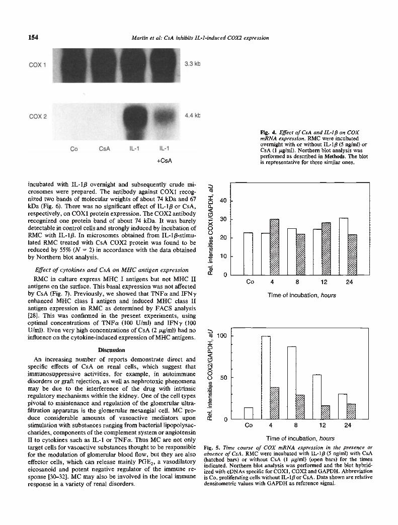
incubated with IL-1f3 overnight and subsequently crude mi-crosomes were prepared. The antibody against COX1 recog-nized two bands of molecular weights of about 74 kDa and 67kDa (Fig. 6). There was no significant effect of IL-1/3 or CsA,respectively, on COX1 protein expression. The COX2 antibodyrecognized one protein band of about 74 kDa. It was barelydetectable in control cells and strongly induced by incubation ofRMC with IL-1$. In microsomes obtained from IL-1/3-stimu-lated RMC treated with CsA COX2 protein was found to bereduced by 55% (N = 2) in accordance with the data obtainedby Northern blot analysis.
Effect of cytokines and GsA on MHC antigen expressionRMC in culture express MHC I antigens but not MHC II
antigens on the surface. This basal expression was not affectedby CsA (Fig. 7). Previously, we showed that TNFa and IFN7enhanced MHC class I antigen and induced MHC class IIantigen expression in RMC as determined by FACS analysis[281. This was confirmed in the present experiments, usingoptimal concentrations of TNFa (100 U/ml) and IFNy (100UIml). Even very high concentrations of CsA (2 pg/m1) had noinfluence on the cytokine-induced expression of MHC antigens.
Discussion
An increasing number of reports demonstrate direct andspecific effects of CsA on renal cells, which suggest thatimmunosuppressive activities, for example, in autoimmunedisorders or graft rejection, as well as nephrotoxic phenomenamay be due to the interference of the drug with intrinsicregulatory mechanisms within the kidney. One of the cell typespivotal to maintenance and regulation of the glomerular ultra-filtration apparatus is the glomerular mesangial cell. MC pro-duce considerable amounts of vasoactive mediators uponstimulation with substances ranging from bacterial lipopolysac-charides, components of the complement system or angiotensinII to cytokines such as IL-i or TNFa. Thus MC are not onlytarget cells for vasoactive substances thought to be responsiblefor the modulation of glomerular blood flow, but they are alsoeffector cells, which can release mainly PGE2, a vasodilatoryeicosanoid and potent negative regulator of the immune re-sponse [30—32]. MC may also be involved in the local immuneresponse in a variety of renal disorders.
154 Martin et a!: CsA inhibits IL-I -induced COX2 expression
+CsA
FIg. 4. Effect of CsA and IL-I/3 on COXmRNA expression. RMC were incubatedovernight with or without IL-l/3 (5 ng/mI) orCsA (1 tg/ml). Northern blot analysis wasperformed as described in Methods. The blotis representative for three similar ones.
Co 4 8 12 24
400
30x0020
a)
Cl)C10C
a)0
C' 100I0
o 50U)a)
Ci)Ca)C
Time of incubation, hours
Co 4 8 12 24
Time of incubation, hours
Fig. 5. Time course of COX mRNA expression in the presence orabsence of CsA. RMC were incubated with IL-1/3 (5 ng/ml) with CsA(hatched bars) or without CsA (1 g/ml) (open bars) for the timesindicated. Northern blot analysis was performed and the blot hybrid-ized with eDNAs specific for COXI, COX2 and GAPDH. Abbreviationis Co, proliferating cells without IL-ip or CsA. Data shown are relativedensitometric values with GAPDH as reference signal.
COX 1
COX 2
Co CsA IL-i IL-i
3.3 kb
4.4 kb'4

Martin et a!: CsA inhibits IL-I -induced COX2 expression 155
Table 2. Regulation of COX2 mRNA in RMC
Stimulation of COX2mRNA expression
Inhibition by CsA%
IL-l/35ng/ml 6.4±0.7 53±9LPS 100 ng/ml 9.5 2.1 67 7A 23 187 1 rsg/ml 4.7 0.2 40 10
RMC were incubated with the stimuli indicated for 4 hours in theabsence or presence of CsA (1 g/ml). COX2 mRNA was detected byNorthern blot analysis. To confirm equal quantities of RNA, all blotswere hybridized with a cDNA probe for GAPDH. Densitometric valuesof COX2 mRNA were corrected for GAPDH signals. Stimulation wascalculated as fold increase compared to nontreated RMC. Data aremeans SD (N = 3, stimulus IL-1/3) or means range (N = 2, stimuliLPS or A 23187).
Cell culture approaches have enabled us and others to studythe effects of drugs on the behavior of glomerular cells in clearlydefined in vitro systems. CsA effectively inhibited the vasocon-strictor-induced POE2 synthesis in mesangial cells [1], thusinterfering with the balance of vasoconstrictor and vasodilatorinfluences. Now, we could show that CsA also interfered withthe cytokine-induced release of the main arachidonate metabo-lite of MC in culture, PGE2. IL-i was chosen to induceprostaglandin synthesis, because it is a central mediator ofinflammatory processes and has also been shown to be involvedin the pathogenicity of renal disorders [33]. All experimentswere performed with cycling cells, that is, in the presence ofFCS, as in inflammatory situations mesangial cells frequentlyproliferate.
IL-I and TNFa enhanced the basal prostaglandin formationin a concentration dependent manner. CsA reduced the en-hanced PGE2 release of RMC by about 50%. In none of theexperiments did CsA inhibit IL-i or TNFa-induced POE2release to control levels, even if the drug was given for 72hours. These observations suggest a molecular mechanism notonly different from the direct COX inhibition by nonsteroidalanti-inflammatory drugs (such as acetylsalicylic acid), but alsodifferent from the receptor-mediated effect of glucocorticoids,which reduce cytokine-induced POE2 synthesis to backgroundslevels [19]. Suppression of PGE2 synthesis was observed atearly time points of stinftilation by IL-i, that is, as early as fourto six hours after onset of stimulation, and also during theprolonged release of POE2 between 24 and 48 hours of incuba-tion. This time course suggested a pronounced inhibitory effecton the molecular mechanisms underlying induction of PGE2synthesis, not just an attenuating effect of CsA on the existingenzymatic machinery.
Previous experiments studying antiproliferative effects ofCsA on rat MC in culture [7] have shown that 125 to 250 ng/mlCsA yielded detectable suppression of thymidine incorporation,which was reversible after withdrawal of the drug. The sameconcentration range of CsA inhibited IL-i-induced PGE2 re-lease when added simultaneously with the cytokine, but higherconcentrations (500 to 1000 ng/ml) were necessary to obtainpronounced effects. This is in accordance with other studieswith RMC [17]. The concentrations of CsA used were relativelyhigh when compared to the amount required for effectiveinhibition of T-lymphocyte proliferation (100 ng/ml) and ex-ceeded conventional therapeutic levels in humans. However, asdiscussed by Stahl et al [17] and by us [7], tissue levels in the
kidney may be considerably higher than CsA levels determinedin serum. Furthermore, the threshold for nephrotoxicity seemsto be higher in rats than in humans. Therefore, the concentra-tions used in this study are likely to be within the concentrationrange which leads to nephrotoxicity in vivo.
The mechanism by which CsA interferes with POE2 synthesishas been discussed at different levels. The inhibitory effect ofCsA on phorbol ester-stimulated POE2 release of RMC wasdiscussed as direct interference of the lipophilic substance CsAwith the activation process of the protein kinase C [34]. Theformation and release of prostanoids from mesangial cells is amulti-step process requiring a series of enzymatic conversionsof the educt arachidonic acid to the final product, in this study,POE2. Arachidonic acid is liberated upon stimulation fromplasma membrane phospholipids. Pfeilschifter et al [15] showedin RMC that phospholipase A2 activity is enhanced by IL-1/3and TNFa treatment. In those studies CsA inhibited the cyto-kine-induced phospholipase A2 synthesis and release. Stahl etal [17] reported that CsA inhibited the release of radiolabeledarachidonate in RMC after stimulation with the calcium iono-phore A23 187 or angiotensin II. No effect of CsA was observedon the activity of the microsomal cyclooxygenase. Similarresults were also obtained by Skorecki, Rutledge and Schrier[35] using vasopressin-stimulated MC, indicating an effect ofCsA at the level of arachidonic acid liberation.
In our studies, we focused on the second step of the arachi-donate cascade. Cyclooxygenase (prostaglandin 0/H synthase,COX) utilizes arachidonic acid as a substrate to produce ametastable intermediate product, which is then further pro-cessed to different prostanoids, depending on the enzymaticequipment of the respective cell type. Recently, it was shownthat IL-i/3 and TNFa induced de novo synthesis of COXmolecules in mesangial cells [19]. This was paralleled by anenhancement of POE2 release into the conditioned media ofthese cultures and an increased enzymatic activity measured inmicrosomes obtained from IL-113 or TNFa-treated MC.
Here we demonstrate that COX activities were reduced inmicrosomes obtained from IL-113 plus CsA-stimulated RMC. Inaccordance with the data of Stahl et al [17], we could not detectany effects on COX activity in RMC microsomes, when themicrosomes were incubated with CsA. This indicated an effectof CsA on COX biosynthesis, which was confirmed on proteinand mRNA level.
The two isoforms of COX presently known are regulated in acell type and stimulus specific manner [20]. IL-i/3 was shown toinduce COXI mRNA in human endothelial cells and COX2mRNA in human monocytes [36, 37]. Now we showed that inRMC IL-1,3 had no significant effect on COX! expression butstrongly induced COX2 mRNA and protein, which were barelydetectable in nonstimulated proliferating cells. These data are inaccordance with those of Coyne et a! [19], assuming that theantibody used by this group might cross react with both, COX1and COX2. A maximal COX2 mRNA signal was obtained afterfour hours of incubation with IL-l)3. Thereafter, COX2 mRNAlevels slowly declined, but remained elevated for more than 20hours.
CsA interfered at the mRNA level with the induction ofCOX2 without effecting the constitutive expression of COX1.The effect of CsA was not restricted to the IL-1f3-inducedCOX2 mRNA expression. It was previously shown that LPS

— —
156 Martin et a!: CsA inhibits IL-I-induced COX2 expression
10680
49.5
coxi COX2
18.5
St. Ko GsA IL-i IL-i Ko CsA IL-i+CsA
HkA
ALog fluorescence intensity
Fig. 6. Effect of GsA and IL-i f3 on COXprotein expression. RMC were incubatedovernight with or without IL-113 (5 ng/ml) orCsA (1 pg/mI). Western blot analysis wasperformed as described in Methods.Abbreviations are St, molecular weightstandard; Ko, control. The blot isrepresentative of three independentexperiments.
transduction pathway between receptor and transcription of thespecific gene might be the target of CsA action.
The missing effect of CsA on the constitutively expressedD COX1 isozyme explained the minor effects of CsA on basal
POE2 release. Other mechanisms such as an effect on phospho-lipase A2 may also contribute to the marginal reduction in basalPGE2 release. The striking concurrence of CsA-mediated inhi-
GSA bition of POE2 release, COX activity and COX2 protein andmRNA expression strongly suggests that the interference ofCsA at this step of the biosynthetic cascade is the most
TNF relevant, which, however, does not exclude effects of CsA at+ other levels. The interference of CsA with the induction ofIFN prostaglandin synthesis was specific, as other induction path-
ways in RMC were not effected. As an example, we measuredGsA the induction of MHC class I and class II antigens by IFN7 and+ TNFa. CsA had no effect on the basal or induced expression of
INF these antigens.PFN The interference with an induction pathway resulting in
inhibition of mRNA expression and of de novo protein synthe-sis of specific proteins, while leaving others unaffected, resem-bles the action of CsA on mitogen activated T-lymphocytes. Inthis system CsA blocked completely the production of theautocrine growth factor IL-2 by activated T cells, while otherproteins were still produced [38].
It is thus tempting to speculate that the mechanisms of CsAaction on a molecular level may be very similar in MC and Tcells. On the contrary, cell specific regulation by CsA hasrecently been demonstrated with respect to the expression ofphosphoenolpyruvate carboxykinase, which was inhibited byCsA in the kidney but not in the liver [391.
In summary, we demonstrate that the regulation of cytokineinduction of and CsA interference with PGE2 synthesis inmesangial cells occurs at the level of COX2 mRNA. COX2 isthus a new member of the group of genes whose expression isCsA sensitive. There are multiple reports in the literature thatCsA in addition to inhibiting the synthesis of the vasodilator
32.527.5
FITC
IL-i+CsA
MHC II MHC I
EC
0
Fig. 7. Induction of MHC antigen expression in the presence of CsA.RMC were incubated for 72 hours in the presence of 0.1% DMSO (D),2 pg/mi CsA (CsA), 100 U/mI TNFa plus 100 U/mi IFN-y (TNF + IFN)or CsA plus cytokines (CsA ÷ TNF + IFN). Expression of anirreievant antigen (FITC) and of MHC class I or MHC class II antigenswas determined by FACS analysis. Data shown are from one experi-ment representative of three identical ones.
and ionophore were inducers of PGE2 synthesis in RMC [17]. Inour experiments, both compounds induced the expression ofCOX2 mRNA to a similar extent as IL-lp in RMC. Again, CsAwas an effective partial inhibitor of the induction.
It is unclear at present, whether the observed regulation ofCOX2 mRNA in mesangial cells is due to an increase in mRNAsynthesis and or an enhanced stability of the mRNA. Further-more, it remains to be evaluated which step of the signal

Martin et al: CsA Inhibits IL-I -induced COX2 expression 157
PGE2 enhanced the synthesis of the vasoconstrictor thrombox-ane, most likely in extrarenal cells [1]. One might speculate thatother effects of CsA, such as the enhancement of intracellularcalcium or inhibition of the reacylating system of membranephospholipids [40], might be essential in cells without thecapacity of de novo protein biosynthesis, such as platelets. Thiswould explain the adverse effects of CsA on prostanoid synthe-sis which in summary contribute to the interference of CsA withrenal hemodynamics.
Acknowledgments
The technical assistance of Mrs. M. Golombek and H. Wesche isgratefully acknowledged. We thank R. Schwinzer for performing theFACS analyses. T.H. is recipient of a grant from the "Fonds derChemischen Industrie."
Reprint requests to Dr. M. Martin, Molecular Pharmacology, OE5320, Medical School Hannover, D-30623 Hannover, Germany.
Appendix. Abbreviations
COX, cyclooxygenase = prostaglandin endoperoxide synthase =prostaglandin GH synthase; CsA, cyclosporin A; IL-i, interleukin 1;POE2, prostaglandin E2; RMC, rat mesangial cells; TNFa, tumornecrosis factor a; IFN7, interferon gamma; MHC, major histocompat-ibiity complex.
References
1. MYERS BD: Cyclosporine nephrotoxicity. Kidney mt 36:964—974,1986
2. Ko JB, KLOTMAN PE: Cellular and molecular mechanisms ofcyclosporin nephrotoxicity. JAm Soc Nephrol 1:162—179, 1990
3. Biutos EJG, B0IM MA, AJZEN H, RAMOS OL, SCHNOR N:Glomerular hemodynamics and hormonal participation in cyclospo-rine nephrotoxicity. Kidney mt 32:19—25, 1987
4. ENGLISH J, EVAN A, HOUGHTON DC, BENNErF WM: Cyclosporin-induced acute renal dysfunction in the rat. Transplantation 44:135—141, 1987
5. CHAPMAN JR, HARDING NGL, GRIFFITHS D, Moiuus PJ: Revers-ibility of Cyclosporin nephrotoxicity after three month treatment.Lancet 19:128—130, 1985
6. MYERS BD, SIBLEY R, NEWTON L, TOMLANOVICH SJ, B05HK0sC,STINSON E, LUETSCHER JA, WHITNEY DJ, KRASNY D, COPLONNS, PERLROTH MG: The long-term course of cyclosporine-associ-ated chronic nephropathy. Kidney mt 32:590—600, 1988
7. MARTIN M, KRICHBAUM M, KAEVER V, GOPPELT-STRUBE M,RESCH K: Cyclosporin A suppresses proliferation of renal mesan-gial cells in culture. Biochem Pharmacol 37:1083—1088, 1988
8. PFEILSCHIFTEIt J: Cyclosporin A augments vasoconstrictor-in-duced rise in intracellular free calcium in rat mesangial cells.Biochem Pharmacol 37:4205—4210, 1988
9. GOLDBERG HJ, WONG PY, COLE EH, LEVY GA, SKORECKI KL:Dissociation between the immunosuppressive activity of cyclospo-rine derivatives and their effects on intracellular calcium signallingin mesangial cells. Transplantation 47:731—744, 1989
10. MEYER-LEHNERT H, SCHRIER RW: Cyclosporin A enhances vaso-pressin-induced Ca2 mobilization and contraction in mesangialcells. Kidney mt 34:89—97, 1988
11. RODRIGUEZ-PUYOL D, LAMAS S, OLIVERA A, LOPEZ-FARRE A,ORTEGA 0, HERNADO L, LOPEZ-NOVOA JM: Actions of cyclospo-rin A on cultured mesangial cells. Kidney mt 35:632—637, 1989
12. LOVETT DH, RESCH K, GEMSA D: Interleukin 1 and the glomerularmesangium II. Monokine stimulation of mesangial cell prostanoidsecretion. Am J Pathol 129:543—551, 1987
13. BAUD L, PEREZ J, FRIEDLANDER 0, ARDAILLOU R: Tumor necro-sis factor stimulates prostaglandin production and cyclic AMPlevels in rat glomerular mesangial cells. FEBS Lett 239:50—54, 1988
14. TOPLEY N, FLOEGE J, WES5EL K, HAss R, RADEKE HH, KAEVERV, RESCH K: Prostaglandin E2 production is synergistically inducedin cultured human glomerular mesangial cells by combinations ofIL-I and TNFa. Jlmmunol 143:1989—1995, 1989
15. PFEILSCHIFTER J, PIGNAT W, VOSBECK K, MARKI F, WJESENBERGJ: Susceptibility of Interleukin 1- and Tumor Necrosis Factor-
induced prostaglandin E2 and phospholipase A2 release from ratrenal mesangial cells to different drugs. Bioch Soc Trans 17:916—917, 1989
16. KREMER S, MARGOLIS B, HARPTER P, SKORECKI K: Cyclosporineinduced alterations in vasopressin signalling in the glomerular cell.Clin Invest Med 12:201—206, 1989
17. STAHL RAK, ADLER S, BAKER PJ, JOHNSON RJ, CHEN Y-P,PRITZL R, COU5ER WG: Cyclosporin A inhibits prostaglandin E2formation by rat mesangial cells in culture. Kidney mt 35:1161—1167, 1989
18. BUNKE M, WILDER L, MARTIN A: The effect of cylosporine onagonist-stimulated glomerular and mesangial cell vasodilatory pros-taglandin production. Transplantation 52(4):718—722, 1991
19. COYNE DW, NIcK0L5 M, BERTRAND W, MoluusoN AR: Regula-tion of mesangial cell cyclooxygenase synthesis by cytokines andglucocorticoids. Am J Physiol 263:97—102, 1992
20. SMITH WL: Prostanoid biosynthesis and mechanisms of action. AmJPhysiol 263:181—191, 1992
21. REDINBAUGH MR, CAMPBELL WH: Adaption of the dyebinding
protein assay to microtiter plates. Anal Biochem 147:144-147, 198522. REINKE M, PILLER M, BRUNE K: Development of an enzyme-
linked immunosorbent assay of thromboxane B2 using a monoclo-nal antibody. Prostaglandins 5:577—586, 1989
23. KOEHLER L, HASS R, WESSEL K, DEWI1-F DL, KAEVER V, RESCHK, GOPPELT-STRUEBE M: Altered arachidonic acid metabolismduring differentiation of the human monoblastoid cell line U937.
Biochim Biophys Acta 1042:395—403, 199024. LAEMMLI UK: Cleavage of structural proteins during the assembly
of the head of the bacteriophage T4. Nature 227:680—685, 197025. REGIER MK, DEWITT DL, SCHINDLER MS. SMITH WL: Subcellu-
larlocalizytion of prostaglandin endoperoxide synthase-2 in murine3T3 cells. Arch Biochem Biophys (in press)
26. CHOMCZYNSKI P, SACCHI N: Single-step method of RNA isolationby acid guanidinium thiocyanate-phenol-chioroform extraction.Anal Biochem 612:156—159, 1987
27. HOFF T, SPENCICER T, EMMENDOERFFER A, GOPPELT-STRUEBE M:Effects of glucocorticoids on the TPA-induced monocytic differen-tiation. J Leuk Biol 52:173—182, 1992
28. MARTIN M, SCHWINZER R, SCHELLEKENS H, RESCH K: Glomeru-lar mesangial cells in local inflammation: Induction of MHC class IIantigens by interferon-gamma. J Immunol 142:1887—1894, 1989
29. HOFF T, DEWITT D, KAEVER V, RESCH K, GOPPELT-STRUEBE M:Differentiation-associated expression of prostaglandin G/H syn-thase in monocytic cells. FEBS Lett 320:38—42, 1993
30. SCHLONDORF D: The glomerular mesangial cell: an expanding rolefor a specialized pericyte. FASEB J 1:272—281, 1987
31. MENE P, CINOTFI GA: Paracnne and autocrine functions of gb-merular mesangial cell. J Endocrinol Invest 12:497—509, 1989
32. BADR KF: Arachidonate cyclo-oxygenase and lipoxygenase prod-ucts in the mediation of glomerular immune injury. Nephrol DialTransplant 6:662—669, 1991
33. LAN HY, NIKOLIC-PATERSON DJ, ZARAMA M, VANNICE JL, AT-KINS RC: Suppression of experimental crescentic glomerulonephri-tis by the interleukin-1 receptor antagonist. Kidneymt 43:479—485,1993
34. WALKER RI, LAZZARO VA, DUGGIN GO, HORVATH JS, TILLER DJ:Cyclosporin A inhibits protein kinase C activity: A contributingmechanism in the development of nephrotoxicity? Biochem Bio-phys Res Commun 160:409—415, 1989
35. SKORECKI KL, RUTLEDGE WP, SCHRIEIt RW: Acute cyclosporinenephrotoxicity—prototype for a renal membrane signalling disor-
der. Kidney mt 42: 1—10, 199236. MAIER JAM, HLAT, MAClAG T: Cyclooxygenase is an immediate-
early gene induced by Interleukin-l in human endothelial cells. JBiol Chem 265(19): 10805—10808, 1990

158 Martin et a!: GsA inhibits IL-I-induced COX2 expression
37. O'BANION MK, WINN VD, YOUNG DA: eDNA cloning and func-tional activity of a glucocorticoid-regulated inflammatory cycloox-ygenase. Proc Nat! Acad Sci USA 89:4888—4892, 1992
38. SCHREIBER SL, CRABTREE GR: The mechanism of action of cyclo-sporin A and FK 506. Immunol Today 13:136—142, 1992
39. Moiuus SM, KEPKA-LENRART D, MCGILL RL, CURTHOYS NP,
ADLER S: Specific disruption of renal function and gene transcrip-tion by cyclosponn A. J Biol Chem 267(19):13768—13771, 1992
40. KROOGEL R, GOPPELT-STRUEBE M, MARTIN M, RESCH K: Theimmunosuppressive activities of different cyclosporins are cone-lated to inhibition of the early membrane phospholipid metabolismin activated lymphocytes. Immunobiology 175:159—171, 1987