Embed Size (px)
Citation preview

1
Interlaboratory Collaborative Study Validation Protocol for the Quantitative Three Step Method Prepared by: Stephen F. Tomasino, Ph.D., Study Director Rebecca M. Fiumara, Co-Study Director U.S. Environmental Protection Agency Office of Pesticide Programs Microbiology Laboratory Environmental Science Center 701 Mapes Road Fort Meade, Maryland 20755-5350 Study Director Phone: 410-305-2976 FAX: 410-305-3094 Email: [email protected] CONTROL COPY #: 7.20.06 (final)
2
TABLE OF CONTENTS SECTION PAGE 1. INTRODUCTION 3 2. COLLABORATORS 5 3. STUDY DESIGN 7 4. TEST SAMPLE PREPARTION 11 5. QUALITY ASSURANCE 12 6. REFERENCE METHOD – Revised AOAC Method 966.04 14 7. QUANTITATIVE THREE STEP METHOD 23 8. REPORTING RAW DATA 36 9. STATISTICAL ANALYSIS 36 10. REFERENCES 39 11. LIST OF APPENDICES 40 A. EPA Quality Assurance Project Plan B. AOAC Method Data Sheets C. TSM Data Sheets D. Media and Reagent Preparation Sheets E. Parameters for Testing Chemicals F. TSM Log Reduction Data G. Material Safety Data Sheets (MSDS) H. Safety Checklist
3
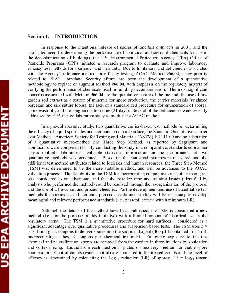
Section 1. INTRODUCTION In response to the intentional release of spores of Bacillus anthracis in 2001, and the associated need for determining the performance of sporicidal and sterilant chemicals for use in the decontamination of buildings, the U.S. Environmental Protection Agency (EPA) Office of Pesticide Programs (OPP) initiated a research program to evaluate and improve laboratory efficacy test methods for sporicides and sterilants. Due to limitations and deficiencies associated with the Agency's reference method for efficacy testing, AOAC Method 966.04, a key priority related to EPA's Homeland Security efforts has been the development of a quantitative methodology to replace or augment Method 966.04, with emphasis on the regulatory aspects of verifying the performance of chemicals used in building decontamination. The most significant concerns associated with Method 966.04 are the qualitative nature of the method, the use of raw garden soil extract as a source of minerals for spore production, the carrier materials (unglazed porcelain and silk suture loops), the lack of a standardized procedure for enumeration of spores, spore wash-off, and the long incubation time (21 days). Several of the deficiencies were recently addressed by EPA in a collaborative study to modify the AOAC method. In a pre-collaborative study, two quantitative carrier-based test methods for determining the efficacy of liquid sporicides and sterilants on a hard surface, the Standard Quantitative Carrier Test Method – American Society for Testing and Materials (ASTM) E 2111-00 and an adaptation of a quantitative micro-method (the Three Step Method) as reported by Sagripanti and Bonifacino, were compared (1). By conducting the study in a comparative, standardized manner across multiple laboratories, valuable statistical information on the performance of two quantitative methods was generated. Based on the statistical parameters measured and the additional test method attributes related to logistics and human resources, the Three Step Method (TSM) was determined to be the more suitable method, and will be advanced to the AOAC validation process. The flexibility in the TSM for incorporating coupon materials other than glass was considered as an advantage, and that the practice time and training issues (identified by analysts who performed the method) could be resolved through the re-organization of the protocol and the use of a flowchart and process checklist. As the development and use of quantitative test methods for sporicides and sterilants proceeds, additional studies will be necessary to develop meaningful and relevant performance standards (i.e., pass/fail criteria with a minimum LR). Although the details of the method have been published, the TSM is considered a new method (i.e., for the purpose of this initiative) with a limited amount of historical use in the regulatory arena. The TSM is a quantitative procedure for hard surfaces – considered as a significant advantage over qualitative procedures and suspension-based tests. The TSM uses 5 × 5 × 1 mm glass coupons to deliver spores into the sporicidal agent (400 µL) contained in 1.5 mL microcentrifuge tubes, 3 coupons per chemical treatment. Following exposure to the test chemical and neutralization, spores are removed from the carriers in three fractions by sonication and vortex-mixing. Liquid from each fraction is plated on recovery medium for viable spore enumeration. Control counts (water control) are compared to the treated counts and the level of efficacy is determined by calculating the Log10 reduction (LR) of spores; LR = log10 (mean

4
spores/control carrier) - log10 (mean spores/treated carrier). The original procedures for the TSM were reported by Sagripanti and Bonifacino (2). However, during the course of the pre-collaborative studies, EPA refined and altered the method slightly, and finalized the protocol for the purpose of this validation study. It should be noted that following the completion of the pre-collaborative study, a similar version of the TSM was accepted and published by ASTM International (3). The purpose of this Interlaboratory Collaborative Study (CS) is to evaluate the TSM according to AOACI Official Methods of Analysis (OMA) procedures for official method validation. The applicability will be limited to one spore-forming microorganism (Bacillus subtilis), one hard surface (glass), liquid formulations of sporicides and sterilants, and without organic burden added to the spore inoculum. The test chemicals used in the CS represent three chemical classes of sporicides: sodium hypochlorite, a combination of peracetic acid and hydrogen peroxide, and glutaradehyde. The suitability of the TSM for porous materials and gaseous formulations will require additional collaborative studies. If the TSM is validated for B. subtilis, the Study Director will propose language in the validation report to allow the testing of other Bacillus species, such as B. anthracis, with the TSM. The overall objective of the CS is to evaluate the performance of the TSM by generating and comparing control counts and efficacy data and assessing the degree and source(s) of variability associated with the data, both within and between laboratories, when the method is used in actual practice. AOAC Method 966.04, Sporicidal Activity of Disinfectants, is recognized as the reference method. Method 966.04 provides a qualitative measure of product efficacy against spores of B. subtilis and Clostridium sporogenes dried on two types of carriers, porcelain penicylinders and silk suture loops. Sixty-carrier tests on three lots of product are required for an EPA registration – all carriers must show no growth to support a sporicidal claim. For the purpose of this CS, only the Bacillus and hard surface (porcelain penicylinders) components of Method 966.04 will be evaluated. The comparative testing with the reference method is problematic due to its qualitative nature. In this study, chemical treatments from one of three replications per product will be tested using both the reference method and TSM. The cost of conducting Method 966.04 is a limiting factor and it would not be feasible to generate enough inoculated porcelain carriers to do each treatment and replication. Prior to initiation of this collaborative effort, the AOAC INTERNATIONAL will assemble a review panel, the AOAC Sporicidal Method Expert Review Panel (ERP), to evaluate the CS protocol. The ERP, along with members of the AOAC INTERNATIONAL Methods Committee on Microbiology, will be engaged early in the development of the study design to ensure the protocol format and test design are acceptable and meet the goals established for the AOACI Official Methods process.
5
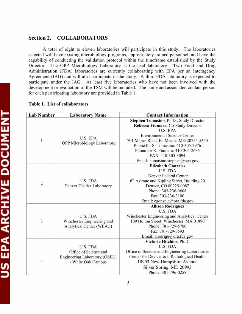
Section 2. COLLABORATORS A total of eight to eleven laboratories will participate in this study. The laboratories selected will have existing microbiology programs, appropriately trained personnel, and have the capability of conducting the validation protocol within the timeframe established by the Study Director. The OPP Microbiology Laboratory is the lead laboratory. Two Food and Drug Administration (FDA) laboratories are currently collaborating with EPA per an Interagency Agreement (IAG) and will also participate in the study. A third FDA laboratory is expected to participate under the IAG. At least five laboratories who have not been involved with the development or evaluation of the TSM will be included. The name and associated contact person for each participating laboratory are provided in Table 1. Table 1. List of collaborators Lab Number Laboratory Name Contact Information
1 U.S. EPA OPP Microbiology Laboratory
Stephen Tomasino, Ph.D., Study Director Rebecca Fiumara, Co-Study Director
U.S. EPA Environmental Science Center
701 Mapes Road, Ft. Meade, MD 20755-5350 Phone for S. Tomasino: 410-305-2976 Phone for R. Fiumara: 410-305-2635
FAX: 410-305-3094 Email: [email protected]
2 U.S. FDA Denver District Laboratory
Elizabeth Gonzales U.S. FDA
Denver Federal Center 6th Avenue and Kipling Street, Building 20
Denver, CO 80225-0087 Phone: 303-236-9608
Fax: 303-236-3100 Email: [email protected]
3 U.S. FDA
Winchester Engineering and Analytical Center (WEAC)
Allison Rodriguez U.S. FDA
Winchester Engineering and Analytical Center 109 Holton Street, Winchester, MA 01890
Phone: 781-729-5700 Fax: 781-729-3593
Email: [email protected]
4
U.S. FDA Office of Science and
Engineering Laboratory (OSEL) - White Oak Campus
Victoria Hitchins, Ph.D. U.S. FDA
Office of Science and Engineering Laboratories Center for Devices and Radiological Health
10903 New Hampshire Avenue Silver Spring, MD 20993
Phone: 301-796-0258
6
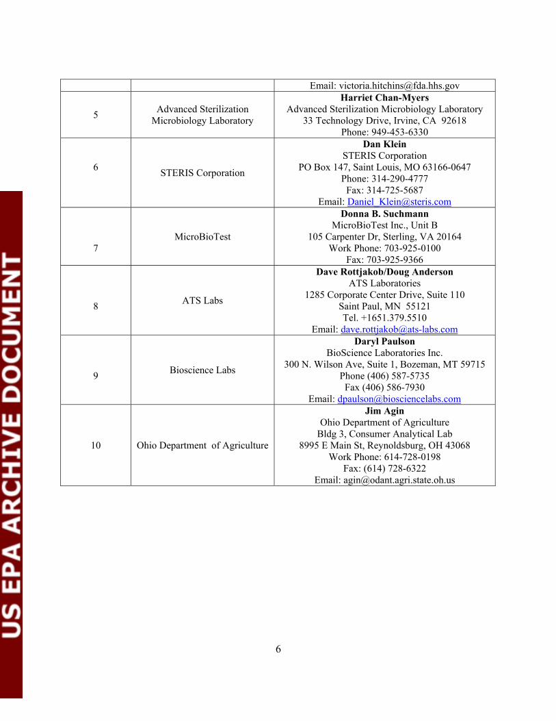
Email: [email protected]
5 Advanced Sterilization Microbiology Laboratory
Harriet Chan-Myers Advanced Sterilization Microbiology Laboratory
33 Technology Drive, Irvine, CA 92618 Phone: 949-453-6330
6 STERIS Corporation
Dan Klein STERIS Corporation
PO Box 147, Saint Louis, MO 63166-0647 Phone: 314-290-4777
Fax: 314-725-5687 Email: [email protected]
7 MicroBioTest
Donna B. Suchmann MicroBioTest Inc., Unit B
105 Carpenter Dr, Sterling, VA 20164 Work Phone: 703-925-0100
Fax: 703-925-9366
8 ATS Labs
Dave Rottjakob/Doug Anderson ATS Laboratories
1285 Corporate Center Drive, Suite 110 Saint Paul, MN 55121 Tel. +1651.379.5510
Email: [email protected]
9 Bioscience Labs
Daryl Paulson BioScience Laboratories Inc.
300 N. Wilson Ave, Suite 1, Bozeman, MT 59715 Phone (406) 587-5735
Fax (406) 586-7930 Email: [email protected]
10 Ohio Department of Agriculture
Jim Agin Ohio Department of Agriculture
Bldg 3, Consumer Analytical Lab 8995 E Main St, Reynoldsburg, OH 43068
Work Phone: 614-728-0198 Fax: (614) 728-6322
Email: [email protected]

7
Section 3. STUDY DESIGN
A. The Study Director, Dr. Stephen Tomasino, is responsible for organizing the CS and assessing the preparedness of each collaborating laboratory prior to initiation of research. Toward that effort, the Study Director and the OPP Microbiology Laboratory Quality Assurance Unit will conduct a readiness review of each participating laboratory to ensure compliance with EPA's Quality Assurance Project Plan 2003-01 (Appendix A). The Co-Study Director, Rebecca Fiumara, will serve as the technical lead for the TSM.
B. The method protocols, standardized test forms and data sheets (Appendices B and C),
media preparation sheets (Appendix D), selected media and reagents, test chemicals, and inoculated porcelain penicylinders will be provided by the Study Director. Test parameters for each chemical treatment describing the conditions for testing (e.g., dilution, neutralizer, contact time, temperature, etc.) will be provided to each laboratory by the Study Director (Appendix E).
C. The Study Director recognizes that it is desirable to distribute the chemical treatments to the testing laboratories in containers that are marked only with a treatment code. However, due to the instability of test chemicals such as diluted sodium hypochlorite, it will be necessary for each lab to prepare the actual test formulations (i.e., perform dilutions) on-site. The test chemicals will be provided by the Study Director. The test scheme will be randomized to account for potential subjectivity by the analysts.
D. In order to have a balanced design that will be conducive to a straight-forward statistical
analysis, the labs will be asked to perform the same number of tests per day. The study design calls for testing three chemicals, three levels each (high, medium and low), one chemical per day. Three replications are required. Assuming no repeat testing, 9 test days will be required to complete the CS. Water controls (control carrier counts) will be included each test day for the TSM. Each treatment will be evaluated by the TSM; however, Method 966.04 will be conducted concurrently on the first replication only for a total of nine 30-carrier AOAC tests per laboratory.
E. Testing must be initiated within 4 weeks after the readiness review, practice/training and receiving supplies, and must be completed within 12-14 weeks after initiation of the first test.
F. Two or three technicians will be required per test day for approximately 6 hours. Each laboratory will be encouraged to establish a technician team which will conduct all tests, i.e., it is important to design the study so that the differences among technician teams do not affect the outcome. If the same technician team always conducted the testing, the technician effect will not confound the results. Practice runs will be encouraged in
8
advance of testing to ensure analyst proficiency in performing each method.
G. It is important to randomize any steps in which subjective decisions or unknown factors could affect the conclusions. The experimental results could be criticized because the order of experimentation was systematic; any systematic trend in environmental conditions would affect the results. It is preferable to have a numbering system or positioning arrangement that gives each item a unique identification. In this study, the Study Director will provide the randomized order of testing of chemicals for each lab using an acceptable method of randomization. The randomization will be done before the experimentation is initiated.
H. Test chemicals used in the study are:
1. Sodium hypochlorite (reagent grade, Sigma-Aldrich sodium hypochlorite solution, 12% available chlorine)
2. A combination of peracetic acid and hydrogen peroxide (Spor-Klenz Ready to Use, an EPA-registered commercial sporicide)
3. 2.6% glutaraldehyde (Metricide 14-Day, a commercially available sterilant).
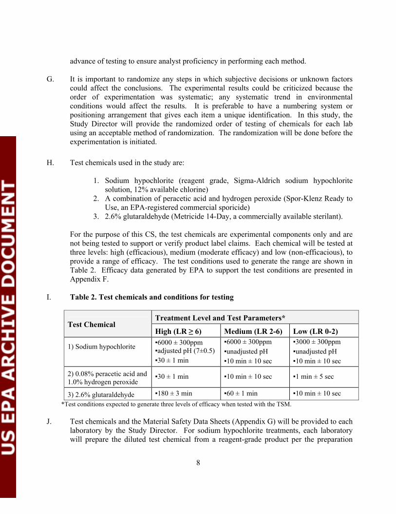
For the purpose of this CS, the test chemicals are experimental components only and are not being tested to support or verify product label claims. Each chemical will be tested at three levels: high (efficacious), medium (moderate efficacy) and low (non-efficacious), to provide a range of efficacy. The test conditions used to generate the range are shown in Table 2. Efficacy data generated by EPA to support the test conditions are presented in Appendix F.
I. Table 2. Test chemicals and conditions for testing
Treatment Level and Test Parameters*
Test Chemical High (LR ≥ 6) Medium (LR 2-6) Low (LR 0-2)
1) Sodium hypochlorite
▪6000 ± 300ppm ▪adjusted pH (7±0.5) ▪30 ± 1 min
▪6000 ± 300ppm ▪unadjusted pH ▪10 min ± 10 sec
▪3000 ± 300ppm ▪unadjusted pH ▪10 min ± 10 sec
2) 0.08% peracetic acid and 1.0% hydrogen peroxide
▪30 ± 1 min ▪10 min ± 10 sec ▪1 min ± 5 sec
3) 2.6% glutaraldehyde ▪180 ± 3 min ▪60 ± 1 min ▪10 min ± 10 sec *Test conditions expected to generate three levels of efficacy when tested with the TSM.
J. Test chemicals and the Material Safety Data Sheets (Appendix G) will be provided to each laboratory by the Study Director. For sodium hypochlorite treatments, each laboratory will prepare the diluted test chemical from a reagent-grade product per the preparation
9
sheets provided by the Study Director – HACH Test Kits will be used to verify available chlorine for diluted samples. The Study Director will request the use of a single lot for each test chemical. In advance of testing, an analysis of formulation chemistry will be performed on each lot (one container) by OPP chemists to confirm the percent active ingredient.
K. The test microbe is Bacillus subtilis (ATCC # 19659) obtained directly from a reputable
supplier (e.g., ATCC). Each lab will initiate a new stock culture. Note: An existing stock culture may be used if it meets the quality control standards (e.g., proper documentation, confirmation testing) – Study Director approval is required in this case.
L. Presque Isle Cultures, 3804 West Lake Rd, Erie, PA 16505 will provide inoculated
porcelain carriers (approx. 4,000 total for the entire study) to each laboratory per the modified methodology for AOAC Method 966.04. The vendor, per instructions provided by the Study Director, will follow the revised method (i.e., the use of nutrient agar amended with manganese sulfate) for generation of spore suspensions and inoculation of carriers. No organic burden will be added to the spore inoculum. Carrier counts and HCl resistance will be determined by the vendor, and must meet AOAC method specifications, in advance of shipping the carriers to the collaborative laboratories. A minimum of 1.0 × 10⁵ (log10 density = 5.0) and a maximum of approximately 1.0 × 10⁶ spores/carrier will be required. Multiple lots of inoculated carriers are anticipated.
M. Spores for use in the TSM will be produced by each collaborating laboratory per the
method provided. The mean target carrier load for the TSM is 1.0 × 10⁷ spores/carrier or 7.0 logs per carrier – a level suitable for measuring a log reduction of ≥ 6 logs. No organic burden will be added to the spore inoculum. Carriers will be inoculated from one spore preparation per laboratory; enough carriers will be inoculated to perform the entire study (approx. 150).
N. The basic CS design:
o 8-11 laboratories o One microbe o Three chemicals, each with three levels of efficacy o Water control for TSM (control carriers) o Three replications for TSM, one replication for AOAC method o One carrier type for the TSM (glass) o One carrier type for the AOAC method (porcelain) o TSM uses 3 carriers per treatment o Each AOAC test will use 30 carriers o Target carrier counts established for each method
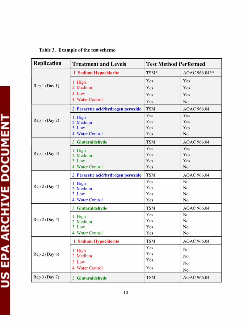
O. Example of a test scheme for one laboratory with test chemicals randomized for three replications – see Table 3.
10
Table 3. Example of the test scheme
Replication Treatment and Levels Test Method Performed 1. Sodium Hypochlorite TSM* AOAC 966.04**
Rep 1 (Day 1) 1. High 2. Medium 3. Low 4. Water Control
Yes Yes Yes Yes
Yes Yes Yes No
2. Peracetic acid/hydrogen peroxide TSM AOAC 966.04
Rep 1 (Day 2) 1. High 2. Medium 3. Low 4. Water Control
Yes Yes Yes Yes
Yes Yes Yes No
3. Glutaraldehyde TSM AOAC 966.04
Rep 1 (Day 3) 1. High 2. Medium 3. Low 4. Water Control
Yes Yes Yes Yes
Yes Yes Yes No
2. Peracetic acid/hydrogen peroxide TSM AOAC 966.04
Rep 2 (Day 4) 1. High 2. Medium 3. Low 4. Water Control
Yes Yes Yes Yes
No No No No
3. Glutaraldehyde TSM AOAC 966.04
Rep 2 (Day 5) 1. High 2. Medium 3. Low 4. Water Control
Yes Yes Yes Yes
No No No No
1. Sodium Hypochlorite TSM AOAC 966.04
Rep 2 (Day 6) 1. High 2. Medium 3. Low 4. Water Control
Yes Yes Yes
Yes
No No No No
Rep 3 (Day 7) 3. Glutaraldehyde TSM AOAC 966.04

11
1. High 2. Medium 3. Low 4. Water Control
Yes Yes Yes Yes
No No No No
1. Sodium Hypochlorite TSM AOAC 966.04
Rep 3 (Day 8) 1. High 2. Medium 3. Low 4. Water Control
Yes Yes Yes
Yes
No No No No
2. Peracetic acid/hydrogen peroxide TSM AOAC 966.04
Rep 3 (Day 9) 1. High 2. Medium 3. Low 4. Water Control
Yes Yes Yes Yes
No No No No
*Three carriers per treatment; **30-carriers per treatment
P. Table 4. Randomized Order of Testing (based on 10 labs)
Random Order of Test Chemicals** Rep* Lab 1 Lab 2 Lab 3 Lab 4 Lab 5 Lab 6 Lab 7 Lab 8 Lab 9 Lab 10Rep 1 2, 3, 1 1, 3, 2 2, 3, 1 1, 2, 3 2, 3, 1 2, 1, 3 1, 2, 3 2, 3, 1 1, 2, 3 3, 2, 1 Rep 2 1, 3, 2 3, 1, 2 2, 3, 1 3, 2, 1 1, 2, 3 1, 3, 2 3, 2, 1 1, 2, 3 1, 3, 2 1, 2, 3 Rep 3 2, 3, 1 2, 3, 1 3, 2, 1 2, 3, 1 1, 3, 2 3, 2, 1 1, 2, 3 3, 2, 1 3, 2, 1 2, 3, 1
*Three total tests days per replication; one chemical class tested per day **1 = sodium hypochlorite, 2 = hydrogen peroxide/peracetic acid, and 3 = glutaraldehyde; order within a
test day will be High, Medium, Low, and Water Control
Section 4. Test Sample Preparation Prepare test chemicals as follows: 1. Sodium hypochlorite (Sigma Aldrich reagent grade solution, approx. 12% available
chlorine, product no. 425044), diluted to 6000 ± 300ppm with reagent grade water, pH adjusted (pH 7.0 ± 0.5) with 5% acetic acid. pH adjusted treatment must be used in test within approximately 120 min after preparation; used for the high treatment.
2. Sodium hypochlorite (Sigma Aldrich reagent grade solution), diluted to 6000 ± 300ppm
with reagent grade water, unadjusted pH (pH ~10.0). Test within approximately 120 min after preparation; used for medium treatment.
12
3. Sodium hypochlorite (Sigma Aldrich reagent grade solution), diluted to 3000 ± 300ppm
with reagent grade water, unadjusted pH (pH ~10.0). Test within approximately 120 min after preparation; used for low treatment.
4. 0.08% peracetic acid/1.0% hydrogen peroxide product; ready to use product, must initiate testing within approximately 3 hr after dispensing. 5. 2.6% glutaraldehyde product, activate according to product directions, 14-day shelf-life post activation. The activated product may used for testing during the 14-day period. Section 5. Quality Assurance The sponsor of this project is the EPA Office of Research and Development. Document archiving will be adequate to ensure that all studies are supported by complete, accurate, consistent, and chronological records from initial collection of raw data to final analysis interpretation and reporting of results. The preparedness of each laboratory will be assessed by the Study Director and Quality Assurance Unit (QAU) for each laboratory prior to initiation of the study to ensure compliance with a project-specific EPA Quality Assurance Project Plan (Appendix A). The expected level of quality assurance is consistent with EPA Good Laboratory Practices. Numerous guidance documents, standard methods and Standard Operating Procedures (SOPs) will be used to maintain data quality. Proper record keeping and archiving will be performed to ensure the defensibility and reconstructibility or reanalysis of the study. No specific certification is required for this study; however, staff performing the assays must be familiar with standard microbiological techniques such as aseptic transfer, serial dilutions, plate counts and microbe identification. Scientists and analysts involved in testing shall be familiar with each efficacy method and associated procedure (e.g., carrier counts, neutralization confirmation) and will be proficient in conducting each designated efficacy test method. In-house practice sessions will be required for each laboratory to build proficiency with each method prior to official testing. The Lead laboratory (OPP Microbiology Laboratory) will conduct a series of conference calls with the participating laboratories to discuss the details of each method protocol. Documentation of practice and training for projects will be maintained in a training file for each participating scientist or analyst. Research documentation shall include project identification, data, and researcher. Pre-printed forms will be used. Research documentation shall be in ink and the use of a single line for correcting entries with the date and initials of the person making the correction and the reason for the change. Project-specific documentation shall be maintained in a project file, or the project file must identify where the documents are stored. Where possible, EPA SOPs and standard forms shall be used for those operations which have become or will become routine, including test
13
methodology, analytical procedures and calibration procedures. SOPs that are comparable to EPA’s may be used; however, this will require concurrence by the Study Director or the EPA Quality Assurance Unit. The purpose of the SOPs is to facilitate the uniform performance of routine procedures. The Standard Operating Procedures for quality control activities are located at: http://www.epa.gov/oppbead1/methods/atmpa2z.htm. Selected electronic spreadsheets and email will be considered as official documentation and will be maintained and archived accordingly. All preparations of test chemicals, media and reagents will be tracked using an assigned media preparation number. Samples, test chemicals, will be maintained to ensure their integrity. Test chemicals will be stored away from standards, media, and reagents to prevent cross-contamination. No official chain of custody documentation will be required for test chemicals evaluated in this research; however specific information on source, identification, and volume received will be maintained and archived for all test chemicals. All supplies and materials considered “critical” to the quality of the research such as media, reagents, carriers, and test chemicals shall be inspected prior to use to ensure that the shipment has not been damaged or compromised in any way. For pre-sterilized lab supplies, the manufacturer’s statement of sterility is acceptable for quality control documentation for sterility; no further testing is required. For growth media, performance testing (sterility and suitability to support growth of B. subtilis) must be performed a minimum of one time, preferably on the first batch prepared per lot. Suppliers (vendors) of testing materials and components with specific requirements such as sodium hypochlorite and inoculated carriers will be requested to provide verification of the desired specifications. The information and data will be maintained and archived in the project file. Upon completion of each study, a peer review of the data entry/tabulation will be performed by laboratory personnel. A draft report of the findings or data summary will be compiled and forwarded to each lab’s Quality Assurance personnel for review. The designated QAO at each facility will review and comment on the data and supporting information before submission to the statistician. Critical findings will be immediately communicated to the Study Director. Data may be rejected if microbial contamination occurs at a level unacceptable to the Study Director.
14
Section 6. The Reference Method Note: Laboratories will only conduct the efficacy component (see C. g)
AOAC Official Method 966.04; Sporicidal Activity of Disinfectants
Modified Method
First Action 2006
Applicable to testing sporicidal activity of liquid disinfectants using modified method 966.04 against Bacillus subtilis on a hard surface (porcelain carrier). Performance criteria for product efficacy are not impacted. This method has been validated for products containing sodium hypochlorite, peracetic acid/hydrogen peroxide, and glutaraldehyde. See results of the collaborative study supporting the modifications to Method 966.04. (1) All manipulations of the test organism are required to be performed in accordance with biosafety practices stipulated in the institutional biosafety regulations. Use the equipment and facilities indicated for the test organism. For recommendations on safe handling of microorganisms refer to the Centers for Disease Control/National Institutes of Health (CDC/NIH) Biosafety in Microbiological and Biomedical Laboratories manual. (2) Disinfectants may contain a number of different active ingredients, such as heavy metals, aldehydes, peroxides, and phenol. Personal protective clothing or devices are recommended during the handling of these items for purpose of activation, dilution, or efficacy testing. A chemical fume hood or other containment equipment may be employed when appropriate during performing tasks with concentrated products. The study analyst may wish to consult the Material Safety Data Sheet for the specific product/active ingredient to determine best course of action. (3) References to water mean reagent-grade water, except where otherwise specified. (4) Commercial dehydrated media made to conform to the specified recipes may be substituted. (5) These microbiological methods are very technique sensitive and technique-oriented, thus exact adherence to the method, good laboratory practices, and quality control (QC) are required for proficiency and validity of the results. (6) Detergents used in washing glassware may leave residues which are bacteriostatic. Test for inhibitory residues on glassware periodically. For procedure, refer to Standard Methods for the Examination of Water and Wastewater, Section 9020, Quality Assurance/Quality Control. A. Media and Reagents (a) Culture Media.–(1) Nutrient broth.–For use in preparing nutrient agar. Add 5 g beef extract (paste or powder), 5 g NaCl, and 10 g peptone (anatone, peptic hydrolysate of pork tissues, manufactured by American Laboratories, Inc., 4410 S 102nd St., Omaha, NE 68127) to approximately 1 L water. Boil mixture for 20 minutes with constant stirring. Readjust volume to 1 L with water and allow cooling to around 50ºC. Adjust pH to 6.8 ± 0.2 with 1N HCL or 1N NaOH, if necessary. Filter through paper (e.g., Whatman filter paper No. 4). Dispense 10 mL portions into 20 × 150 mm culture tubes or 20 mL portions into 25 × 150 mm culture tubes.
15
Dehydrated nutrient broth may be substituted – prepare according to the manufacturer's instructions. (2) Nutrient agar.–For stock cultures slants. Add 1.5% (w/v) Bacto-agar to unsterilized nutrient broth. Boil mixture until agar is dissolved. Adjust pH to 7.2 ± 0.2 if necessary. Dispense 5 mL portions into 16 x 100 mm screw cap tubes. Larger tubes may be used as well. Autoclave for 20 minutes at 121ºC. Remove from autoclave and slant tubes to form agar slopes. (3) Nutrient agar with 5µg/mL MnSO4:H20 (amended nutrient agar).–For spore production. Suspend 11.5 g nutrient agar in 495 mL water, add 5 mL 500 ppm MnSO4:H20. Dissolve by boiling. Adjust pH to 6.8 ± 0.2 if necessary. Autoclave for 15 minutes at 121ºC. Pour agar into plates. (4) Trypticase soy agar.–Suspend 40 g dehydrated trypticase soy agar in 1 L water and heat gently while stirring. Boil one minute or until completely dissolved. Adjust pH to 7.3 ± 0.2. Autoclave 15 minutes at 121ºC. Pour agar into plates. (5) Fluid thioglycollate medium (FTM).–Suspend 29.5 g of dehydrated fluid thioglycollate medium in 1 L water. Heat to boiling to dissolve completely. Adjust pH to 7.1 ± 0.2 if necessary. Dispense 10 mL portions into 20 ×150 mm culture tubes and autoclave for 15 minutes at 121ºC. Store at room temperature. Protect from light. Note: If after autoclaving the aerated portion of media consumes more than one third of tube, media must be re-boiled by placing tubes in beaker of boiling water. Media can only be re-boiled once. (6) Fluid thioglycollate medium with 1M NaOH (modified FTM).– For subculturing spores exposed to 2.5 M HCl. Suspend 29.5 g of fluid thioglycollate medium in 1 L water. Heat boiling to dissolve completely. Cool and adjust pH to 7.1 ± 0.2 if necessary. Add 20 mL 1M NaOH, mix well. Check final pH and record (pH between 8 and 9 is typical). Dispense 10 mL into 20 × 150 mm culture tubes and autoclave for 15 minutes at 121ºC. Store at room temperature. Protect from light. Note: If after autoclaving the aerated portion of media consumes more than one third of tube, media must be re-boiled by placing tubes in beaker of boiling water. Media can only be re-boiled once. Note: Media can be stored for up to two months.
(b) Manganese Sulfate Monohydrate.–500 ppm. Add 0.25 g of manganese sulfate to 500 mL water. Filter sterilize for use. (c) Dilute hydrochloric acid.–2.5M. Use to determine resistance of dried spores. Standardize and adjust to 2.5M as in 936.15. (d) Sterile water.–Use reagent-grade water. Reagent-grade water should be free of substances that interfere with analytical methods. Any method of preparation of reagent-grade water is acceptable provided that the requisite quality can be met. Reverse osmosis, distillation, and deionization in various combinations all can produce reagent-grade water when used in the proper arrangement. See Standard Methods for the Examination of Water and Wastewater for details on reagent-grade water. (e) Triton X-100 (f) Ethanol (40%) (g) Test organism.–Bacillus subtilis (ATCC No. 19659) obtained directly from a commercial supplier (e.g., ATCC).
16
B. Apparatus (a) Carriers.–Penicylinders, porcelain, 8 ± 1 mm OD, 6 ± 1 mm ID, 10 ± 1 mm length (Available from CeramTec Ceramic, Laurens, SC, www.ceramtec.com, Cat. No. LE15819.)
(b) Glassware.– For disinfectant, 25 × 150 mm or 25 × 100 mm culture tubes (Bellco Glass Inc., Vineland, NJ; reusable or disposable 20 × 150 mm (for cultures/subcultures); 16 × 100 mm screw cap tubes for stock cultures. Cap with closures before sterilizing. Sterilize all glassware 2 hr in hot air oven at 180o C or steam sterilize for a minimum of 20 min at 121oC with drying cycle. (c) Sterile centrifuge tubes.–Polypropylene, 15 mL conical tubes with conical bottoms (Corning), from Fisher, or equivalent. (d) Water bath/chiller unit.–Constant temperature for test chemical, capable of maintaining 20 ± 1ºC temperature or specified temperature for conducting the test.
(e) Petri dishes.–Plastic (sterile) (f) Filter paper.–Whatman filter paper #2; placed in Petri dishes for storing carriers.
(g) Test tube racks.–Any convenient style. (h) Inoculating loop.–Any convenient inoculation/transfer loop for culture transfer.
(i) Wire hook.–For carrier transfer. Make 3 mm right angle bend at end of 50 – 75 mm nichrome wire No. 18 B&S gage. Have other end in suitable holder.
(j) Centrifuge.–Non-refrigerated (e.g., Eppendorf 5804 R).
(k) Sonicator.–Ultrasonic cleaner (e.g., Branson Model 1510).
(l) Orbital shaker.–speed range from 25 to 500 rpm (e.g., VWR DS 500).
(m)Vacuum desiccator.–For carrier storage. With adequate gauge for measuring 27” (69 cm) of Hg and fresh desiccant.
(n) Certified biosafety cabinet (Class I or II).–Recommended for use to maintain aseptic work environment. (o) Certified Timer.–For managing timed activities, any certified timer that can display time in seconds.
17
C. Operating Technique (a) Culture initiation.– Initiate B. subtilis culture (e.g., use nutrient broth to re-hydrate a lyophilized culture, and incubate the broth culture for 24 ± 2 hours at 36 ± 1ºC prior to streak inoculation). Streak inoculate a set (e.g., six) nutrient agar slopes and incubate 24 ± 2 hours at 36 ± 1ºC. Concurrently, perform purity and identification confirmation testing for QC (e.g., colony morphology on TSA, Gram stain, or use of other identification systems). Following incubation, store at 2-5ºC. Maintain stock culture on nutrient agar slants by monthly (30 ± 2 days) transfers.
(b) Production of B. subtilis spore suspension.–Using growth from a stock culture tube, inoculate 10 mL tubes (e.g., 2 tubes, depending on the amount of spore preparation desired) of nutrient broth and incubate tubes on an orbital shaker for 24 ± 2 hours at approximately 150 rpm at 36 ± 1ºC. Use this culture to inoculate amended nutrient agar plates. Inoculate each plate with 500 µl of broth culture and spread the inoculum with a sterile bent glass rod or suitable spreading device. Wrap each plate with parafilm or place in plastic bags. Incubate plates inverted for 12-14 days at 36 ± 1ºC. Following incubation, harvest the spores by adding 10 mL cold sterile water to each plate. Using a spreader (e.g. bent glass rod), remove growth from plates and pipet suspensions into 15 mL sterile conical tubes (10 plates = 14 tubes, ~10 mL each). Centrifuge tubes at 5000 rpm for approximately 10 minutes at room temperature. Remove and discard supernatant. Re-suspend pellet in each tube with 10 mL cold sterile water and centrifuge at 5000 rpm for approximately 10 minutes. Remove and discard supernatant. Repeat twice. Re-suspend the pellet in each tube with 10 mL sterile water. Store the spore suspension at 2-5ºC. Examine spore suspension with a phase contrast microscope or by staining to assess quality of the spores. Examine a minimum of five fields and determine ratio of spores to vegetative cells (or sporangia). Percentage of spores versus vegetative cells should be at least 95%. Spore suspension from multiple plates can be combined and re-aliquoted into tubes for uniformity. Prior to inoculation of carriers, determine spore titer of the concentrated spore suspension by plating serial dilutions (e.g., 1.0 × 10⁻⁶ through 1.0 × 10⁻⁸) using pour or spread plating on TSA plates. For pour plating, add molten TSA tempered to 45-55ºC to each plate, swirl, and allow agar to solidify. Incubate plates for 24 ± 2 hours at 36 ± 1ºC and determine titer. Note: When harvested and processed, ten plates of amended nutrient agar should provide 80 - 100 mL of concentrated spore suspension (approx. 109 CFU/mL). Diluting the suspension prior to carrier inoculation will be necessary; a titer of 1.0 × 10⁸ to 5.0 × 10⁸ CFU/mL should be adequate to achieve the target carrier count. (c) Preparation of porcelain carriers.–Prior to use, examine porcelain carriers individually and discard those with scratches, nicks, spurs, or discolorations. Rinse unused carriers gently in water three times to remove loose material and drain. Place rinsed carriers into Petri dishes matted with 2 layers of filter paper in groups of 15 carriers per Petri dish. Sterilize 20 minutes at 121ºC. Cool and store at room temperature. Note: Handle porcelain carriers with care when placing in Petri dishes. Minimize carrier movement and avoid excessive contact between carriers that might result in chips and cracks. Wash carriers with Triton X-100 and rinse with water 4 times for reuse.
18
(d) Inoculation of Porcelain Carriers.–Dilute the concentrated spore suspension as necessary with sterile water to achieve carrier counts between 1.0 × 10⁵ and approximately 1.0 × 10⁶ spores/carrier. Dispense 10 mL diluted spore suspension into an appropriate number of 25 × 150 mm tubes. Add 10 sterile carriers to each tube containing 10 mL spore suspension, slightly agitate, and let stand 10-15 minutes. Remove each carrier with sterile hook and place upright in sterile Petri dish lined with two sheets of filter paper, no more than 30 carriers per Petri dish. Air dry in biological safety cabinet for approximately 30 ± 2 minutes. Place Petri dishes containing inoculated carriers in vacuum desiccator containing CaCl2 and draw vacuum of 69 cm (27”) Hg. Dry carriers under vacuum for 24 ± 2 hours before use in HCl resistance, efficacy testing or carrier counts. Maintain under vacuum for up to three months. Carriers may be used after three months if they meet the acceptable HCl resistance and carrier count criteria. Inoculated carriers should not be used after one year of storage. Sterilize and reuse if necessary (see C.c). (e) Spore Enumeration (Carrier Counts).–Prior to use, determine the carrier counts for each preparation of carriers. Assay 5 randomly selected carriers per preparation. Place each inoculated carrier into a 50 mL plastic, polypropylene conical centrifuge tube containing 10 mL of sterile water. Sonicate carriers for 5 minutes ± 30 seconds. Note: For sonication, place tubes into an appropriately sized beaker with tap water to the level of sterile water in the tubes. Place beaker in sonicator (ultrasonic cleaner) so that water level in the beaker is even with water level fill line on sonicator tank. Fill tank with tap water to water level fill line. Suspend beaker in sonicator tank so it does not touch bottom of tank and so all three water levels (inside test tubes, inside beaker, and sonicator tank) are the same. Following sonication, vortex tubes for 2 minutes ± 5 seconds. Dilute spore suspensions by transferring 1 mL aliquots to tubes containing 9 mL sterile water. Dilute spore suspensions out to 1.0 ×10⁻⁵ and plate dilutions 1.0 × 10⁻² through 1.0 × 10⁻⁵. Plate each dilution in duplicate using pour or surface spread plating with TSA. For pour plating, add molten TSA tempered to 45-55ºC to each plate. Swirl pour plates to distribute spores evenly and allow agar to solidify. Invert plates and incubate for 24-48 hours at 36 ± 1ºC. Count colonies (by hand or with colony counter). Use dilutions yielding between 30 and 300 CFU per plate (target counts) for enumeration; however, record all counts less than 30. Report plates with colony counts over 300 as TNTC (Too Numerous to Count). Average spore counts per carrier should be between 1.0 × 10⁵ and approximately 1.0 × 10⁶ spores/carrier. Do not use carriers with counts outside this range. (f) HCl resistance.–Equilibrate water bath to 20 ± 1ºC. Pipet 10 mL of 2.5M HCl into two 25 × 100 mm tubes, place into water bath, and allow to equilibrate. Start timer and rapidly transfer 4 inoculated penicylinders into an acid tube (2.5 M HCl) with flamed hooks or forceps. Do not allow carriers or transfer device to contact inside of wall of acid tube. Transfer individual carriers after 2, 5, 10, and 20 minutes of HCl exposure to a separate tube of modified FTM. Rotate each tube vigorously by hand for approximately 20 seconds and then transfer carrier to a second tube of modified FTM. For viability control, place one unexposed inoculated carrier in a separate tube of modified FTM. For media sterility, use one tube of modified FTM. Incubate all test and control tubes for 21 days at 36 ± 1ºC. Record results as growth (+) or no growth (0) at each time period. Spores should resist HCl for ≥ 2 minutes to be qualified as resistant test spores. Discard carriers if not resistant and repeat preparation of carriers as previously described.
19
(g) Efficacy Test.–Aseptically prepare disinfectant samples as directed. Prepare all dilutions with sterile standardized volumetric glassware. For diluted products, use 1.0 mL or 1.0 g of sample disinfectant to prepare the use-dilution to be tested. Use v/v dilutions for liquid products and w/v dilutions for solids. For a 30-carrier test, place 10 mL product at dilution recommended for use or under investigation into each of six 25 × 150 mm or 25 × 100 mm test tubes, or use appropriate number of tubes assuming 5 test carriers per tube of test chemical. Place tubes in 20 ± 1ºC water bath and let equilibrate to temperature. Using a sterile hook (or forceps), transfer inoculated carriers sequentially at 2 minute intervals in groups of 5 from Petri dish to test tubes containing sporicidal agent. Use a certified timer to monitor time. Flame hook and allow cooling after each transfer. When lowering carriers into test tube, neither carriers nor wire hook may touch sides of tubes. If interior sides are touched, note tube number. Do not count carrier set if any carrier from that group of 5 yields a positive result; testing another set of five carriers is recommended. Carriers must be deposited into test tubes within ± 5 seconds of the prescribed drop time. Return tubes to water bath immediately after adding carriers. After contact period has been achieved, transfer carriers in same sequential timed fashion into primary subculture tubes containing appropriate neutralizer (10 mL in 20 × 150 mm test tubes). Remove the carriers one at a time from the test tube with sterile hook, tap against interior side of tube to remove excess sporicidal agent, and transfer into neutralizer tube (primary tube). All five carriers must be transferred during each 2 minute interval. Flame hook between each carrier transfer. Move remaining carriers into their corresponding neutralizer tubes at appropriate time. Carriers may touch interior sides of neutralizer tube during transfer, but contact should be minimized. After each carrier is deposited, recap neutralizer tube and gently shake to facilitate adequate mixing and efficient neutralization. Within one hour from when last carrier was deposited into primaries, transfer carriers using sterile wire hook to second subculture tube (secondary tube) containing 10 mL of appropriate recovery medium, one carrier per tube. Move carriers in order, but movements do not have to be timed. Gently shake entire rack of secondary tubes after all carriers have been transferred. Incubate primary (neutralizer) and secondary subculture tubes for 21 days at 36 ± 1ºC. Report results as growth (+) or no growth (0). A positive result is one in which medium appears turbid. A negative result is one in which medium appears clear. Shake each tube prior to recording results to determine presence or absence of growth/turbidity. Primary and secondary subculture tubes for each carrier represent a “carrier set”. A positive result in either primary or secondary subculture tube is considered a positive result for the carrier set. Media sterility controls and system controls (check for aseptic technique during carrier transfer process) are recommended. For media controls, incubate 1-3 unopened subculture medium tubes with the test sample tubes for 21 days at 36 ± 1ºC. For system controls, use sterile forceps or needle hooks to transfer 3 sterile carriers into a tube of test chemical. Transfer system control carriers to neutralizer medium as follows: at start of sample test (prior to first tube), transfer 1 sterile carrier to tube of neutralizer medium. After one half of test carriers have been transferred to neutralizer tubes, transfer a second sterile carrier to tube of neutralizer medium. After all test carriers (last tube) have been transferred to neutralizer tubes, transfer third sterile carrier to tube of neutralizer medium. Transfer system control carriers to secondary subculture medium as follows: immediately prior to initiating transfer of test carriers into secondary
20
subculture medium tubes, transfer first system control sterile carrier from neutralizer medium to tube of subculture medium. After one half of test carriers have been transferred to secondary subculture medium tubes, transfer second system control sterile carrier to tube of subculture medium. After all test carriers have been transferred to secondary subculture medium tubes, transfer third system control sterile carrier to tube of subculture medium. For each test, include a positive carrier control by placing one inoculated carrier into tube of secondary subculture medium. Incubate controls and test sample tubes together for 21 days at 36 ± 1ºC. Perform identification confirmation on a minimum of three positive carrier sets per test, if available, using Gram stain and/or plating on TSA. Additional confirmation may be performed using VITEK, API analysis or comparable method. If fewer than three positive carrier sets, confirm growth from each positive carrier set. If both tubes are positive in carrier set, select only one tube for confirmatory testing. For test with 20 or more positive carrier sets, confirm at least 20% by Gram stain. If Gram stains are performed from growth taken directly from positive tubes, the staining should be performed within 5-7 days of conducting the efficacy test. (h) Neutralization Confirmation Procedure.– A neutralization confirmation test must be performed in advance or in conjunction with efficacy testing. This assay is designed to simulate the conditions (i.e., neutralizer, subculture medium, contact time, diluent, concentration of test substance) of the efficacy test and to demonstrate the recovery of a low level of spores (e.g., 5 – 100). Diluted inoculum (e.g., spores of B. subtilis) is added directly to the various sets of subculture media tubes (see Table 1). This assay provides for a quantitative approach to assessing the effectiveness of the neutralizer and any bacteriostatic action resulting from the neutralizer itself or neutralizer – disinfectant interactions. Produce a spore preparation according to the procedure for amended nutrient agar. Harvest growth from plates (e.g., five plates) per the method, except re-suspend pellet after final centrifugation step in approximately 100 ml aqueous (40%) ethanol. Determine spore count by serial dilution and plating on TSA. Desirable target of the initial working suspension is 1.0 × 10⁸ to 1.0 × 10⁹ CFU/mL. The suspension may require adjustment to reach target titer. Prepare serial ten-fold dilutions of the inoculum in sterile water out to 10-8. Use 10-6, 10-7 and 10-8 dilutions to inoculate the neutralizer and subculture media tubes – the target number of spores to be delivered per tube in this assay is 5–100 per tube. Determine spore titer by plating (spread plate or pour plate) each of three dilutions in duplicate on TSA agar. Incubate plates inverted for 24-48 hours at 36 ± 1ºC. Count colonies (by hand or with colony counter). Report plates with colony counts over 300 as TNTC (Too Numerous to Count). Note: A standardized spore preparation adjusted to deliver 5–100 spores/mL may be substituted for the three dilutions of spore inoculum. In addition, spores sheared from inoculated carriers may be used as a working suspension. Use 5 sterile porcelain carriers (only 3 to be used in the assay). Within 5 seconds, place a set of 5 carriers into a test tube (25 × 150 mm or 25 × 100 mm) containing test chemical; transfer carriers according to section (g). Allow carriers to remain in test chemical per the specified contact time and temperature. After the contact time is complete, aseptically transfer three of the five carriers individually into tubes containing the neutralizer per section (g). This set of tubes is

21
the Neutralizer/Primary Subculture treatment. Following the transfer of the last carrier into neutralizer tube, transfer each carrier, in sequence, into tube containing secondary subculture medium. This portion of assay is not timed, but should be made as soon as possible. This set is the Secondary Subculture treatment. Following carrier transfer, inoculate each tube (Neutralizer/Primary and Secondary Subculture treatment tubes) with one mL of each of three inoculum dilutions (10-6, 10-7 and 10-8). For controls, use three fresh unexposed tubes of neutralizer and three tubes of the secondary subculture medium; also inoculate each control tube with one mL of each of three inoculum dilutions. Include one uninoculated tube of neutralizer and secondary subculture media to serve as sterility controls. See Table 1 for tube inoculation scheme. Incubate all tubes 5-7 days at 36 ± 1ºC. Record results as growth (+) or no growth (0). Note: The lack of complete neutralization of the disinfectant or bacteriostatic activity of the neutralizer itself may be masked when a high level of inoculum (spores) is added to the subculture tubes. Table 1. Neutralization confirmation procedure – inoculating treatment and control tubes with diluted spore suspension* Neutralizer-Primary Subculture Treatment
Secondary Subculture Treatment (with Carrier)
Neutralizer-Primary Inoculated Control
Secondary Subculture Inoculated Control
1 mL of 10-6 ÿ Tube 1 1 mL of 10-7 ÿ Tube 2
1 mL of 10-8 ÿ Tube 3
1 mL of 10-6 ÿ Tube 1 1 mL of 10-7 ÿ Tube 2 1 mL of 10-8 ÿ Tube 3
1 mL of 10-6 ÿ Tube 1 1 mL of 10-7 ÿ Tube 2 1 mL of 10-8 ÿ Tube 3
1 mL of 10-6 ÿ Tube 1 1 mL of 10-7 ÿ Tube 2 1 mL of 10-8 ÿ Tube3
*1.0 × 10-6 through 1.0 × 10⁻⁸ based on an approx. starting suspension of 108 spores/mL Confirm a minimum of one positive per treatment and control (if available) using Gram staining and colony morphology on TSA. For each treatment and control group, conduct confirmation testing on growth from tube with fewest spores delivered. B. subtilis is a Gram positive rod and colonies on TSA are opaque, rough, dull, round, with irregular margins, and low convex. Colonial variation may be observed and is typical for this strain. Growth in the inoculated controls verifies the presence of the spores, performance of the media, and provides a basis for comparison of growth in the neutralizer and subculture treatment tubes. Note: There may be cases when the neutralizer is significantly different from the secondary subculture media; in these cases, growth may not be comparable. The uninoculated control tubes are used to determine sterility, and must show no growth for the test to be valid. The occurrence of growth in the Neutralizer/Primary Subculture and Secondary Subculture treatment tubes is used to assess the effectiveness of the neutralizer. No growth or growth only in tubes which received a high level of inoculum (e.g., the dilution with plate counts which are too numerous to count) indicates poor neutralization and/or presence of bacteriostatic properties of the neutralizer or neutralizer-disinfectant interactions. For a neutralizer to be deemed effective, growth must occur in the Secondary Subculture treatment tubes which received lower levels of inoculum (e.g., 5-100 CFU/mL). Growth in the Secondary Subculture inoculated Control verifies the presence of the spores, performance of the media, and provides a basis for comparison of growth in the neutralizer and subculture treatment tubes. No growth or only
22
growth in tubes which received high levels of inoculum (e.g., a dilution with plate counts which are too numerous to count) indicates poor media performance. Growth in the Neutralizer-Primary inoculated Control should be comparable to the Secondary Subculture inoculated Control if the neutralizer is the same as the secondary subculture media. There may be cases when the neutralizer is significantly different from the secondary subculture media. In these cases, growth may not be comparable to the Secondary Subculture inoculated Control. The Neutralizer-Primary and Secondary Subculture uninoculated Control tubes are used to determine sterility, and must show no growth for the test to be valid. Note: For product registration, the U.S. EPA requires the following to demonstrate sporicidal/sterilant-level efficacy: Using AOAC method 966.04, sixty carriers representing each of two types of surfaces (porcelain penicylinders and silk suture loops) must be tested separately against spores of both Bacillus subtilis (ATCC 19659) and Clostridium sporogenes (ATCC 3584) on three samples representing three different batches of product, one of which must be at least 60 days old (2 carrier types × 2 test microorganisms × 60 carriers/type = 240 carriers per batch sample; 3 product batches × 240 carriers/batch = total of 720 carriers). The product must kill all of the test spores on all of the 720 carriers without any failures.
References ASTM International Method E 1054 – Standard Test Methods for Evaluation of Inactivators of Antimicrobial Agents Standard Methods for the Examination of Water and Wastewater. 21st Ed. American Public Health Association, 1015 15th Street, NW, Washington, DC Biosafety in Microbiological and Biomedical Laboratories. 4th Ed. U.S. Department of Health and Human Services, Public Health Service, Centers for Disease Control and Prevention and National Institutes of Health
23
Section 7. Quantitative Three Step Method Note: Laboratories will conduct the method in its entirety. Determining the Efficacy of Liquid Sporicides and Sterilants Against Spores of Bacillus subtilis on a Hard Surface Using the Quantitative Three Step Method The Quantitative Three Step Method (TSM) is suitable for determining the sporicidal activity of liquid sporicidal agents against the genus Bacillus on a hard surface. For the purpose of this protocol, the terms sporicide and sterilant are considered synonymous. The TSM is an adaptation of a quantitative micro-method as reported by Sagripanti and Bonifacino (1). See results of the pre-collaborative study supporting the use of the TSM (2). A similar version of the TSM was recently accepted and published by ASTM International (3). Note: All manipulations of the test organism are required to be performed in accordance with biosafety practices stipulated in the institutional biosafety regulations. Use the equipment and facilities indicated for the test organism. For recommendations on safe handling of microorganisms refer to the CDC/NIH Biosafety in Microbiological and Biomedical Laboratories manual (4). Disinfectants may contain a number of different active ingredients, such as heavy metals, aldehydes, peroxides, and phenol. Personal protective clothing or devices are recommended during the handling of these items for purpose of activation, dilution, or efficacy testing. A chemical fume hood or other containment equipment may be employed when appropriate during performing tasks with concentrated products. The study analyst may wish to consult the Material Safety Data Sheet for the specific product/active ingredient to determine best course of action. References to water mean reagent-grade water, except where otherwise specified (5). The methods are technique sensitive and technique-oriented, thus exact adherence to the method, good laboratory practices, and quality control (QC) are required for proficiency and validity of the results. A. Media and Reagents
(a) Media.–(1) Nutrient broth (NB).–Dehydrated NB. For use in re-hydrating test organism and preparing nutrient agar. (2) Nutrient agar (NA).–For stock cultures slants and plating. Add 1.5% (w/v) Bacto-agar to un-sterilized nutrient broth. Boil mixture until agar is dissolved. If necessary, adjust pH to 7.2 ± 0.2. Dispense 5 mL portions into 16 × 100 mm screw cap tubes. Larger tubes may be used as well. Autoclave for 20 min at 121ºC. Remove from autoclave and slant tubes to form agar slopes. Dehydrated nutrient agar may be substituted – suspend 23 g nutrient agar per L water, dissolve by boiling. If necessary, adjust pH to 6.8 ± 0.2. Autoclave for 15 min at 121ºC. (3) Nutrient agar with 5µg/mL MnSO4:H20 (amended nutrient agar).–For spore production. Suspend 11.5 g nutrient agar in 495 mL water, add 5 mL 500 ppm MnSO4:H20. Dissolve by boiling. If necessary, adjust pH to 6.8 ± 0.2. Autoclave for 15 min at 121ºC. Pour agar into plates. (4) Trypticase soy agar (TSA).–Poured in plates for microbe isolation and spread
24
plating. (5) Luria-Bertani broth (LB broth).-Dehydrated LB broth (e.g., Difco); suspend 25 g LB broth in 1 L water, mix well, if necessary adjust pH to 7.0 ± 0.2, dispense in bottles and autoclave for 15 min at 121°C; use as neutralizer. (6) Modified Luria-Bertani broth.-neutralizer in HCl resistance test, add 20 mL 1M NaOH to 1 L LB broth, mix well, dispense in bottles and autoclave for 15 min at 121°C.
(b) Manganese sulfate monohydrate.–500 ppm. Add 0.25 g of manganese sulfate to 500 mL
water. Filter sterilize for use.
(c) Sterile water.–Use sterile reagent-grade water. Reagent-grade water should be free of substances that interfere with analytical methods. Any method of preparation of reagent-grade water is acceptable provided that the requisite quality can be met. Reverse osmosis, distillation, and deionization in various combinations all can produce reagent-grade water when used in the proper arrangement. See Standard Methods for the Examination of Water and Wastewater for details on reagent-grade water (5). (d) Test organism.-Bacillus subtilis (ATCC No. 19659) obtained directly from a commercial supplier (e.g., ATCC). B. Apparatus (a) Certified biosafety cabinet (Class I or II).–Recommended to maintain an aseptic work environment. (b) Glass coupon.-Hard surface carrier, 5 × 5 × 1 mm, Erie Scientific Company, Portsmouth, NH; custom order part number EPA-1101 (minimum order of 1000 pieces), single use. (c) Sterile 1.5 mL microcentrifuge tubes.-For exposing carrier to disinfectant, Fisherbrand cat. #05-408-129. (d) Sterile centrifuge tubes.–For preparation of spore suspension, polypropylene, 15 mL conical tubes with conical bottoms, Fisher, cat. #05-538-53D. (e) Dissecting forceps.-For the transfer of carriers, sterile, VWR cat. #25607-195 or Fisher cat. #13-812-42. (f) Micropipette.-Used to make serial dilutions, calibrated. (g) Positive displacement pipette.-For carrier inoculation. (h) Desiccator.–For carrier storage.
25
(i) Water bath/chiller unit.-Constant temperature for test chemical and controls, capable of maintaining 20 ± 1ºC temperature or specified temperature; e.g., Neslab RTE-221 or Nalgene Labtop Cooler. (j) Orbital shaker. (k) Microcentrifuge. (l) Microcentrifuge tube lid openers.-USA Scientific #1400-1508. (m) Sonicator.-Ultrasonic cleaner (Branson Model 1510 Bath Sonicator, or equivalent). (n) Floating microcentrifuge tube holder.-For sonication, VWR: #60986-099. (o) Hematology rotator.-For fraction C recovery, Hematology Chemistry Mixer 346–Fisher Scientific; or a suitable mixer/shaker to provide gentle agitation during incubation. (p) Vortex mixer. (also an option for fraction C recovery using a vortex adapter). (q) Vortex adapters.-Fisher Scientific cat. #1281161 and 1281211. (r) Certified timer.-For managing timed activities, any certified timer that can display time in seconds. (s) Test tubes.-For sterilizing carriers, 25 × 150 mm. (t) 95% ethyl alcohol.- For cleaning carriers. C. Operating Technique (a) Culture initiation.–Initiate B. subtilis culture (e.g., use nutrient broth to rehydrate a lyophilized culture, and incubate the broth culture for 24 ± 2 hours at 36 ± 1ºC prior to streak inoculation). Streak inoculate a set (e.g., six) nutrient agar slopes and incubate 24 ± 2 hours at 36 ± 1ºC. Perform purity and identification confirmation testing for QC (e.g., colony morphology on TSA, Gram stain, or use of other identification systems). Following incubation, store at 2-5ºC. Maintain stock culture on nutrient agar slants by monthly (30 ± 2 days) transfers. (b) Production of B. subtilis spore suspension.–Using growth from a stock culture tube, inoculate 10 mL tubes (e.g., 2 tubes, depending on the amount of spore preparation desired) of nutrient broth and incubate tubes 24 ± 2 hr on an orbital shaker at approximately 150 rpm at 36 ± 1ºC. Use this culture to inoculate amended nutrient agar plates. Inoculate each plate with 500 µl of broth culture and spread the inoculum with a sterile bent glass rod or suitable spreading device.
26
Wrap each plate with parafilm or place in plastic bags. Incubate plates inverted for 12-14 days at 36 ± 1ºC. Following incubation, harvest the spores by adding 10 mL cold sterile water to each plate. Using a spreader (e.g., bent glass rod), remove growth from plates and pipet suspensions into 15 mL sterile conical tubes (10 plates = 14 tubes, ~10 mL each). Centrifuge tubes at 5000 rpm for approximately 10 min at room temperature. Remove and discard supernatant. Resuspend pellet in each tube with 10 mL cold sterile water and centrifuge at 5000 rpm for 10 ± 1 min. Remove and discard supernatant. Repeat twice. Resuspend the pellet in each tube with 10 mL sterile water. Store the spore suspension at 2-5ºC. Examine spore suspension with a phase contrast microscope or by staining to assess quality of the spores. Examine a minimum of 5 fields and determine ratio of spores to vegetative cells (or sporangia). Percentage of spores versus vegetative cells should be at least 95%. Spore suspension harvested from multiple plates can be combined and re-aliquoted into tubes for uniformity. Prior to inoculation of carriers, determine spore titer of the concentrated spore suspension by plating serial dilutions (e.g., 1.0 × 10⁻⁶ through 1.0 × 10⁻⁸) on TSA or NA. Incubate plates for 24 ± 2 hr at 36 ± 1ºC and determine titer. Note: When harvested and processed, 10 plates of amended nutrient agar should provide 80 - 100 mL of concentrated spore suspension. Diluting the suspension prior to carrier inoculation will be necessary; a spore titer of approx. 1.0 × 10⁹ CFU/mL in the suspension should be adequate to achieve the target carrier count. (c) Carrier Preparation.-Visually screen glass coupons (carriers) for scratches, chips, or cracks. Discard those which are damaged or defective. Rinse carriers once with water, rinse 3 times with 95% ethyl alcohol, and finally rinse 3 times with water. Allow carriers to dry. Place in glass tubes (25 × 150 mm) 40 carriers per tube. Steam sterilize 45 min at 121ºC with a 30 min dry cycle or sterilize for 2 hr in hot air oven at 180o C. Cool. Transfer carriers to sterile plastic Petri dishes for inoculation (approx. 40 carriers per dish). (d) Carrier Inoculation.-Transfer 10 µL of spore suspension with a micropipette using aerosol barrier tips or positive displacement pipette onto a 5 × 5 × 1 mm sterile, dry glass coupon. Apply to one central spot on each carrier. Inoculate the necessary number of carries to complete the validation study (approx. 140). Allow carriers to dry for minimum of 1 hour in open Petri dish in a biosafety cabinet, then for a minimum of 12 ± 2 hr in a desiccator. Store inoculated carriers under desiccation for up to 30 days. Glass carriers must be discarded after use. Note: During carrier inoculation, vortex mix inoculum frequently to ensure uniform distribution of spores. Recommend verification of carrier counts (per the method for control carriers) prior to test day; mean counts must be 5.0 × 10⁶ to 5.0 × 10⁷ spores/carrier. (e) Disinfectant Sample Preparation.-Aseptically prepare disinfectant samples as directed. Prepare all dilutions with sterile standardized volumetric glassware. For diluted products, use 1.0 mL or 1.0 g of sample disinfectant to prepare the use-dilution to be tested. Use v/v dilutions for liquid products and w/v dilutions for solids. Place approximately 1.5 mL of each disinfectant or control (sterile water) in microcentrifuge tubes. Allow to equilibrate to appropriate temperature for 15-30 min. (f) Test Procedure Overview.-A minimum of 3 carriers per disinfectant and 3 carriers for the
27
water control (control carriers) are required per product test. Three carriers per treatment will be required for the validation study. Use 1 pair of sterile forceps per fraction for each disinfectant. Fractions may be refrigerated briefly to allow for processing of other fractions. If possible, it is recommended that two analysts perform this method so that dilution and plating of the multiple fractions may be conducted as soon as possible. See Appendices TSM-1, TSM-2 and TSM-3 for additional guidance. Note: It is recommended that no more than 3 disinfectant treatments (9 test carriers) plus the water control (3 control carriers) should be tested during the same test period. Using sterile forceps, carefully transfer 1 inoculated carrier into each microcentrifuge tube labeled fraction A. Avoid touching inoculated area of carrier and sides of microcentrifuge tube. Discard carrier and tube if carrier touches sides of tube. Place fraction A tubes containing carriers and tubes containing disinfectant(s) and sterile water (control) into chiller water bath at 20 ± 1ºC, or use a labtop cooler to maintain temperature of the tubes. Equilibrate approximately 10 min. Add 400 µL disinfectant (test carriers) or 400 µL sterile water (control carriers) at 15 or 30 ± 5 sec intervals to appropriate microcentrifuge tube (in triplicate). Allow contact of the carriers to the disinfectant or water in fraction A tubes for the appropriate exposure period. Following the exposure period, add 600 µL of appropriate ice-cold neutralizer (e.g., LB broth) to each disinfectant fraction A tube. Add 600 µL LB broth as neutralizer for water control fraction A tubes. Slightly agitate tubes to thoroughly mix liquid components. Transfer each carrier using 1 pair sterile forceps per carrier set (i.e., 3 carriers) from fraction A tube to corresponding fraction B tube. Fraction B tubes contain 400 µL ice-cold (0-5ºC) sterile water. Place fraction A tubes in microcentrifuge, centrifuge for 6 min ± 30 sec at 13,000 rpm (15,500 × g). Remove 900 µL from each tube without disturbing pellet. Discard supernatant. Carefully add 900 µL ice-cold LB broth to each tube. Repeat 2 additional times. After third centrifugation, remove 900 µL from each tube. Carefully add 100 µL ice-cold LB Broth to each fraction A tube and resuspend pellet by vortex mixing 5 min ± 30 sec (use the vortex adapter) at midrange speed. Add 800 µL ice-cold LB Broth to each fraction A tube. Proceed to dilution and plating if another analyst is available, or store fraction A tubes in refrigerator. Note: Fluid remaining in the fraction A tubes contains spores dislodged from carrier by exposure to disinfectant or water control. Consistent orientation of the microcentrifuge tubes in the microcentrifuge is important in locating the pellet. The pellet may range in size and be difficult to visualize depending on the treatment. Fraction B and fraction C tubes can be evaluated while fraction A tubes are being centrifuged. Sonicate fraction B tubes 5 min ± 30 sec using a floating microcentrifuge tube holder placed inside an ultrasonic cleaner. After sonication is complete, add 600 µL ice-cold LB Broth to fraction B tubes. Vortex approx. 1 minute. Transfer each carrier using 1 pair sterile forceps per carrier set from fraction B tube to corresponding fraction C tube (fraction C tubes contain 400 µL ice-cold LB broth). Proceed to dilution and plating if another analyst is available, or store fraction B tubes in refrigerator; however, storage should be limited to 2 hrs. Note: Fluid remaining in the fraction B tubes contains spores dislodged from the carrier by sonication.
28
Place fraction C tubes in a hematology rotator inside incubator for 30 ± 2 min at 36 ± 1ºC. Remove fraction C tubes after 30 ± 2 min rotation/incubation from incubator. Add 600 µL ice-cold LB Broth to each tube. The carriers remain in the fraction C tubes. Proceed to dilution and plating if another analyst is available, or store fraction C tubes in refrigerator. Note: Fluid remaining in fraction C tubes contains spores dislodged from the carrier by gentle agitation for 30 min. Vortex mix each microcentrifuge tube thoroughly prior to making dilutions. For each fraction and control tube, remove 100 µL and serially dilute 10-fold in 900 μL ice-cold LB broth. For each carrier, direct plate 100 µL of the sufficient dilutions onto TSA or NA to ensure obtaining counts within the target range of 30-300 CFU/plate. Incubate plates a minimum of 24 ± 2 hr at 36 ± 1ºC. Record control counts at 24 ± 2 hr. Record treated carrier counts at 24 ± 2 hr and at 48 ± 2 hr. Confirm the identity of a minimum of one representative colony taken from at least one plate per treatment level (if available) using Gram staining, general growth media (e.g., TSA or NA) or other confirmation procedure. B. subtilis is a large Gram positive rod. On general growth media B. subtilis colonies are opaque, rough, round low convex colonies with irregular margins. Notes: After plating, dilution tubes may be stored at 2-5 ºC until the results are recorded; the tubes may be used for additional plating if initial plate counts are beyond the recommended target range. Use counts which fall within 0-300 CFU/plate for calculations. Obtain the total number of spores per fraction by dividing the number of colonies counted in each fraction by its dilution, and account for volume plated. Obtain the total number of spores per carrier by adding the total number of viable spores per fraction for fractions A, B, and C. Determine log density (LD) of total number of viable spores per carrier by taking Log10 (total number of spores per carrier). Determine log reduction (LR) of test carriers by subtracting log density of test carriers from log density of control carriers. Determine average LD and LR for each disinfectant. (g) Neutralization Confirmation (EPA Laboratory only).-Prepare 12 microcentrifuge tubes. Add 400 µL sterile water to tubes 1-6 and 400 µL of disinfectant to tubes 7-12. Allow tubes to equilibrate approximately 10 min at 20 ± 1ºC (or other specified temperature). Add 600 µL neutralizer in ice-cold Luria-Bertani (LB) broth to tubes 4-6 (neutralizer controls). Add 600 µL neutralizer in ice-cold LB broth to tubes 7-9 (ability of neutralizer to inactivate the disinfectant). Gently mix. Add 10 µL of B. subtilis spore suspension (approx. 109 spores/mL) to each tube and vortex mix for approximately 15 sec. Incubate tubes for 30 min ± 2 min at 20 ± 1ºC (or temperature specified by disinfectant manufacturer). After incubation, add 600 µL ice-cold LB broth to tubes 1-3 (survival controls). Add 600 µL ice-cold LB Broth to tubes 10-12 (disinfectant controls). Serially dilute each tube (10 µL into 990 µL ice-cold LB broth) to achieve plate counts of 30-300 CFU/plate. Plate 100 µL of each dilution onto NA or TSA. Incubate 24 ± 2 hr at 36 ± 1ºC. Count colonies on each plate. Log densities in tubes 1-3 and 4-6 reflect the spore suspension titer and should be within one log of each other. If log densities between tubes 1-3 and 4-6 are greater than one log, then the neutralizer has a sporicidal effect. If the disinfectant is highly effective, log densities in tubes 10-12 should be approximately 6 logs lower than log densities in tubes 1-6. To be an effective neutralizer, log densities in tubes 7-9 should be within 1 log of the
29
log densities in tubes 1-6. Note: The lead laboratory, the OPP Microbiology Laboratory, will perform this assay on each of the high treatments prior to the initiation of the study to verify the effectiveness of the chosen neutralizers. For this assay, produce a spore preparation according to the procedure for amended nutrient agar. Harvest growth from plates (e.g., five plates) per the method, except re-suspend pellet after final centrifugation step in approximately 100 ml aqueous (40%) ethanol. (h) HCl resistance.– Perform on each preparation of inoculated carriers. Conduct TSM procedure on 2.5 M HCl. Follow procedure as specified in part (f) with 2 and 5 min exposure periods with three inoculated carriers per time period. Include three control (sterile water) carriers to determine control carrier counts. Use modified LB broth (addition of NaOH) as the neutralizer instead of LB broth for HCl treatments. Perform test at 20 ± 1ºC. Calculate log reduction. Spores should resist HCl for ≥ 2 min (i.e., based on presence of viable spores after 2 min) to be qualified as resistant test spores. Discard carriers if not resistant and repeat preparation of carriers as previously described. Note: Compared to the water control, anticipate 1-2 log reduction of viable spores at 2 min exposure and 3-5 log reduction following the 5 min exposure. References Associated with the Three Step Method (1) Sagripanti, J.L. & Bonifacino, A. (1996) Am. J. Infect. Control 24, 364 – 371 (2) Tomasino, S.F. & Hamilton, M.A. (2006) Unpublished Report. Comparative Evaluation of Two Quantitative Test Methods for Determining the Efficacy of Liquid Sporicides and Sterilants on a Hard Surface: A Pre-Collaborative Study (3) Standard Test Method for Quantitative Sporicidal Three-Step Method (TSM) to Determine Sporicidal Efficacy of Liquids, Liquid Sprays, and Vapor or Gases on Contaminated Surfaces. (2005) ASTM Designation E 2414 – 05 (4) Biosafety in Microbiological and Biomedical Laboratories (1999) 4th Ed. U.S. Department of Health and Human Services, Public Health Service, Centers for Disease Control and Prevention and National Institutes of Health (5) Standard Methods for the Examination of Water and Wastewater. 21st Ed. American Public Health Association, 1015 15th Street, NW, Washington, DC

30
Appendix: TSM-1 Three Step Method Processing Sheet Analyst(s): ______________________________ Test Date: ___/_____/_____ Test Chemical(s): _____________________________________________________________________ NOTE: Carriers exposed to the disinfectant(s) and water control will be tested in triplicate. It is recommended that no more than three disinfectants plus the water control be tested during the same test period. The contents of microcentrifuge tubes prior to processing fractions for 1 disinfectant and control.
Treatment Tube Contents
A1-A3 400 μL disinfectant + 600 μL LB broth w/neutralizer (after exposure period)
B1-B3 400 μL sterile water + 600 μL LB broth (after sonication) Disinfectant
C1-C3 400 μL LB broth + 600 μL LB broth (after incubation/rotation)
A4-A6 400 μL sterile water + 600 μL LB broth (after exposure period)
B4-B6 400 μL sterile water + 600 μL LB broth (after sonication) Control
C4-C6 400 μL LB broth + 600 μL LB broth (after incubation/rotation)
Prior to Testing (i.e. day before test); as you proceed, add initials to each step in the space provided. ____ Label fraction A, B and C microcentrifuge tubes with fraction letter (e.g. A, B, or C) and carrier
number (e.g., 1, 2, 3, etc.). • Fraction tube examples: A1, A2, A3, B1, B2, B3, C1, C2, C3 etc.
____ In advance of testing, prepare fraction B and fraction C microcentrifuge tubes: • Add 400 μL of ice-cold sterile water to fraction B tubes. • Add 400 μL of ice-cold LB broth to fraction C tubes. • Store these tubes in a refrigerator (2-5°C) until ready for use.
____ Label dilution microcentrifuge tubes (serial dilution blanks) with fraction letter (e.g. A, B, or C),
carrier number (e.g. 1, 2, 3, etc.), and dilution (e.g., 10-1, 10-2, 10-3, etc.). • Serial dilution blank examples: A1-10-1, A1-10-2, A1-10-3, etc.
____ Prepare serial dilution blanks for A, B, and C fractions for all test carriers.
• Add 900 μL of ice-cold LB broth to each dilution blank for test carriers.
31
• Store these tubes in a refrigerator (2-5°C) until ready for use.
____ Prepare serial dilution blanks for A, B, and C fractions for all control carriers. • Add 900 μL of ice-cold LB broth to each dilution blank for control carriers. • Store these tubes in a refrigerator (2-5°C) until ready for use.
____ Sterilize forceps (two pair of forceps for each disinfectant/control tested plus 3 extra). ____ Prepare a 50 mL conical tube with the appropriate neutralizer (place it on ice on test day). ____ Turn on the recirculating chiller and water bath and allow them to reach 20 ± 1°C or the
temperature specified. ____ On test day, prepare the disinfectant(s) and place ~1.5 mL of each disinfectant and water control
into a microcentrifuge tube • Be sure to prepare and use the disinfectant within its specified period. • If the disinfectant requires a dilution, a minimum of 1 mL of the product must be used. • Ready-to-use disinfectants are tested as received; no dilution is required.
……………………………………………………………………………………………………… Fraction A ____ Using sterile forceps, carefully place one inoculated carrier inside each microcentrifuge tube
labeled fraction A. • Avoid touching the inoculated area of the carrier and the sides of the microcentrifuge tube. If the
carrier touches the sides of the microcentrifuge tube, the carrier and tube must be discarded. • These forceps should not be used for any additional transfers, unless sterilized appropriately.
____ Place the fraction A tubes containing the carriers and the tubes containing the disinfectant(s) and water control into a chiller water bath equilibrated at 20 ± 1°C.
• Allow the carriers, disinfectant(s), and water control to equilibrate for approx. 10 min. ____ Using a 1000 μL micropipette, add 400 μL of the disinfectant or 400 μL sterile water control at 15
or 30 ± 5 sec intervals to the appropriate microcentrifuge tubes (in triplicate). ____ Expose fraction A tubes in the chiller water bath for the appropriate exposure period. ____ After exposure is complete add 600 μL of the appropriate ice-cold neutralizer to each disinfectant
and water control fraction A tube. • Slight agitation (e.g., light tapping) may be necessary to thoroughly mix the liquid components.
____ Using one pair of sterile forceps per set of 3 carriers (e.g., per disinfectant), transfer each carrier
from its fraction A tube to its corresponding fraction B tube. • Avoid touching the sides of the tube if possible. • These forceps should not be used for any additional transfers.
Note: Processing of the fraction B and fraction C tubes can be completed by a second
32
analyst while the fraction A tubes are being centrifuged (e.g., sonication of the fraction B tubes, transfer of the carrier to the fraction C tubes, rotation of the fraction C tubes.
____ Place the fraction A tubes into the microcentrifuge for 6 min ± 30 sec at 13,000 rpm (15,500 × g).
• Centrifugation #1 ____ After fraction A tubes complete centrifugation #1, carefully remove 900 μL from each fraction A
tube without disturbing the pellet. Discard the supernatant. ____ Carefully add 900 μL of ice-cold LB broth into each fraction A tube and place the fraction A tubes
into the microcentrifuge for 6 min ± 30 sec at 13,000 rpm (15,500 × g). • Centrifugation #2
Note: The final volume in each fraction A tube is 1000 μL.
____ After fraction A tubes complete centrifugation #2, carefully remove 900 μL from each fraction A
tube without disturbing the pellet. Discard the supernatant. ____ Carefully add 900 μL of ice-cold LB broth into each fraction A tube and place the fraction A tubes
into the microcentrifuge for 6 min at 13,000 rpm (15,500 × g). • Centrifugation #3
____ After fraction A tubes complete centrifugation #3, carefully remove 900 μL from each fraction A tube without disturbing the pellet. Discard the supernatant.
____ Carefully add 100 μL of ice-cold LB broth into each fraction A tube. ____ Resuspend the pellet in the fraction A tubes by vortex mixing for 5 min ± 30 sec at a midrange speed.
• The use of a vortex adapter is recommended.
____ Add 800 μL of ice-cold LB broth into each fraction A tube. ____ Proceed to the Dilutions and Plating section.
• Place fraction A tubes in a refrigerator if dilutions are not made immediately. Processing should proceed as quickly as possible.
......................................................................................................................................................... Fraction B ____ Sonicate fraction B tubes for 5 min ± 30 sec using a floating microcentrifuge tube holder. ____ After sonication is complete, add 600 μL of ice-cold LB broth to the fraction B tubes and vortex
for one minute. ____ Using one pair of sterile forceps per set of 3 carriers (e.g., per disinfectant), transfer each carrier
from its fraction B tube to its corresponding fraction C tube.
33
• These forceps should not be used for any additional transfers, unless sterilized appropriately. ____ Proceed to the Dilutions and Plating section.
• Place fraction B tubes in a refrigerator if dilutions are not made immediately. Processing should proceed as quickly as possible.
……………………………………………………………………………………………………… Fraction C ____ Place the fraction C tubes in a hematology rotator inside a 36 ± 1°C incubator for 30 ± 2 min. ____ After the 30 min ± 2 min rotation/incubation, remove the fraction C tubes from the incubator and
add 600 μL of ice-cold LB broth to each tube. • The carriers can remain in the fraction C tubes.
____ Proceed to the Dilutions and Plating section.
• Place fraction C tubes in a refrigerator if dilutions are not made immediately. Processing should proceed as quickly as possible.
………………………………………………………………………………………………………. Dilutions and Plating ____ Remove dilution blanks from the refrigerator immediately prior to making dilutions. ____ Vortex mix each microcentrifuge tube thoroughly prior to making any dilutions. ____ Serially dilute fractions A, B, and C for test carriers.
• Vortex mix each tube prior to making the next dilution. • Appropriate dilutions for test carriers will be determined via a screen prior to testing.
____ Serially dilute fractions A, B, and C for control carriers.
• Vortex mix each tube prior to making the next dilution. • Appropriate dilutions for control carriers will be determined via a screen prior to testing.
____ Directly plate 100 μL of the dilutions that will yield counts within the target range (30-300) for
each carrier. • Spread using sterile spreader.
____ Incubate all plates at 36 ± 1°C for a minimum of 24 ± 2 hr (see text for details)
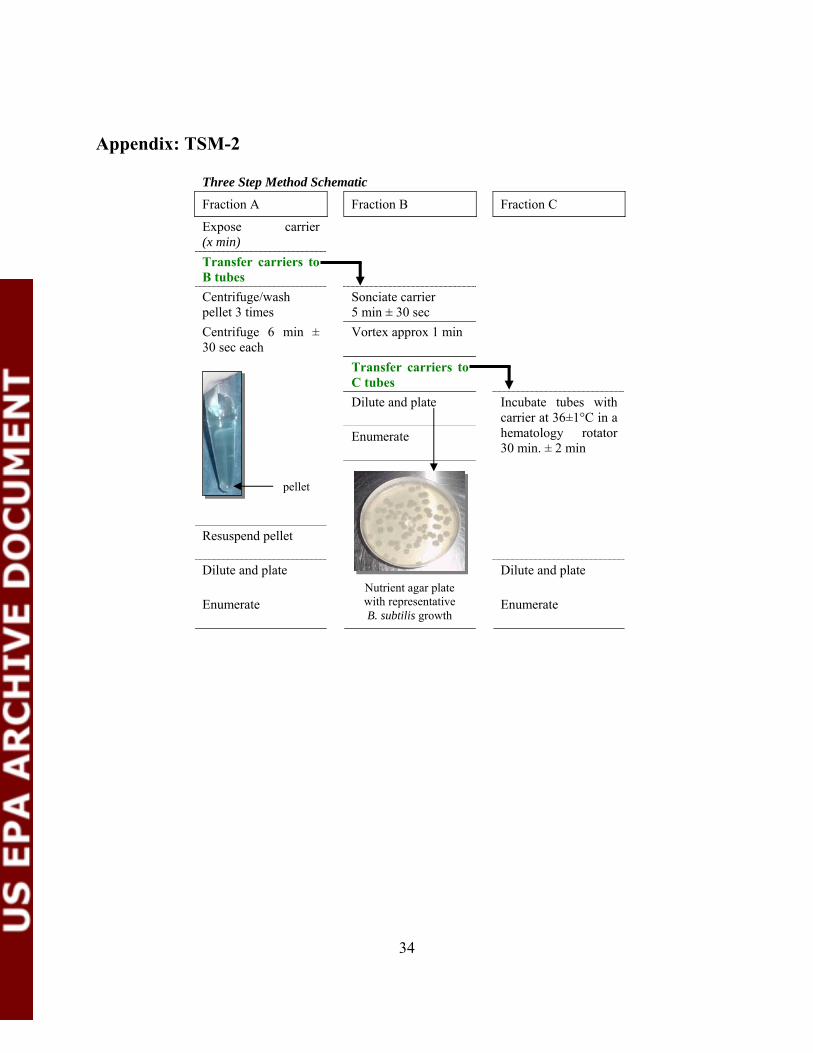
34
Appendix: TSM-2
Three Step Method Schematic
Fraction A Fraction B Fraction C
Expose carrier (x min) Transfer carriers to B tubes
Centrifuge/wash pellet 3 times
Sonciate carrier 5 min ± 30 sec
Centrifuge 6 min ± 30 sec each
Vortex approx 1 min
Transfer carriers to C tubes
Dilute and plate
Enumerate
Resuspend pellet
Incubate tubes with carrier at 36±1°C in a hematology rotator 30 min. ± 2 min
Dilute and plate Dilute and plate
Enumerate
Enumerate
pellet
Nutrient agar plate with representative B. subtilis growth
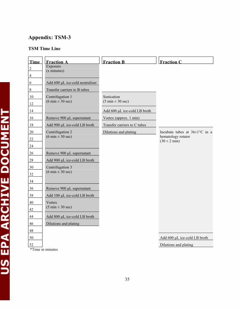
35
Appendix: TSM-3 TSM Time Line
Time Fraction A Fraction B Fraction C 2
4
Exposure (x minutes)
6 Add 600 μL ice-cold neutralizer
8 Transfer carriers to B tubes
10
12
Sonication (5 min ± 30 sec)
14
Centrifugation 1 (6 min ± 30 sec)
Add 600 μL ice-cold LB broth
16 Remove 900 μL supernatant Vortex (approx. 1 min)
18 Add 900 μL ice-cold LB broth Transfer carriers to C tubes
20 Dilutions and plating
22
24
Centrifugation 2 (6 min ± 30 sec)
26 Remove 900 μL supernatant
28 Add 900 μL ice-cold LB broth
30
32
34
Centrifugation 3 (6 min ± 30 sec)
36 Remove 900 μL supernatant
38 Add 100 μL ice-cold LB broth
40
42
Vortex (5 min ± 30 sec)
44 Add 800 μL ice-cold LB broth
46 Dilutions and plating
48
Incubate tubes at 36±1°C in a hematology rotator (30 ± 2 min)
50 Add 600 μL ice-cold LB broth
52 Dilutions and plating*Time in minutes
36
Section 8. REPORTING RAW DATA Information and raw data will be recorded on the test forms and data sheets provided by the Study Director (Appendices B and C). Electronic spreadsheets, provided by the Study Director, will be populated with the data from the hard copy data sheets, peer-reviewed for accuracy, and forwarded to the statistician for analysis. The preparation of media and reagents will be recorded on Media Preparation Sheets provided by the Study Director (Appendix D). Section 9. STATISTICS AND ANALYZING RAW DATA For statistical analysis, the Study Director will utilize the services of Dr. Martin Hamilton at the Center for Biofilm Engineering, Montana State University – Bozeman. The statistical analysis will produce estimates of the repeatability standard deviation, denoted by Sr, and the reproducibility standard deviation, denoted by SR, for each treatment (disinfectant × efficacy level combination) and for each quantitative response (log reduction value and the control carrier log spores per carrier).
In studies such as this, it is not unusual for the dilution series to miss occasionally the counting range of dilutions, thereby providing anomalous counts, either all zeros or all “too numerous to count” (TNTC). For such anomalous data, artificial counts will be substituted. If all dilutions produce TNTC, 300 will be substituted at the last dilution for the fraction (A, B, or C). If all dilutions produce zeros, 0.5 will be substituted at the first dilution.
Overview The statistical analyses will provide the following information. More detailed descriptions of the statistical methods are presented in the next section. C Raw data plots – the individual data points will be plotted for visual inspection to see
trends and effects and to detect outliers or influential observations. C Analysis of control carrier spore titers – the log transformed spores per control carrier
will be analyzed using an analysis of variance (ANOVA) two factor, nested, random effects model (details below). These results will describe the “normal range” of control carrier titers for each test method as well as estimates of the Sr and the SR and the within-test, intra-laboratory, and inter-laboratory sources of variability.
C log reduction (LR) value – LR is the primary quantitative response and most of the
statistical work will focus on the LR data. The LR value will be calculated using the formulas appropriate for each laboratory test (details below). For each combination of test method and test treatment, a one factor, random effects model ANOVA will be conducted to estimate the Sr, the SR, and the intra-laboratory versus inter-laboratory sources of
37
variability. C Mean LR – for each chemical × efficacy level combination, the mean LR will be
calculated along with the associated standard error and confidence interval. For each chemical treatment, a statistical trend test will determine whether the mean LR values increase significantly with increasing efficacy level.
C Diagnostic plots and tests – performed routinely to check whether the observations
conform to the mathematical assumptions underlying the ANOVA calculations. C Presentation of results – tables and figures will be created to present the results. • LR for the AOAC test – To calculate an LR value that is consistent with each AOAC test
result, the P/N formula will be applied (1, 4). Those LR values will be used to calculate SR for the AOAC test.
• Percent of the Total Counts by Fraction – An assessment of the percentage of the total
spore counts for each fraction of the TSM will be calculated – the contribution of fraction C will be assessed for importance.
Statistical analysis details • Analysis of control carrier spore titers – The ANOVA will be based on a nested, two-
factor, random effects model, similar to the models used in (5, 6). For each combination of test method, the response variable is the log spores per control carrier, the main effect is laboratory (a random effect), the nested effect is replication (a random effect) and the “chance error” is due to the variation among the carriers within the replication. The laboratories taking part in this collaborative study are assumed to be statistically representative of all laboratories that will be conducting these types of sporicide tests in the future. Because the ten participating laboratories were not in fact randomly selected from a population of testing laboratories, the assumption is inaccurate; nevertheless, it is required for the conventional approach to analyzing collaborative studies.
In standard statistical notation [see for example (7)], let Yijk denote the log spores for the kth carrier in the jth trial (replication) in the ith laboratory, k=1,2, or 3, j=1,2, or 3 and i = 1,2, …, 10. Then the model is Yij = : + Li + Tj(i) + gijk , where : denotes the true mean log spores per carrier for that test method and treatment, Li denotes the effect of the ith laboratory, Tj(i) denotes the effect of the jth trial (replication) in laboratory i, and gijk denotes the chance error for the kth carrier in the jth replicate test in the ith laboratory. The parameter : is deterministic; it is a specific, but unknown, numerical value. The quantities Li, Tj(i), and gij are random variables. According to conventional assumptions,
38
Li follows a normal probability distribution with mean zero and variance FL2, Tj(i) follows
a normal probability distribution with mean zero and variance FT2, and gijk follows a
normal probability distribution with mean zero and variance F2 . This model implies that Yij is a random variable following a normal probability distribution with mean : and variance FL
2 + FT2 + F2. Let FR
2 (= FL2 + FT
2 + F2) denote the total variance of Yijk. Conventionally, FR = /FR
2 is called the reproducibility standard deviation and Fr = [FT2 +
F2]½ is called the repeatability standard deviation (5, 8). The ANOVA will provide numerical estimates of the parameters :, FL
2, FT2, F2, Fr
2, and FR2.
• log reduction (LR) values – For the TSM, the LR is the mean of log10 spores per control
carrier minus the mean of log10 spores per treated carrier. Formulas for calculating the LR and associated within-test standard error are presented in Zelver et al. (9). For AOAC 966.04, the LR value will be calculated using the P/N formula presented in the report to AOAC by Tomasino and Hamilton (4).
The ANOVA will be based on a one-way, random effects linear statistical model. For
each combination of test method and chemical, the LR is the response variable, the main effect is laboratory (a random effect) and the “chance error” is due to the variation among independent repeats of the test within laboratories. The laboratories taking part in this collaborative study are assumed to be statistically representative of all laboratories that will be conducting these types of sporicide tests in the future.
In standard statistical notation [see for example (7)], let Yij denote the LR value for a specific combination of test method and chemical at the jth replication in the ith laboratory, j=1, 2, or 3 and i = 1, 2, or 3. Then the model is Yij = : + Li + gij , where : denotes the true mean LR for that test method and chemical, Li denotes the effect of the ith laboratory, and gij denotes the chance error for the jth replicate test in the ith laboratory. The parameter : is deterministic; it is a specific, but unknown, numerical value. The quantities Li and gij are random variables. According to conventional assumptions, Li follows a normal probability distribution with mean zero and variance FL
2 and gij follows a normal probability distribution with mean zero and variance Fr
2 . This model implies that Yij is a random variable following a normal probability distribution with mean : and variance FL
2 + Fr
2 . Let FR2 (= FL
2 + Fr2 ) denote the variance of Yij ; FR
2 is called the “total variance” in ANOVA textbooks. In the context of germicide tests, FR = /FR
2 is called the reproducibility standard deviation and Fr = /Fr
2 is called the repeatability standard deviation (5, 8). The ANOVA will provide numerical estimates of the parameters :, FL
2, Fr
2, and FR2. If the estimates of the variances differ insignificantly among test chemicals,
the data may be combined across chemicals for purposes of running one ANOVA for that test method. If it is appropriate to do so, combining the data will produce more reliable estimates of the parameters. For the TSM, by using the within-test standard error associated with each LR, it will be possible to partition out the within-test component of variance from Fr
2.
39
C Mean LR – The mean for each test chemical and test method combination will be estimated by the ANOVA. The formula for the standard error of the mean depends on the results of the ANOVA and the equations will be derived by the statistician, as in (10). The confidence intervals will be based on normal distribution theory.
It is of interest to determine whether each method is sensitive enough to correctly
order treatments known to have low, medium, and high efficacy. For each method and disinfectant, a trend test will be conducted to determine whether the log spores per treated carrier increases significantly with known efficacy. The trend test will be a test for a significant mean slope based on a simple linear regression model. The efficacy levels will be coded as 1, 2, and 3, going from low to high efficacy.
• Sr and SR of LR values for the TSM – It is desirable for the standard deviations to be
small. For disinfectant tests, the AOAC has no specifications for concluding that a standard deviation is acceptably small. Some guidance is provided by a recent literature review which showed that, for established suspension and dried surface disinfectant tests, Sr ranged from 0.2 to 1.2 with a median of 0.4 and SR ranged from 0.3 to 1.5 with a median of 0.8 (6). It would be reasonable to claim that the Sr and SR are acceptably small if they fall within these ranges.
C Diagnostic plots and tests – conventional plots and tests of residuals will be used to check
the homogeneous variance and normality assumptions underlying the ANOVA. The Anderson-Darling test will be used to check normality.
C The % of the total spore counts for each fraction of the TSM will be evaluated – the
contribution of fraction C will be assessed for importance. Section 10. REFERENCES ASSOCIATED WITH THE VALIDATION PROTOCOL (1) Tomasino, S.F. & Hamilton, M.A. (2006) Unpublished Report. Comparative Evaluation of Two Quantitative Test Methods for Determining the Efficacy of Liquid Sporicides and Sterilants on a Hard Surface: A Pre-Collaborative Study (2) Sagripanti, J.L. & Bonifacino, A. (1996) Am. J. Infect. Control 24, 364 – 371 (3) Standard Test Method for Quantitative Sporicidal Three-Step Method (TSM) to Determine Sporicidal Efficacy of Liquids, Liquid Sprays, and Vapor or Gases on Contaminated Surfaces. (2005) ASTM Designation E 2414 – 05 (4) Tomasino, S.F. & Hamilton, M.A. (2006) Unpublished Report. Modifications to the
AOAC Sporicidal Activity of Disinfectants Test, Method 966.04: Collaborative Study (5) Youden, W.J. and Steiner, E.H. (1975) Statistical Manual of the AOAC. AOAC:
Arlington, VA (6) Tilt, N. and Hamilton, M.A. (1999) JAOAC Int., 82, 384 – 389 (7) Neter et al., (1996) Applied Linear Statistical Models – 4th edition, McGraw-Hill:Boston
40
(8) Helrich, K., editor, (1990) Official Methods of the AOAC, AOAC: Arlington, VA, p. 681, 1990
(9) Zelver, N. et al. (2001) Methods in Enzymology - Biofilms II, R.J. Doyle, editor, 337:363 – 376
(10) Johnson, K., Lundman, R., and Hamilton, M. (1993) Efficient Sampling Designs for Microbial Processes: A Case Study. Journal of Microbiological Methods, 18:69 – 81
Section 11. APPENDICES A. EPA Quality Assurance Project Plan B. AOAC Method Data Sheets C. TSM Data Sheets D. Media and Reagent Preparation Sheets E. Parameters for Testing Chemicals F. TSM Log Reduction Data G. Material Safety Data Sheets H. Safety Checklist