Embed Size (px)
Citation preview
Chapter 25
Interdisciplinary Surgical Management ofOrbital and Maxillo-Ethmoidal Complex Disorders
Jarosław Paluch, Jarosław Markowski, Jan Pilch,Agnieszka Piotrowska – Seweryn,Robert Kwiatkowski, Joanna Lewin-Kowalik,Czesław Zralek and Agnieszka Gorzkowska
Additional information is available at the end of the chapter
http://dx.doi.org/10.5772/53486
1. Introduction
Surgical management of the orbital and of maxillo-ethmoidal complex disorders is usuallyperformed in patients with trauma, inflammation and/or neoplasms. Depending on the des‐tructed craniofacial region rhinotomy, sinusotomy, orbitotomy, maxillectomy and othertypes of operations are performed. In case of skull base extension the situation becomesmore complicated leading to the necessity of co-operation of several specialists as well asmodifications of surgical technique. Surgical procedures on eyeball are undertaken mainlyby opthalmologists,but no particular specialty has been yet dedicated for surgical treatmentof other orbital regions. However, an attempt is made by surgeons, such as maxillo-facialsurgeons, ENT surgeons, neurosurgeons, trauma surgeons, oncologic surgeons and rarelyopthalmologists. Patients, in whom operation is performed, are ‘border-line‘ patients andthe anatomical structures that are traumatised belong topographically to above specialties. Itis very uncommon that there is an interdisciplinary team of surgeons available permanentlyin hospital to treat the described cases.
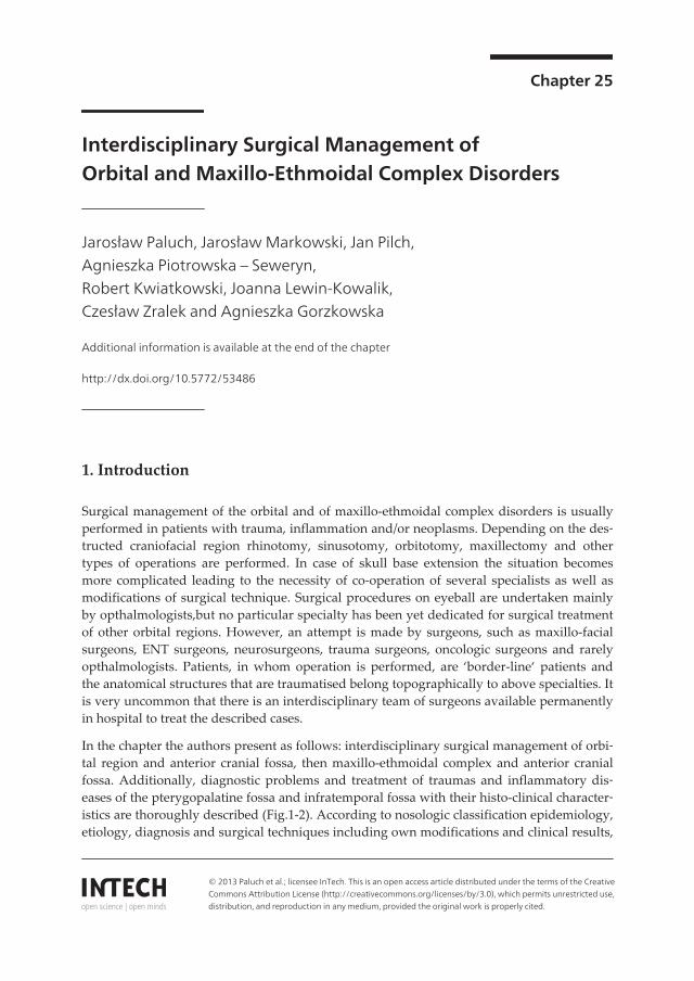
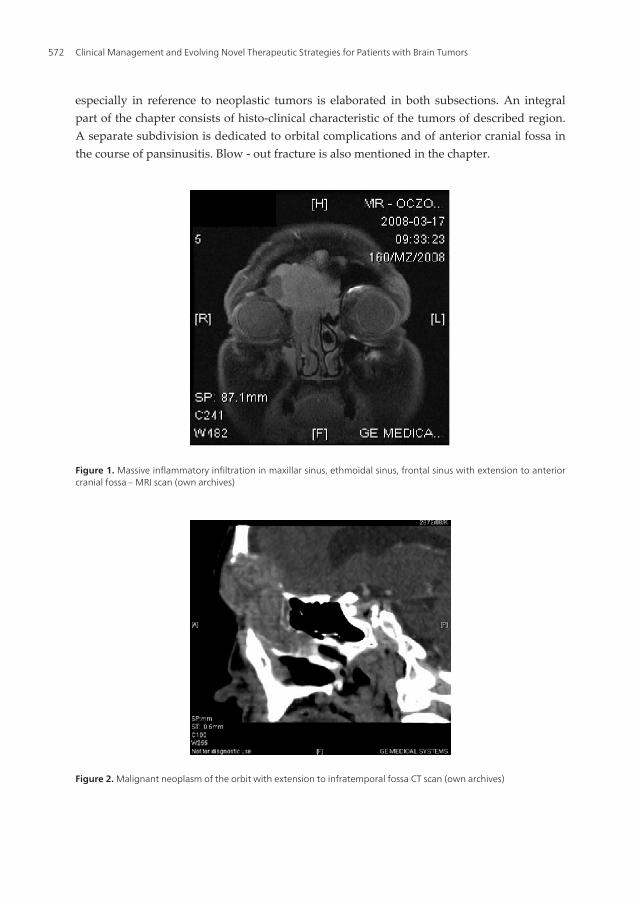
In the chapter the authors present as follows: interdisciplinary surgical management of orbi‐tal region and anterior cranial fossa, then maxillo-ethmoidal complex and anterior cranialfossa. Additionally, diagnostic problems and treatment of traumas and inflammatory dis‐eases of the pterygopalatine fossa and infratemporal fossa with their histo-clinical character‐istics are thoroughly described (Fig.1-2). According to nosologic classification epidemiology,etiology, diagnosis and surgical techniques including own modifications and clinical results,
© 2013 Paluch et al.; licensee InTech. This is an open access article distributed under the terms of the CreativeCommons Attribution License (http://creativecommons.org/licenses/by/3.0), which permits unrestricted use,distribution, and reproduction in any medium, provided the original work is properly cited.
especially in reference to neoplastic tumors is elaborated in both subsections. An integralpart of the chapter consists of histo-clinical characteristic of the tumors of described region.A separate subdivision is dedicated to orbital complications and of anterior cranial fossa inthe course of pansinusitis. Blow - out fracture is also mentioned in the chapter.
Figure 1. Massive inflammatory infiltration in maxillar sinus, ethmoidal sinus, frontal sinus with extension to anteriorcranial fossa – MRI scan (own archives)
Figure 2. Malignant neoplasm of the orbit with extension to infratemporal fossa CT scan (own archives)
Clinical Management and Evolving Novel Therapeutic Strategies for Patients with Brain Tumors572
2. Epidemiology
Nowadays, pansinusitis is one of the most common diseases that occurs almost as often asarthritis and high blood preassure. Morbidity of chronic pansinusitis is 10,9% (according toHastan D et al. ).
Traumas and intoxications are the third causes of deaths in Poland. Higher mortalitypresent cardiovascular diseases and neoplasms. Traumas of head and neck are responsiblefor 60-72% of multi-organ traumas. They usually affect elderly men. They occur mainly dueto collapses and accidents.
According to Szyfter et al. 3% of head and neck neoplasms and 05% of all neoplasms is lo‐calized in maxillo-ethmoidal complex. They occur mainly in men, in their 60s-80s. They in‐filtrate maxillar sinus in 50-70% of cases, nasal cavity in 15-30 and ethmoidal sinus in10-20%. According to anatomical topography and terminology neoplasms of maxillo-eth‐moidal complex and orbit with skull base extension expand in the region above Ohngren’splane that divides the maxillary sinus into an anterior-inferior part and superior-posterior.Tumors that arise in the first part have better prognosis.
3. Diagnostic methods
Diagnosis of traumas, inflammatory diseases and carcinomas of the described region has tobe very precise in order to use appropriate surgical approach. Apart from basic diagnosticmethods such as thorough anamnesis and examination, the authors emphasise a great roleof nasal and nasopharyngeal endoscopy perfectly suited for the assessment of inflammatoryand tumor penetration of nasal cavity and paranasal sinuses. However, in terms of carcino‐mas of orbit and the maxillo-ethmoidal complex with skull base penetration it is recom‐mended to use imaging techniques that improve both the preclinical research and clinicaltreatment, such as computed tomography (CT), magnetic resonance imaging (MRI), posi‐tron emission tomography (PET) and ultrasound imaging. Recently various modalities ofthese techniques have been introduced to investigate the progression and treatment resultsof brain tumors. Among them we can name CT three-dimensional reconstruction, electronbeam CT, dynamic CT enhancement, CT angiography, CT perfusion enhancement, high-re‐slution computed tomography (HRCT), diffusion-weighted imaging (DWI-MR), diffusiontensor imaging (DTI-MR) and many others. However, due to financial reasons conventionalCT and MRI remains the most common diagnostic imaging method in the management ofdiseases of the described region.
Also, the authors focus on a great role of fine-needle biopsy as a determinant in diagnosis oforbital neoplasms and of infratemporal fossa. It has been found that histological types ofmaxillo-ethmoidal complex including pterygopalatine fossa and infratemporal fossa are asfollows: pseudotumor, angioma, lymphoma, malignant epithelial neoplasms, neuromas andneurofibromas. Histological classification of intraorbital tumors has been thoroughly descri‐bed by Handerson.
Interdisciplinary Surgical Management of Orbital and Maxillo-Ethmoidal Complex Disordershttp://dx.doi.org/10.5772/53486
573
Histopathological results of biopsied tumors are essential for the following treatment, espe‐cially surgical management.
3.1. Fine-needle aspiration biopsy of orbital tumors
Fine-needle aspiration biopsy of orbital tumors verifies whether the tumor is neoplasticor non-neoplastic as well as it gives information about its malignancy. The most com‐mon orbital tumors are pseudotumors. The possibility to perform a biopsy depends onthe localization of a tumor. In case of meningiomas localized in orbital conus in patientwith good vision biopsy should be avoided due to the risk of damaging the optic nerve.Also, biopsy of cavernous hemangioma might develop complications such as bleedingand even haemorrhage.
A 25G hollow needle (length- 25 or 40mm)is used in fine-needle biopsy. The place of its in‐sertion into the examined mass is verified with palpatation. The insertion is carried outthrough the skin of eyelids without local anaesthesia. In case of impalpable tumors CT orMRI is used and the needle is continuously inserted until the resistance.
The consistence od most orbital tumors is solid. In case of cyst there is a liquid found and interatomas – sebaceous masses.
The authors emphasise that diagnostic results obtained by fine-needle aspiration biopsy arenot unequivocal and in lymphomas immunohistochemical examination is essential.
Also, it is worth mentioning that the described diagnostic methods are limited by ‘diagnos‐tic window’ (short time in traumas, more time in inflammatory diseases and neoplasms).
4. Surgical management
Surgical treatment in cases of trauma of craniofacial region, pterygopalatine fossa and infra‐temporal fossa is undertaken using an approach that gives access to damaged structures
4.1. Common approaches
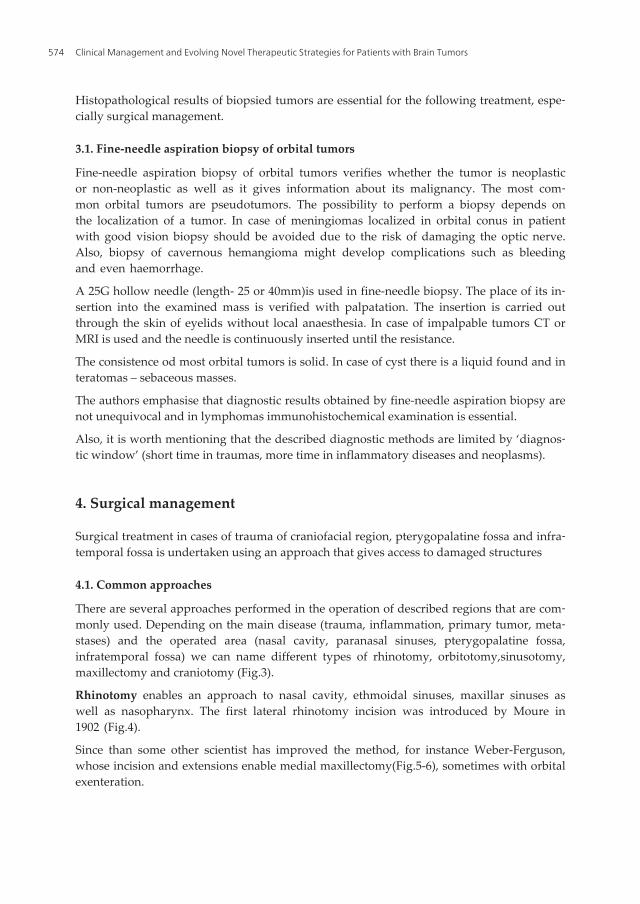
There are several approaches performed in the operation of described regions that are com‐monly used. Depending on the main disease (trauma, inflammation, primary tumor, meta‐stases) and the operated area (nasal cavity, paranasal sinuses, pterygopalatine fossa,infratemporal fossa) we can name different types of rhinotomy, orbitotomy,sinusotomy,maxillectomy and craniotomy (Fig.3).
Rhinotomy enables an approach to nasal cavity, ethmoidal sinuses, maxillar sinuses aswell as nasopharynx. The first lateral rhinotomy incision was introduced by Moure in1902 (Fig.4).
Since than some other scientist has improved the method, for instance Weber-Ferguson,whose incision and extensions enable medial maxillectomy(Fig.5-6), sometimes with orbitalexenteration.
Clinical Management and Evolving Novel Therapeutic Strategies for Patients with Brain Tumors574
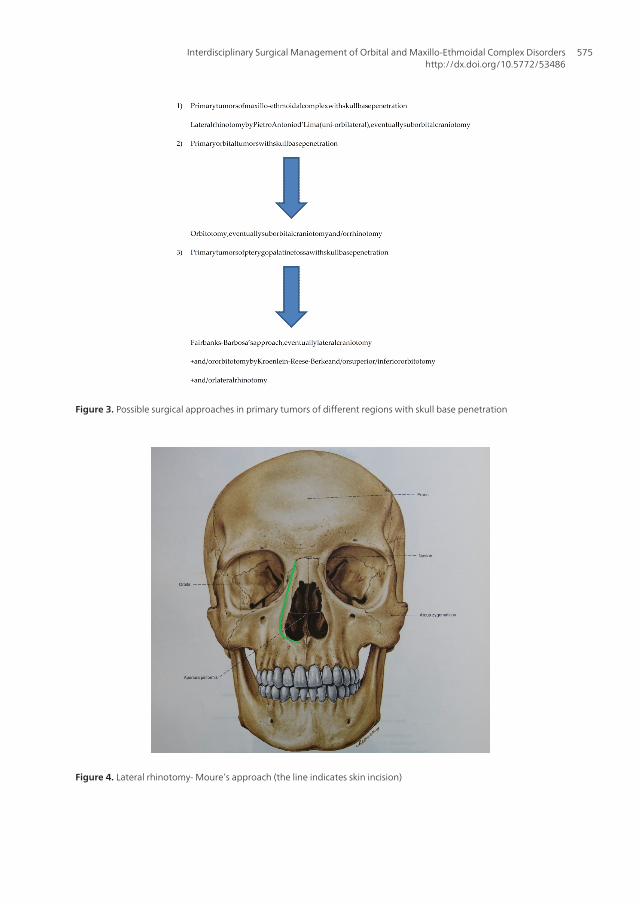
Figure 3. Possible surgical approaches in primary tumors of different regions with skull base penetration
Figure 4. Lateral rhinotomy- Moure’s approach (the line indicates skin incision)
Interdisciplinary Surgical Management of Orbital and Maxillo-Ethmoidal Complex Disordershttp://dx.doi.org/10.5772/53486
575
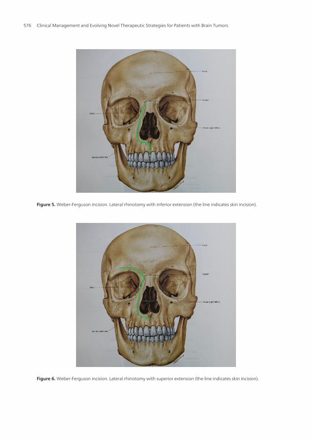
Figure 5. Weber-Ferguson incision. Lateral rhinotomy with inferior extension (the line indicates skin incision).
Figure 6. Weber-Ferguson incision. Lateral rhinotomy with superior extension (the line indicates skin incision).
Clinical Management and Evolving Novel Therapeutic Strategies for Patients with Brain Tumors576
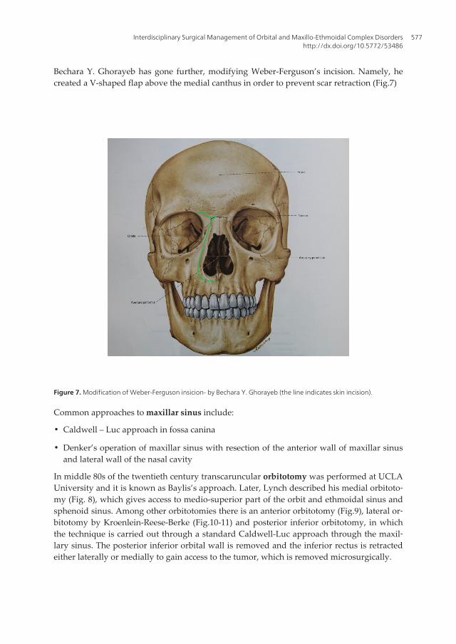
Bechara Y. Ghorayeb has gone further, modifying Weber-Ferguson’s incision. Namely, hecreated a V-shaped flap above the medial canthus in order to prevent scar retraction (Fig.7)
Figure 7. Modification of Weber-Ferguson insicion- by Bechara Y. Ghorayeb (the line indicates skin incision).
Common approaches to maxillar sinus include:
• Caldwell – Luc approach in fossa canina
• Denker’s operation of maxillar sinus with resection of the anterior wall of maxillar sinusand lateral wall of the nasal cavity
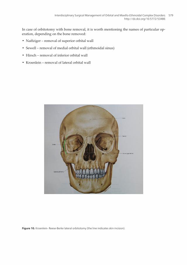
In middle 80s of the twentieth century transcaruncular orbitotomy was performed at UCLAUniversity and it is known as Baylis’s approach. Later, Lynch described his medial orbitoto‐my (Fig. 8), which gives access to medio-superior part of the orbit and ethmoidal sinus andsphenoid sinus. Among other orbitotomies there is an anterior orbitotomy (Fig.9), lateral or‐bitotomy by Kroenlein-Reese-Berke (Fig.10-11) and posterior inferior orbitotomy, in whichthe technique is carried out through a standard Caldwell-Luc approach through the maxil‐lary sinus. The posterior inferior orbital wall is removed and the inferior rectus is retractedeither laterally or medially to gain access to the tumor, which is removed microsurgically.
Interdisciplinary Surgical Management of Orbital and Maxillo-Ethmoidal Complex Disordershttp://dx.doi.org/10.5772/53486
577
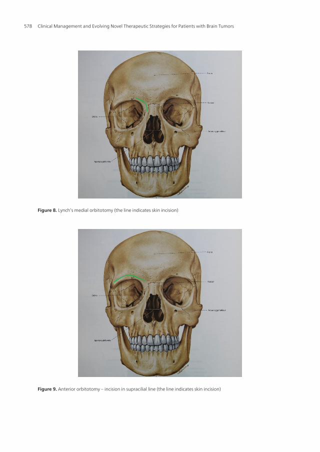
Figure 8. Lynch’s medial orbitotomy (the line indicates skin incision)
Figure 9. Anterior orbitotomy – incision in supracilial line (the line indicates skin incision)
Clinical Management and Evolving Novel Therapeutic Strategies for Patients with Brain Tumors578
In case of orbitotomy with bone removal, it is worth mentioning the names of particular op‐eration, depending on the bone removed:
• Naffziger – removal of superior orbital wall
• Sewell – removal of medial orbital wall (ethmoidal sinus)
• Hirsch – removal of inferior orbital wall
• Kroenlein – removal of lateral orbital wall
Figure 10. Kroenlein- Reese-Berke lateral orbitotomy (the line indicates skin incision).
Interdisciplinary Surgical Management of Orbital and Maxillo-Ethmoidal Complex Disordershttp://dx.doi.org/10.5772/53486
579
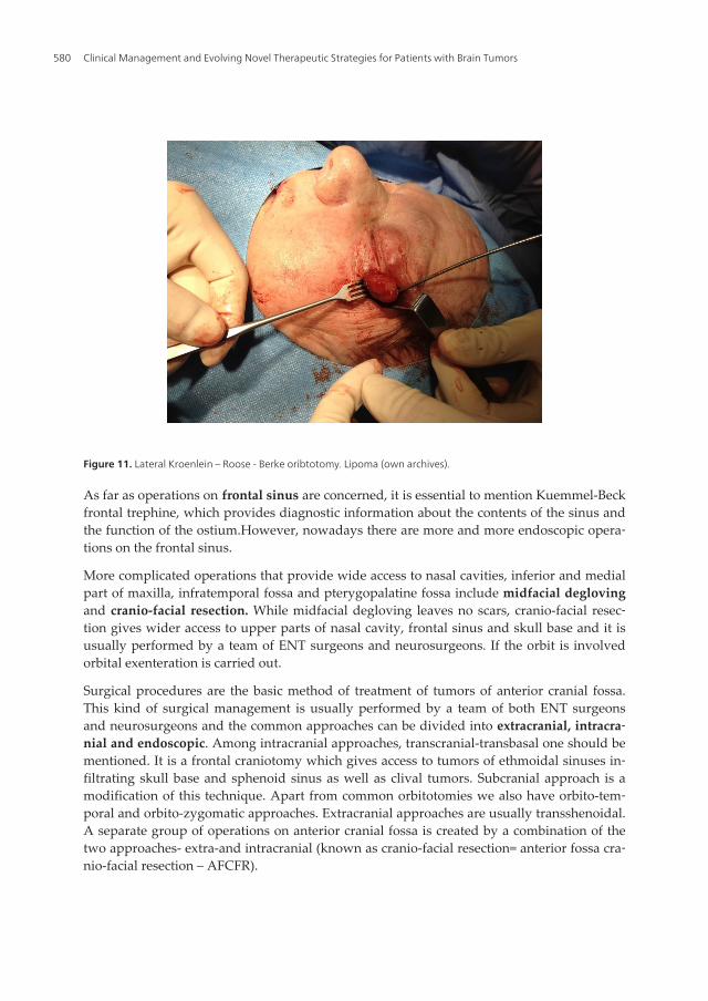
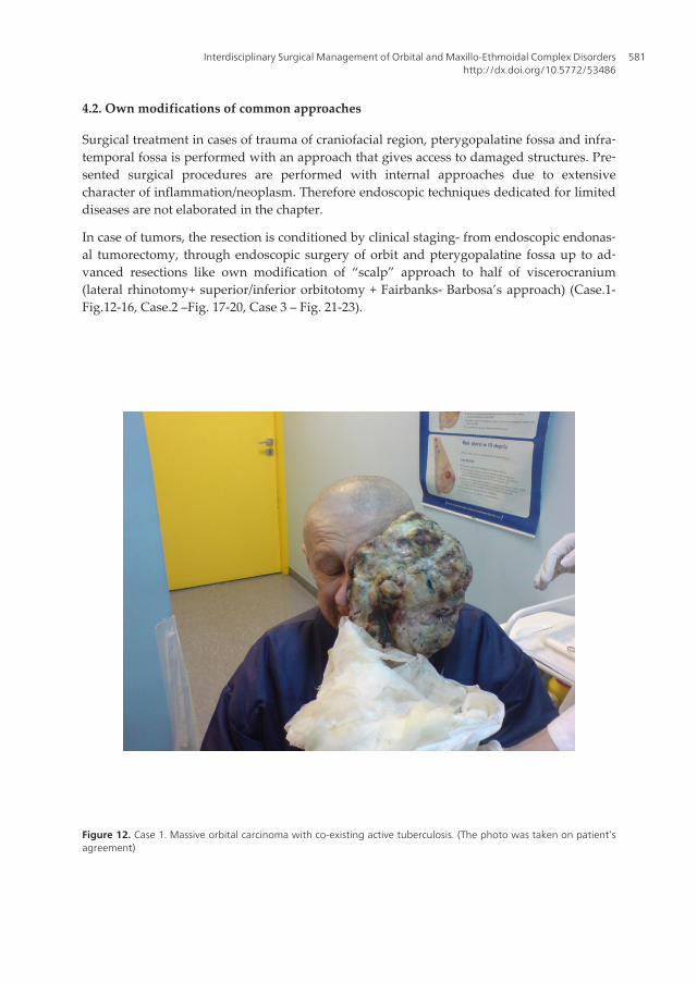
Figure 11. Lateral Kroenlein – Roose - Berke oribtotomy. Lipoma (own archives).
As far as operations on frontal sinus are concerned, it is essential to mention Kuemmel-Beckfrontal trephine, which provides diagnostic information about the contents of the sinus andthe function of the ostium.However, nowadays there are more and more endoscopic opera‐tions on the frontal sinus.
More complicated operations that provide wide access to nasal cavities, inferior and medialpart of maxilla, infratemporal fossa and pterygopalatine fossa include midfacial deglovingand cranio-facial resection. While midfacial degloving leaves no scars, cranio-facial resec‐tion gives wider access to upper parts of nasal cavity, frontal sinus and skull base and it isusually performed by a team of ENT surgeons and neurosurgeons. If the orbit is involvedorbital exenteration is carried out.
Surgical procedures are the basic method of treatment of tumors of anterior cranial fossa.This kind of surgical management is usually performed by a team of both ENT surgeonsand neurosurgeons and the common approaches can be divided into extracranial, intracra‐nial and endoscopic. Among intracranial approaches, transcranial-transbasal one should bementioned. It is a frontal craniotomy which gives access to tumors of ethmoidal sinuses in‐filtrating skull base and sphenoid sinus as well as clival tumors. Subcranial approach is amodification of this technique. Apart from common orbitotomies we also have orbito-tem‐poral and orbito-zygomatic approaches. Extracranial approaches are usually transshenoidal.A separate group of operations on anterior cranial fossa is created by a combination of thetwo approaches- extra-and intracranial (known as cranio-facial resection= anterior fossa cra‐nio-facial resection – AFCFR).
Clinical Management and Evolving Novel Therapeutic Strategies for Patients with Brain Tumors580
4.2. Own modifications of common approaches
Surgical treatment in cases of trauma of craniofacial region, pterygopalatine fossa and infra‐temporal fossa is performed with an approach that gives access to damaged structures. Pre‐sented surgical procedures are performed with internal approaches due to extensivecharacter of inflammation/neoplasm. Therefore endoscopic techniques dedicated for limiteddiseases are not elaborated in the chapter.
In case of tumors, the resection is conditioned by clinical staging- from endoscopic endonas‐al tumorectomy, through endoscopic surgery of orbit and pterygopalatine fossa up to ad‐vanced resections like own modification of “scalp” approach to half of viscerocranium(lateral rhinotomy+ superior/inferior orbitotomy + Fairbanks- Barbosa’s approach) (Case.1-Fig.12-16, Case.2 –Fig. 17-20, Case 3 – Fig. 21-23).
Figure 12. Case 1. Massive orbital carcinoma with co-existing active tuberculosis. (The photo was taken on patient’sagreement)
Interdisciplinary Surgical Management of Orbital and Maxillo-Ethmoidal Complex Disordershttp://dx.doi.org/10.5772/53486
581
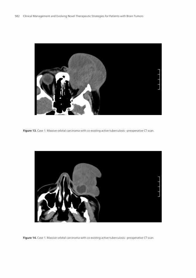
Figure 13. Case 1. Massive orbital carcinoma with co-existing active tuberculosis –preoperative CT scan.
Figure 14. Case 1. Massive orbital carcinoma with co-existing active tuberculosis– preoperative CT scan.
Clinical Management and Evolving Novel Therapeutic Strategies for Patients with Brain Tumors582
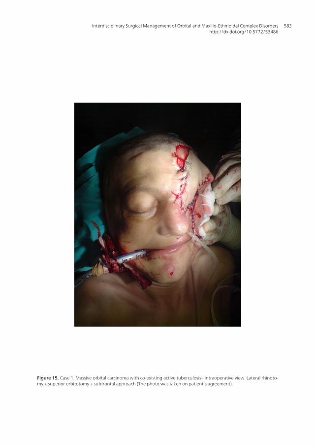
Figure 15. Case 1. Massive orbital carcinoma with co-existing active tuberculosis– intraoperative view. Lateral rhinoto‐my + superior orbitotomy + subfrontal approach (The photo was taken on patient’s agreement).
Interdisciplinary Surgical Management of Orbital and Maxillo-Ethmoidal Complex Disordershttp://dx.doi.org/10.5772/53486
583

Figure 16. Case 1. Massive orbital carcinoma with co-existing active tuberculosis– postperative CT scan.
Figure 17. Case 2. Incission line (lateral rhinotomy+ infrerior orbitotomy + Fairbanks- Barbosa’s approach)of modifiedapproach to massive carcinoma of maxillo-ethmoidal complex – pre-operative image (The photo was taken on pa‐tient’s agreement).
Clinical Management and Evolving Novel Therapeutic Strategies for Patients with Brain Tumors584
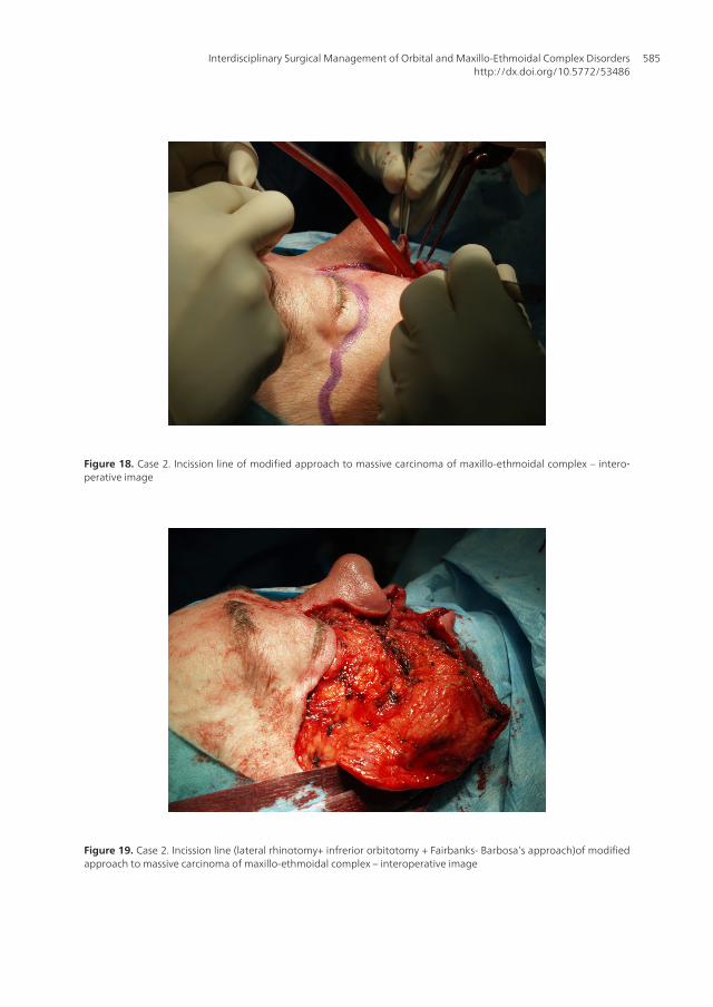
Figure 18. Case 2. Incission line of modified approach to massive carcinoma of maxillo-ethmoidal complex – intero‐perative image
Figure 19. Case 2. Incission line (lateral rhinotomy+ infrerior orbitotomy + Fairbanks- Barbosa’s approach)of modifiedapproach to massive carcinoma of maxillo-ethmoidal complex – interoperative image
Interdisciplinary Surgical Management of Orbital and Maxillo-Ethmoidal Complex Disordershttp://dx.doi.org/10.5772/53486
585
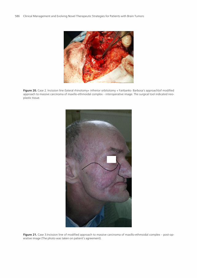
Figure 20. Case 2. Incission line (lateral rhinotomy+ infrerior orbitotomy + Fairbanks- Barbosa’s approach)of modifiedapproach to massive carcinoma of maxillo-ethmoidal complex – interoperative image. The surgical tool indicated neo‐plastic tissue.
Figure 21. Case 3.Incission line of modified approach to massive carcinoma of maxillo-ethmoidal complex – post-op‐erative image (The photo was taken on patient’s agreement).
Clinical Management and Evolving Novel Therapeutic Strategies for Patients with Brain Tumors586
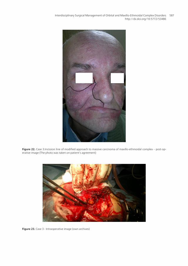
Figure 22. Case 3.Incission line of modified approach to massive carcinoma of maxillo-ethmoidal complex – post-op‐erative image (The photo was taken on patient’s agreement)
Figure 23. Case 3 - Intraoperative image (own archives)
Interdisciplinary Surgical Management of Orbital and Maxillo-Ethmoidal Complex Disordershttp://dx.doi.org/10.5772/53486
587
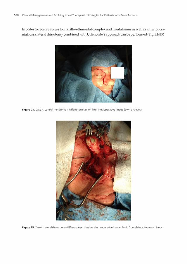
In order to receive access to maxillo-ethmoidal complex and frontal sinus as well as anterior cra‐nial fossa lateral rhinotomy combined with Uffenorde’s approach can be performed (Fig. 24-25)
Figure 24. Case 4. Lateral rhinotomy + Uffenorde scission line- intraoperative image (own archives).
Figure 25. Case 4. Lateral rhinotomy + Uffenorde section line – intraoperative image. Pus in frontal sinus. (own archives).
Clinical Management and Evolving Novel Therapeutic Strategies for Patients with Brain Tumors588
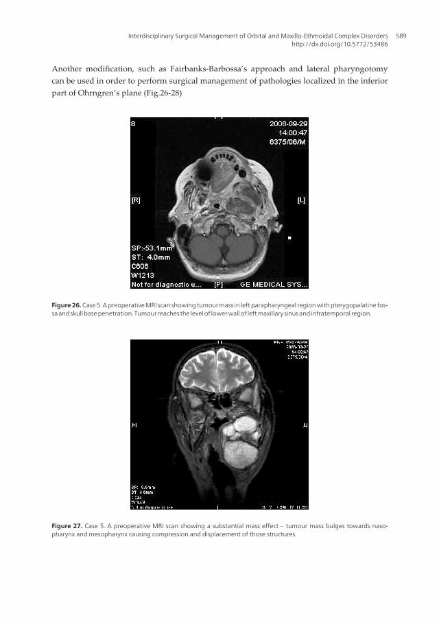
Another modification, such as Fairbanks-Barbossa’s approach and lateral pharyngotomycan be used in order to perform surgical management of pathologies localized in the inferiorpart of Ohrngren’s plane (Fig.26-28)
Figure 26. Case 5. A preoperative MRI scan showing tumour mass in left parapharyngeal region with pterygopalatine fos‐sa and skull base penetration. Tumour reaches the level of lower wall of left maxillary sinus and infratemporal region.
Figure 27. Case 5. A preoperative MRI scan showing a substantial mass effect – tumour mass bulges towards naso‐pharynx and mesopharynx causing compression and displacement of those structures.
Interdisciplinary Surgical Management of Orbital and Maxillo-Ethmoidal Complex Disordershttp://dx.doi.org/10.5772/53486
589
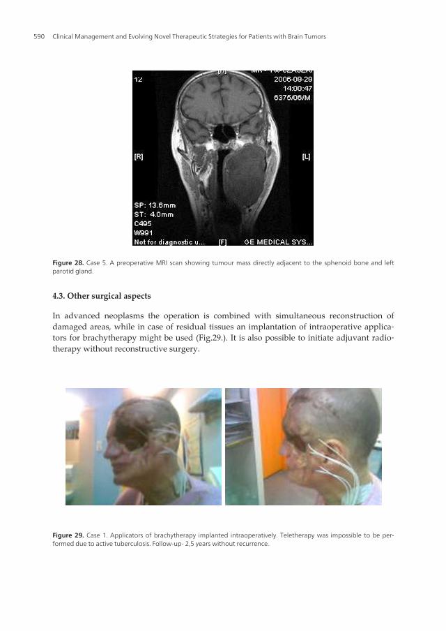
Figure 28. Case 5. A preoperative MRI scan showing tumour mass directly adjacent to the sphenoid bone and leftparotid gland.
4.3. Other surgical aspects
In advanced neoplasms the operation is combined with simultaneous reconstruction ofdamaged areas, while in case of residual tissues an implantation of intraoperative applica‐tors for brachytherapy might be used (Fig.29.). It is also possible to initiate adjuvant radio‐therapy without reconstructive surgery.
Figure 29. Case 1. Applicators of brachytherapy implanted intraoperatively. Teletherapy was impossible to be per‐formed due to active tuberculosis. Follow-up- 2,5 years without recurrence.
Clinical Management and Evolving Novel Therapeutic Strategies for Patients with Brain Tumors590
However, reconstructive surgery with vascular microanastomosis seems to be the pricelessmethod of treatment, resulting in good functional and aesthetic effect.
Occasionally, it is essential to obliterate mechanically the vessel providing blood to trau‐matised region and/or use temporary or permanent endovascular embolization (esp. in‐ternal jugular vein) as the first step of therapeutic procedure. A wide drainage to nasalcavities is crucial for proper healing of inflammations or inflammatory complications oforbit and pterygopalatine fossa.
Also, the authors indicate that in the described region there is a risk of expanding the neo‐plastic and/or inflammatory tissue into ‘critical structures’ such as meninges, optic nerve, in‐ternal carotid artery, facial nerve and sinus cavernosus limits safe course of surgicalprocedures. The situation reveals due to natural extension of neoplastic and/or inflammato‐ry tissue, intraoperative failure or high-dose radiotherapy. The presence of critical structureslimits safe course of surgical procedure.
It is also worth mentioning that in the course of chronic and, rarely, acute pansinusitisorbital complications are observed in 3,7-11 % of all patients hospitalized due to pansi‐nusitis and they are associated with symptoms such as blurred vision, diplopia, deterio‐ration of visual acuity, oedema of palpebra and oedema of the tissues of medial angleof the eye. In advanced situations exophthalmia may occur. The process can extend intoextraorbital structures resulting in their oedema and phlegmons and/or abscess of theorbit. HRCT plays the greatest role in diagnosis. Surgical treatment consists of externalopening of many sinuses and excision of the pathological tissues in the orbit. Opthal‐mological examination with assessment of visual acuity, morphology of the eyeball andpossible damage of optical nerve is essential in preoperative diagnosis of above cases.Optic nerve and sinus cavernosus are ‘critical structures in surgical management of thislimited region.
A separate subdivision should be dedicated to blow-out fracture which is an orbital floorfracture due to blunt trauma of the head. Very fine bones of this region, in case of their frac‐ture, cause an entrapment of the orbital content (i.e.extraocular muscles, esp.inferior rectusand inferior oblique) in maxillar sinus. A damage of infraorbital canal with infraorbitalnerve is often observed and in such case numbness in the region of lower eyelid, cheek, lat‐eral part of nose, upper labium, upper teeth and gingivia can be diagnosed.
Symptoms include:
• enopthalmos
• diplopia
• pain during eye movement
• limited movements of eyeball
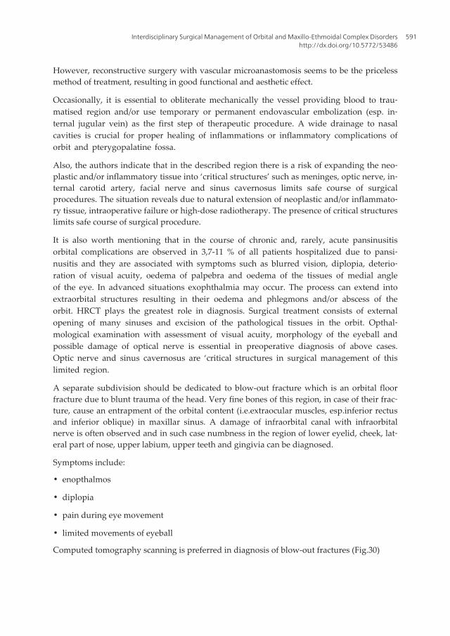
Computed tomography scanning is preferred in diagnosis of blow-out fractures (Fig.30)
Interdisciplinary Surgical Management of Orbital and Maxillo-Ethmoidal Complex Disordershttp://dx.doi.org/10.5772/53486
591
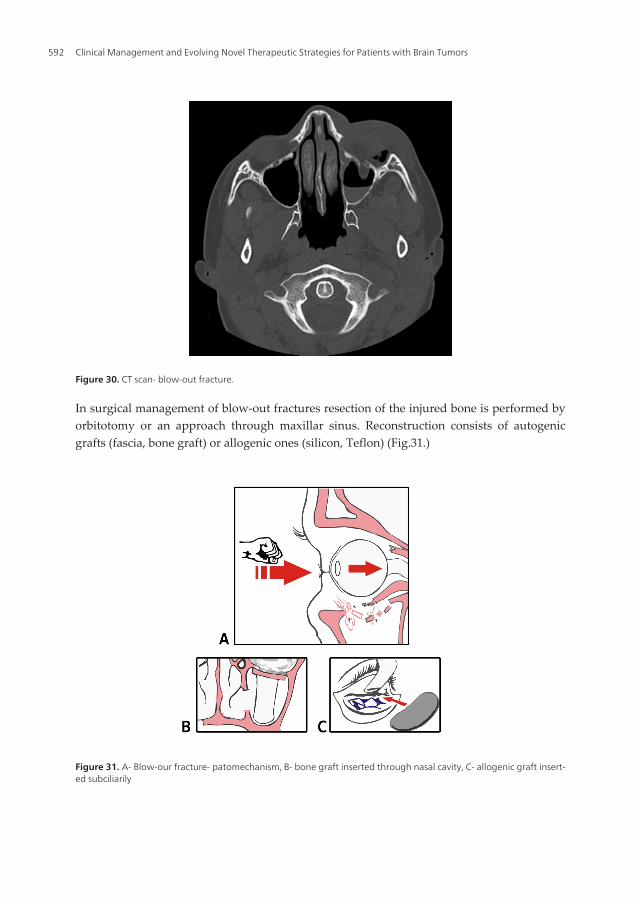
Figure 30. CT scan- blow-out fracture.
In surgical management of blow-out fractures resection of the injured bone is performed byorbitotomy or an approach through maxillar sinus. Reconstruction consists of autogenicgrafts (fascia, bone graft) or allogenic ones (silicon, Teflon) (Fig.31.)
Figure 31. A- Blow-our fracture- patomechanism, B- bone graft inserted through nasal cavity, C- allogenic graft insert‐ed subciliarily
Clinical Management and Evolving Novel Therapeutic Strategies for Patients with Brain Tumors592
In most cases oculoplastic surgeons will wait 10-14 days following the trauma in order toenable an associated oedema and/or heamorrage to be absorbed.
5. Results
The results of surgical management of tumors of described region depend on primary clini‐cal staging assessed with TNM scale, the stage of malignancy, presence of associated disor‐ders and patient’s age. Survival rates depend on above factors and application of adjuvantoncologic treatment utilizing teletherapy, brachytherapy, chemical cytoreduction and com‐bination of suggested methods.
In summary the authors emphasise the necessity of interdisciplinary treatment of orbit andmaxillo-ethmoidal complex.
Author details
Jarosław Paluch1, Jarosław Markowski1, Jan Pilch1, Agnieszka Piotrowska – Seweryn1,Robert Kwiatkowski2, Joanna Lewin-Kowalik3, Czesław Zralek4 andAgnieszka Gorzkowska5
1 Department of Laryngology, Medical University of Silesia, Katowice, Poland
2 Radiotherpay Division, Katowice, Poland
3 Department of Physiology, Medical University of Silesia, Katowice, Poland
4 Department of Neurosurgery, Medical University of Silesia, Katowice, Poland
5 Department of Neurology, Medical University of Silesia, Katowice, Poland
References
[1] Bulsara, Ketan R.; Al-Mefty: Skull Base Surgery for Benign Skull Base Tumors; Ossa‐ma.Journal of Neuro-Oncology vol. 69 issue 1-3 August 2004. p. 181 – 189.
[2] Byron J.Bailey : Head and neck surgery – Otolaryngology, Volume Two, J.B. Lippin‐cott Company Philadalphia, 1993, 1110-1125
[3] Chen T. William: Oculoplastic surgery. The essentials; Thieme New York 2001; ISBN1-58890 – 027-4; 419-451
[4] Handerson J.W.: Orbital Tumors, ed 3. New York: Raven Pres, 1994
Interdisciplinary Surgical Management of Orbital and Maxillo-Ethmoidal Complex Disordershttp://dx.doi.org/10.5772/53486
593
[5] Hastan D, Fokkens WJ, Bachert C et al. Chronic rhinosinusitis in Europe - an under‐estimated disease. A GA2LEN study. Allergy 2011; Published online ahead of print,May 2011
[6] Hussain A, Hulmi OJ, Murray DP: Lateral rhinotomy through nasal aesthetic subu‐nits. Improved cosmetic outcome; J Laryngol Otol. 2002 Sep;116(9):703-6.
[7] Kennerdell JS, Maroon JC, Celin SE. :The posterior inferior orbitotomy; Ophthal PlastReconstr Surg. 1998 Jul;14(4):277-80.
[8] Kyuha C., Taeyun K., Kyungsun C., Myunghwan C., Jonghee Y., Chulhee C.: Diag‐nostic Techniques and Surgical Management of Brain Tumors, Current Optical Imag‐ing Techniques for Brain Tumor Research: Application of in vivo Laser ScanningMicroscopy Imaging with a Cranial Window System, ISBN: 978-953-307-589-1 , In‐Tech 2011:155-172
[9] Lund VJ, Stammberger H, Nicolai P, Castelnuovo P, Beal T, Beham A, Bernal-Spre‐kelsen M, Braun H, Cappabianca P, Carrau R, Cavallo L, Clarici G, Draf W, EspositoF, Fernandez-Miranda J, Fokkens W, Gardner P, Gellner V, Hellquist H, Hermann P,Hosemann W, Howard D, Jones N, Jorissen M, Kassam A, Kelly D, Kurschel-LacknerS, Leong S, McLaughlin N, Maroldi R, Minovi A, Mokry M, Onerci M, Ong YK, Pre‐vedello D, Saleh H, Sehti DS, Simmen D, Snyderman C, Solares A, Spittle M, StammA, Tomazic P, Trimarchi M, Unger F, Wormald PJ, Zanation A; European RhinologicSociety Advisory Board on Endoscopic Techniques in the Management of Nose, Par‐anasal Sinus and Skull Base Tumours: European position paper on endoscopic man‐agement of tumours of the nose, paranasal sinuses and skull base; Rhinol Suppl. 2010Jun 1;(22):1-143.
[10] Margarino G., Scala M. , Mereu P., Comandin D.i, Schenone G., Galli A., FrancavigliaN., Gipponi M.: Combined craniofacial approach to facial tumours involving the an‐terior skull base; European Journal of Surgical Oncology (EJSO)Volume 22, Issue 4,August 1996, Pages 361–365
[11] Mingkun Y., Wei Y., Xiangqian Q, Jun Q., Zhenyang L., Wenfeng F. : "DiagnosticTechniques and Surgical Management of Brain Tumors", Imaging Techniques inBrain Tumor, ISBN 978-953-307-589-1, InTech 2011:43-66
[12] Morioka M, Hamada J, Yano S, Kai Y, Ogata N, Yumoto E, Ushio Y, Kuratsu J: Fron‐tal skull base surgery combined with endonasal endoscopic sinus surgery; Surg Neu‐rol. 2005 Jul;64(1):44-9; discussion 49.
[13] Neumann H.H., Tardy M.E.,Jr, Kastenbauer E.R.: Head and Neck Surgery.Volume 1,Face, Nose and Facial Skull,Part II; Georg Thieme Verlag, Stuttgart, New York 1995,ISBN 3-13-547102-0, ISBN 0-86577-586-9: 476-498, 555-609
[14] Paluch J., Markowski J., Gierek T., Pencak P., Witkowska M., Kajor M., GorzkowskaA., Piotrowska A.: Resonance Tractography in Neuroradiological Diagnostic Aspects,Diagnostic Techniques and Surgical Management of Brain Tumors, ISBN 978-953-307-589-1, InTech 2011: 199-204
Clinical Management and Evolving Novel Therapeutic Strategies for Patients with Brain Tumors594
[15] Silver C.E. Atlas of Head and neck surgery; Churchill Livingstone, New York, Edin‐burgh, London, Melbourne 1986
[16] Słoniewski P., Dzierżanowski J, Lipowski P.,Szmuda T., Czapiewski P.: Orbital tu‐mours operated by orbito-cranial ap roaches– results of treatment of 38 patients;Ann. Acad. Med. Gedan., 2010, 40, 81–89
[17] Snyderman CH, Carrau RL, Kassam AB, Zanation A, Prevedello D, Gardner P, MintzA: Endoscopic skull base surgery: principles of endonasal oncological surgery; J SurgOncol. 2008 Jun 15;97(8):658-64.
[18] Spaeth G.L:. Chirurgia okulistyczna. Wyd.I polskie pod redakcją Jerzego Szaflika,Urban & Partner, Wrocław 2006, ISBN -10-8389581-1, ISBN -13-978-83-89581-53-2;457-471
[19] Szyfter W.:Nowotwory w otorynolaryngologii; Termedia Wydawnictwa Medyczne,Poznań 2012, ISBN: 978-83-62138-80-7; 139-144, 366-369
[20] Ulgen T, Turhan T, Yurtseven T, Oner K.: Simple anterior orbitotomy; Minim Inva‐sive Neurosurg. 2004 Apr;47(2):115-8.
[21] Zhou W, Fang J, Ni X, Huang Z, Wang Q, Chen X, Chen XZ, Xu H: The modifiedrhinotomy for treatment of tumors involving skull base; Lin Chung Er Bi Yan HouTou Jing Wai Ke Za Zhi. 2010 Apr;24(7):301-3.
Interdisciplinary Surgical Management of Orbital and Maxillo-Ethmoidal Complex Disordershttp://dx.doi.org/10.5772/53486
595