Embed Size (px)
Citation preview
Intercalary Elements, Treefrogs, and theEarly Differentiation of a Complex
System in the NeobatrachiaADRIANA S. MANZANO,1* MARISSA FABREZI,2 AND MIGUEL VENCES3
1Consejo Nacional de Investigaciones Cientıficas y Tecnicas (CICyTTP-CONICET),Diamante, Entre Rıos, Republica Argentina
2Instituto de Bio y Geociencias - Museo de Ciencias Naturales, Universidad Nacional deSalta, Salta, Republica Argentina
3Zoological Institute, Technical University of Braunschweig, Braunschweig,Germany
ABSTRACTIntercalary elements are additional skeletal structures of digits of
many anuran amphibians. Twelve terminal clades in the neobatrachianlineage of frogs have intercalary elements revealing it is a homoplasticcharacter with five to seven gains and two to four losses along aconsensus phylogeny of the Neobatrachia. We analyzed anatomical var-iation of intercalary elements, related structures (distal phalanges, ten-dons, and muscles), and articulations of digits of 45 anuran species,representing eight suprageneric terminal taxa. The intercalary ele-ments are integrated in a complex system that is probably related todifferent types of movements, which are produced by a similar set ofmuscles and tendons with limited variation among the studied taxa.Species in the clades Hyloides and Ranoides show distinctive patternsof morphostructural features in their intercalary elements that are usu-ally wedge-shaped and composed of hyaline cartilage in Ranoides, andbiconcave and composed of embryonic cartilage in Hyloides. Featuresderived from the typical hyloid condition may only be interpreted insome Hylidae (Pseudis and Lysapsus) and Centrolenidae. In Ranoides,the described features of the intercalary elements are found in all taxaexamined with the exception of Leptopelis, which have an intercalaryelement similar to the other Ranoides but formed by connective tissue.Several features are shared by all taxa having intercalary elements: (1)the intercalary elements differ from the phalanges by lacking terminalepiphyses, (2) they are present in hands and feet, and (3) they appearin all digits. This finding suggests that the genetic basis for presence ofintercalary elements may be homologous in all these taxa and mayhave evolved only once early in neobatrachian history. Anat Rec,290:1551–1567, 2007. � 2007 Wiley-Liss, Inc.
Key words: intercalary elements; muscles; digits; treefrogs;anurans
Received 3 April 2007; Accepted 4 September 2007
DOI 10.1002/ar.20608Published online 24 October 2007 in Wiley InterScience (www.interscience.wiley.com).
*Correspondence to: Adriana S.Manzano, CICyTTP-CONICET,Matteri y Espana, 3105 - Diamante, Entre Rıos, Republica Argen-tina. Fax: 54-343-4983087. E-mail: [email protected]
Grant sponsor: Agencia Nacional de Promocion Cientıfica yTecnologica; Grant number: PICT/2002 12418; Grant sponsor:Consejo Nacional de Investigaciones Cientıficas y Tecnicas;Grant number: PIP 6347; Grant sponsor: Universidad Auton-oma de Entre Rıos; Grant number: PIDP Res. 387-07.
� 2007 WILEY-LISS, INC.
THE ANATOMICAL RECORD 290:1551–1567 (2007)
The intercalary element is an additional skeletalstructure located between the terminal and subterminal(penultimate) phalanges in the digits of many anuranamphibians. The presence of this element has beenhypothesized to be related to other morphological spe-cializations associated with arboreal habits, such as welldeveloped adhesive digit pads (e.g., Paukstis and Brown,1987, 1991; Wiens et al., 2005). The intercalary may bepart of an integrated system of articulations allowingangular movements responsible for the attachment anddetachment of the adhesive digit pads (Hanna andBarnes, 1991).Analyses of the variation of the intercalary element in
frogs were mainly focused on their presence or absenceand resulted in different proposals on the monophyly ofparticular suprageneric groups (Lynch, 1973; Laurent,1986; Duellman and Trueb, 1986; Ohler and Dubois,1989; Ford and Cannatella, 1993; Duellman, 2001; Frostet al., 2006).Only few studies were carried out on particular line-
ages analyzing the variation of the element itself.Drewes (1984) described four character states in hypero-liid frogs on the basis of structural features (juvenilecartilage without intracellular matrix, cartilaginous andunmineralized, cartilaginous and peripherally mineral-ized, completely mineralized). Vences et al. (2003) forthe same taxon recognized intercalary elements formedby cartilage or not and concluded that the degree of min-eralization can differ within a single species and repre-sents only a gradual modification of basically identicalstructures. Scott (2005) in her analysis of ranoid rela-tionships described some variation in the morphology ofintercalary elements, and Faivovich et al. (2005) consid-ered an elongated shape of intercalary elements in Neo-tropical treefrogs as a synapomorphy of the clade Lysap-sus 1 Pseudis, and the partial mineralization of inter-calary elements as a synapomorphy of the Scinaxcatharinae group.Paukstis and Brown (1987), studying the intercalary
elements in Pseudacris, recognized three morphologicaltypes (wide intercalaries with concave surfaces; cuboi-dal intercalaries; and very thin and reduced intercala-ries), and concluded that reduction of this structurecould be interpreted as a degenerative condition, fur-ther evolution toward burrowing habits possibly result-ing in the loss of the intercalary. These authors,furthermore, analyzed the correlation between themorphology of the intercalary elements and the frog’shabits, and hypothesized that the enlargement of theintercalary could lead to increased digit length for abetter development of webbing in aquatic species(Paukstis and Brown, 1991).Based on the most comprehensive reconstruction of
amphibian phylogeny (Frost et al., 2006), the occurrenceof the intercalary elements is known in five terminaltaxa of Hyloides and five terminal taxa of Ranoides,which both are the major lineages in the clade Neobatra-chia (Fig. 1). Frost et al. (2006) reconstructed intercalaryelements to be synapomorphies for Centrolenidae (Cen-troleninae), Microhylidae (Phrynomantis), Artholeptidae(Leptopelinae), and Rhacophoroidea. These authors dis-cussed intercalary element occurrence and include themas potential synapomorphies; nevertheless, in most ofthe cases, the character was not optimized and thebranches were defined on molecular data.
Drewes (1984) noted that hyperoliid and leptopelineintercalary elements are histologically quite differentfrom each other. Leptopelinae (Leptopelis) is distin-guished morphologically by histological distinct interca-lary phalangeal elements. Rhacophoroidea (Mantellidaeand Rhacophoridae) is a sister group of Ranoidea (Nycti-batrachidae and Ranidae, sensu stricto), and one charac-ter that definitely optimizes on Rhacophoroidea is theintercalary element present.Wiens et al. (2005) found evidence for a significant
influence of homoplasy in the intercalary element onconflicts between morphological and molecular data sets.These authors characterized the Neotropical family Hyli-dae by several traits that presumably represent adapta-tions to the use of arboreal habitat (e.g., expanded toepads and intercalary elements) and concluded that theplacement of hemiphractines with other hylids may becaused by the convergent acquisition of traits associatedwith arboreality (e.g., offset terminal phalanges; claw-shaped terminal phalanges, intercalary elements; modi-fied base of metacarpal III), in contrast to the strongmolecular evidence that hemiphractines are not closelyrelated to other hylids. Several other aspects in a phylo-genetic reconstruction based on morphological traitswere also seen as possible results of the misleadingeffects of this suite of traits: (1) the placement of theclade Centrolenidae 1 Allophryne with hylids, bothgroups containing taxa with intercalary elements; (2)the basal placement of Pseudis, Lysapsus, Acris, andsome Pseudacris within hylids, which may be associatedwith a reversion to terrestrial and/or aquatic lifestyle inthese clades, with a concomitant loss of one or more ofthe characters associated with arboreality (e.g., offsetterminal phalanges, expanded toe pads); and (3) the lossof these traits in the terrestrial/fossorial hylid Cyclorana(i.e., terminal phalanges not offset, not claw-shaped, lossof intercalary elements, toe pads not expanded) whichmay contribute to the erroneous placement of this taxonoutside of the Hylidae (Wiens et al., 2005).Here we present a comparative analysis of different
morphological traits related to intercalary elements inrepresentative hyloid and ranoid taxa in which interca-laries are present. Because intercalary elements aremorphologically and functionally integrated in anuranlimbs, we describe and analyze both (1) the variation inshape and structure of the intercalaries and their articu-lations with phalanges, and (2) patterns of variation inassociated muscles, in particular in the Extensoresbreves profundus, Extensores breves distalis tendons,and Tendo superficialis. Our goal in providing detailedcomparative descriptions of these patterns is to obtain abasis for understanding their evolution in the light ofrecent phylogenetic hypotheses.
MATERIALS AND METHODS
Specimens of 45 species of Neobatrachia with interca-lary elements were studied. They belong to eight termi-nal suprageneric taxa following the classificationproposed by Frost et al. (2006): Amphignathodontidae,Hylidae, Centrolenidae, Microhylidae, Mantellidae,Rhacophoridae, Arthroleptidae, and Hyperoliidae. Thespecies examined, voucher specimens, and collectiondata are listed in Table 1. Three different kinds of datawere gathered on these specimens: (1) Morphological
1552 MANZANO ET AL.
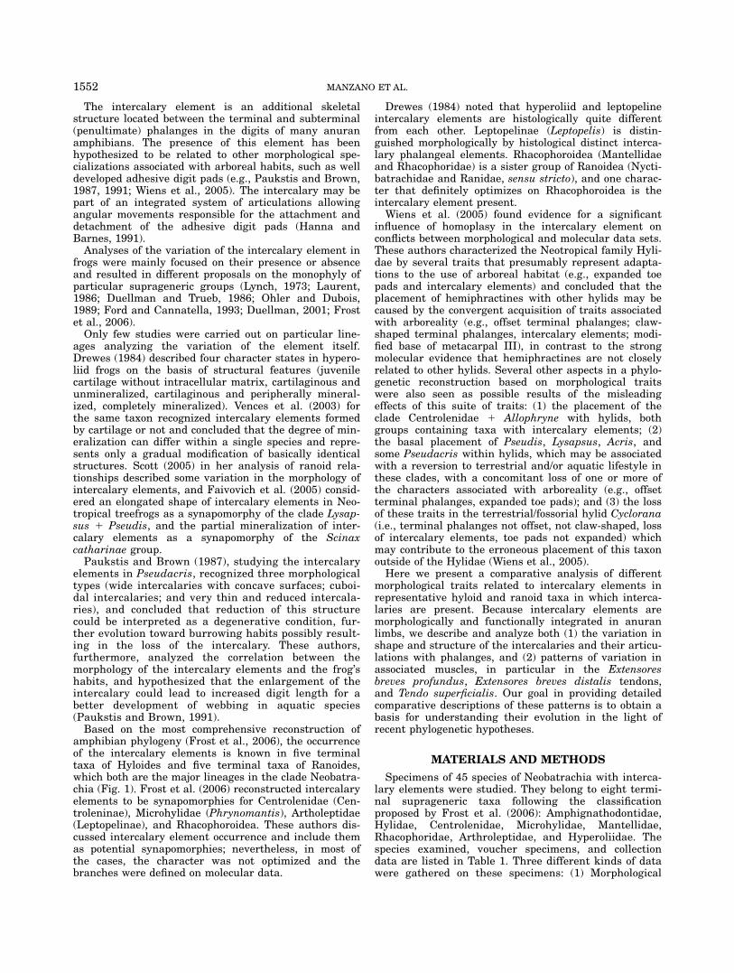
Fig. 1. Neobatrachian relationships based on recent molecular evi-dence. The tree topology in general follows Frost et al. (2006), withmodifications in ranoid phylogeny following Van der Meijden et al.(2005, 2007) and Van Bocxlaer et al. (2006). Note that the nonmono-phyly of marsupial frogs and their division into three families is contro-versial (Wiens, 2007). Similarly, the placement of Phrynomantis asmost basal microhylid (Van Bocxlaer et al., 2006) is not supported(Van der Meijden et al., 2007) but its possible deeper nestednesswithin microhylids would only indicate more strongly a parallel evolu-tion of the intercalary element in this taxon. Calyptocephalidae is the
correct name for the taxon named Batrachophrynidae in Frost et al.(2006), according to Frost (2007). Terminal taxa are families except incases where the presence or absence of the intercalary element infamilies is not universal. Underlined taxa are those having intercalaryelements. Major clades are symbolized by black circles; secondaryclades as used by Frost et al. (2006) and mentioned in the text aremarked by white circles. Black branches are reconstructed as havingan intercalary element, gray branches as lacking this element, andcross-hatched segments are differently reconstructed underACCTRAN and DELTRAN transformations in MacClade.
1553INTERCALARY ELEMENTS IN ANURANS

TABLE 1. Specimens examined in this study
Family Species Collection dataaNumber ofspecimens Study
Amphignathodontidae Flectonotus fitzgeraldi MCN 017 2 SkeletonAmphignathodontidae Gastrotheca christiani MCN 437 1 Skeleton, muscles, and histologyAmphignathodontidae Gastrotheca gracilis FML 2995 1 SkeletonArthroleptidae Leptopelis christyi MCN 829 4 Skeleton
MCN s/n 1 Muscles and histologyCentrolenidae Centrolene geckoideum DIAM 314 1 Skeleton, muscles and histology.Centrolenidae Centrolene grandisonae DIAM 0320 2 Skeleton and musclesCentrolenidae Centrolene robledoi DIAM 0315 2 Skeleton and muscles.Centrolenidae Centrolene notostictum UIS A 863 1 Skeleton
UIS A 410 1 SkeletonCentrolenidae Cochranella griffithsi DIAM 0319 1 MusclesCentrolenidae Cochranella ignota DIAM 0321 2 Skeleton and muscles.Centrolenidae Cochranella savagei DIAM 0322 1 MusclesCentrolenidae Hyalinobatrachium
aureoguttatumDIAM 0318 3 Skeleton and muscles
Hylidae Argenteohyla siemersi FML 3954 1 SkeletonHylidae Dendropsophus minutus DIAM 066 1 MusclesHylidae Dendropsophus nanus MCN 791 1 SkeletonHylidae Hylomantis lemur MCN 012 2 SkeletonHylidae Hypsiboas andinus MCN 937 1 Skeleton
MCN 092 1 HistologyHylidae Isthmohyla rivularis MCN 013 1 SkeletonHylidae Litoria caerulea DIAM 313 1 Skeleton, muscles, and histology.Hylidae Lysapsus limellum FML 0716 1 Skeleton
DIAM 019 2 Skeleton and muscles.Hylidae Phyllomedusa sauvagii MCN 795 1 Skeleton, muscles, histology
MCN 392 1 HistologyDIAM 0337 1 Skeleton and muscles
Hylidae Pseudis minuta DIAM 0338 2 Skeleton and musclesHylidae Pseudis paradoxa MCN 812 1 Skeleton
MACN 37698 1 Skeleton and musclesMACN 37699 1 Skeleton and musclesMCN 567 1 Histology
Hylidae Scinax acuminatus MCN 1006 1 Skeleton and histologyHylidae Scinax fuscovarius MCN 813 1 Skeleton and histologyHylidae Scinax nasicus MCN 1005 1 Skeleton
DIAM 095 1 SkeletonHylidae Scinax squalirostris DIAM 021 3 Skeleton and musclesHylidae Trachycephalus venulosus FML 2712 2 Skeleton
DIAM 024 1 Skeleton and musclesHyperoliidae Afrixalus quadrivittatus MCN 943 2 SkeletonHyperoliidae Afrixalus laevis MCN 993 2 SkeletonHyperoliidae Afrixalus osorioi MCN 994 2 SkeletonHyperoliidae Hyperolius castaneus MCN 833 5 SkeletonHyperoliidae Hyperolius kivuensis MCN 804 2 Skeleton, muscles, and histologyHyperoliidae Kassina senegalensis MCN 823 2 SkeletonHyperoliidae Opisthothylax immaculatus MCN 825 1 SkeletonHyperoliidae Phlyctimantis verrucosus MCN 832 3 SkeletonMantellidae Aglyptodactylus
madagascariensisDIAM 0333 - 0324 2 Skeleton, muscles, and histology
Mantellidae Boophis goudoti DIAM 0332 1 Skeleton, muscles and histologyMantellidae Boophis madagascariensis DIAM 0325 1 Skeleton, muscles and histologyMantellidae Mantella auriantica DIAM 0334 1 Skeleton, muscles and histologyMantellidae Mantidactylus curtus DIAM 0328 - 0335 2 Skeleton, muscles, and histologyMantellidae Guibemantis liber DIAM 0330 2 Skeleton, muscles, and histologyMantellidae Guibemantis timidus DIAM 0340 1 HistologyMicrohylidae Phrynomantis bifasciatus MCN 830 4 SkeletonMicrohylidae Phrynomantis microps DIAM 0331 1 Skeleton, muscles, and histologyRhacophoridae Chiromantis rufescens MCN 831 4 Skeleton
DIAM 0339 2 Skeleton, muscles, and histology
aMuseum abbreviations: FML, Instituto de Herpetologıa, Fundacion Miguel Lillo, Tucuman, Argentina; MCN, Museo deCiencias Naturales, Universidad Nacional de Salta, Argentina; UIS, Coleccion Herpetologica, Escuela de Biologıa, Universi-dad Industrial de Santander, Colombia; MACN, Museo Argentino de Ciencias Naturales Bernardino Rivadavia, BuenosAires, Argentina; DIAM, Coleccion Herpetologica CICyTTP-CONICET, Diamante, Entre Rıos, Argentina.
1554 MANZANO ET AL.
descriptions of distal phalanges and intercalary ele-ments in skeletal whole-mounts cleared and doublestained with Alcian Blue and Alizarin Red S (Was-sersug, 1976); (2) anatomical descriptions of musclesand tendons based on dissections of whole specimensthat were double stained with Alcian Blue and AlizarinRed S but not cleared. These preparations were pre-served in 70% ethanol and, at the time of observation,temporarily stained with iodine solution to obtain a bet-ter contrast of the structures (Bock and Shear, 1972);and (3) histological serial sections of 7-mm thick of par-affin-embedded digit tips (Anderson and Bancroft, 2002)were stained with hematoxylin—eosin (Wilson andGamble, 2002).Most descriptions were based on features observed in
finger and toe IV. Observations, illustrations, and photo-
graphs were made with Nikon SMZ1000 and Olympusstereo dissection microscopes and with a Leica DM lightmicroscope equipped with digital camera and cameralucida.Movements of the articulations were interpreted by
moving structures, pulling tendons, and muscles in eachdigit of preserved samples. Variation of terminal pha-langes, intercalary elements, articulations, muscles, andtendons in fore- and hindlimbs are summarized inTables 2–4. Standard muscle terminology follows Gaupp(1896) and Burton (1998a,b); arthrology nomenclaturefollows Dyce et al. (1996).To reconstruct the phylogenetic history of intercalary
elements, we compiled an informal consensus tree fromrecent molecular phylogenetic studies (Frost et al., 2006;Van Bocxlaer et al., 2006; Van der Meijden, 2005, 2007).
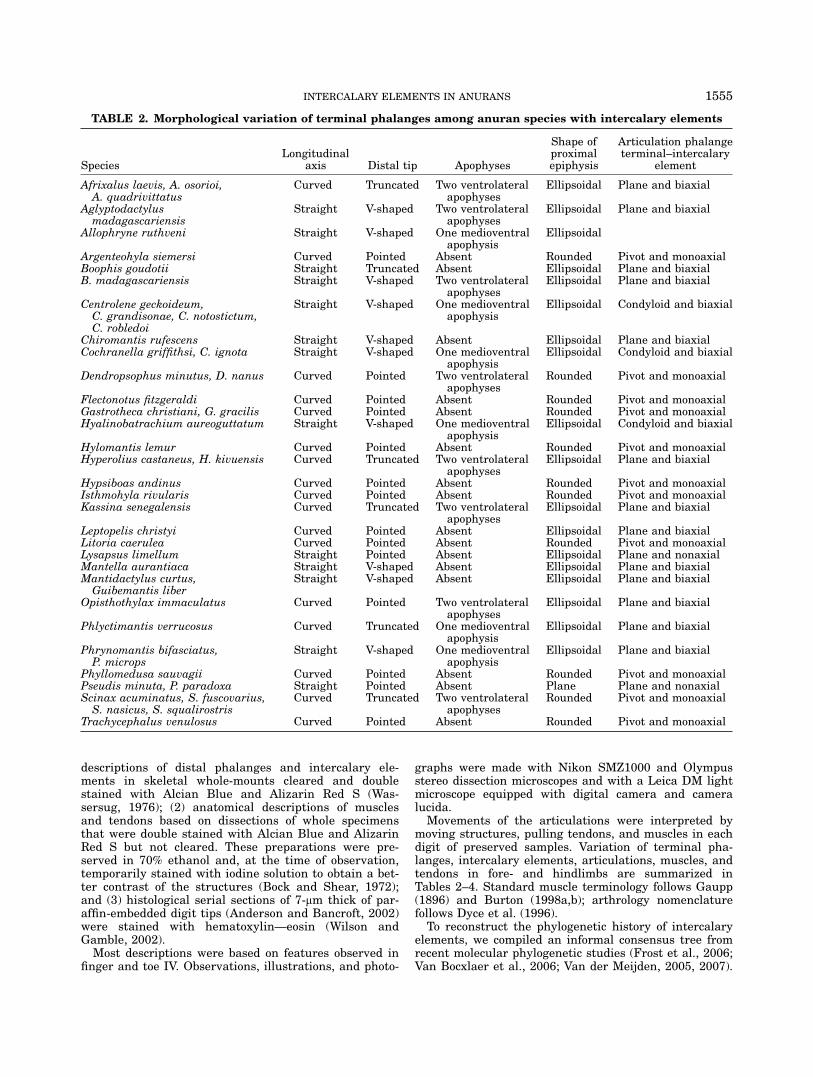
TABLE 2. Morphological variation of terminal phalanges among anuran species with intercalary elements
SpeciesLongitudinal
axis Distal tip Apophyses
Shape ofproximalepiphysis
Articulation phalangeterminal–intercalary
element
Afrixalus laevis, A. osorioi,A. quadrivittatus
Curved Truncated Two ventrolateralapophyses
Ellipsoidal Plane and biaxial
Aglyptodactylusmadagascariensis
Straight V-shaped Two ventrolateralapophyses
Ellipsoidal Plane and biaxial
Allophryne ruthveni Straight V-shaped One medioventralapophysis
Ellipsoidal
Argenteohyla siemersi Curved Pointed Absent Rounded Pivot and monoaxialBoophis goudotii Straight Truncated Absent Ellipsoidal Plane and biaxialB. madagascariensis Straight V-shaped Two ventrolateral
apophysesEllipsoidal Plane and biaxial
Centrolene geckoideum,C. grandisonae, C. notostictum,C. robledoi
Straight V-shaped One medioventralapophysis
Ellipsoidal Condyloid and biaxial
Chiromantis rufescens Straight V-shaped Absent Ellipsoidal Plane and biaxialCochranella griffithsi, C. ignota Straight V-shaped One medioventral
apophysisEllipsoidal Condyloid and biaxial
Dendropsophus minutus, D. nanus Curved Pointed Two ventrolateralapophyses
Rounded Pivot and monoaxial
Flectonotus fitzgeraldi Curved Pointed Absent Rounded Pivot and monoaxialGastrotheca christiani, G. gracilis Curved Pointed Absent Rounded Pivot and monoaxialHyalinobatrachium aureoguttatum Straight V-shaped One medioventral
apophysisEllipsoidal Condyloid and biaxial
Hylomantis lemur Curved Pointed Absent Rounded Pivot and monoaxialHyperolius castaneus, H. kivuensis Curved Truncated Two ventrolateral
apophysesEllipsoidal Plane and biaxial
Hypsiboas andinus Curved Pointed Absent Rounded Pivot and monoaxialIsthmohyla rivularis Curved Pointed Absent Rounded Pivot and monoaxialKassina senegalensis Curved Truncated Two ventrolateral
apophysesEllipsoidal Plane and biaxial
Leptopelis christyi Curved Pointed Absent Ellipsoidal Plane and biaxialLitoria caerulea Curved Pointed Absent Rounded Pivot and monoaxialLysapsus limellum Straight Pointed Absent Ellipsoidal Plane and nonaxialMantella aurantiaca Straight V-shaped Absent Ellipsoidal Plane and biaxialMantidactylus curtus,Guibemantis liber
Straight V-shaped Absent Ellipsoidal Plane and biaxial
Opisthothylax immaculatus Curved Pointed Two ventrolateralapophyses
Ellipsoidal Plane and biaxial
Phlyctimantis verrucosus Curved Truncated One medioventralapophysis
Ellipsoidal Plane and biaxial
Phrynomantis bifasciatus,P. microps
Straight V-shaped One medioventralapophysis
Ellipsoidal Plane and biaxial
Phyllomedusa sauvagii Curved Pointed Absent Rounded Pivot and monoaxialPseudis minuta, P. paradoxa Straight Pointed Absent Plane Plane and nonaxialScinax acuminatus, S. fuscovarius,S. nasicus, S. squalirostris
Curved Truncated Two ventrolateralapophyses
Rounded Pivot and monoaxial
Trachycephalus venulosus Curved Pointed Absent Rounded Pivot and monoaxial
1555INTERCALARY ELEMENTS IN ANURANS

Terminal taxa were families according to Frost et al.(2006) with some modifications, i.e., acceptance of Ranix-alidae according to Van Bocxlaer et al. (2006) and usageof Calyptocephalidae instead of Batrachophrynidaeaccording to Frost (2007). Families that contain somegenera with and some genera without intercalaries werefurther split into terminal taxa to account for these dif-ferences. Ancestral states along the tree were tracedusing both Acctran and Deltran models in MacClade(Maddison and Maddison, 1998).We used the concentrated changes test of Maddison
(1990) to test the association of changes in the presenceof intercalaries and arboreal habits, coding both as
binary characters. This test determines the probabilitythat various numbers of gains and losses of the depend-ent variable (terminal phalanx morphology) would occurin certain distinguished areas of the clade selected(defined by arboreal habits), given that a certain numberof gains and losses occur in the whole clade, and giventhe null model that changes are randomly distributedamong the branches of the clade. Because many groupsof frogs contain species of somewhat climbing habits(e.g., on rocks), we used a rather strict definition ofarboreality, and coded as arboreal only groups that con-tain a large proportion of real treefrogs that spend mostof their life (except during breeding) on the vegetation,
TABLE 3. Morphological and structural variation of intercalary elements among anurans
Species Shape Structure
Articulation intercalaryelement–penultimate
phalange
Afrixalus laevis, A. osorioi,A. quadrivittatus
Wedge-shaped Hyaline cartilage Sellaris and biaxial
Aglyptodactylusmadagascariensis
Wedge-shaped Hyaline cartilage fullymineralized
Sellaris and biaxial
Argenteohyla siemersi Thick biconcave disc Embryonic cartilage Ginglymus and monoaxialBoophis goudoti, B.madagascariensis
Wedge-shaped Hyaline cartilage fullymineralized
Sellaris and biaxial
Centrolene geckoideum,C. grandisone,C. notostictum, C. robledoi
Thick biconcave disc Embryonic cartilage with alarge mineralized nucleus
Ginglymus and monoaxial
Chiromantis rufescens Wedge-shaped Hyaline cartilage with peripheralmineralization
Sellaris and biaxial
Cochranella griffithsi, C. ignota Thick biconcave disc Embryonic cartilage with a largemineralized nucleus
Ginglymus and monoaxial
Dendropsophus minutus Thick biconcave disc Embryonic cartilage Ginglymus and monoaxialD. nanus Thick biconcave disc Embryonic cartilage with a small
mineralized nucleusGinglymus and monoaxial
Flectonotus fitzgeraldi Thick biconcave disc Embryonic cartilage Ginglymus and monoaxialGastrotheca christiani,G. gracilis
Thick biconcave disc Embryonic cartilage Ginglymus and monoaxial
Hyalinobatrachiumaureoguttatum
Thick biconcave disc Embryonic cartilage with a largemineralized nucleus
Ginglymus and monoaxial
Hylomantis lemur Thick biconcave disc Embryonic cartilage with a smallmineralized nucleus
Ginglymus and monoaxial
Hyperolius castaneus,H. kivuensis
Wedge-shaped Hyaline cartilage with peripheralmineralization
Sellaris and biaxial
Hypsiboas andinus Thick biconcave disc Embryonic cartilage Ginglymus and monoaxialIsthmohyla rivularis Thick biconcave disc Embryonic cartilage with a small
mineralized nucleusGinglymus and monoaxial
Kassina angeli Wedge-shaped Hyaline cartilage Sellaris and biaxialLeptopelis christyi Wedge-shaped Dense connective tissue Sellaris and biaxialLitoria caerulea Thick biconcave disc Embryonic cartilage Ginglymus and monoaxialLysapsus limellum Cylindrical Hyaline cartilage fully
mineralizedPlane and nonaxial
Mantella auriantica Wedge-shaped Hyaline cartilage fullymineralized
Sellaris and biaxial
Mantidactylus curtus,Guibematis liber
Wedge-shaped Hyaline cartilage fullymineralized
Sellaris and biaxial
Opisthothylax immaculatus Wedge-shaped Hyaline cartilage Sellaris and biaxialPhlyctimantis verrucosus Wedge-shaped Hyaline cartilage Sellaris and biaxialPhrynomantis bifasciatus,P. microps
Wedge-shaped Hyaline cartilage fullymineralized
Sellaris and biaxial
Phyllomedusa sauvagii Thick biconcave disc Embryonic cartilage with a smallmineralized nucleus
Ginglymus and monoaxial
Pseudis minuta, P. paradoxa Cylindrical Hyaline cartilage fully mineralized Plane and nonaxialScinax acuminatus,S. fuscovarius, S. nasicus,S. squalirostris
Thick biconcave disc Embryonic cartilage Ginglymus and monoaxial
Trachycephalus venulosus Thick biconcave disc Embryonic cartilage Ginglymus and monoaxial
1556 MANZANO ET AL.

TABLE
4.Obse
rved
variation
inpenultim
ate
phalange,TendoSuperficia
lisand
M.extenso
rbrevesdigitoriu
mamong
anuranswith
intercalaryelements
Species
Lon
gitudinal
crests
ondistal
epiphysisof
pen
ultim
ate
phalange
Shapeof
Ten
do
Sperficialisat
insertion
Ten
do
Sperficialis
insertion
Relation
ships
betwee
nintercalary
elem
entandTen
do
Superficialis
Len
gth
oftendon
ofm.extensor
breves
profundus
Hand
M.extensor
brevis
distalis
Foo
tM.extensor
brevis
distalis
Aglyptodactylus
madagascarien
sis
Present
Tru
ncated
Onapop
hysis
Absent
Lon
gPresent
Absent
Boo
phis
gou
doti
Present
Fan-shaped
med
ial
Absent
Lon
gAbsent
Absent
B.madagascarien
sis
Present
Tru
ncated
Onapop
hysis
Few
fiberson
intercalary
Lon
gPresent
Absent
Cen
trolen
egeckoideu
mPresent
Bifurcated
Onapop
hysis
Throughintercalary
capsu
leSom
eare
short
Present
Present
C.gra
ndison
ae,
C.robledoi
Present
Bifurcated
Infron
tof
apop
hysis
Few
fiberson
intercalary
Som
eare
short
Present
Present
Chirom
antisru
fescen
sPresent
Fan-shaped
Med
ial
Few
fiberson
intercalary
Lon
gPresent
Present
Cochra
nella
griffithsi
Present
Tru
ncated
Onapop
hysis
andin
fron
tof
apop
hysis
Few
fiberson
intercalary
Som
eare
short
Present
Present
C.ignota
Present
Tru
ncated
Infron
tof
apop
hysis
Few
fiberson
intercalary
Som
eare
short
Present
Present
Den
dropsophusminutus
Present
Twodistinct
branch
esOnapop
hysis
Absent
Lon
gPresent
Present
Gastrothecach
ristiani
Present
Fan-shaped
Med
ial
Few
fiberson
intercalary
Lon
gPresent
Present
Hya
linob
atrach
ium
aureog
uttatum
Present
Tru
ncated
Infron
tof
apop
hysis
Few
fiberson
intercalary
Som
eare
short
Present
Present
Hyp
eroliuskivuen
sis
Present
Tru
ncated
Onapop
hysis
Few
fiberson
intercalary
Lon
gPresent
Present
Lep
topelis
christyi
Present
Tru
ncated
Med
ial
few
fiberson
intercalary
Lon
gPresent
Present
Litoria
caerulea
Present
Fan-shaped
with
anincipient
bifurcation
Onep
iphysis
Absent
Lon
gPresent
Present
Lysapsu
slimellus
Present
Twodistinct
branch
esMed
ial
Few
fiberson
intercalary
Lon
gPresent
Absent
Mantellaaura
ntiaca
Absent
Fan-shaped
Med
ial
Absent
Lon
gAbsent
Absent
Mantidactyluscu
rtus
Present
Fan-shaped
Med
ial
Few
fiberson
intercalary
Lon
gAbsent
Absent
Guibem
antisliber
Present
Tru
ncated
Med
ial
Few
fiberson
intercalary
Lon
gPresent
Absent
Phrynom
antismicrops
Present
Fan-shaped
Onapop
hysis
Absent
Largo
Absent
Absent
Phyllomed
usa
sauvagii
Present
Fan-shaped
Med
ial
Absent
Lon
gPresent
Present
Pseudis
minuta,
P.para
dox
aAbsent
Twodistinct
branch
esMed
ial
Few
fiberson
intercalary
Lon
gPresent
Absent
Scinaxnasicu
sPresent
Twodistinct
branch
esOnapop
hysis
Absent
Lon
gPresent
Present
Scinaxsqualirostris
Present
Twodistinct
branch
esOnapop
hysis
Absent
Lon
gPresent
Absent
Tra
chycep
halus
ven
ulosu
sPresent
Twodistinct
branch
esOnep
iphysis
Absent
Lon
gPresent
Absent
1557INTERCALARY ELEMENTS IN ANURANS
namely, the families Hylidae, Hemiphractidae, Cryptoba-trachidae, Amphignathodontidae, Hylidae, Centroleni-dae, Hyperoliidae, Ceratobatrachidae, Rhacophoridae,mantelline and boophine Mantellidae; the Arthrolepti-dae genus Leptopelis; as well as nonphrynomerineMicrohylidae (arboreal taxa in subfamilies Cophylinaeand Asterophryinae).
RESULTS
Phylogenetic Character Tracing
According to character tracing along our informal con-sensus tree (Fig. 1) using parsimony criteria imple-mented in MacClade, intercalary elements have beenindependently gained 5 times and lost 4 times underACCTRAN reconstructions, and gained 7 times and lost2 times under DELTRAN reconstructions. Plotting thesegains and losses onto trees where the evolution of arbor-eality (as defined here in the Materials and Methods sec-tion) is traced, 3 gains and 2 losses (ACCTRAN) and 6gains and 1 loss (DELTRAN) occur in tree sections char-acterized by arboreality. The concentrated changes testindicated that changes indeed occur in significantlyhigher proportions in arboreal clades (P < 0.05) and,thus, supported a possible evolutionary correlationbetween arboreal habits and intercalaries.
Morphology of Terminal Phalanges
Terminal phalanges are characterized by a cartilagi-nous proximal epiphysis, which sometimes is mineral-ized; and an ossified diaphysis. The shape of the tip isthe most known feature of terminal phalanx variationamong anurans (e.g., Liem, 1970; Lynch, 1971, Heyer,1975; Clarke, 1981; Drewes, 1984; Laurent, 1986;Fabrezi, 1996; Kamermans and Vences, in press).In the species examined here, we considered four
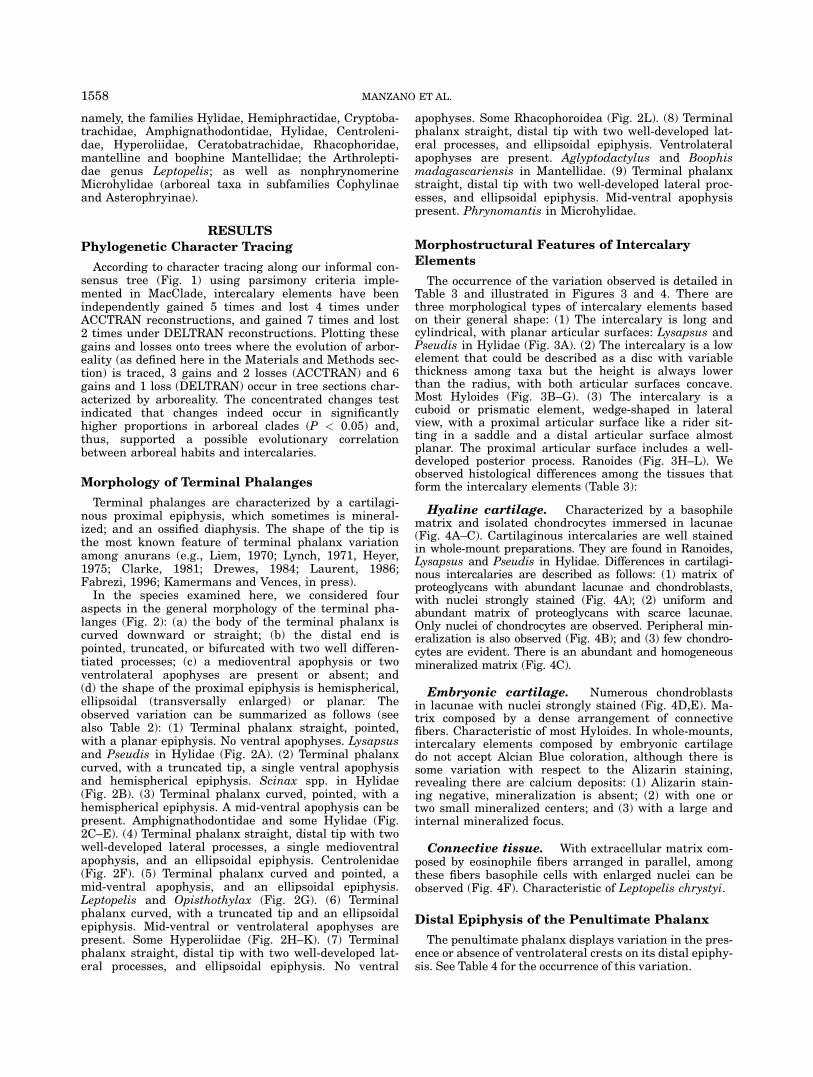
aspects in the general morphology of the terminal pha-langes (Fig. 2): (a) the body of the terminal phalanx iscurved downward or straight; (b) the distal end ispointed, truncated, or bifurcated with two well differen-tiated processes; (c) a medioventral apophysis or twoventrolateral apophyses are present or absent; and(d) the shape of the proximal epiphysis is hemispherical,ellipsoidal (transversally enlarged) or planar. Theobserved variation can be summarized as follows (seealso Table 2): (1) Terminal phalanx straight, pointed,with a planar epiphysis. No ventral apophyses. Lysapsusand Pseudis in Hylidae (Fig. 2A). (2) Terminal phalanxcurved, with a truncated tip, a single ventral apophysisand hemispherical epiphysis. Scinax spp. in Hylidae(Fig. 2B). (3) Terminal phalanx curved, pointed, with ahemispherical epiphysis. A mid-ventral apophysis can bepresent. Amphignathodontidae and some Hylidae (Fig.2C–E). (4) Terminal phalanx straight, distal tip with twowell-developed lateral processes, a single medioventralapophysis, and an ellipsoidal epiphysis. Centrolenidae(Fig. 2F). (5) Terminal phalanx curved and pointed, amid-ventral apophysis, and an ellipsoidal epiphysis.Leptopelis and Opisthothylax (Fig. 2G). (6) Terminalphalanx curved, with a truncated tip and an ellipsoidalepiphysis. Mid-ventral or ventrolateral apophyses arepresent. Some Hyperoliidae (Fig. 2H–K). (7) Terminalphalanx straight, distal tip with two well-developed lat-eral processes, and ellipsoidal epiphysis. No ventral
apophyses. Some Rhacophoroidea (Fig. 2L). (8) Terminalphalanx straight, distal tip with two well-developed lat-eral processes, and ellipsoidal epiphysis. Ventrolateralapophyses are present. Aglyptodactylus and Boophismadagascariensis in Mantellidae. (9) Terminal phalanxstraight, distal tip with two well-developed lateral proc-esses, and ellipsoidal epiphysis. Mid-ventral apophysispresent. Phrynomantis in Microhylidae.
Morphostructural Features of Intercalary
Elements
The occurrence of the variation observed is detailed inTable 3 and illustrated in Figures 3 and 4. There arethree morphological types of intercalary elements basedon their general shape: (1) The intercalary is long andcylindrical, with planar articular surfaces: Lysapsus andPseudis in Hylidae (Fig. 3A). (2) The intercalary is a lowelement that could be described as a disc with variablethickness among taxa but the height is always lowerthan the radius, with both articular surfaces concave.Most Hyloides (Fig. 3B–G). (3) The intercalary is acuboid or prismatic element, wedge-shaped in lateralview, with a proximal articular surface like a rider sit-ting in a saddle and a distal articular surface almostplanar. The proximal articular surface includes a well-developed posterior process. Ranoides (Fig. 3H–L). Weobserved histological differences among the tissues thatform the intercalary elements (Table 3):
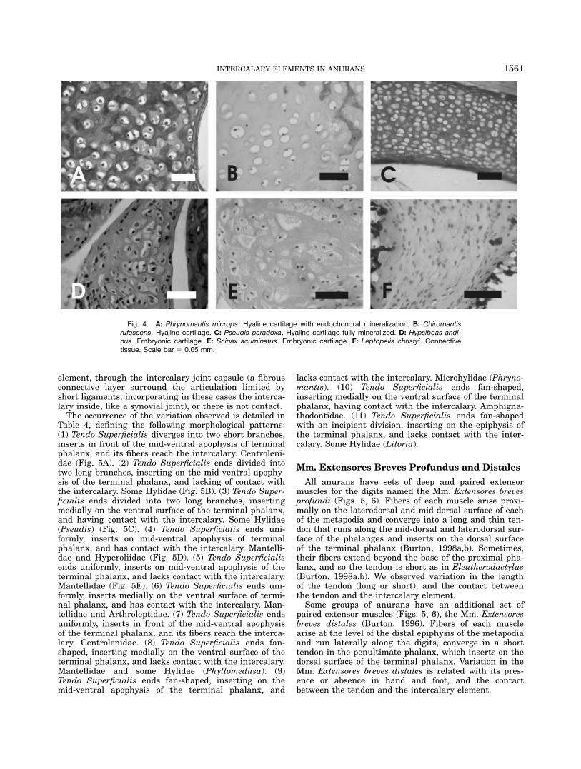
Hyaline cartilage. Characterized by a basophilematrix and isolated chondrocytes immersed in lacunae(Fig. 4A–C). Cartilaginous intercalaries are well stainedin whole-mount preparations. They are found in Ranoides,Lysapsus and Pseudis in Hylidae. Differences in cartilagi-nous intercalaries are described as follows: (1) matrix ofproteoglycans with abundant lacunae and chondroblasts,with nuclei strongly stained (Fig. 4A); (2) uniform andabundant matrix of proteoglycans with scarce lacunae.Only nuclei of chondrocytes are observed. Peripheral min-eralization is also observed (Fig. 4B); and (3) few chondro-cytes are evident. There is an abundant and homogeneousmineralized matrix (Fig. 4C).
Embryonic cartilage. Numerous chondroblastsin lacunae with nuclei strongly stained (Fig. 4D,E). Ma-trix composed by a dense arrangement of connectivefibers. Characteristic of most Hyloides. In whole-mounts,intercalary elements composed by embryonic cartilagedo not accept Alcian Blue coloration, although there issome variation with respect to the Alizarin staining,revealing there are calcium deposits: (1) Alizarin stain-ing negative, mineralization is absent; (2) with one ortwo small mineralized centers; and (3) with a large andinternal mineralized focus.
Connective tissue. With extracellular matrix com-posed by eosinophile fibers arranged in parallel, amongthese fibers basophile cells with enlarged nuclei can beobserved (Fig. 4F). Characteristic of Leptopelis chrystyi.
Distal Epiphysis of the Penultimate Phalanx
The penultimate phalanx displays variation in the pres-ence or absence of ventrolateral crests on its distal epiphy-sis. See Table 4 for the occurrence of this variation.
1558 MANZANO ET AL.
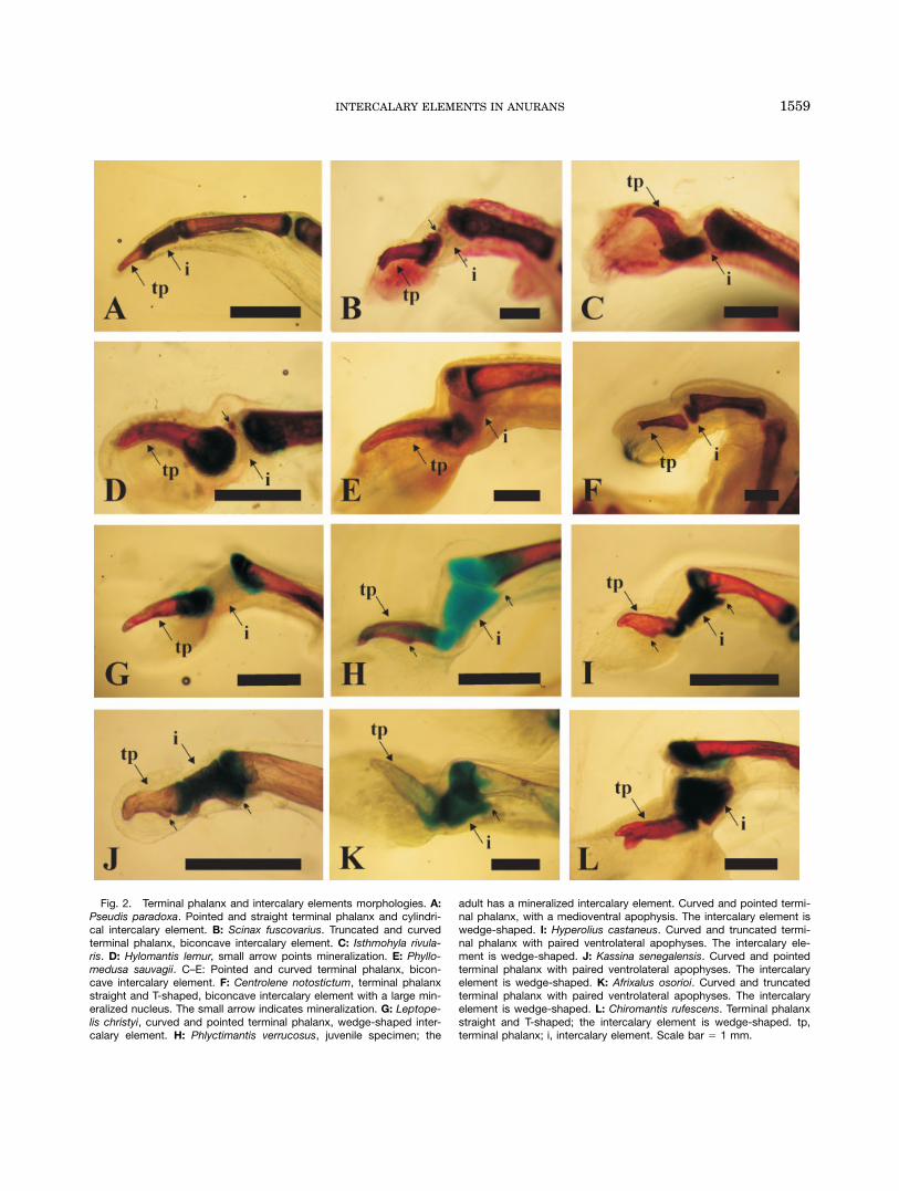
Fig. 2. Terminal phalanx and intercalary elements morphologies. A:Pseudis paradoxa. Pointed and straight terminal phalanx and cylindri-cal intercalary element. B: Scinax fuscovarius. Truncated and curvedterminal phalanx, biconcave intercalary element. C: Isthmohyla rivula-ris. D: Hylomantis lemur, small arrow points mineralization. E: Phyllo-medusa sauvagii. C–E: Pointed and curved terminal phalanx, bicon-cave intercalary element. F: Centrolene notostictum, terminal phalanxstraight and T-shaped, biconcave intercalary element with a large min-eralized nucleus. The small arrow indicates mineralization. G: Leptope-lis christyi, curved and pointed terminal phalanx, wedge-shaped inter-calary element. H: Phlyctimantis verrucosus, juvenile specimen; the
adult has a mineralized intercalary element. Curved and pointed termi-nal phalanx, with a medioventral apophysis. The intercalary element iswedge-shaped. I: Hyperolius castaneus. Curved and truncated termi-nal phalanx with paired ventrolateral apophyses. The intercalary ele-ment is wedge-shaped. J: Kassina senegalensis. Curved and pointedterminal phalanx with paired ventrolateral apophyses. The intercalaryelement is wedge-shaped. K: Afrixalus osorioi. Curved and truncatedterminal phalanx with paired ventrolateral apophyses. The intercalaryelement is wedge-shaped. L: Chiromantis rufescens. Terminal phalanxstraight and T-shaped; the intercalary element is wedge-shaped. tp,terminal phalanx; i, intercalary element. Scale bar 5 1 mm.
1559INTERCALARY ELEMENTS IN ANURANS
Tendo Superficialis
The Tendo Superficialis is a flexor tendon that arisesfrom the palmar/plantar aponeurosis, runs ventrally tothe digit, and inserts on the ventral surface of the termi-nal phalange (Gaupp, 1896). It presents three types ofvariation at the insertion: (a) The Tendo Superficialisfibers are either parallel and end uniformly, diverge and
expand in a fan-shaped end displaying an incipient divi-sion into two short branches, or diverge into two longbranches. (b) The insertion of Tendo Superficialis iseither positioned on the epiphysis of the terminal phalanx,at the base of the phalanx, on the mid-ventral surface ofthe phalanx, on the ventral/apophyses, or in front of themid-ventral apophysis. (c) Fibers of the Tendo Superfi-cialis may have a direct contact with the intercalary
Fig. 3. Cross-longitudinal section of digit tips. A: Pseudis para-doxa. B: Phyllomedusa sauvagi. C: Litoria caerulea. D: Gastrothecachristiani. E: Scinax fuscovarius. F: Hypsiboas andinus. G: Cochranellasauvagei. H: Chiromantis rufescens. I: Guibemantis timidus. J: Hypero-lius kivuensis. K: Leptopelis christyi. L: Phrynomantis microps. A,I,L:
The cartilaginous intercalary fully mineralized. B–G: Embryonic carti-lage with numerous chondrocytes in lacuna without matrix. H,J: Hya-line cartilage with peripheral mineralization. K: Intercalary elementcomposed by connective tissue. Scale bar 5 0.2 mm.
1560 MANZANO ET AL.
element, through the intercalary joint capsule (a fibrousconnective layer surround the articulation limited byshort ligaments, incorporating in these cases the interca-lary inside, like a synovial joint), or there is not contact.The occurrence of the variation observed is detailed in
Table 4, defining the following morphological patterns:(1) Tendo Superficialis diverges into two short branches,inserts in front of the mid-ventral apophysis of terminalphalanx, and its fibers reach the intercalary. Centroleni-dae (Fig. 5A). (2) Tendo Superficialis ends divided intotwo long branches, inserting on the mid-ventral apophy-sis of the terminal phalanx, and lacking of contact withthe intercalary. Some Hylidae (Fig. 5B). (3) Tendo Super-ficialis ends divided into two long branches, insertingmedially on the ventral surface of the terminal phalanx,and having contact with the intercalary. Some Hylidae(Pseudis) (Fig. 5C). (4) Tendo Superficialis ends uni-formly, inserts on mid-ventral apophysis of terminalphalanx, and has contact with the intercalary. Mantelli-dae and Hyperoliidae (Fig. 5D). (5) Tendo Superficialisends uniformly, inserts on mid-ventral apophysis of theterminal phalanx, and lacks contact with the intercalary.Mantellidae (Fig. 5E). (6) Tendo Superficialis ends uni-formly, inserts medially on the ventral surface of termi-nal phalanx, and has contact with the intercalary. Man-tellidae and Arthroleptidae. (7) Tendo Superficialis endsuniformly, inserts in front of the mid-ventral apophysisof the terminal phalanx, and its fibers reach the interca-lary. Centrolenidae. (8) Tendo Superficialis ends fan-shaped, inserting medially on the ventral surface of theterminal phalanx, and lacks contact with the intercalary.Mantellidae and some Hylidae (Phyllomedusa). (9)Tendo Superficialis ends fan-shaped, inserting on themid-ventral apophysis of the terminal phalanx, and
lacks contact with the intercalary. Microhylidae (Phryno-mantis). (10) Tendo Superficialis ends fan-shaped,inserting medially on the ventral surface of the terminalphalanx, having contact with the intercalary. Amphigna-thodontidae. (11) Tendo Superficialis ends fan-shapedwith an incipient division, inserting on the epiphysis ofthe terminal phalanx, and lacks contact with the inter-calary. Some Hylidae (Litoria).
Mm. Extensores Breves Profundus and Distales
All anurans have sets of deep and paired extensormuscles for the digits named the Mm. Extensores brevesprofundi (Figs. 5, 6). Fibers of each muscle arise proxi-mally on the laterodorsal and mid-dorsal surface of eachof the metapodia and converge into a long and thin ten-don that runs along the mid-dorsal and laterodorsal sur-face of the phalanges and inserts on the dorsal surfaceof the terminal phalanx (Burton, 1998a,b). Sometimes,their fibers extend beyond the base of the proximal pha-lanx, and so the tendon is short as in Eleutherodactylus(Burton, 1998a,b). We observed variation in the lengthof the tendon (long or short), and the contact betweenthe tendon and the intercalary element.Some groups of anurans have an additional set of
paired extensor muscles (Figs. 5, 6), the Mm. Extensoresbreves distales (Burton, 1996). Fibers of each musclearise at the level of the distal epiphysis of the metapodiaand run laterally along the digits, converge in a shorttendon in the penultimate phalanx, which inserts on thedorsal surface of the terminal phalanx. Variation in theMm. Extensores breves distales is related with its pres-ence or absence in hand and foot, and the contactbetween the tendon and the intercalary element.
Fig. 4. A: Phrynomantis microps. Hyaline cartilage with endochondral mineralization. B: Chiromantisrufescens. Hyaline cartilage. C: Pseudis paradoxa. Hyaline cartilage fully mineralized. D: Hypsiboas andi-nus. Embryonic cartilage. E: Scinax acuminatus. Embryonic cartilage. F: Leptopelis christyi. Connectivetissue. Scale bar 5 0.05 mm.
1561INTERCALARY ELEMENTS IN ANURANS

Fig. 5. Lateral (on left) and ventral (on right) views of digits showingterminal phalanx, intercalary element, and subterminal phalanx. A:Centrolene geckoideum, digit IV, m. extensor brevis distalis is present,m. extensor brevis profundus has a short tendon, and Tendo Superfi-cialis inserts on the ventral apophysis with two short branches. B: Sci-nax squalirostris, digit IV, m. extensor brevis profundus has a long ten-don and Tendo Superficialis is divided in two long branches at theinsertion. C: Pseudis minuta, digit II, m. extensor brevis profundus hasa long tendon and Tendo Superficialis is divided in two long branches
at the insertion. D: Chiromantis rufescens, digit IV, m. extensor brevisprofundus has a long tendon, m. extensor brevis distalis is presentand Tendo Superficialis is truncated at the insertion. E: Aglyptodacty-lus madagascariensis, digit IV, m. extensor brevis profundus has a longtendon, m. extensor brevis distalis is present and Tendo Superficialisis truncated at insertion. Aph, apophysis; i, intercalary element; Ebd,m. Extensor brevis distalis; Ebp, m. Extensor brevis profundi; Ebpt, m.Extensor brevis profundi tendon; TS, Tendo Superficialis. Scale bar 51 mm.
1562 MANZANO ET AL.
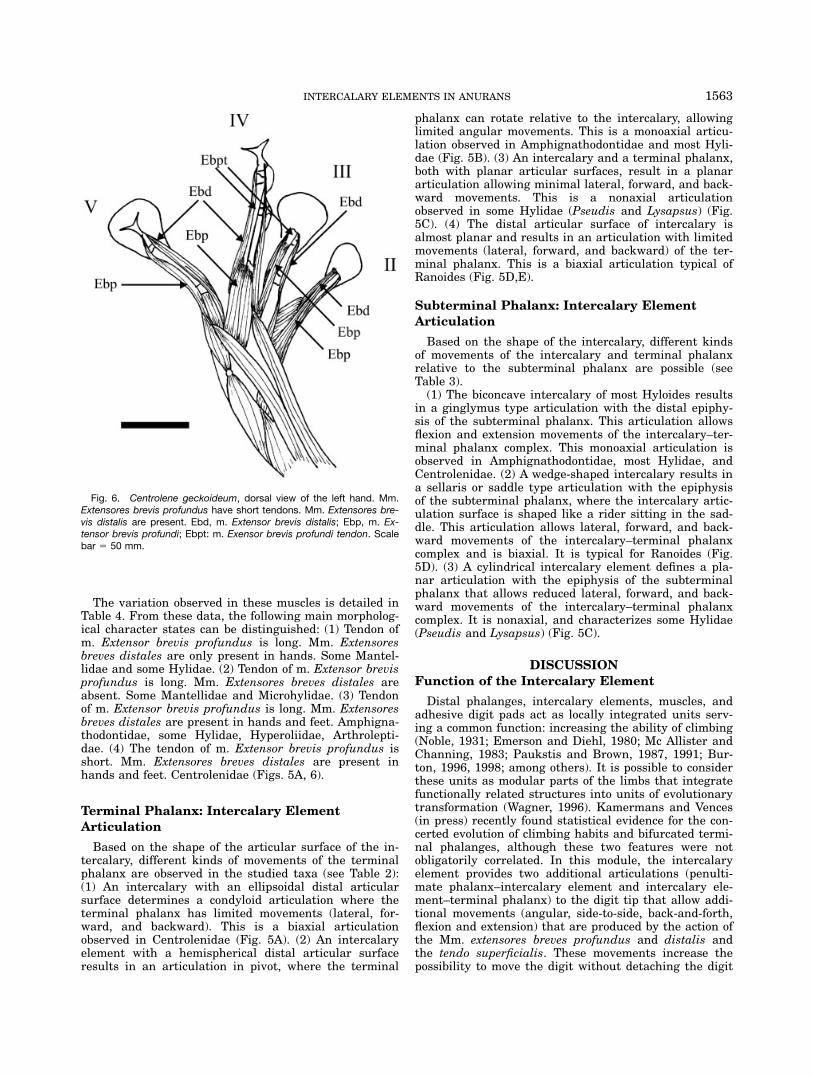
The variation observed in these muscles is detailed inTable 4. From these data, the following main morpholog-ical character states can be distinguished: (1) Tendon ofm. Extensor brevis profundus is long. Mm. Extensoresbreves distales are only present in hands. Some Mantel-lidae and some Hylidae. (2) Tendon of m. Extensor brevisprofundus is long. Mm. Extensores breves distales areabsent. Some Mantellidae and Microhylidae. (3) Tendonof m. Extensor brevis profundus is long. Mm. Extensoresbreves distales are present in hands and feet. Amphigna-thodontidae, some Hylidae, Hyperoliidae, Arthrolepti-dae. (4) The tendon of m. Extensor brevis profundus isshort. Mm. Extensores breves distales are present inhands and feet. Centrolenidae (Figs. 5A, 6).
Terminal Phalanx: Intercalary Element
Articulation
Based on the shape of the articular surface of the in-tercalary, different kinds of movements of the terminalphalanx are observed in the studied taxa (see Table 2):(1) An intercalary with an ellipsoidal distal articularsurface determines a condyloid articulation where theterminal phalanx has limited movements (lateral, for-ward, and backward). This is a biaxial articulationobserved in Centrolenidae (Fig. 5A). (2) An intercalaryelement with a hemispherical distal articular surfaceresults in an articulation in pivot, where the terminal
phalanx can rotate relative to the intercalary, allowinglimited angular movements. This is a monoaxial articu-lation observed in Amphignathodontidae and most Hyli-dae (Fig. 5B). (3) An intercalary and a terminal phalanx,both with planar articular surfaces, result in a planararticulation allowing minimal lateral, forward, and back-ward movements. This is a nonaxial articulationobserved in some Hylidae (Pseudis and Lysapsus) (Fig.5C). (4) The distal articular surface of intercalary isalmost planar and results in an articulation with limitedmovements (lateral, forward, and backward) of the ter-minal phalanx. This is a biaxial articulation typical ofRanoides (Fig. 5D,E).
Subterminal Phalanx: Intercalary ElementArticulation
Based on the shape of the intercalary, different kindsof movements of the intercalary and terminal phalanxrelative to the subterminal phalanx are possible (seeTable 3).(1) The biconcave intercalary of most Hyloides results
in a ginglymus type articulation with the distal epiphy-sis of the subterminal phalanx. This articulation allowsflexion and extension movements of the intercalary–ter-minal phalanx complex. This monoaxial articulation isobserved in Amphignathodontidae, most Hylidae, andCentrolenidae. (2) A wedge-shaped intercalary results ina sellaris or saddle type articulation with the epiphysisof the subterminal phalanx, where the intercalary artic-ulation surface is shaped like a rider sitting in the sad-dle. This articulation allows lateral, forward, and back-ward movements of the intercalary–terminal phalanxcomplex and is biaxial. It is typical for Ranoides (Fig.5D). (3) A cylindrical intercalary element defines a pla-nar articulation with the epiphysis of the subterminalphalanx that allows reduced lateral, forward, and back-ward movements of the intercalary–terminal phalanxcomplex. It is nonaxial, and characterizes some Hylidae(Pseudis and Lysapsus) (Fig. 5C).
DISCUSSION
Function of the Intercalary Element
Distal phalanges, intercalary elements, muscles, andadhesive digit pads act as locally integrated units serv-ing a common function: increasing the ability of climbing(Noble, 1931; Emerson and Diehl, 1980; Mc Allister andChanning, 1983; Paukstis and Brown, 1987, 1991; Bur-ton, 1996, 1998; among others). It is possible to considerthese units as modular parts of the limbs that integratefunctionally related structures into units of evolutionarytransformation (Wagner, 1996). Kamermans and Vences(in press) recently found statistical evidence for the con-certed evolution of climbing habits and bifurcated termi-nal phalanges, although these two features were notobligatorily correlated. In this module, the intercalaryelement provides two additional articulations (penulti-mate phalanx–intercalary element and intercalary ele-ment–terminal phalanx) to the digit tip that allow addi-tional movements (angular, side-to-side, back-and-forth,flexion and extension) that are produced by the action ofthe Mm. extensores breves profundus and distalis andthe tendo superficialis. These movements increase thepossibility to move the digit without detaching the digit
Fig. 6. Centrolene geckoideum, dorsal view of the left hand. Mm.Extensores brevis profundus have short tendons. Mm. Extensores bre-vis distalis are present. Ebd, m. Extensor brevis distalis; Ebp, m. Ex-tensor brevis profundi; Ebpt: m. Exensor brevis profundi tendon. Scalebar 5 50 mm.
1563INTERCALARY ELEMENTS IN ANURANS
pad (Hanna and Barnes, 1991). Similar to the shape ofterminal phalanges, there appears to be no obligatorycorrelation between the presence of intercalary elementsand climbing habits. Several taxa, such as the mantellidgenus Aglyptodactylus, do have intercalaries but arefully terrestrial. The microhylids of the genus Phryno-mantis, with intercalaries, comprise species climbing onrocks such as Phrynomantis annectens, but no fully ar-boreal species. In contrast, there are many strictly arbo-real taxa (e.g., in the ceratobatrachid genus Platymantisor the microhylid genera Cophyla and Platypelis) with-out intercalaries. In this context, it is interesting thatWu (1994), in a so far unpublished thesis, mentions thepresence of weakly expressed, uncalcified intercalariesin some species of Cophyla and Platypelis. If confirmedby further studies, this finding would constitute a fur-ther instance of independent occurrence of intercalariesin concert with arboreal habits.
Structural Variation of Intercalary Elements
Structural differences in the histology of the interca-lary elements can be interpreted as different states ofcartilage differentiation, including the states of minerali-zation. Intercalary elements of most Hyloides are formedby embryonic cartilage with numerous chondrocytes,with an extracellular matrix lacking chondroitin sulfate.On the contrary, most Ranoides have intercalariesformed by cartilage with a well-differentiated extracellu-lar matrix with proteoglycans.The extracellular matrix of the cartilage is a complex
formed by glycosaminoglycans and collagen fibril/fibers,rates of synthesis of which are regulated independently.The appearance of type II collagen protein in limb mes-enchyme coincides with differentiation of precursor cellsinto chondroblasts, whereas the synthesis of chondroitinsulfate neither is an exclusive property of, nor diagnosticfor, chondrogenic cells (Hall, 2005). Hence, histologicaldifferences between the intercalaries of Hyloides andRanoides seem to depend on the rates of differentiationof cells and of the extracellular matrix, which are slowin Hyloides and faster in Ranoides. Within Hyloides, theintercalary elements of Pseudis and Lysapsus would rep-resent a derived condition resulting from fast differen-tiation rate of cartilaginous tissue as compared withmost other Hyloides. Within the Ranoides, the connec-tive intercalary element of Leptopelis would result froma slower differentiation rate, in which cells and matrixremain as a condensed mesenchyme. Data from cartilagein rats show a decrease in the number of chondrocytes,increase in glycogen and lipid contents, decrease inchondroitin sulfate but increase in keratan sulfate, anincrease of thickness of collagen fibers, and increase ofmineralization with increasing age of the animal (Hall,2005). Some of these trends show remarkable parallelsto the differences among the intercalary elements ofRanoides. The intercalary element of Hyperolius has fewchondrocytes, whereas the intercalary elements of Man-tellidae are strongly mineralized. Because several genesand cellular processes play a role in regulating pathwaysof cartilage differentiation (Hall, 2005), the structuraldifferences exhibited in the intercalary elements of anu-rans, are probably results from differential differentia-tion rates that appear to be largely conserved in eachlineage (Ranoides and Hyloides).
Intercalary and Terminal PhalanxMorphologies
In Hyloides, with the exceptions of the cartilaginousand cylindrical elements of Pseudis and Lysapsus, inter-calary elements are morphologically and structurallyvery similar. In most representatives of this clade, theproximal and distal articulations of the intercalary ele-ment show the same pattern. In contrast, the centrole-nids have a distal ellipsoidal articulation similar to thatfound in all ranoids. Pseudis and Lysapsus display dis-tinctive patterns with planar articulations limiting themovements. Morphological variation of the terminalphalanx within Hyloides is limited to the following: Cen-trolenidae, having a straight terminal phalanx with twowell-developed lateral processes; Scinax spp., with acurved and truncated terminal phalanx; and Pseudisand Lysapsus, with a straight and pointed terminal pha-lanx. The truncated terminal phalanx in Scinax fusco-varius arises from the reduction of two lateral processesduring larval development, similar to the terminal pha-lanx development observed in some Leptodactylus spp.(Noble, 1917; Fabrezi, 1996). Many Hyloides have acurved and pointed terminal phalanx that was consid-ered as a synapomorphy to join them with the Hylidae(Laurent, 1986; Ford and Cannatella, 1993). Theobserved variation in the morphology of terminal pha-langes and articular surfaces of the intercalary in Cen-trolenidae suggests coordinated changes in both skeletalcomponents.The morphology of the intercalary within Ranoides
displays scarce variation defining the same distal andproximal articulations. In this clade, the shape of theterminal phalanx with two lateral processes is wide-spread, although there are pointed and truncated termi-nal phalanx within Laurentobatrachia (Liem, 1970;Drewes, 1984; Kamermans and Vences, in press).
Variation of Muscles and Tendons
The Mm. Extensores breves profundus are very uni-form, and commonly their fibers extend along the meta-podials up to form a tendon at the level of the proximalphalanx to insert dorsally on the terminal one. The onlyvariation related to these muscles was observed in Cen-trolenidae (including Allophrynae), which displays elon-gated muscles forming a short tendon up to the level ofthe terminal phalanx. This condition has also beendescribed for other anurans reported to be climbers orat least scansorials in their habits, such as the hyloidgenus Eleutherodacylus (Burton, 1996, 1998a,b), whichhowever, lack intercalaries. Differently from Eleuthero-dactylus, the Centrolenidae have also additional setsof muscles Extensores breves distalis. The presence ofMm. Extensores breves distalis in hands has been foundto be strongly related to arboreality (Burton, 1996,1998a,b, 2004). They are present in Hyloides and someRanoides, their presence is variable within Mantellidae,and they are absent in Phrynomantis. When thesemuscles are present, there is no variation among thespecies. Summarizing, our results showed the morpho-logical variation recorded in intercalary elements,terminal phalanx, Tendo superficialis, and musclesExensores digitorum seems to be constrained to fewcombinations only.
1564 MANZANO ET AL.

Phylogenetic Interpretations
In recent hypotheses of anuran phylogeny, the neoba-trachians are composed of two sister clades, Hyloidesand Ranoides (Frost et al., 2006), with 12 terminal taxawith intercalary elements (Fig. 1). As shown herein,there are morphostructural differences between the in-tercalary of Hyloides (patterns 1, 2, and 3) and Ranoides(patterns 5, 6, and 7). Within hyloids, intercalary ele-ments are present in five taxa of Nobleobatrachia; theywere probably lost in Brachycephalidae and twice withinthe Leptodactyliformes. Centrolenidae (including Allo-phryne) and also some leptodactylids have the set ofmuscles that integrate the articulation with intercalaryelements even though the intercalary element is absentin some of them. The morphostructural characteristicsof the intercalary elements have only been stronglymodified in two hylid genera, Pseudis and Lysapsus (pat-tern 4). All Hyloides share a biconcave intercalary ele-ment formed by embryonic cartilage, except for thesetwo genera. Variations in the morphostructural patternsdescribed by us are mainly related to intercalary miner-alization. The Tendo superficialis appears with a charac-teristic bifurcated pattern at insertion in Hylidae, andthis same pattern is observed also in Centrolenidae.Among Hyloides, the articulation of the intercalary ele-ment and terminal phalanx, shape of terminal phalanx,and characteristics of the Mm. Extensores breves profun-dus and distalis observed in Centrolenidae seem to bederived in an integrated manner, which is in agreementwith phylogenetic hypotheses placing centrolenids as notclosely related to other Neotropical treefrogs (e.g., Frostet al., 2006).Different from Hyloides, Ranoides share a wedge-
shaped intercalary element composed of hyaline carti-lage, with the exception of Leptopelis. Among theRanoides, the intercalary would have appeared inde-pendently in three clades Laurentobatrachia, Microhyli-dae (genus Phrynomantis), and Rhacophoroidea. The in-tercalary elements of Rhacophoroidea have been inter-preted as a synapomorphy for the clade (Frost et al.,2006). The genus Laliostoma, which is nested withinRhacophoroidea, has no intercalary element (Glaw et al.,1998) and its condition, therefore, is to be interpreted assecondary loss. In Laurentobatrachia, the intercalary ofLeptopelis presents the same shape of the intercalary ofRanoides but is formed by dense connective tissue.Drewes (1984) suggested that the histological differencesbetween the intercalary elements of Leptopelis andhyperoliids are arguments to reject their homology. Evenif the intercalary elements of Rhacophoroidea appear assynapomorphy, their reappearance does not suggestchanges from the ancient or plesiomorphic condition.
Evolutionary Interpretations
From an evolutionary perspective, morphological nov-elty was defined as ‘‘. . . a structure that is neither ho-mologous to any structure in the ancestral species norhomonymous to any other structure of the same orga-nism.’’ (Muller and Wagner, 1991, p. 243). In accordancewith this definition, the intercalary element of anuransseems to be a morphological novelty that appeared inneobatrachians. No extant or fossil species belonging toany basal anuran lineage (i.e., not belonging to the Neo-
batrachia) is known to have intercalary elements. Theintercalary element is not equivalent or homologous toother limb skeletal components and is not incorporatedin the autopodia limb plan. Some invariable featurescharacterize the intercalary elements: (1) they arealways present in hands as well as feet, (2) they arealways present between the penultimate and terminalphalanges, (3) they are present only in those taxa hav-ing a complete set of phalanges. These features suggestsome level of homology and indicate they may havearisen only once, early in the neobatrachian history.Paukstis and Brown (1987, 1991) explained quantita-
tive variation of intercalary elements related to habitator life style. They noticed a decrease in the thickness ofthe intercalary and size of digital pad in some speciesof Hyloides, and in Mantella aurantiaca, and the loss ofdigital pads in species of Kassina that may be associatedwith terrestrial habitat. The unusual features of the in-tercalary of the aquatic Pseudis and Lysapsus wereinterpreted as advantages to increase digit lengths forsupporting extensive webbing on the feet, and for grasp-ing aquatic vegetation with the hands. However, such ascenario fails to explain the presence of intercalary ele-ments and well-developed digital pads in the aquaticLitoria aurea and the terrestrial L. lesueuri, where theloss of arboreal habits may be recent or the evolutionaryrate of change in the intercalary is low (Paukstis andBrown, 1991). In our character tracing, under bothACCTRAN and DELTRAN reconstructions, more gainsof intercalary elements than losses were reconstructed(Fig. 1). The most unequivocal loss is that of Laliostoma,a species that is phylogenetically nested in a clade (Rha-cophoroidea) where otherwise intercalaries are univer-sally present. However, there seem to be many instancesof fully terrestrial or aquatic frogs with intercalaries,nested in otherwise largely arboreal clades, indicatingthat these elements do not cause any strong disadvan-tages to nonarboreal frogs and, therefore, are not understrong selective pressure to be lost to the evolutionaryloss of arboreal habits.The intercalary element is a modular part functionally
integrated with other musculoskeletal structures andspecializations of the tegument. Explaining the loss ofits functional capability is not easy. The presence ofintercalaries reveals strong constraints: (1) The interca-lary elements are integrated in the developmental limbprograms in those lineages in which they are present.This fact was demonstrated for experiments of digitregeneration in Hyperolius viridiflavus (Richards et al.,1975). (2) These elements show conservative patterns ofvariation, similar to other related structures. Occasionalchanges in this modular unit sometimes seem to be dis-sociated. For example: the absence of digital padsin Pseudis paradoxus is not related to the reduction ofintercalary elements, and the absence of ventrolateralcrests in the penultimate phalanx limits extension andflexion movements in the articulation in Phyllomedusa,Mantella, Pseudis, and Lysapsus, independently of themorphostructural features of the intercalary and devel-opment of the digital pad. Even if these taxa share thecapability to oppose fingers, the absence of ventrolateralcrests in the penultimate phalanx in all digits cannot beassociated with this feature. In other cases, reappearan-ces of this system, which are interpreted in Centroleni-dae and Rhacophoroidea, may suggest coordinated
1565INTERCALARY ELEMENTS IN ANURANS
derived changes in distal articulation and muscles (Cen-trolenidae) or not (Rhacophoroidea); (3) Allophryne ruth-veni lacks intercalary elements but presents a well-developed digital pad, short tendons of Mm. extensoresbreves profundus, a biaxial articulation between penulti-mate–terminal phalanges (Fabrezi and Langone, 2000),and Mm. extensores breves distalis. These features couldbe either interpreted as remnants from a condition withthe intercalary element present ancestrally but that istoday lost, or as a first step before a reappearance of anintercalary. However, both alternatives involve a similarlevel of speculation.Due to the absence of intercalaries in possible out-
groups, it is currently not possible to polarize any of theintercalary shapes typical for ranoids or hyloids (i.e.,wedge-shaped hyaline cartilage vs. biconcave embryoniccartilage) as plesiomorphic or apomorphic. However,because in ranoids there are three phylogenetically inde-pendent origins of the intercalary and in each casewedge-shaped elements are observed, it is unlikely thatthis state represents a derived condition. In hyloids, itseems more likely that the intercalaries evolved onlyonce and were subsequently lost in two lineages, re-evolving again in centrolenids. As a conclusion, if indeedearly neobatrachians were characterized by intercalaryelements or by the genetic basis to develop these, then itcould by hypothesized that (1) the ranoid ancestor haslost this original neobatrachian state but this state wasthen re-acquired three times independently in Ranoides,and that (2) the biconcave element of hyloids is derivedfrom this original neobatrachian state.From our analysis, we conclude that the intercalary
element is a morphological novelty and forms part of amodular unit in which other musculoskeletal structuresand the tegument are functionally integrated. This mod-ular unit, or its developmental genetic basis, may haveoriginated early in the history of the Neobatrachia andhas further evolved in parallel in Hyloides and Ranoides.We have here described morphological variation in thismodular unit, pointing out those traits that could be usefulfor systematic analyses. However, variation in the modularunit formed by the intercalary elements is limited andsuggests few opportunities of change.
ACKNOWLEDGMENTS
We thank Virginia Abdala, Javier Goldberg, and Mar-celo Cabada for critically reading the first draft of thismanuscript. We also thank the editor and the anony-mous reviewer for their valuables suggestions toimprove the manuscript.
LITERATURE CITED
Anderson G, Bancroft J. 2002. Tissue processing and microtomy. In:Bancroft J, Gamble M, editors. Theory and practice of histologicaltechniques, 5th ed. London: Churchill Livingston, ElsevierScience Limited. p 85–107.
Bock WJ, Shear R. 1972. A staining method for gross dissection ofvertebrate muscles. Anat Anz 130:222–227.
Burton TC. 1996. Adaptation and evolution in the hand muscles ofAustralo-Papuan hylid frogs (Anura: Hylidae: Pelodryadinae).Aust J Zool 44:611–623.
Burton TC. 1998a. Are the distal extensor muscles of the fingers ofanuran an adaptation to arboreality? J Herpetol 32:611–617.
Burton TC. 1998b. Variation in the hand and superficial throatmusculature of the Neotropical leptodactylid frogs. Herpetologica54:53–72.
Burton TC. 2004. Muscles of the pes of hylid frogs. J Morphol260:209–233.
Clarke BT. 1981. Comparative osteology and evolutionary relation-ships in the African Raninae (Anura: Ranidae) Monitore Zool ItalSuppl (NS) 15:285–331.
Drewes RC. 1984. A phylogenetic analysis of the Hyperoliidae(Anura): treefrogs of Africa, Madagascar, and the SeychellesIslands. Occas Pap Calif Acad Sci 139:1–70.
Duellman W. 2001. The hylid frogs of middle America. Vol. 2.Ithaca, NY: Society for the Study of Amphibians and Reptiles.
Duellman W, Trueb L. 1986. Biology of amphibians. New York:McGraw-Hill.
Dyce KM, Sack WO, Wensing CJG. 1996. Text book of veterinary anat-omy, 2nd ed. Philadelphia: WB Saunders Company. p 528–529.
Emerson SB, Diehl D. 1980. Toe pad morphology and mechanismsof sticking in frogs. Biol J Linn Soc 13:199–216.
Fabrezi M. 1996. Las falanges terminales en la clasificacion de losanuros. Cuad Herpetol 10:1–9.
Fabrezi M, Langone J. 2000. Los caracteres morfologicos del contro-vertido Neobatrachia arborıcola Allophryne ruthveni Gaige 1926.Cuad Herpetol 14:47–59.
Faivovich J, Haddad CF, Garcia PCA, Frost DR, Campbell JA,Wheeler WC. 2005. Systematic review of the frog family Hylidae,with special reference to Hylinae: Phylogenetic analysis and taxo-nomic revision. Bull Am Mus Nat Hist 294:1–240.
Ford L, Cannatella DC. 1993. The major clades of frogs. HerpetolMonogr 7:94–117.
Frost DR. 2007. Amphibian species of the world: an online refer-ence. Version 3.0 (22 August, 2004). Available at: http://research.amnh.org/herpetology/amphibia/index.html.
Frost DR, Grant T, Faivovich J, Bain RH, Haas A, Haddad CFB, DeSa RO, Channing A, Wilkinson M, Donnellan SC, Raxworthy CJ,Campbell JA, Blotto BL, Moler P, Drewes RC, Nussbaum RA,Lynch JD, Green DM, Wheeler WC. 2006. The amphibian tree oflife. Bull Am Mus Nat Hist 297:1–370.
Gaupp E. 1896. A. Ecker’s und R. Wiedersheim’s Anatomie desFrosches. Braunschweg Friedrich: Friedrich Vieweg und Sohn.Vol. 2:1–961.
Glaw F, Vences M, Bohme W. 1998. Systematic revision of the genusAglyptodactylus Boulenger, 1919 (Amphibia: Ranidae), and analysisof its phylogenetic relationships to other Madagascan ranid genera(Tomopterna, Boophis, Mantidactylus, and Mantella). Journal ofZoological Systematics and Evolutionary Research 36:17–37.
Hall BK. 2005. Bones and cartilages. Developmental and evolution-ary skeletal biology. New York: Elsevier, Academic Press.
Hanna G, Barnes WJP. 1991. Adhesion and detachment of the toepads of tree frogs. J Exp Biol 155:103–125.
Heyer R. 1975. A preliminary analysis of the intergeneric relationshipsof the frog family Leptodactylidae. Smith Contrib Zool 199:1–55.
Kamermans M, Vences M. Terminal phalanges in ranoid frogs: mor-phological diversity and evolutionary correlation with climbinghabits. Alytes. (in press).
Laurent RF. 1986. Souss classe lissamphibiens (Lissamphibia). Sys-tematique. Tome XIV, Batraciens, Fasc. 1B. Masson, pp. 594–798.In: Grasse PP, Delsol M, editors. Traite de Zoologie. Anatomie,Systematique, Biologie. Paris.
Liem SS. 1970. The morphology, systematics and evolution of theOld World treefrogs (Rhacophoridae and Hyperoliidae). Field Zool57:1–45.
Lynch JD. 1971. Evolutionary relationships, osteology and zoogeog-raphy of leptodactyloid frogs. Univ Kansas Publ Mus Nat Hist53:1–238.
Lynch JD. 1973. The transition from archaic to advanced frogs. In:Vial JL, editor. Evolutionary biology of the anurans. Columbia,MO: University of Missouri Press. p 133–182.
Mc Allister W, Channing A. 1983. Comparison of toe pads of somesouthern African climbing frogs. S Afr Tydskr Dierk 18:110–114.
Maddison W. 1990. A method for testing the correlated evolution oftwo binary characters: are gains or losses concentrated on certainbranches of a phylogenetic tree? Evolution 44:539–557.
1566 MANZANO ET AL.
MaddisonWP, Maddison DR. 1998. MacClade: analysis of phylogeny andcharacter evolution, version 3.08. Sunderland, MA: Sinauer.
Muller GB, Wagner GP. 1991. Novelty in evolution: restructuringthe concept. Annu Rev Ecol Syst 22:229–256.
Noble GL. 1917. The systematic status of some batrachians fromSouth America. Bull Am Mus Nat Hist 37:793–815.
Noble GK. 1931. The biology of the amphibians. New York:McGraw-Hill. vii1577 p.
Ohler A, Dubois A. 1989. Demonstration de l’origine independantedes ventouses digitales dans deux lignees phylogenetiques deRanidae (Amphibiens, Anoures). C R Acad Sci Paris 309:419–422.
Paukstis GL, Brown LE. 1987. Evolution of the intercalary cartilagein Chorus frogs, genus Pseudacris (Salientia: Hylidae). Brim-leyana 13:55–61.
Paukstis GL, Brown LE. 1991. Evolutionary trends in the morphol-ogy of the intercalary phalanx of anuran amphibians. Can J Zool69:1297–1301.
Richards CM, Carlson BM, Rogers SL. 1975. Regeneration of digitsand forelimbs in the Kenyan reed frog Hyperolius viridiflavusferniquei. J Morphol 146:431–446.
Scott E. 2005. A phylogeny of ranid frogs (Anura: Ranoidea: Rani-dae), based on a simultaneous analysis of morphological andmolecular data. Cladistics 21:507–574.
Van Bocxlaer I, Roelants K, Biju SD, Nagaraju J, Bossuyt F. 2006.Late Cretaceous vicariance in Gondwanan amphibians. PLoS ONE1:e74.
Van der Meijden A, Vences M, Hoegg S, Meyer A. 2005. A previ-ously unrecognized radiation of ranid frogs in southern Africa
revealed by nuclear and mitochondrial DNA sequences. Mol PhylEvol 37:674–685.
Van der Meijden A, Vences M, Hoegg S, Boistel R, Channing A,Meyer A. 2007. Nuclear gene phylogeny of narrow-mouthed toads(Family: Microhylidae) and a discussion of competing hypothesesconcerning their biogeographical origins. Mol Phylogenet Evol44:1017–1030.
Vences M, Kosuch J, Glaw F, Bohme W, Veith M. 2003. Molecularphylogeny of hyperoliid treefrogs: biogeographic origin of Mala-gasy and Seychellean taxa and re-analysis of familial paraphyly.J Zool Syst Evol Res 41:205–215.
Wagner GP. 1996. Homologues, natural kinds and the evolution ofmodularity. Am Zool 36:36–43.
Wassersug RJ. 1976. A procedure for differential staining of carti-lage and bone in whole formalin fixed vertebrates. Stain Technol51:131–134.
Wiens JJ. 2007. Book review: the amphibian tree of life. Q Rev Biol82:55–56.
Wiens JJ, Fetzner JW Jr, Parkinson CL, Reeder TW. 2005. Hylidfrog phylogeny and sampling strategies for species for specioseclades. Syst Biol 54:719–748.
Wilson I, Gamble M. 2002. The hematoxylins and eosins. In. Ban-croft J, Gamble M, editors. Theory and practice of histologicaltechniques, 5th ed. London: Churchill Livingston, Elsevier Sci-ence Limited. p 125–137.
Wu SH. 1994. Phylogenetic relationships, higher classification, andhistorical biogeography of the microhyloid frogs (Lissamphibia:Anura: Brevicipitidae and Microhylidae). Unpublished PhD the-sis. The University of Michigan.
1567INTERCALARY ELEMENTS IN ANURANS


![Molecular phylogeny of stream treefrogs (Hylidae: Hyloscirtus ...home.gwu.edu/~rpyron/publications/Guayasamin_et_al_2015.pdf · Faivovich et al. [3], Crawford et al. [27], Coloma](https://img.pdfslide.us/doc/110x75/5fa5c116f3f86023b05ce7ea/molecular-phylogeny-of-stream-treefrogs-hylidae-hyloscirtus-homegwuedurpyronpublicationsguayasaminetal2015pdf.jpg)