Embed Size (px)
Citation preview
APPLIED AND ENVIRONMENTAL MICROBIOLOGY, Sept. 2009, p. 5991–5995 Vol. 75, No. 180099-2240/09/$08.00�0 doi:10.1128/AEM.01170-09Copyright © 2009, American Society for Microbiology. All Rights Reserved.
Interactions of Typical and Atypical Enteropathogenic Escherichia coliStrains with the Calf Intestinal Mucosa Ex Vivo�
Francis Girard,1 Francis Dziva,2 Mark P. Stevens,2 and Gad Frankel1*Centre for Molecular Microbiology and Infection, Division of Cell & Molecular Biology, Imperial College London,
London,1 and Enteric Bacterial Pathogens Group, Division of Microbiology, Institute for Animal Health,Compton, Berkshire,2 United Kingdom
Received 20 May 2009/Accepted 20 July 2009
Enteropathogenic Escherichia coli (EPEC) can be found in healthy and diarrheic cattle; however, little isknown about the role of attaching and effacing (A/E) lesion formation in colonization of bovine intestinalmucosa by such strains. We show that typical and atypical EPEC induce A/E lesions on calf intestinal explantsindependently of Tir tyrosine phosphorylation and TccP. Our data support the existence of conserved Tir- andTccP-independent mechanisms of A/E lesion formation in a range of hosts and reinforce the zoonotic potentialof EPEC in cattle.
Enteropathogenic Escherichia coli (EPEC) and the closelyrelated enterohemorrhagic E. coli (EHEC) are importanthuman pathogens (28). EPEC strains are divided into typi-cal EPEC and atypical EPEC (aEPEC) based on the pres-ence or absence of the EAF plasmid, respectively (15). Typ-ical EPEC strains, which belong mainly to 1 of 12 Oserogroups, are further divided into EPEC lineage 1(EPEC-1), which is characterized by expression of flagellarantigen H6 or H34 and intimin �, and EPEC-2, which com-monly expresses H2 (or H�), intimin �, and the type IIIsecretion system effector TccP2 (40). aEPEC strains aremuch more diverse and may belong to one of many sero-groups.
EPEC, aEPEC, and EHEC are diarrheal pathogens capableof forming attaching and effacing (A/E) lesions (reviewed inreferences 8 and 9). A/E lesions are characterized by efface-ment of the brush border microvilli and intimate bacterialattachment to the host cell plasma membrane (23). The genesrequired for A/E lesion formation are carried on the locus ofenterocyte effacement (27), which encodes transcriptional reg-ulators, the adhesin intimin (20), a type III secretion system(19), chaperones, translocators, and several effector proteins(reviewed in references 9 and 11).
One of the major hallmarks of EPEC and EHEC strains istheir ability to trigger actin polymerization at the site ofbacterial attachment to cultured cells (23). The principaleffector protein needed for A/E lesion formation on muco-sal surfaces and actin polymerization in vitro is Tir (22, 33).Once translocated, Tir is integrated into the host cell plasmamembrane in a hairpin loop topology (16), and the extra-cellular loop serves as an intimin receptor (reviewed inreference 10). In EPEC-1 (represented by strain E2348/69,O127:H6), actin polymerization in vitro is triggered by phos-
phorylation of a Tir tyrosine (Y) residue at position 474(21), which recruits the adaptor protein Nck, leading toactivation of the neuronal Wiskott-Aldrich syndrome pro-tein (N-WASP) and actin polymerization via the actin-re-lated protein 2/3 (Arp2/3) complex (reviewed in reference7). Tir from E2348/69 can also trigger weak Nck-indepen-dent actin polymerization (5) via a universally conservedNPY Tir motif (4), which was recently shown to recruitinsulin receptor tyrosine kinase substrate p53 (39) and/orinsulin receptor tyrosine kinase substrate (36). In EHECO157:H7, binding of insulin receptor tyrosine kinase sub-strate p53/insulin receptor tyrosine kinase substrate to Tir(which lacks an Y474 equivalent) leads to the recruitment ofTccP (aka EspFU), which in turn activates N-WASP (6, 12,36). Strains belonging to EPEC-2 (represented by strainB171, O111:NM) express both Tir containing a Y474 equiv-alent and TccP2 (24, 40), which is interchangeable with TccPof EHEC O157 (40).
aEPEC strains can trigger actin polymerization in vitro bydiverse mechanisms involving Tir-Nck and/or Tir-TccP/TccP2pathways. However, a significant proportion of aEPEC (rep-resented by strain ICC223, O125:H6) strains cannot triggeractin polymerization in vitro, as they express Tir lacking a Y474equivalent and TccP/TccP2 (3). However, these strains cantrigger typical A/E lesions to form on human in vitro organcultures (hIVOC) (33).
Fecal excretion of EPEC by healthy and diarrheic calveshas been reported in the United States (18), Europe (2, 26),Australia (17), India (38), and Brazil (1); however, the zoo-notic and pathogenic potential of such strains is ill defined.While A/E lesion formation is known to play a role inintestinal colonization of ruminants by EHEC O157 andO26 (35), not much is known about EPEC or aEPEC patho-genesis on bovine intestinal mucosa or what role actin nu-cleation may play in the efficiency of adherence. Here, weinvestigated the interactions of EPEC-1 strain E2348/69,EPEC-2 strain B171, and aEPEC strain ICC223 with the calfgut mucosa using a bovine IVOC (bIVOC) model. All thestrains used in this study (listed in Table 1) were grown
* Corresponding author. Mailing address: Centre for Molecular Mi-crobiology and Infection, Division of Cell & Molecular Biology, Flow-ers Building, Imperial College London, London SW7 2AZ, UnitedKingdom. Phone: 44 020 2594 525. Fax: 44 020 5794 3069. E-mail:[email protected].
� Published ahead of print on 24 July 2009.
5991
on June 2, 2018 by guesthttp://aem
.asm.org/
Dow
nloaded from
overnight in tryptic soy broth, then transferred into fresh,sterile tryptic soy broth, and grown to early log phase for 2.5to 3 h. Kanamycin was used at a final concentration of 50 �gml�1 where appropriate. The calf gut IVOC model wasused, as previously described (14), and five Friesian bullcalves that were 6 to 9 weeks of age were used in fiveseparate experiments in accordance with the Animals (Sci-entific Procedures) Act 1986 under license 30/2463. Unin-fected explants were also cultured in each experiment inorder to confirm the absence of endogenous infection andthat no external contamination occurred during the experi-mental process. Each strain was tested on explants derivedfrom at least two animals. The terminal ileum and terminalrectum were used in this study.
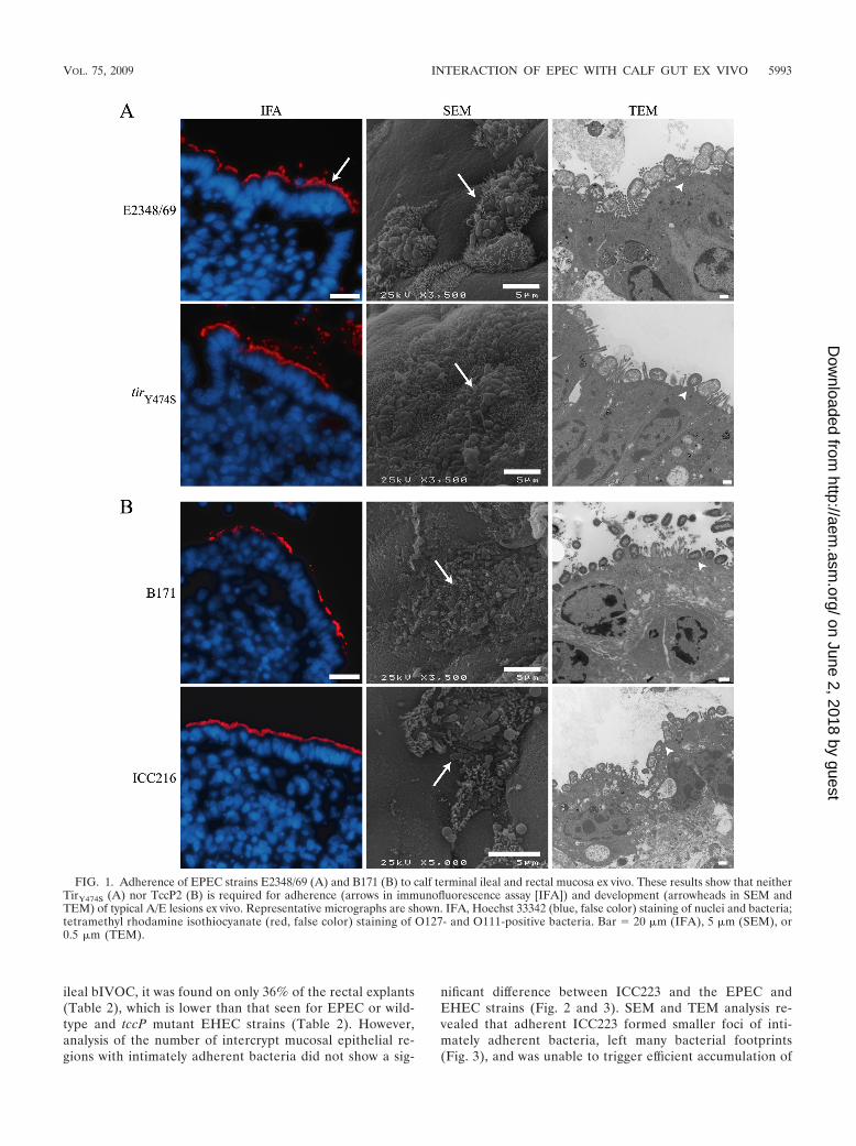
We first investigated the ability of typical EPEC to adhereto and induce A/E lesion formation on bIVOC. BothE2348/69 and B171 were found to be intimately associatedwith the epithelia of explants derived from the terminalileum and terminal rectum, following staining of formalin-fixed, paraffin-embedded sections with their respective Oantigen antisera (rabbit anti-O127 and rabbit anti-O111)(13, 14) (Table 2 and Fig. 1). Further analysis of the colo-nized bIVOC indicated that the number of intercrypt mu-cosal epithelial regions with intimately adherent bacteriawas comparable for E2348/69 and B171 in the ileal andrectal explants, although adherence of B171 to rectal ex-plants occurred at a lower level (Table 2). Scanning electronmicroscopy (SEM) and transmission electron microscopy(TEM) showed that the intimately adherent bacteria formedtypical A/E lesions, with accumulation of electron-densematerial at the site of bacterial attachment (Fig. 1).
We next investigated the role of Tir residue Y474 inEPEC adherence and A/E lesion formation on bIVOC.E2348/69 tirY474S (33), which cannot assemble the Tir-Nckcomplex, colonized the bIVOC mucosa (Table 2; Fig. 1 and2), induced the formation of A/E lesions, and triggered theaccumulation of electron-dense material (Fig. 1) at wild-type levels. However, the number of ileal intercrypt mucosalepithelial regions with intimately adherent E2348/69 tirY474S
bacteria was lower than that of E2348/69 (but is not re-flected in the percentage of explants that are positive inTable 2 or in the intercrypt mucosal epithelial values for therectum) (Fig. 2), suggesting that the Tir-Nck complex mayinfluence the efficiency of adherence at this site. These find-ings are consistent with the report of Schuller et al. (33),
showing that strain E2348/69 tirY474S induces typical A/Elesions to form on hIVOC, suggesting the existence of analternative actin polymerization pathway in mucosal sur-faces of both cattle and humans. Infection with wild-typeE2348/69 but not with E2348/69 tirY474S led to the recruit-ment of the mammalian adaptor protein Nck underneathadherent bacteria (data not shown), supporting the conclu-sion that A/E lesion formation by E2348/69 tirY474S involvedan Nck-independent mechanism.
In order to investigate how expression of TccP2 mightimpact on the interaction of EPEC with bIVOC, we infectedbIVOC with B171 and B171 �tccP strain ICC216 (40).Staining of infected explants with anti-O111 antiserum re-vealed that ICC216 colonized the ileal mucosa less effi-ciently than the wild-type B171 strain, although the differ-ence was not statistically significant (P � 0.0554) (Table 2).Moreover, the number of intercrypt mucosal epithelial re-gions with intimately adherent bacteria was significantlylower for ICC216 in the terminal rectum (the rectum had aP value of 0.0437, unlike the ileum) (Fig. 2). Nevertheless,adherent ICC216 induced the formation of typical A/E le-sions (Fig. 1), similar to that triggered by the wild-typestrain; electron-dense material was visible underneath inti-mately adherent bacteria (Fig. 1). Taken together, our datashow that EPEC can colonize and induce formation of A/Elesions independently of the Tir-Nck and Tir-TccP2 signal-ing complexes, although they appear to influence the bind-ing efficiency.
Finally, we investigated if aEPEC can also colonize andinduce formation of A/E lesions on bIVOC. We selectedstrain ICC223 for this analysis, which expresses an EHEC-O157-like Tir (lacking an equivalent of Y474) and is natu-rally tccP or tccP2 gene negative. Accordingly, this straincannot trigger actin polymerization in vitro (3). As a controlfor this infection, we have used EHEC O157:H7 strains85-170 and TUV 93-0 and their respective isogenic tccPmutant strains ICC185 and ICC203, which resembleICC223. Although ICC223 was found in 92% of the infected
TABLE 2. Adherence of EPEC and EHEC to calf gutmucosa ex vivo
Strain
No. of explants with intimately adherentbacteria/total no. of explants (% of
explants with intimatelyadherent bacteria)a
Terminal ileum Terminal rectum
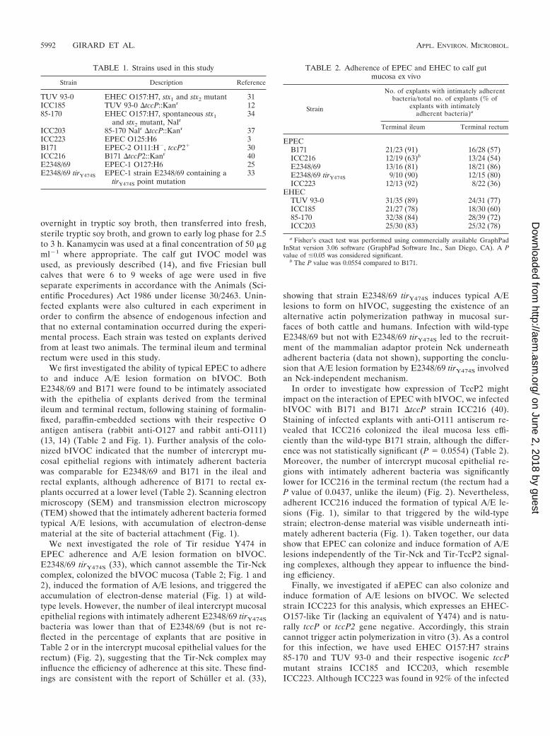
EPECB171 21/23 (91) 16/28 (57)ICC216 12/19 (63)b 13/24 (54)E2348/69 13/16 (81) 18/21 (86)E2348/69 tirY474S 9/10 (90) 12/15 (80)ICC223 12/13 (92) 8/22 (36)
EHECTUV 93-0 31/35 (89) 24/31 (77)ICC185 21/27 (78) 18/30 (60)85-170 32/38 (84) 28/39 (72)ICC203 25/30 (83) 25/32 (78)
a Fisher’s exact test was performed using commercially available GraphPadInStat version 3.06 software (GraphPad Software Inc., San Diego, CA). A Pvalue of �0.05 was considered significant.
b The P value was 0.0554 compared to B171.
TABLE 1. Strains used in this study
Strain Description Reference
TUV 93-0 EHEC O157:H7, stx1 and stx2 mutant 31ICC185 TUV 93-0 �tccP::Kanr 1285-170 EHEC O157:H7, spontaneous stx1
and stx2 mutant, Nalr34
ICC203 85-170 Nalr �tccP::Kanr 37ICC223 EPEC O125:H6 3B171 EPEC-2 O111:H�, tccP2� 30ICC216 B171 �tccP2::Kanr 40E2348/69 EPEC-1 O127:H6 25E2348/69 tirY474S EPEC-1 strain E2348/69 containing a
tirY474S point mutation33
5992 GIRARD ET AL. APPL. ENVIRON. MICROBIOL.
on June 2, 2018 by guesthttp://aem
.asm.org/
Dow
nloaded from
ileal bIVOC, it was found on only 36% of the rectal explants(Table 2), which is lower than that seen for EPEC or wild-type and tccP mutant EHEC strains (Table 2). However,analysis of the number of intercrypt mucosal epithelial re-gions with intimately adherent bacteria did not show a sig-
nificant difference between ICC223 and the EPEC andEHEC strains (Fig. 2 and 3). SEM and TEM analysis re-vealed that adherent ICC223 formed smaller foci of inti-mately adherent bacteria, left many bacterial footprints(Fig. 3), and was unable to trigger efficient accumulation of
FIG. 1. Adherence of EPEC strains E2348/69 (A) and B171 (B) to calf terminal ileal and rectal mucosa ex vivo. These results show that neitherTirY474S (A) nor TccP2 (B) is required for adherence (arrows in immunofluorescence assay [IFA]) and development (arrowheads in SEM andTEM) of typical A/E lesions ex vivo. Representative micrographs are shown. IFA, Hoechst 33342 (blue, false color) staining of nuclei and bacteria;tetramethyl rhodamine isothiocyanate (red, false color) staining of O127- and O111-positive bacteria. Bar � 20 �m (IFA), 5 �m (SEM), or0.5 �m (TEM).
VOL. 75, 2009 INTERACTION OF EPEC WITH CALF GUT EX VIVO 5993
on June 2, 2018 by guesthttp://aem
.asm.org/
Dow
nloaded from
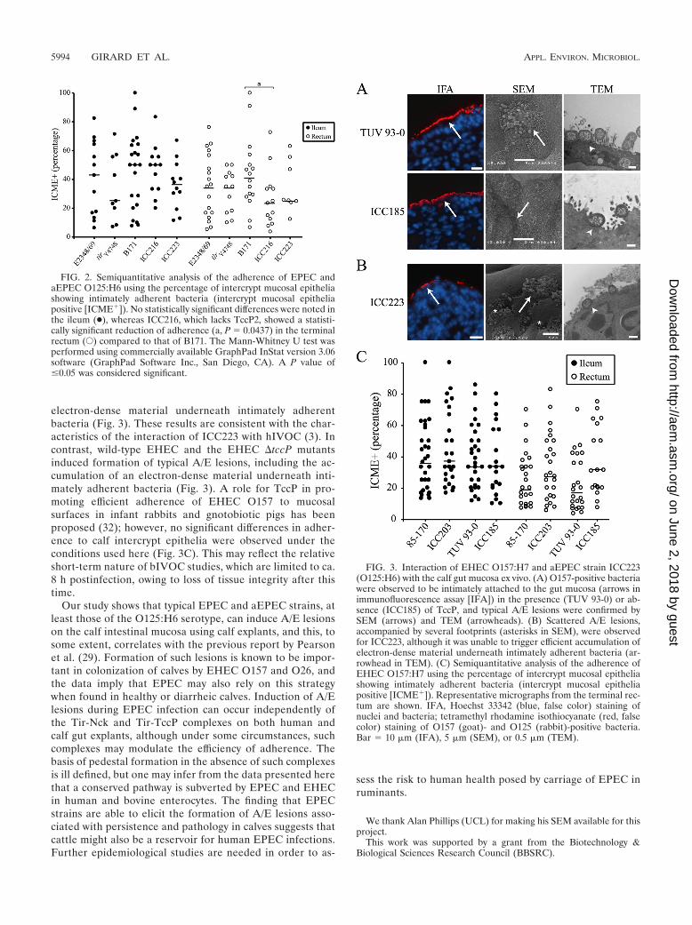
electron-dense material underneath intimately adherentbacteria (Fig. 3). These results are consistent with the char-acteristics of the interaction of ICC223 with hIVOC (3). Incontrast, wild-type EHEC and the EHEC �tccP mutantsinduced formation of typical A/E lesions, including the ac-cumulation of an electron-dense material underneath inti-mately adherent bacteria (Fig. 3). A role for TccP in pro-moting efficient adherence of EHEC O157 to mucosalsurfaces in infant rabbits and gnotobiotic pigs has beenproposed (32); however, no significant differences in adher-ence to calf intercrypt epithelia were observed under theconditions used here (Fig. 3C). This may reflect the relativeshort-term nature of bIVOC studies, which are limited to ca.8 h postinfection, owing to loss of tissue integrity after thistime.
Our study shows that typical EPEC and aEPEC strains, atleast those of the O125:H6 serotype, can induce A/E lesionson the calf intestinal mucosa using calf explants, and this, tosome extent, correlates with the previous report by Pearsonet al. (29). Formation of such lesions is known to be impor-tant in colonization of calves by EHEC O157 and O26, andthe data imply that EPEC may also rely on this strategywhen found in healthy or diarrheic calves. Induction of A/Elesions during EPEC infection can occur independently ofthe Tir-Nck and Tir-TccP complexes on both human andcalf gut explants, although under some circumstances, suchcomplexes may modulate the efficiency of adherence. Thebasis of pedestal formation in the absence of such complexesis ill defined, but one may infer from the data presented herethat a conserved pathway is subverted by EPEC and EHECin human and bovine enterocytes. The finding that EPECstrains are able to elicit the formation of A/E lesions asso-ciated with persistence and pathology in calves suggests thatcattle might also be a reservoir for human EPEC infections.Further epidemiological studies are needed in order to as-
sess the risk to human health posed by carriage of EPEC inruminants.
We thank Alan Phillips (UCL) for making his SEM available for thisproject.
This work was supported by a grant from the Biotechnology &Biological Sciences Research Council (BBSRC).
FIG. 2. Semiquantitative analysis of the adherence of EPEC andaEPEC O125:H6 using the percentage of intercrypt mucosal epitheliashowing intimately adherent bacteria (intercrypt mucosal epitheliapositive [ICME�]). No statistically significant differences were noted inthe ileum (●), whereas ICC216, which lacks TccP2, showed a statisti-cally significant reduction of adherence (a, P � 0.0437) in the terminalrectum (E) compared to that of B171. The Mann-Whitney U test wasperformed using commercially available GraphPad InStat version 3.06software (GraphPad Software Inc., San Diego, CA). A P value of�0.05 was considered significant.
FIG. 3. Interaction of EHEC O157:H7 and aEPEC strain ICC223(O125:H6) with the calf gut mucosa ex vivo. (A) O157-positive bacteriawere observed to be intimately attached to the gut mucosa (arrows inimmunofluorescence assay [IFA]) in the presence (TUV 93-0) or ab-sence (ICC185) of TccP, and typical A/E lesions were confirmed bySEM (arrows) and TEM (arrowheads). (B) Scattered A/E lesions,accompanied by several footprints (asterisks in SEM), were observedfor ICC223, although it was unable to trigger efficient accumulation ofelectron-dense material underneath intimately adherent bacteria (ar-rowhead in TEM). (C) Semiquantitative analysis of the adherence ofEHEC O157:H7 using the percentage of intercrypt mucosal epitheliashowing intimately adherent bacteria (intercrypt mucosal epitheliapositive [ICME�]). Representative micrographs from the terminal rec-tum are shown. IFA, Hoechst 33342 (blue, false color) staining ofnuclei and bacteria; tetramethyl rhodamine isothiocyanate (red, falsecolor) staining of O157 (goat)- and O125 (rabbit)-positive bacteria.Bar � 10 �m (IFA), 5 �m (SEM), or 0.5 �m (TEM).
5994 GIRARD ET AL. APPL. ENVIRON. MICROBIOL.
on June 2, 2018 by guesthttp://aem
.asm.org/
Dow
nloaded from
REFERENCES
1. Aidar-Ugrinovich, L., J. Blanco, M. Blanco, J. E. Blanco, L. Leomil, G.Dahbi, A. Mora, D. L. Onuma, W. D. Silveira, and A. F. Pestana de Castro.2007. Serotypes, virulence genes, and intimin types of Shiga toxin-producingEscherichia coli (STEC) and enteropathogenic E. coli (EPEC) isolated fromcalves in Sao Paulo, Brazil. Int. J. Food Microbiol. 115:297–306.
2. Aktan, I., K. A. Sprigings, R. M. La Ragione, L. M. Faulkner, G. A. Paiba,and M. J. Woodward. 2004. Characterisation of attaching-effacing Esche-richia coli isolated from animals at slaughter in England and Wales. Vet.Microbiol. 102:43–53.
3. Bai, L., S. Schuller, A. Whale, A. Mousnier, O. Marches, L. Wang, T. Ooka,R. Heuschkel, F. Torrente, J. B. Kaper, T. A. Gomes, J. Xu, A. D. Phillips,and G. Frankel. 2008. Enteropathogenic Escherichia coli O125:H6 triggersattaching and effacing lesions on human intestinal biopsy specimens inde-pendently of Nck and TccP/TccP2. Infect. Immun. 76:361–368.
4. Brady, M. J., K. G. Campellone, M. Ghildiyal, and J. M. Leong. 2007.Enterohaemorrhagic and enteropathogenic Escherichia coli Tir proteins trig-ger a common Nck-independent actin assembly pathway. Cell. Microbiol.9:2242–2253.
5. Campellone, K. G., and J. M. Leong. 2005. Nck-independent actin assemblyis mediated by two phosphorylated tyrosines within enteropathogenic Esch-erichia coli Tir. Mol. Microbiol. 56:416–432.
6. Campellone, K. G., D. Robbins, and J. M. Leong. 2004. EspFU is a translo-cated EHEC effector that interacts with Tir and N-WASP and promotesNck-independent actin assembly. Dev. Cell 7:217–228.
7. Caron, E., V. F. Crepin, N. Simpson, S. Knutton, J. Garmendia, and G.Frankel. 2006. Subversion of actin dynamics by EPEC and EHEC. Curr.Opin. Microbiol. 9:40–45.
8. Frankel, G., and A. D. Phillips. 2008. Attaching effacing Escherichia coli andparadigms of Tir-triggered actin polymerisation: getting off the pedestal.Cell. Microbiol. 10:549–556.
9. Frankel, G., A. D. Phillips, I. Rosenshine, G. Dougan, J. B. Kaper, and S.Knutton. 1998. Enteropathogenic and enterohemorrhagic Escherichia coli:more subversive elements. Mol. Microbiol. 30:911–921.
10. Frankel, G., A. D. Phillips, L. R. Trabulsi, S. Knutton, G. Dougan, and S.Matthews. 2001. Intimin and the host cell-is it bound to end in Tir(s)? TrendsMicrobiol. 9:214–218.
11. Garmendia, J., G. Frankel, and V. F. Crepin. 2005. Enteropathogenic andenterohemorrhagic Escherichia coli infections: translocation, translocation,translocation. Infect. Immun. 73:2573–2585.
12. Garmendia, J., A. D. Phillips, M. F. Carlier, Y. Chong, S. Schuller, O.Marches, S. Dahan, E. Oswald, R. K. Shaw, S. Knutton, and G. Frankel.2004. TccP is an enterohaemorrhagic Escherichia coli O157:H7 type IIIeffector protein that couples Tir to the actin-cytoskeleton. Cell. Microbiol.6:1167–1183.
13. Girard, F., V. F. Crepin, and G. Frankel. 2009. Modelling EPEC-2 andEPEC-4 infection ex vivo and in vivo using Citrobacter rodentium expressingTccP. Infect. Immun. 77:1304–1314.
14. Girard, F., F. Dziva, P. van Diemen, A. D. Phillips, M. P. Stevens, and G.Frankel. 2007. Adherence of enterohemorrhagic Escherichia coli O157, O26,and O111 strains to bovine intestinal explants ex vivo. Appl. Environ. Mi-crobiol. 73:3084–3090.
15. Giron, J. A., A. S. Ho, and G. K. Schoolnik. 1991. An inducible bundle-forming pilus of enteropathogenic Escherichia coli. Science 254:710–713.
16. Hartland, E. L., M. Batchelor, R. M. Delahay, C. Hale, S. Matthews, G.Dougan, S. Knutton, I. Connerton, and G. Frankel. 1999. Binding of intiminfrom enteropathogenic Escherichia coli to Tir and to host cells. Mol. Micro-biol. 32:151–158.
17. Hornitzky, M. A., K. Mercieca, K. A. Bettelheim, and S. P. Djordjevic. 2005.Bovine feces from animals with gastrointestinal infections are a source ofserologically diverse atypical enteropathogenic Escherichia coli and Shigatoxin-producing E. coli strains that commonly possess intimin. Appl. Envi-ron. Microbiol. 71:3405–3412.
18. Ishii, S., K. P. Meyer, and M. J. Sadowsky. 2007. Relationship betweenphylogenetic groups, genotypic clusters, and virulence gene profiles of Esch-erichia coli strains from diverse human and animal sources. Appl. Environ.Microbiol. 73:5703–5710.
19. Jarvis, K. G., and J. B. Kaper. 1996. Secretion of extracellular proteins byenterohemorrhagic Escherichia coli via a putative type III secretion system.Infect. Immun. 64:4826–4829.
20. Jerse, A. E., J. Yu, B. D. Tall, and J. B. Kaper. 1990. A genetic locus ofenteropathogenic Escherichia coli necessary for the production of attachingand effacing lesions on tissue culture cells. Proc. Natl. Acad. Sci. USA87:7839–7843.
21. Kenny, B. 1999. Phosphorylation of tyrosine 474 of the enteropathogenicEscherichia coli (EPEC) Tir receptor molecule is essential for actin nucle-ating activity and is preceded by additional host modifications. Mol. Micro-biol. 31:1229–1241.
22. Kenny, B., R. DeVinney, M. Stein, D. J. Reinscheid, E. A. Frey, and B. B.Finlay. 1997. Enteropathogenic Escherichia coli (EPEC) transfers its recep-tor for intimate adherence into mammalian cells. Cell 91:511–520.
23. Knutton, S., D. R. Lloyd, and A. S. McNeish. 1987. Adhesion of entero-pathogenic Escherichia coli to human intestinal enterocytes and culturedhuman intestinal mucosa. Infect. Immun. 55:69–77.
24. Lacher, D. W., H. Steinsland, T. E. Blank, M. S. Donnenberg, and T. S.Whittam. 2007. Molecular evolution of typical enteropathogenic Escherichiacoli: clonal analysis by multilocus sequence typing and virulence gene allelicprofiling. J. Bacteriol. 189:342–350.
25. Levine, M. M., J. P. Nataro, H. Karch, M. M. Baldini, J. B. Kaper, R. E.Black, M. L. Clements, and A. D. O’Brien. 1985. The diarrheal response ofhumans to some classic serotypes of enteropathogenic Escherichia coli isdependent on a plasmid encoding an enteroadhesiveness factor. J. Infect.Dis. 152:550–559.
26. Mainil, J. G., E. R. Jacquemin, A. E. Kaeckenbeeck, and P. H. Pohl. 1993.Association between the effacing (eae) gene and the Shiga-like toxin-encod-ing genes in Escherichia coli isolates from cattle. Am. J. Vet. Res. 54:1064–1068.
27. McDaniel, T. K., K. G. Jarvis, M. S. Donnenberg, and J. B. Kaper. 1995. Agenetic locus of enterocyte effacement conserved among diverse enterobac-terial pathogens. Proc. Natl. Acad. Sci. USA 92:1664–1668.
28. Nataro, J. P., and J. B. Kaper. 1998. Diarrheagenic Escherichia coli. Clin.Microbiol. Rev. 11:142–201.
29. Pearson, G. R., C. A. Watson, G. A. Hall, and C. Wray. 1989. Naturalinfection with an attaching and effacing Escherichia coli in the small and largeintestines of a calf with diarrhoea. Vet. Rec. 124:297–299.
30. Riley, L. W., L. N. Junio, L. B. Libaek, and G. K. Schoolnik. 1987. Plasmid-encoded expression of lipopolysaccharide O-antigenic polysaccharide in en-teropathogenic Escherichia coli. Infect. Immun. 55:2052–2056.
31. Riley, L. W., R. S. Remis, S. D. Helgerson, H. B. McGee, J. G. Wells, B. R.Davis, R. J. Hebert, E. S. Olcott, L. M. Johnson, N. T. Hargrett, P. A. Blake,and M. L. Cohen. 1983. Hemorrhagic colitis associated with a rare Esche-richia coli serotype. N. Engl. J. Med. 308:681–685.
32. Ritchie, J. M., M. J. Brady, K. N. Riley, T. D. Ho, K. G. Campellone, I. M.Herman, A. Donohue-Rolfe, S. Tzipori, M. K. Waldor, and J. M. Leong.2008. EspFu, a type III-translocated effector of actin assembly, fosters epi-thelial association and late-stage intestinal colonization by E. coli O157:H7.Cell. Microbiol. 10:836–847.
33. Schuller, S., Y. Chong, J. Lewin, B. Kenny, G. Frankel, and A. D. Phillips.2007. Tir phosphorylation and Nck/N-WASP recruitment by enteropatho-genic and enterohaemorrhagic Escherichia coli during ex vivo colonization ofhuman intestinal mucosa is different to cell culture models. Cell. Microbiol.9:1352–1364.
34. Stevens, M. P., A. J. Roe, I. Vlisidou, P. M. van Diemen, R. M. La Ragione,A. Best, M. J. Woodward, D. L. Gally, and T. S. Wallis. 2004. Mutation oftoxB and a truncated version of the efa-1 gene in Escherichia coli O157:H7influences the expression and secretion of locus of enterocyte effacement-encoded proteins but not intestinal colonization in calves or sheep. Infect.Immun. 72:5402–5411.
35. van Diemen, P. M., F. Dziva, M. P. Stevens, and T. S. Wallis. 2005. Identi-fication of enterohemorrhagic Escherichia coli O26:H- genes required forintestinal colonization in calves. Infect. Immun. 73:1735–1743.
36. Vingadassalom, D., A. Kazlauskas, B. Skehan, H. C. Cheng, L. Magoun, D.Robbins, M. K. Rosen, K. Saksela, and J. M. Leong. 2009. Insulin receptortyrosine kinase substrate links the E. coli O157:H7 actin assembly effectorsTir and EspF(U) during pedestal formation. Proc. Natl. Acad. Sci. USA106:6754–6759.
37. Vlisidou, I., F. Dziva, R. M. La Ragione, A. Best, J. Garmendia, P. Hawes, P.Monaghan, S. A. Cawthraw, G. Frankel, M. J. Woodward, and M. P. Stevens.2006. Role of intimin-Tir interactions and the Tir-cytoskeleton couplingprotein in the colonization of calves and lambs by Escherichia coli O157:H7.Infect. Immun. 74:758–764.
38. Wani, S. A., I. Hussain, A. Nabi, I. Fayaz, and Y. Nishikawa. 2007. Variantsof eae and stx genes of atypical enteropathogenic Escherichia coli and non-O157 Shiga toxin-producing Escherichia coli from calves. Lett. Appl. Micro-biol. 45:610–615.
39. Weiss, S. M., M. Ladwein, D. Schmidt, J. Ehinger, S. Lommel, K. Stading,U. Beutling, A. Disanza, R. Frank, L. Jansch, G. Scita, F. Gunzer, K.Rottner, and T. E. Stradal. 2009. IRSp53 links the enterohemorrhagic E. colieffectors Tir and EspFU for actin pedestal formation. Cell Host Microbe5:244–258.
40. Whale, A. D., R. T. Hernandes, T. Ooka, L. Beutin, S. Schuller, J. Garmen-dia, L. Crowther, M. A. Vieira, Y. Ogura, G. Krause, A. D. Phillips, T. A.Gomes, T. Hayashi, and G. Frankel. 2007. TccP2-mediated subversion ofactin dynamics by EPEC 2—a distinct evolutionary lineage of enteropatho-genic Escherichia coli. Microbiology 153:1743–1755.
VOL. 75, 2009 INTERACTION OF EPEC WITH CALF GUT EX VIVO 5995
on June 2, 2018 by guesthttp://aem
.asm.org/
Dow
nloaded from