Embed Size (px)
Citation preview
BIOCHEMICAL AND BIOPHYSICAL RESEARCH COMMUNICATIONS 235, 83–88 (1997)ARTICLE NO. RC976741
Interaction of p59fyn with Interferon-Activated Jak Kinases
Shahab Uddin,* Dorie A. Sher,* Yazan Alsayed,* Sebastian Pons,† Oscar R. Colamonici,‡Eleanor N. Fish,§ Morris F. White,† and Leonidas C. Platanias*,1
*Section of Hematology-Oncology, Department of Medicine, University of Illinois at Chicago and West Side VeteransAffairs Hospital, Chicago, Illinois 60607; †Research Division, Joslin Diabetes Center, Harvard Medical School,Boston, Massachusetts 02215; ‡Department of Pathology, University of Tennessee, Memphis, Tennessee 38163; and§Department of Medical Genetics and Microbiology, University of Toronto, Toronto, Ontario M5S 3E2, Canada
Received May 5, 1997
and C-cbl (17) proto-oncogenes. Furthermore, engage-During IFNa stimulation, p59fyn associates with the ment of Jak kinases during IFNa stimulation appears
Type I IFNR-associated Tyk-2 kinase in several human to regulate activation of the lipid (14) and serine (18)hematopoietic cell lines in vivo. This interaction is di- kinase activities of the phosphatidylinositol 3 *-kinase.rect, and is mediated by the SH2 domain in p59fyn, as On the other hand, binding of IFNg to the Type II IFNshown by binding studies using glutathione-S-tranfer- receptor results in activation of the Jak-2 and Jak-ase fusion proteins and far western blots. Further-
1 kinases (3,4,19) and tyrosine phosphorylation andmore, in response to IFNa-treatment of cells, the SH2homodimerization of Stat-1 (reviewed in 13).domain of Fyn interacts with the Tyk-2-associated c-
Although the involvement of Jak kinases in inter-cbl proto-oncogene product. In a similar manner, dur-feron signaling is well documented, the role of kinasesing IFNg stimulation, p59fyn associates via its SH2 do-of other families is unknown. In the present study wemain with the activated form of the IFNg-dependentsought to determine whether the Src-related tyrosineJak-2 kinase. These data suggest that p59fyn is a com-kinase Fyn is engaged in interferon-signaling. This ki-mon element in IFNa and IFNg signaling, and is selec-nase has been previously implicated in signal transduc-tively engaged by the Type I or II IFN receptors viation by various receptors, including the PDGF, GSF-1,specific interactions with dis-tinct Jak kinases. q 1997
Academic Press and B and T cell-antigen receptors (20-23). Our datademonstrate that p59fyn interacts via its SH2 domainwith the activated forms of Tyk-2 or Jak-2, in responseto IFNa or IFNg stimulation respectively, suggesting
Interferons (IFNs) are pleiotropic cytokines that ex- that this kinase may be involved in the regulation ofert antiproliferative, antiviral and immunomodulatory interferon-signaling pathways downstream of Jaks.activities on normal and neoplastic cells (1). Althoughthe precise mechanisms by which these cytokines exert
EXPERIMENTAL PROCEDUREStheir biological effects remain unknown, significant ad-vances have been recently made in our understanding
Cells and reagents. The Molt-16 (acute T-cell lymphocytic leuke-of the signaling events that take place during binding mia), Molt-4 (acute T-cell lymphocytic leukemia), Daudi (lymphoblas-of interferons to their receptors. Binding of IFNa to the toid), and U-266 (multiple myeloma) cell lines were grown in RPMIType I IFN receptor results in activation of the Janus 1640 (Life Technologies, Inc.) supplemented with 10% (v/v) fetal bo-
vine serum (Life Technologies, Inc.) or 10% (v/v) defined calf serumfamily Tyk-2 and Jak-1 kinases (2-6), and tyrosine(Hyclone Laboratories, Logan, UT) and antibiotics. Human recombi-phosphorylation of various signaling elements, includ-nant IFNa2 was provided by Hoffmann Laroche. Human recombi-ing the a and b subunits of the Type I IFNR (7-9), Stat- nant IFNcon1 (IFNa) was provided by Amgen Inc. Human recombi-
proteins (10-13), IRS-proteins (14,15), and the Vav (16) nant IFNg was provided by Genentech Inc. The antiphosphotyrosinemonoclonal antibody (4G-10) and the anti-Jak-2 polyclonal antibodywere obtained from Upstate Biotechnology (Lake Placid, NY). Thepolyclonal anti-Tyk-2 antibody has been raised against a synthetic1 Corresponding author at Section of Hematology-Oncology, The
University of Illinois at Chicago, MBRB, MC-734, Rm. 3150, 900 S. peptide corresponding to the C-terminal 15 aminoacids of Tyk-2(8,24). The monoclonal antibody against Tyk-2 was obtained fromAshland Ave, Chicago, IL 60607-7173. Fax:(312)413-7963, E-
mail:[email protected]. Transduction Laboratories (Lexin-gton, KY) and was used for immu-noblotting. The polyclonal anti-body against the tyrosine kinase FynAbbreviations: ISGs, interferon-stimulated genes; PAGE, poly-
acrylamide gel electrophoresis; IFNR, interferon receptor; Stat, sig- has been described elsewhere (25). A polyclonal antibody against C-cbl was obtained from Santa Cruz Biotechnology (Santa Cruz, CA).nal transducer and activator of transcription.
0006-291X/97 $25.00Copyright q 1997 by Academic PressAll rights of reproduction in any form reserved.
83
AID BBRC 6741 / 692e$$$401 05-21-97 09:32:23 bbrcg AP: BBRC
Vol. 235, No. 1, 1997 BIOCHEMICAL AND BIOPHYSICAL RESEARCH COMMUNICATIONS
Immunoprecipitations and immunoblotting. Cells were stimu-lated with the indicated IFNs (104 U/ml) for the indicated time peri-ods. In some experiments the cells were serum starved by incubationin serum-free DMEM (Life Technologies Inc.) for 2 hours prior tointerferon stimulation. After stimulation, the cells were rapidly cen-trifuged and lysed as previously described (26). In some experimentsthe cell lysates were pre-cleared with nonimmune rabbit serum priorto immunoprecipitation. Immunopreci-pitations and immunoblotingwere performed as previously descri-bed (26).
Preparation of glutathione-S-transferase fusion proteins and bind-ing studies. The generation of the pGEX-FynSH2 and pGEX-FynSH3 constructs has been described elsewhere (25). Production ofglutathione-S-transferase fusion proteins and binding experimentsusing lysates from cells stimulated with 104 U/ml of IFNa or IFNgwere performed as previously described (14,27).
Far western blots. Cells were treated in the presence or absenceof IFNa as indicated, cell lysates were immunoprecipitated with theindicated antibodies, analyzed by SDS-PAGE and transferred toPVDF membranes as described in the procedure for immunoblotting.The membranes were subsequently incubated for 1 hr at room tem-perature with approximately 2-4 mg of the FynSH2 fusion protein in10 ml TBST-0.5% BSA, washed extensively with TBST, and thenincubated with a polyclonal anti-GST antibody (Santa Cruz Biotech-nology), prior to developing by the ECL method.
RESULTS AND DISCUSSION
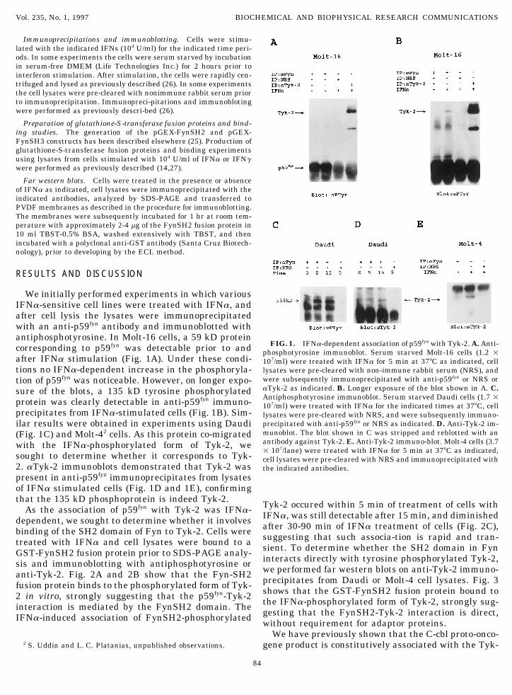
We initially performed experiments in which variousIFNa-sensitive cell lines were treated with IFNa, andafter cell lysis the lysates were immunoprecipitatedwith an anti-p59fyn antibody and immunoblotted withantiphosphotyrosine. In Molt-16 cells, a 59 kD protein
FIG. 1. IFNa-dependent association of p59fyn with Tyk-2. A. Anti-corresponding to p59fyn was detectable prior to and phosphotyrosine immunoblot. Serum starved Molt-16 cells (1.2 1after IFNa stimulation (Fig. 1A). Under these condi- 107/ml) were treated with IFNa for 5 min at 377C as indicated, celltions no IFNa-dependent increase in the phosphoryla- lysates were pre-cleared with non-immune rabbit serum (NRS), and
were subsequently immunoprecipitated with anti-p59fyn or NRS ortion of p59fyn was noticeable. However, on longer expo-aTyk-2 as indicated. B. Longer exposure of the blot shown in A. C.sure of the blots, a 135 kD tyrosine phosphorylatedAntiphosphotyrosine immunoblot. Serum starved Daudi cells (1.7 1protein was clearly detectable in anti-p59fyn immuno- 107/ml) were treated with IFNa for the indicated times at 377C, cell
precipitates from IFNa-stimulated cells (Fig. 1B). Sim- lysates were pre-cleared with NRS, and were subsequently immuno-precipitated with anti-p59fyn or NRS as indicated. D. Anti-Tyk-2 im-ilar results were obtained in experiments using Daudimunoblot. The blot shown in C was stripped and reblotted with an(Fig. 1C) and Molt-42 cells. As this protein co-migratedantibody against Tyk-2. E. Anti-Tyk-2 immuno-blot. Molt-4 cells (3.7with the IFNa-phosphorylated form of Tyk-2, we 1 107/lane) were treated with IFNa for 5 min at 377C as indicated,
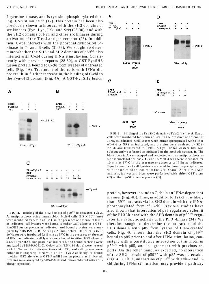
sought to determine whether it corresponds to Tyk- cell lysates were pre-cleared with NRS and immunoprecipitated with2. aTyk-2 immunoblots demonstrated that Tyk-2 was the indicated antibodies.present in anti-p59fyn immunoprecipitates from lysatesof IFNa stimulated cells (Fig. 1D and 1E), confirmingthat the 135 kD phosphoprotein is indeed Tyk-2. Tyk-2 occured within 5 min of treatment of cells withAs the association of p59fyn with Tyk-2 was IFNa- IFNa, was still detectable after 15 min, and diminisheddependent, we sought to determine whether it involves after 30-90 min of IFNa treatment of cells (Fig. 2C),binding of the SH2 domain of Fyn to Tyk-2. Cells were suggesting that such associa-tion is rapid and tran-treated with IFNa and cell lysates were bound to a sient. To determine whether the SH2 domain in FynGST-FynSH2 fusion protein prior to SDS-PAGE analy- interacts directly with tyrosine phosphorylated Tyk-2,sis and immunoblotting with antiphosphotyrosine or we performed far western blots on anti-Tyk-2 immuno-anti-Tyk-2. Fig. 2A and 2B show that the Fyn-SH2 precipitates from Daudi or Molt-4 cell lysates. Fig. 3fusion protein binds to the phosphorylated form of Tyk- shows that the GST-FynSH2 fusion protein bound to2 in vitro, strongly suggesting that the p59fyn-Tyk-2 the IFNa-phosphorylated form of Tyk-2, strongly sug-interaction is mediated by the FynSH2 domain. The gesting that the FynSH2-Tyk-2 interaction is direct,IFNa-induced association of FynSH2-phosphorylated without requirement for adaptor proteins.
We have previously shown that the C-cbl proto-onco-gene product is constitutively associated with the Tyk-2 S. Uddin and L. C. Platanias, unpublished observations.
84
AID BBRC 6741 / 692e$$$401 05-21-97 09:32:23 bbrcg AP: BBRC
Vol. 235, No. 1, 1997 BIOCHEMICAL AND BIOPHYSICAL RESEARCH COMMUNICATIONS
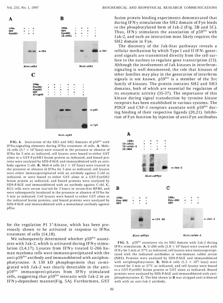
2 tyrosine kinase, and is tyrosine phosphorylated dur-ing IFNa stimulation (17). This protein has been alsopreviously shown to interact with the SH3 domains ofsrc kinases (Fyn, Lyn, Lck, and Src) (28-30), and withthe SH2 domains of Fyn and other src kinases duringactivation of the T-cell antigen receptor (28). In addi-tion, C-cbl interacts with the phosphatidylinositol 3 *-kinase in T- and B-cells (31-33). We sought to deter-mine whether the SH3 and SH2 domains of p59fyn alsointeract with C-cbl during IFNa stimula-tion. Consis-tently with previous reports (28-30), a GST-FynSH3fusion protein bound to C-cbl from lysates of untreatedcells (Fig. 4A). Treatment of the cells with IFNa didnot result in further increase in the binding of C-cbl tothe Fyn-SH3 domain (Fig. 4A). A GST-FynSH2 fusion
FIG. 3. Binding of the FynSH2 domain to Tyk-2 in vitro. A. Daudicells were incubated for 5 min at 377C in the presence or absence ofIFNa as indicated. Cell lysates were immunoprecipitated with eitheraTyk-2 or NRS as indicated, and proteins were analyzed by SDS-PAGE and transferred to PVDF. A FynSH2 far western blot wassubsequently performed as indicated in the methods section. B. Theblot shown in A was stripped and re-blotted with an antiphosphotyro-sine monoclonal antibody. C. and D. Molt-4 cells were incubated for10 min at 377 C in the presence or abscence of IFNa as indicated.Equal amounts of cell lysates were used for immunoprecipitationswith the indicated antibodies for the C or D panel. After SDS-PAGEanalysis, far western blots were performed with either GST alone(C) or the FynSH2 fusion protein (D).
protein, however, bound to C-cbl in an IFNa-dependentmanner (Fig. 4B). Thus, in addition to Tyk-2, it is likelythat p59fyn interacts via its SH2 domain with the IFNa-phosphorylated form of C-cbl. Previous studies havealso shown that interaction of p85 regulatory subunit
FIG. 2. Binding of the SH2 domain of p59fyn to activated Tyk-2.of the PI 3 *-kinase with the SH3 domain of p59fyn regu-A. Antiphosphotyrosine immunoblot. Molt-4 cells (1.5 1 108/ lane)lates the catalytic activity of the PI 3 *-kinase (34). Wewere incubated for 5 min at 377 C in the presence or absence of IFNa
as indicated, cell lysates were bound to either GST alone or a GST- therefore sought to determine the interaction of theFynSH2 fusion protein as indicated, and bound proteins were ana- SH3 domain with p85 from lysates of IFNa-treatedlyzed by SDS-PAGE. B. Anti-Tyk-2 immunoblot. Daudi cells (5 1 cells. Fig. 4C shows that the SH3 domain of p59fyn107/lane) were incubated for 5 min at 377C in the presence or absence
bound to p85 prior to and after IFNa stimulation, con-of IFNa as indicated, cell lysates were bound to either GST alone orsistent with a constitutive interaction of this motif ina GST-FynSH2 fusion protein as indicated, and bound proteins were
analyzed by SDS-PAGE. C. Molt-4 cells (3.51 107/lane) were treated p59fyn with p85, and in agreement with previous re-with IFNa for the indicated times at 377C, and cell lysates were ports. On the other hand, as expected, no interactioneither immunoprecipitated with an anti-Tyk-2 antibody, or bound of the SH2 domain of p59fyn with p85 was detectableto either GST alone or a GST-FynSH2 fusion protein as indicated.
(Fig. 4C). Thus, interaction of p59fyn with Tyk-2 and C-Proteins were analyzed by SDS-PAGE and immunoblotted with anti-phosphotyrosine. cbl during IFNa stimulation, may provide a pathway
85
AID BBRC 6741 / 692e$$$401 05-21-97 09:32:23 bbrcg AP: BBRC
Vol. 235, No. 1, 1997 BIOCHEMICAL AND BIOPHYSICAL RESEARCH COMMUNICATIONS
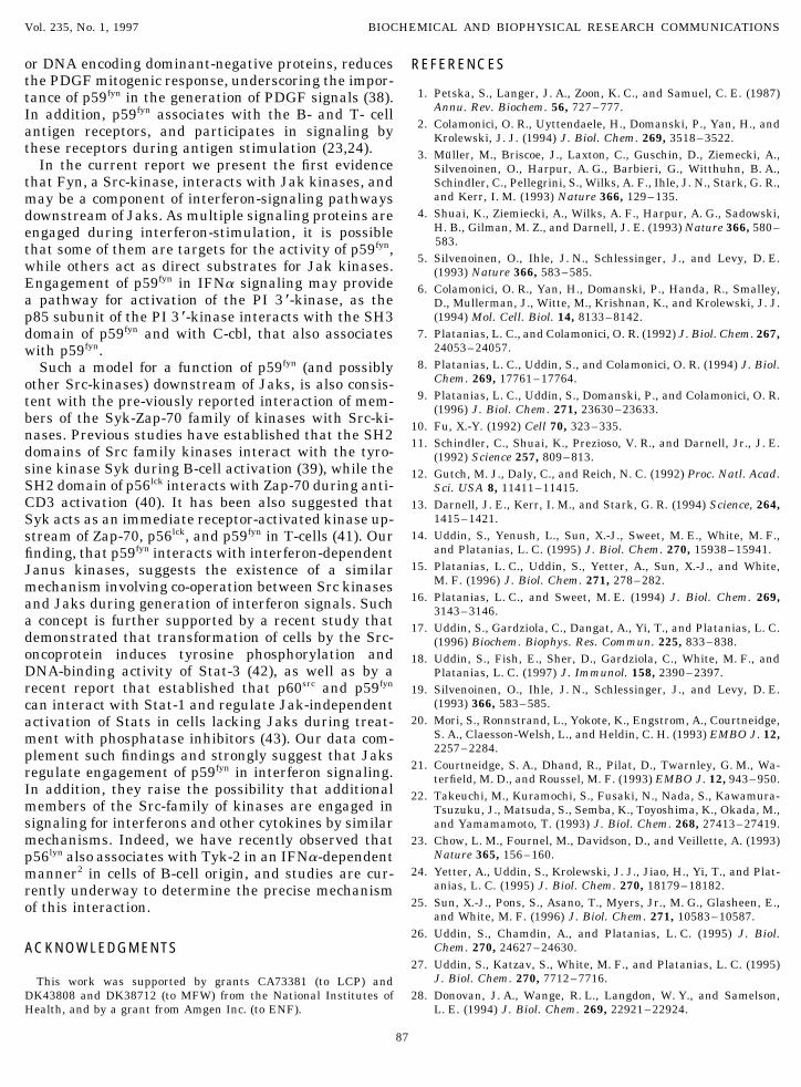
fusion protein binding experiments demonstrated thatduring IFNg stimulation the SH2 domain of Fyn bindsto the phosphorylated form of Jak-2 (Fig. 5B and 5C).Thus, IFNg stimulates the association of p59fyn withJak-2, and such an interaction most likely requires theSH2 domain in Fyn.
The discovery of the Jak-Stat pathways reveals acellular mechanism by which Type I and II IFN- gener-ated signals are transmitted directly from the cell sur-face to the nucleus to regulate gene transcription (13).Although the involvement of Jak kinases in interferon-signaling is well documented, the role that kinases ofother families may play in the generation of interferonsignals is not known. p59fyn is a member of the Srcfamily of kinases. The protein contains SH2 and SH3domains, both of which are essential for regulation ofits enzymatic activity (35-37). The importance of thiskinase during signal transduction by tyrosine kinasereceptors has been established in various systems. ThePDGF and CSF-1 receptors associate with p59fyn dur-ing binding of their respective ligands (20,21). Inhibi-tion of Fyn function by injection of anti-Fyn antibodies
FIG. 4. Interaction of the SH3 and SH2 domains of p59fyn withIFNa-signaling elements during IFNa treatment of cells. A. Molt-16 cells (3.7 1 107/lane) were treated in the presence or absence ofIFNa for 5 min as indicated, cell lysates were bound to either GSTalone or a GST-FynSH3 fusion protein as indicated, and bound pro-teins were analyzed by SDS-PAGE and immunoblotted with an anti-body against C-cbl. B. Molt-4 cells (4.1 1 107/lane) were treated inthe presence or absence of IFNa for 5 min as indicated, cell lysateswere either immunoprecipitated with an antibody against C-cbl asindicated, or were bound to either GST alone or a GST-FynSH2fusion protein as indicated, and bound proteins were analyzed bySDS-PAGE and immunoblotted with an antibody against C-cbl. C.KG1 cells were serum starved for 3 hours in serum-free RPMI, andwere subsequently incubated in the presence or absence of IFNa for5 min as indicated. Cell lysates were bound to either GST alone orthe indicated fusion proteins, and bound proteins were analyzed bySDS-PAGE and immunoblotted with a monoclonal antibody againstp85a.
for the regulation PI 3 *-kinase, which has been pre-viously shown to be activated in response to IFNatreatment of cells (14,18).
We subsequently determined whether p59fyn associ-FIG. 5. p59fyn associates via its SH2 domain with Jak-2 duringates with Jak-2, which is activated during IFNg stimu-
IFNg stimulation. A. U-266 cells (1.8 1 107/lane) were treated withlation (3,4,17). Lysates from IFNg treated U-266 hu- IFNg for 5 min at 377C as indicated, cell lysates were immunoprecipi-man myeloma cells were immuno-precipitated with the tated with the indicated antibodies or non-immune rabbit serumanti-p59fyn antibody and immunoblotted with antiphos- (NRS). Proteins were analyzed by SDS-PAGE and immunoblotted
with antiphosphotyrosine. B. Molt-4 cells (1.3 1 108/ lane) werephotyrosine. A 130 kD phosphoprotein that co-mi-treated for 3 min at 377C as indicated, and cell lysates were boundgrated with Jak-2 was clearly detectable in the anti-to a GST-FynSH2 fusion protein or GST alone as indicated. Boundp59fyn immunopreci-pitates from IFNg stimulated proteins were analyzed by SDS-PAGE and immunoblotted with anti-
cells, suggesting that p59fyn interacts with Jak-2 in an phosphotyrosine. C. The blot shown in B was stripped and re-blottedwith with an anti-Jak-2 antibody.IFNg-dependent manner(Fig. 5A). Furthermore, GST
86
AID BBRC 6741 / 692e$$$401 05-21-97 09:32:23 bbrcg AP: BBRC
Vol. 235, No. 1, 1997 BIOCHEMICAL AND BIOPHYSICAL RESEARCH COMMUNICATIONS
or DNA encoding dominant-negative proteins, reduces REFERENCESthe PDGF mitogenic response, underscoring the impor-
1. Petska, S., Langer, J. A., Zoon, K. C., and Samuel, C. E. (1987)tance of p59fyn in the generation of PDGF signals (38).Annu. Rev. Biochem. 56, 727–777.In addition, p59fyn associates with the B- and T- cell
2. Colamonici, O. R., Uyttendaele, H., Domanski, P., Yan, H., andantigen receptors, and participates in signaling by Krolewski, J. J. (1994) J. Biol. Chem. 269, 3518–3522.these receptors during antigen stimulation (23,24). 3. Muller, M., Briscoe, J., Laxton, C., Guschin, D., Ziemecki, A.,
In the current report we present the first evidence Silvenoinen, O., Harpur, A. G., Barbieri, G., Witthuhn, B. A.,Schindler, C., Pellegrini, S., Wilks, A. F., Ihle, J. N., Stark, G. R.,that Fyn, a Src-kinase, interacts with Jak kinases, andand Kerr, I. M. (1993) Nature 366, 129–135.may be a component of interferon-signaling pathways
4. Shuai, K., Ziemiecki, A., Wilks, A. F., Harpur, A. G., Sadowski,downstream of Jaks. As multiple signaling proteins areH. B., Gilman, M. Z., and Darnell, J. E. (1993) Nature 366, 580–engaged during interferon-stimulation, it is possible583.that some of them are targets for the activity of p59fyn,
5. Silvenoinen, O., Ihle, J. N., Schlessinger, J., and Levy, D. E.while others act as direct substrates for Jak kinases. (1993) Nature 366, 583–585.Engagement of p59fyn in IFNa signaling may provide 6. Colamonici, O. R., Yan, H., Domanski, P., Handa, R., Smalley,a pathway for activation of the PI 3 *-kinase, as the D., Mullerman, J., Witte, M., Krishnan, K., and Krolewski, J. J.
(1994) Mol. Cell. Biol. 14, 8133–8142.p85 subunit of the PI 3 *-kinase interacts with the SH37. Platanias, L. C., and Colamonici, O. R. (1992) J. Biol. Chem. 267,domain of p59fyn and with C-cbl, that also associates
24053–24057.with p59fyn.8. Platanias, L. C., Uddin, S., and Colamonici, O. R. (1994) J. Biol.Such a model for a function of p59fyn (and possibly
Chem. 269, 17761–17764.other Src-kinases) downstream of Jaks, is also consis-9. Platanias, L. C., Uddin, S., Domanski, P., and Colamonici, O. R.tent with the pre-viously reported interaction of mem-
(1996) J. Biol. Chem. 271, 23630–23633.bers of the Syk-Zap-70 family of kinases with Src-ki-
10. Fu, X.-Y. (1992) Cell 70, 323–335.nases. Previous studies have established that the SH2
11. Schindler, C., Shuai, K., Prezioso, V. R., and Darnell, Jr., J. E.domains of Src family kinases interact with the tyro- (1992) Science 257, 809–813.sine kinase Syk during B-cell activation (39), while the 12. Gutch, M. J., Daly, C., and Reich, N. C. (1992) Proc. Natl. Acad.SH2 domain of p56lck interacts with Zap-70 during anti- Sci. USA 8, 11411–11415.CD3 activation (40). It has been also suggested that 13. Darnell, J. E., Kerr, I. M., and Stark, G. R. (1994) Science, 264,
1415–1421.Syk acts as an immediate receptor-activated kinase up-14. Uddin, S., Yenush, L., Sun, X.-J., Sweet, M. E., White, M. F.,stream of Zap-70, p56lck, and p59fyn in T-cells (41). Our
and Platanias, L. C. (1995) J. Biol. Chem. 270, 15938–15941.finding, that p59fyn interacts with interferon-dependent15. Platanias, L. C., Uddin, S., Yetter, A., Sun, X.-J., and White,Janus kinases, suggests the existence of a similar
M. F. (1996) J. Biol. Chem. 271, 278–282.mechanism involving co-operation between Src kinases16. Platanias, L. C., and Sweet, M. E. (1994) J. Biol. Chem. 269,and Jaks during generation of interferon signals. Such 3143–3146.
a concept is further supported by a recent study that 17. Uddin, S., Gardziola, C., Dangat, A., Yi, T., and Platanias, L. C.demonstrated that transformation of cells by the Src- (1996) Biochem. Biophys. Res. Commun. 225, 833–838.oncoprotein induces tyrosine phosphorylation and 18. Uddin, S., Fish, E., Sher, D., Gardziola, C., White, M. F., andDNA-binding activity of Stat-3 (42), as well as by a Platanias, L. C. (1997) J. Immunol. 158, 2390–2397.recent report that established that p60src and p59fyn 19. Silvenoinen, O., Ihle, J. N., Schlessinger, J., and Levy, D. E.
(1993) 366, 583–585.can interact with Stat-1 and regulate Jak-independent20. Mori, S., Ronnstrand, L., Yokote, K., Engstrom, A., Courtneidge,activation of Stats in cells lacking Jaks during treat-
S. A., Claesson-Welsh, L., and Heldin, C. H. (1993) EMBO J. 12,ment with phosphatase inhibitors (43). Our data com-2257–2284.plement such findings and strongly suggest that Jaks
21. Courtneidge, S. A., Dhand, R., Pilat, D., Twarnley, G. M., Wa-regulate engagement of p59fyn in interferon signaling. terfield, M. D., and Roussel, M. F. (1993) EMBO J. 12, 943–950.In addition, they raise the possibility that additional 22. Takeuchi, M., Kuramochi, S., Fusaki, N., Nada, S., Kawamura-members of the Src-family of kinases are engaged in Tsuzuku, J., Matsuda, S., Semba, K., Toyoshima, K., Okada, M.,
and Yamamamoto, T. (1993) J. Biol. Chem. 268, 27413–27419.signaling for interferons and other cytokines by similarmechanisms. Indeed, we have recently observed that 23. Chow, L. M., Fournel, M., Davidson, D., and Veillette, A. (1993)
Nature 365, 156–160.p56lyn also associates with Tyk-2 in an IFNa-dependent24. Yetter, A., Uddin, S., Krolewski, J. J., Jiao, H., Yi, T., and Plat-manner2 in cells of B-cell origin, and studies are cur-
anias, L. C. (1995) J. Biol. Chem. 270, 18179–18182.rently underway to determine the precise mechanism25. Sun, X.-J., Pons, S., Asano, T., Myers, Jr., M. G., Glasheen, E.,of this interaction.
and White, M. F. (1996) J. Biol. Chem. 271, 10583–10587.26. Uddin, S., Chamdin, A., and Platanias, L. C. (1995) J. Biol.
Chem. 270, 24627–24630.ACKNOWLEDGMENTS27. Uddin, S., Katzav, S., White, M. F., and Platanias, L. C. (1995)
J. Biol. Chem. 270, 7712–7716.This work was supported by grants CA73381 (to LCP) andDK43808 and DK38712 (to MFW) from the National Institutes of 28. Donovan, J. A., Wange, R. L., Langdon, W. Y., and Samelson,
L. E. (1994) J. Biol. Chem. 269, 22921–22924.Health, and by a grant from Amgen Inc. (to ENF).
87
AID BBRC 6741 / 692e$$$401 05-21-97 09:32:23 bbrcg AP: BBRC
Vol. 235, No. 1, 1997 BIOCHEMICAL AND BIOPHYSICAL RESEARCH COMMUNICATIONS
29. Marcilla, A., Rivero-Lezcano, O. M., Agarwal, A., and Robbins, 36. Seidel-Dugan, C., Meyer, B. E., Thomas, S. M., and Brugge, J. S.(1992) Mol. Cell. Biol. 12, 1835–1845.K. C. (1995) J. Biol. Chem. 270, 9115–9120.
37. Superti-Furga, G., Fumagalli, S., Koegl, M., Courtneidge, S. A.,30. Tanaka, S., Neff, L., Baron, R., and Levy, J. B. (1995) J. Biol.and Draetta, G. (1993) EMBO J. 12, 2625–2634.Chem. 270, 14347–14351.
38. Veillette, A., Caron, L., Fournel, M., and Pawson, T. (1992) Onco-31. Harley, D., and Corvera, S. (1996) J. Biol. Chem. 271, 21939–gene 7, 971–980.21943.
39. Aoki, Y., Kim, Y.-T., Stilwell, R., Kim, T. J., and Pillai, S. (1995)32. Meisner, H., Conway, B. R., Hartley, D., and Czech, M. P. (1995) J. Biol. Chem. 270, 15658–15653.
J. Biol. Chem. 270, 3561–3578.40. Ting, A. T., Dick, C. J., Schoon, R. A., Karnitz, L. M., Abraham,
33. Panchamoorthy, G., Fukazawa, T., Miyake, S., Soltoff, S., Reed- R. T., and Leibson, P. J. (1995) J. Biol. Chem. 270, 16415–16421.quist, K., Druker, B., Shoelson, S., Cantley, L., and Band, H. 41. Couture, C., Baier, G., Altman, A., and Mustelin, T. (1994) Proc.(1996) J. Biol. Chem. 271, 3187–3194. Natl. Acad. Sci. USA 91, 5301–5305.
34. Pleiman, C. M., Hertz, W. M., and Cambier, J. C. (1994) Science 42. Yu, C.-L., Meyer, D. J., Campbell, G. S., Larner, A. C., Carter-263, 1609–1612. Su, C., Scwartz, J., and Jove, R. (1995) Science 269, 81–83.
43. Haque, S. J., Flati, V., Deb, A., Wu, Q., and Williams, B. R. G.35. Murphy, S. M., Bergman, M., and Morgan, D. O. (1993) Mol.Cell. Biol. 13, 5290–5300. (1995) J. Interf. Cytok. Res. 15 (Suppl. 1), S101 (abstract).
88
AID BBRC 6741 / 692e$$$401 05-21-97 09:32:23 bbrcg AP: BBRC


![Effects of Interferon-a b on HBV Replication Determined by ......infections to inhibit viral replication [1]. After binding to its receptor, IFN-a/b activates the Janus kinase (JAK)](https://img.pdfslide.us/doc/110x75/60162667a0f98871eb4cf039/effects-of-interferon-a-b-on-hbv-replication-determined-by-infections-to.jpg)