Embed Size (px)
Citation preview
Accepted Manuscript
Title: Interaction of nanoparticles with arginine kinase fromTrypanosoma brucei: Kinetic and mechanistic evaluation
Author: Oluyomi Stephen Adeyemi Chris George Whiteley
PII: S0141-8130(13)00481-9DOI: http://dx.doi.org/doi:10.1016/j.ijbiomac.2013.09.008Reference: BIOMAC 3908
To appear in: International Journal of Biological Macromolecules
Received date: 10-7-2013Revised date: 13-9-2013Accepted date: 13-9-2013
Please cite this article as: O.S. Adeyemi, C.G. Whiteley, Interaction ofnanoparticles with arginine kinase from Trypanosoma brucei: Kinetic andmechanistic evaluation., International Journal of Biological Macromolecules (2013),http://dx.doi.org/10.1016/j.ijbiomac.2013.09.008
This is a PDF file of an unedited manuscript that has been accepted for publication.As a service to our customers we are providing this early version of the manuscript.The manuscript will undergo copyediting, typesetting, and review of the resulting proofbefore it is published in its final form. Please note that during the production processerrors may be discovered which could affect the content, and all legal disclaimers thatapply to the journal pertain.
Page 1 of 33
Accep
ted
Man
uscr
ipt
1
Interaction of nanoparticles with arginine kinase from
Trypanosoma brucei: Kinetic and mechanistic evaluation.
Oluyomi Stephen Adeyemi, Chris George Whiteley*
Department of Biochemistry, Microbiology & Biotechnology
Rhodes University, Grahamstown, South Africa
(*author for correspondence) Address for Correspondence: Professor C.G.Whiteley Department of Biochemistry, Microbiology and Biotechnology Rhodes University P.O. Box 94 Grahamstown, 6140 SOUTH AFRICA Tel: +27-46-6038085 Fax: +27-46-6223984 E-Mail: [email protected]
Word Count: Excluding references 3527; Abstract 198
Keywords: Trypanosomiasis; Arginine Kinase; Silver, gold nanoparticles; Kinetic analysis; Mechanism

Page 2 of 33
Accep
ted
Man
uscr
ipt
2
Abstract
Arginine kinase is not only absent from mammalian hosts but is critical to the survival of
trypanosomes under stressful conditions and consequently its inhibition may lead to an
effective treatment for trypanosomiasis. The His-tagged enzyme was cloned from
Trypanosoma brucei genomic DNA, expressed in Escherichia coli BL21 DE3 cells and
purified on a Ni-affinity column and by FPLC on a Superdex 200 HR. The enzyme had a
specific activity of 2.92 µmol.min-1.mg.protein-1, molecular mass of 40 kDa, temperature
and pH optima of 30 oC and 7.8 and Km and Vmax as 2.94 mM and 0.161 μmol.ml-1.min-1
(arginine substrate). The interaction of the enzyme with silver and gold nanoparticles
showed a non-competitive inhibition with, respectively, 75 % and 62 % decrease in
activity; Ki values ranged from 1.5 nM (Ag) to 3.1 nM (Au). A mechanism for this
inhibition was by interaction with Cys271 positioned 3.3 Å from the reactive NH1 of
substrate arginine. This cysteine controls electrophilic and nucleophilic character of the
guanidinium group that is crucial for enzymatic phosphoryl transfer between ADP and
ATP.

Page 3 of 33
Accep
ted
Man
uscr
ipt
3
1. Introduction
Trypanosomes are responsible for the economically important veterinary infections and
severe human diseases. In Africa, Trypanosoma brucei causes sleeping sickness also
known as the African trypanosomiasis, while in America, Trypanosoma cruzi causes
Chagas disease [1-3]. Infection by the T. brucei is fatal if untreated. It is however
unfortunate that the current treatments are beset with several shortcomings including
limited efficacy, unwanted toxicity and emergence of resistant strains of trypanosomes.
These and other factors highlight the need for innovative strategies to combat the
disease. The absence of effective anti-trypanosomal therapies coupled with
unsuccessful efforts at vaccine development occasioned by the phenomenon of
antigenic variation has stimulated the numerous research investigations directed at
understanding the molecular biology and biochemistry of trypanosomes. Among the
biomedical target molecules which have been identified as possible drug targets in
trypanosomatids is arginine kinase (AK), a phosphotransferase enzyme responsible for
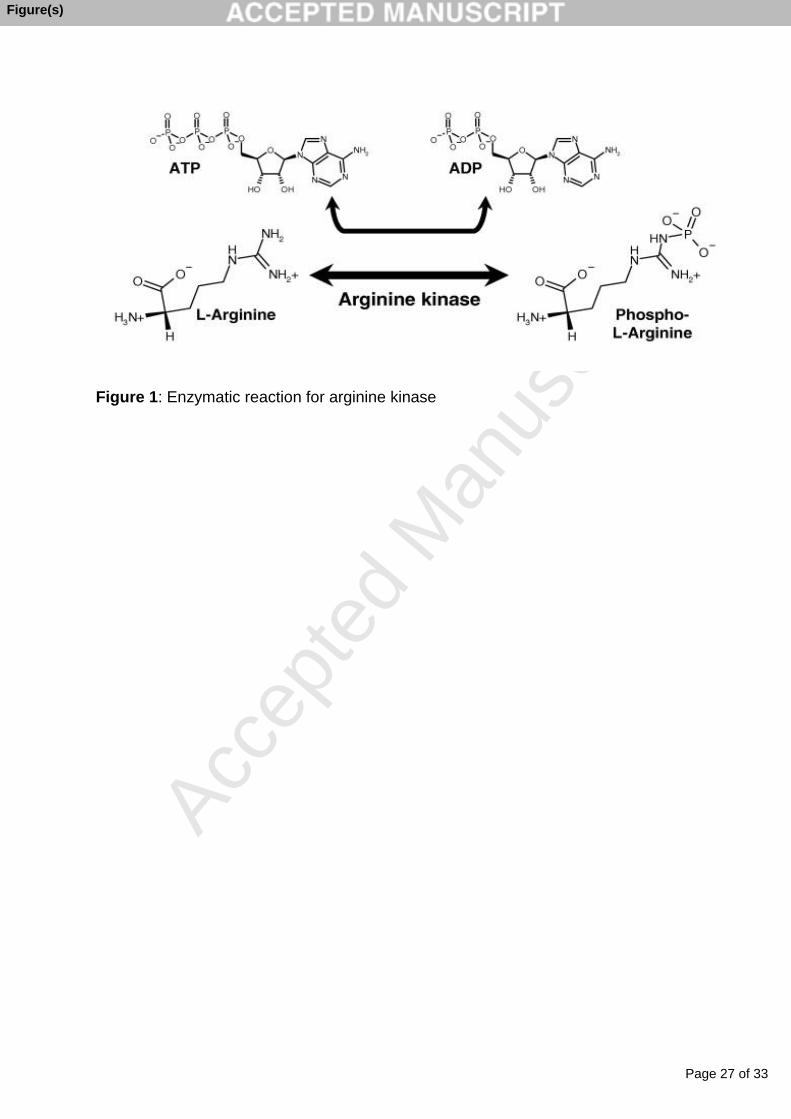
the reversible formation of phosphoarginine using L-arginine and ATP as substrates
[Figure 1][4]. Phospho-arginine can act as an emergency reservoir, not only of ATP but
also for inorganic phosphate [5-7].
The enzyme AK is absent in humans [8], a fact that makes it an attractive target
choice for trypanocide development. These researchers demonstrated an increased
activity of AK in trypanosomes when linked to oxidative stress and its inhibition, in any
way possible, would be lethal to the parasite supporting the importance of AK to the
survival of the trypanosomes. During the parasite life-cycle the trypanosomes are
Page 4 of 33
Accep
ted
Man
uscr
ipt
4
constantly exposed to the pro-oxidants in the blood of mammalian host, and
consequently either the absence of AK or its decrease in activity would present serious
consequences to the growth and survival of the parasites. In light of this, compounds
that selectively inhibit AK are desirable and should become candidates for early
development of trypanocides.
Metal nanoparticles, especially silver and gold, with their increased solubility,
have been reported to possess interesting properties including anti-microbial activity
[9,10], and to cause changes in enzyme activities [11-13].
Engineering nanoparticles that are capable of selective binding to enzymes, is a
new research direction with exciting prospects in biomedical applications and there are
two fundamental approaches used -- either a direct covalent or non-covalent link [14,
15]. In the former, there is multiple non-specific binding of the nanoparticle to cysteine
side chains leading to a loss of enzyme activity, usually with conformational changes to
protein structure. With the second, non-covalent method, either ‘naked’ or fully
functionalised nanoparticles interact with enzymes under normal protein-protein
interactions through the evolution of a loosely attached protein ‘corona’ around the
nanoparticles. The ‘naked’ nanoparticles tend to irreversibly bind and deform an initial
layer of biomolecules that form the first ‘corona’ layer, while functionalized
nanoparticles, which already have an irreversible bound initial layer, act similarly by
binding available biomolecules [14-16]. The binding between nanoparticles and the
enzyme changes the structure of the latter and as the particle gets smaller not only
does its interaction with the protein change but the composition of the protein itself
Page 5 of 33
Accep
ted
Man
uscr
ipt
5
changes. Consequently the ‘nanoparticle-enzyme corona’ have totally different
biological properties in comparison to a native enzyme.
The nanoscale size of these metal nanoparticles allow their unique and
remarkable properties to be exploited within the nanomedical fraternity [17-21] and to
resolve several medical challenges in both infectious (malaria) and neurodegenerative
diseases (Alzheimer). It is against this backdrop that the present study sought to
investigate the interaction of silver and/or gold nanoparticles with a recombinant form of
AK obtained from T. brucei (TbAK).
2. Materials and methods
2.1. Materials
Genomic DNA of T. Brucei (strain: 927/4GUTat 10.1) was a gift from Professor Ullman,
Department of Biochemistry & Biophysics, Oregon Health & Science University,
Portland, Oregon, USA. Enzymes and PCR reagents were provided by Thermo Fischer
Scientific, USA. A Bioflux kit was obtained from Separation Scientific (South Africa).
Oligonucleotides and primers were from the Integrated DNA Technology (IDT) USA.
The clone JET PCR kit, pSMART and pET-28b (+) as well as the BL21 DE3 and JM 109
strain of E. coli were obtained from Fermentas Life Sciences (USA). Absorbance
spectroscopy was performed with a Synergy Mx, (Monochromator Multi-Mode
Microplate Reader, Biotek Instruments, Inc. USA). All other reagents were of analytical

Page 6 of 33
Accep
ted
Man
uscr
ipt
6
grade and obtained from Sigma, USA or from domestic suppliers unless otherwise
stated.
2.2. Cloning of TbAK from genomic DNA [22].
The specific primers of TbAK including the forward (CAT ATG GGC TTC GGA TCA
TCA AAA CCC) with NdeI restriction site underlined and reverse (CTC GAG CTG TTC
CAC GTA CCT GC) with XhoI restriction site underlined were used to amplify the ORF
of the TbAK gene. The PCR reaction was carried out with amplication conditions as
follows 98 oC at 3min, 98oC at 30 sec, 62 oC for 30 sec, 72 oC for 1 min, 72 oC, and held
at 4 oC in a Biorad T100 thermal cycler. The 1000 bp product by PCR, excised and gel
purified using a Bioflux kit was phosphorylated and blunt ligated into a pSMART vector
using a T4 DNA ligase and then transformed into JM 109 cells. The plasmid containing
TbAK was gel purified using the Bioflux kit and sequenced. This product showed 100%
identity with the Trypanosoma brucei expressed product (Accession number –
XP_826998.1) and a high identity with other related guanidino kinases. The plasmid
was double digested and a fragment between NdeI and XhoI restriction sites containing
the TbAK gene was excised from the plasmid and subcloned into pET-28b (+)
expression vector previously treated with NdeI and XhoI. This plasmid transcribes under
the control of T7 promoter and includes a polyhistidine tag.
2.3. Expression and purifcation of TbAK.
Page 7 of 33
Accep
ted
Man
uscr
ipt
7
A method previously described [23] was adopted with slight modification. The
recombinant plasmid pET-28b (+) containing the TbAK gene was transformed into BL21
DE3 cells. The transformed cells were grown overnight, at 37 °C, in 50 ml LB culture
medium containing kanamycin after which an aliquot (5.0 ml) was transferred into a 2 L
flask containing auto-media culture (500 ml). This was grown at 20 oC, at 150 rpm for 36
hours. Cells were then harvested (6,000 x g, 10 min) washed twice with Tris buffered
saline (Tris HCl, 50 mM; NaCl, 150 mM; pH 7.5) and lysed (freeze-thawed [-80 °C/4°C;
2 cycles] in lysis buffer [NaH2PO4 buffer (50 mM, pH 7.6) containing NaCl (300 mM),
glycerol (10 %), Tween (0.25 %), imidazole (10 mM), mercaptoethanol (10 mM),
phenylmethylsulphonylfluoride (1 mM). The lysed cells were centrifuged (2,700 x g, 30
min) and the supernatant centrifuged further (100,000 x g, 90 min) after which the
supernatant (150 ml) was loaded onto a Ni-nitrilotriacetic acid affinity column. The
column was washed with the same lysis buffer until a steady zero-baseline was
obtained and then the fusion protein was eluted with increasing amounts of imidazole
(0-500 mM) in Hepes buffer (10 mM, pH 7.5). The eluted protein was concentrated
using vivaspin (GE Healthcare, Sweden) and the concentrated protein further purified
by FPLC on a Superdex 200 HR 10/30 with Hepes buffer (25 mM, pH 7.6) containing
glycerol (15 %), EDTA (0.1 M), KCL (1 M) at a flow rate (1 ml.min-1). Proteins were
resolved by SDS-PAGE in order to confirm purity of fractions before pooling. All
purification procedures were carried out at 4 oC.
2.4. SDS-PAGE
Page 8 of 33
Accep
ted
Man
uscr
ipt
8
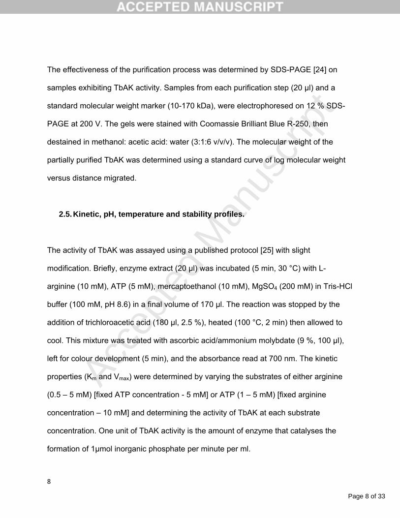
The effectiveness of the purification process was determined by SDS-PAGE [24] on
samples exhibiting TbAK activity. Samples from each purification step (20 µl) and a
standard molecular weight marker (10-170 kDa), were electrophoresed on 12 % SDS-
PAGE at 200 V. The gels were stained with Coomassie Brilliant Blue R-250, then
destained in methanol: acetic acid: water (3:1:6 v/v/v). The molecular weight of the
partially purified TbAK was determined using a standard curve of log molecular weight
versus distance migrated.
2.5. Kinetic, pH, temperature and stability profiles.
The activity of TbAK was assayed using a published protocol [25] with slight
modification. Briefly, enzyme extract (20 µl) was incubated (5 min, 30 °C) with L-
arginine (10 mM), ATP (5 mM), mercaptoethanol (10 mM), MgSO4 (200 mM) in Tris-HCl
buffer (100 mM, pH 8.6) in a final volume of 170 µl. The reaction was stopped by the
addition of trichloroacetic acid (180 µl, 2.5 %), heated (100 °C, 2 min) then allowed to
cool. This mixture was treated with ascorbic acid/ammonium molybdate (9 %, 100 µl),
left for colour development (5 min), and the absorbance read at 700 nm. The kinetic
properties (Km and Vmax) were determined by varying the substrates of either arginine
(0.5 – 5 mM) [fixed ATP concentration - 5 mM] or ATP (1 – 5 mM) [fixed arginine
concentration – 10 mM] and determining the activity of TbAK at each substrate
concentration. One unit of TbAK activity is the amount of enzyme that catalyses the
formation of 1µmol inorganic phosphate per minute per ml.

Page 9 of 33
Accep
ted
Man
uscr
ipt
9
To determine the pH optimum, the enzyme extract (20 µl) was assayed in
different buffers [sodium acetate (pH 3–5.5, 50 mM); Hepes (pH 6-7, 50 mM); Tris-HCl
buffer (pH 7.5-10, 50 mM)] under the standard assay conditions. The temperature
optimum of the partially purified enzyme was determined in Tris-HCl buffer (pH 7.6, 50
mM) over a range of 20 - 70 °C. The reaction was started by addition of enzyme
suspension (20 µl) at the different temperatures. The temperature stability of TbAK was
determined at the optimum temperature and pH and the enzyme activity at time zero
was considered to be 100 % relative activity and aliquots (20 µl) were removed at 15
min intervals and analyzed for activity for a maximum period of 4 h.
2.6. Protein determination
The protein concentration for all experiments was routinely determined, in triplicate,
according to published procedures [26]. In a 96-well titre plate was placed extract (5 μl)
followed by Bradford reagent (245 μl). The mixture was incubated (22 ºC, 10 min), the
absorbance of the solution measured at 595 nm and the concentration of the unknown
sample was determined using a BSA standard curve.
2.7. Gold and silver nanoparticles
2.7.1. Synthesis:

Page 10 of 33
Accep
ted
Man
uscr
ipt
10
The silver/gold nanoparticles (Ag/Au-Nps) were synthesised based on established
protocols [27,28]. Either AgNO3 (5 ml, 100 mM) or AuCl3 (5 ml, 50 mM) was added to
tannic acid [20 ml, 1% (w/v)] with stirring followed by polyvinylpyrrolidone (0.2 g) and
then an adjustment of pH to 8.0 by potassium carbonate (150 mM). The solution turned
pale yellow (Ag-NPs) or wine red (Au-NPs). The Ag/Au-Nps were filtered using the 0.22
µM filter and characterised by UV/Vis spectrophotometry (Biotek Epoch, USA),
inductively coupled plasma optical emission spectrometry (ICP-OES, Cambridge,
United Kingdom), energy dispersive X-ray spectroscopy (EDX) and transmission
electron microscopy (TEM, Brno, Czech Republic).
2.7.2. Characterization
UV/Vis spectroscopy. UV/Vis absorption spectra (250 – 700 nm) of the nanoparticles
were obtained to evaluate the plasmon bands associated with the silver and/or gold
nanoparticles.
Inductively coupled plasma optical emission spectroscopy (ICP OES). Nanoparticles
(250 µl) were dissolved in aqua regia [1:3 nitric to hydrochloric acid, 350 µl (Au); 500 µl
(Ag)] and digested with hydrogen peroxide (1.0 ml, 60 °C, 1 h). Metal quantification was
carried out using an inductively coupled optical emission spectrophotometer after
suitable standard curves were constructed with varying concentrations of respective
metal salt standards (Sigma Aldrich).
Transmission electron microscopy (TEM). Samples for TEM analysis were prepared by
placing a drop of the nanoparticle sample onto carbon coated copper grids, removing

Page 11 of 33
Accep
ted
Man
uscr
ipt
11
excess sample after a minute using blotting paper and the grids air-dried before
analysis. Duplicate samples were prepared and negatively stained using 1 % uranyl
acetate. Mean particle size and standard deviations were determined by the analysis of
200 particles using the computer software “Scandium”.
Energy dispersive X-ray spectroscopy (EDX). Freeze dried samples were placed on
graphite tape which was in turn placed on an aluminium stub. Elemental analysis was
performed with a TESCAN SEM with an EDX scanner (INCAPentalFeTx3, Oxford
Instruments) operating at 20 keV.
2.8. Kinetic analysis of interaction of Ag/Au nanoparticles with TbAK
The analysis was performed in the same way as that described above for TbAK activity
with the same variation in substrate of either arginine (0.5 – 5 mM) [fixed ATP
concentration - 5 mM] or ATP (1 – 5 mM) [fixed arginine concentration – 10 mM] and
either silver or gold nanoparticles (0 nM; 2 nM; 5 nM) in a total volume of 170 µl. The
TbAK activity was assayed, over a 5 min period, at each substrate concentration.
2.9. Statistical analyses
All analyses were carried out in triplicate and values reported as the means with
standard deviation p < 0.05 versus controls. Where necessary analysis of variance was
conducted using Statistica for Windows, version 8 (Statsoft Inc.) and Microsoft Excel
2010.

Page 12 of 33
Accep
ted
Man
uscr
ipt
12
3. Results
3.1. Cloning of TbAK gene.
Following specific amplification of the TbAK gene from using specific primers, a PCR
product of approximately 1000 bp was obtained. Subsequently, the nucleotide
sequencing result revealed absence of mutation and a 100% identity to the
Trypanosoma brucei expressed sequence [Accession number – XP_826998.1]
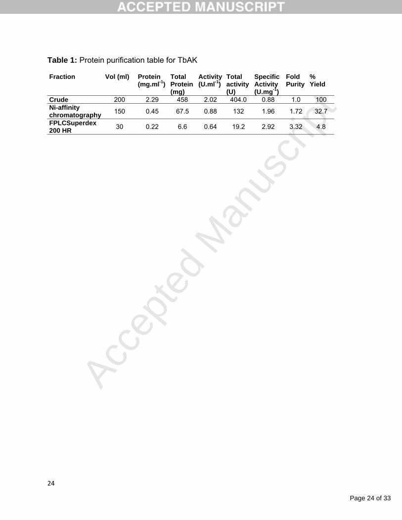
3.2. Expression and purifcation of TbAK.
The recombinant TbAK gene was expressed in E. coli BL21 DE3 harbouring the vector
pET-28b (+)_TbAK. Approximately 9.45 g of cells were harvested from 2 L of auto-
media, lysed and purified on Ni-nitriloacetic acid affinity column. This was further
purified on a Superdex 200 HR 10/30 FPLC column yielding a near homogeneous
protein in a final yield of only 4.8 % [Table 1]. The molecular weight of the fusion protein
was estimated to be around 40 kD as revealed on SDS-PAGE (Figure 2). The specific
activity of the recombinant TbAK was 2.92 µmol.min-1.mg.protein-1. The optimum
temperature and pH for the recombinant TbAK are 30 oC and 7.8 respectively [data not
shown).
3.3. Silver/gold nanoparticles: Synthesis and characterisation.

Page 13 of 33
Accep
ted
Man
uscr
ipt
13
The UV/Vis spectrophotometry with maximal absorance wavelength at 420 and 540 nm
respectively indicated the plasmon resonance absorbance of the Ag/Au nanoparticles
[27,28] [Figure 3]. The TEM analysis of the synthesised nanoparticles revealed them to
be spherical in shapes with sizes ranging from 4 to 9 nm and 7 to 22 nm respectively for
Ag and Au nanoparticles [Figure 3]. After ICP-OES analysis the concentration of the
metal nanoparticles were 30.8 and 76.6 µM for Ag and Au nanoparticles, respectively.
3.4. Kinetic analysis
A study into the effect of substrate concentration on the kinetic activity of TbAK was
investigated by measuring enzyme activity over a range of arginine concentrations (0 –
5 mM). A typical increase in substrate concentration resulted in a proportional increase
in activity as shown by the Hanes-Woolf plot [Figure 4] and with arginine as substrate,
the Vmax and Km were 0.161 μmol.ml-1.min-1 and 2.94 mM, respectively. With a total
amount of enzyme of 340 nmol the turnover number (kcat) and the catalytic efficiency,
calculated using equations 1 and 2 and were found to be 0.0079 s-1 and 2.68 x 10⁻⁶ s-
1.µM-1, respectively.
kcat = Vmax/Et …….(1).
Catalytic efficiency = kcat/Km …..(2)

Page 14 of 33
Accep
ted
Man
uscr
ipt
14
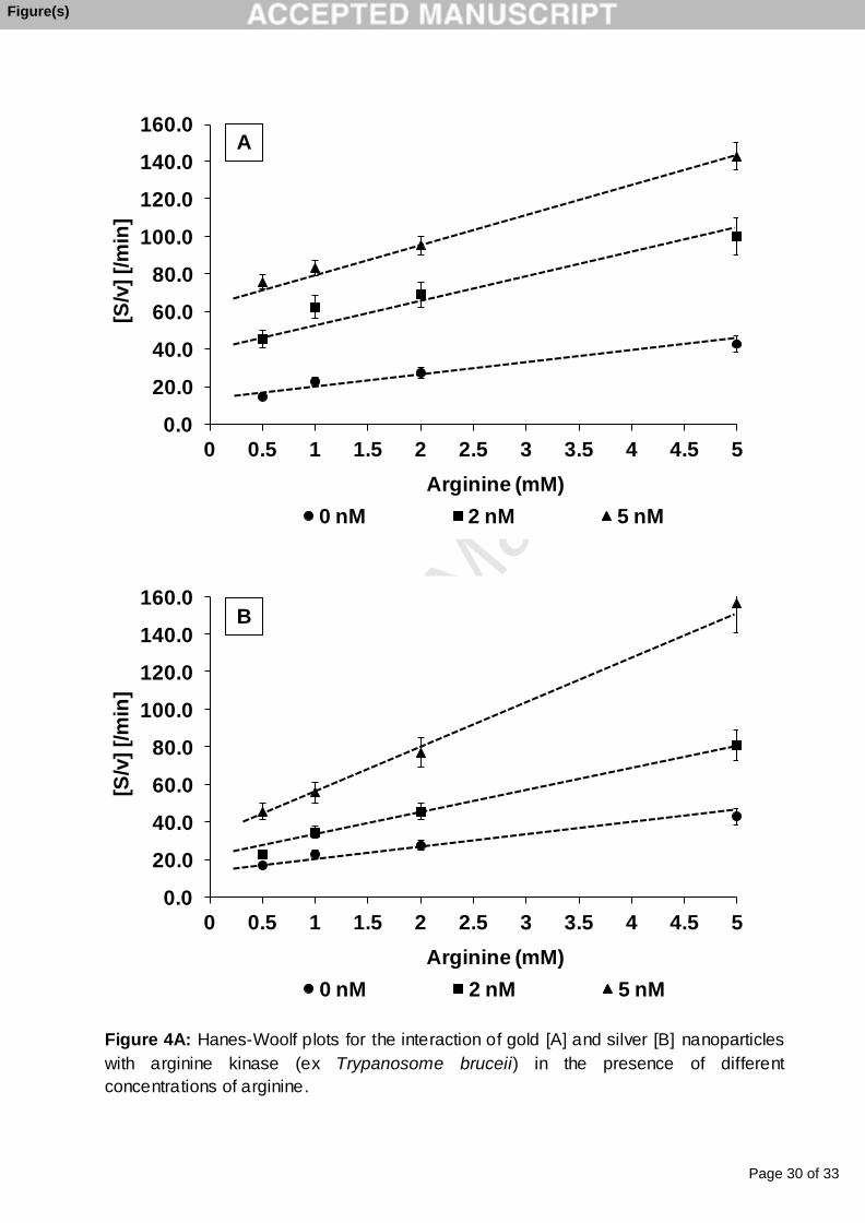
The interaction of the silver and gold nanoparticles with TbAK was determined by
including the particles at a concentration of 2 and 5 nM with either arginine, between 0.5
– 5 mM concentration at fixed levels of ATP (5 mM), or ATP, between 1 – 5 mM
concentration at fixed levels of arginine (10 mM). The results indicated that, with respect
to arginine as a variable substrate, for each type of particle the Vmax decreased [Figure
4A] while the Km virtually remained unchanged. At 5 nM Au nanoparticle the Vmax
decreased from 0.161μmol.min-1.ml-1 to 0.067μmol.min-1.ml-1 – a 62 % decrease. At 2
nM Au nanoparticle the Vmaxapp was 0.082.5μmol.min-1.ml-1 – an approximate decrease
of enzyme activity of 50 %. The decrease is more effective with Ag nanoparticle. At 5
nM Ag nanoparticle the Vmax decreased from 0.161μmol.min-1.ml-1 to 0.041μmol.min-
1.ml-1 – a 75 % decrease. At 2 nM Ag nanoparticle the Vmaxapp was 0.078μmol.min-1.ml-1
– an approximate decrease of enzyme activity of 48 %. The inhibitor constant (Ki)
calculated from equation 3 indicated the binding affinity of TbAK for the nanoparticles.
Ki = [np.Vmax.app]/[Vmax – Vmax
app]…………….(3)
where np = concentration of nanoparticle; Vmaxapp = apparent maximum velocity in the
presence of nanoparticle. Results indicated that Ag nanoparticles inhibited TbAK the
most as reflected by its relatively low Ki value of 1.59 nM; Ki values for Au nanoparticle
was estimated at 3.1 nM.
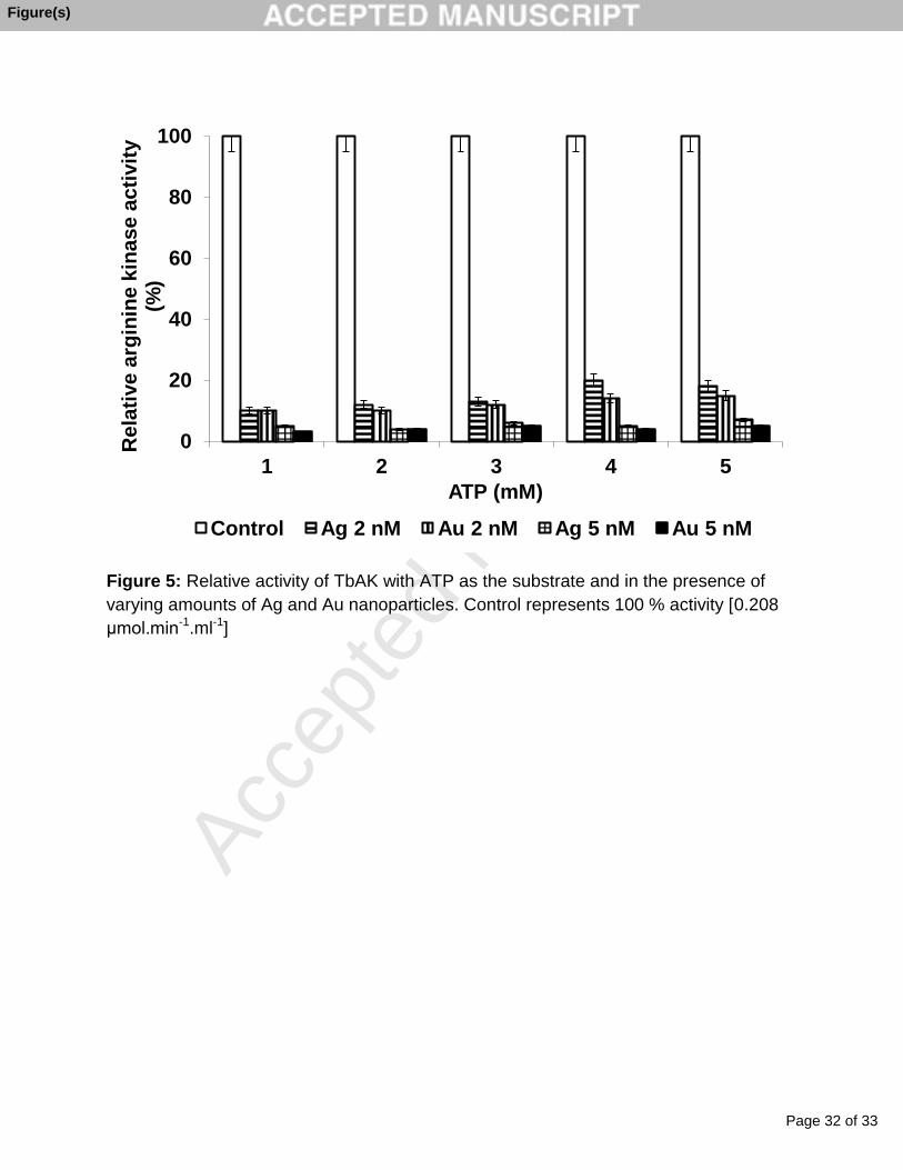
With ATP as the variable substrate [Figure 4B] Vmax and Km are 0.208 μmol.min-
1.ml-1 and 0.06 mM respectively and there was over 90 % inhibition of the enzyme with
Page 15 of 33
Accep
ted
Man
uscr
ipt
15
Ag nanoparticles [Figure 5] and close to 95 % inhibition with Au nanoparticle; respective
Ki values are 4.7 nM and 7.2 nM.
4. Discussion
Since arginine kinase from T. bruceii is absent in mammalian hosts [8] yet is critical for
trypanosome survival under stressful conditions it was clear that studies targeted at
selective inhibition of TbAK had high clinical implications in combating the disease.
The current study has investigated the interaction between metal nanoparticles
(Ag, Au) and TbAK and evidence presented shows decreased enzyme activity by 75 –
50 %. For the case of Au nanoparticles the maximum decrease in Vmax was 62 % [from
0.161 μmol.min-1.ml-1 to 0.067μmol.min-1.ml-1] while with Ag nanoparticles this decrease
was over 75 % [from 0.161 μmol.min-1.ml-1 to 0.041μmol.min-1.ml-1]. With Km values
remaining fairly constant it pointed towards a non-competitive inhibition with respect to
both arginine and ATP substrates suggesting the particles bind to sites on the enzyme
other than the active sites.
Although a thorough computational molecular docking of the Ag/Au nanoparticles
with TbAK is currently being undertaken and will be reported elsewhere, the mechanism
of binding and interaction is as yet unclear. Earlier studies by our group has inferred that
such nanoparticles have strong affinity to the thiol (-SH) group of cysteine residues in
enzymes and were responsible for the change in enzyme conformation and change in
activity [12,13,20,21]. Though it may be regarded as being speculative we offer a
possible mechanism.

Page 16 of 33
Accep
ted
Man
uscr
ipt
16
As far as we were aware no crystallographic structure for arginine kinase from T
bruceii had been deposited in the Protein Data Bank. On the other hand that for T. cruzi
[TcAK] had been reported [29] with 82 % analogy [2] to T. bruceii. The analysis of the
primary structure of TcAK [PDB ID: 2J1Q] reveals that the active site for TbAK consists
of four arginine residues [Arg124, Arg126, Arg280, Arg309] and a glutamic acid residue
[Glu314] all of which enhance the enzymatic rate through the alignment of substrate [30].
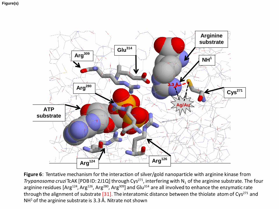
Contrary to this work, however, it is seen that the thiolate sulphur atom from Cys271 is
3.3 Å from the reactive NH1 of the arginine substrate and 4.82 Å from the unreactive Nε
[Figure 6]; it is the NH1 that becomes covalently phosphorylated. These workers imply
that Cys271 is not absolutely essential for TbAK catalysis and does not participate in a
substrate-induced conformational change of the enzyme [29]. It is possible [31] that the
electronegative thiolate would induce overall positive charge to the reactive NH1 of the
arginine substrate disrupting the resonance of the guanidinium group and increasing the
nucleophilicity of this reactive NH1 towards attack on the ϒ-phosphoryl group of ATP.
With the ATP forming reaction (reverse scenario) a proton disposition from the
guanidinium group towards the thiolate should enhance any phosphoryl transfer to ADP.
Our suggestion that Ag/Au nanoparticles interact with this thiolate atom supports our
findings for inhibition of the enzyme and that since the cysteine is not directly involved at
the active site lends credence for a non-competitive mechanism for inhibition.
Reiterating, at this stage that it may be considered speculation to propose a
tentative mechanism it is probable that the following scenarios may occur. Not only do
the noble metallic nanoparticles, especially Au and Ag, have catalytic properties [32,33]
but it is a well known fact that they have a high propensity for sulphur. Consequently,
Page 17 of 33
Accep
ted
Man
uscr
ipt
17
based upon other findings from our laboratories [20,21], the Au/Ag nanoparticles would
then associate with the sulphur atom of Cys271 decreasing its electronegativity. At the
same time, since the NH1 of the arginine substrate is only 3.3 Å away from this cysteine,
there would be disruption of resonance of the guanidinium group and an enhancement
of proton migration. This would, in turn, decrease the nucleophilicity of the NH1 towards
attack on the ϒ-phosphoryl group of ATP. For the enzymatic formation of ATP, a
blocking of the Cys271 thiolate by nanoparticles would prevent its acceptance of a proton
from the guanidinium group decreasing the overall phosphoryl transfer to ADP.
5. Conclusion
To our knowledge, this is the first report demonstrating the inhibitory potentials by the
Ag/Au nanoparticles on the activity of TbAK with Ki values in the nM range: 1.59 nM
(Ag); 3.1 nM (Au) in the presence of arginine as substrate and 4.7 nM (Ag);7.2 nM (Au)
in the presence of ATP as substrate. Overall the decrease in enzyme activity was 75 %
in the presence of Ag nanoparticles and 62 % with Au nanoparticles. The mechanism of
inhibition was consistent with non-competitive interactions with either, or both substrates
[ATP; arginine]. Cys271 is juxtapositioned 3.3 Å from the reactive NH1 of the substrate
arginine and we propose that, in line with other reported evidence, when the
nanoparticles interact with the thiolate group of Cys271 they disrupt, not only its
electronegativity, but also the resonance of the guanidinium group decreasing
nucleophilic potential of the NH1 towards attack on the ϒ-phosphoryl group. Any

Page 18 of 33
Accep
ted
Man
uscr
ipt
18
interference of the thiolate by nanoparticles would also prevent proton transfer from the
guanidinium group decreasing the overall phosphoryl transfer to ADP.
Acknowledgements
The authors wish to thank the National Research Foundation, South Africa and to
Rhodes University, South Africa for financial assistance. We thank Professor Ullman
(Department of Biochemistry & Biophysics, Oregon Health & Sciences University, USA)
for the gift of genomic DNA of T. Brucei (strain: 927/4GUTat 10.1) and Dr. J. van
Marjwik, (Department of Biochemistry, Microbiology & Biotechnology, Rhodes
University, Grahamstown, South Africa), for valuable advice and assistance.
There is no “Conflict of interest".
References
[1] H.B. Tanowitz, L.V. Kirchhoff, D. Simon, S.A. Morris, L.M. Weiss, M. Wittner,
Chaga’s disease, Clin. Microbiol. Rev. 5(4) (1992) 400–419.
[2] C.A. Pereira, G.D. Alonso, H.N. Torres, M.M. Flawiá, Arginine kinase: a common
feature for management of energy reserves in African and American flagellated
trypanosomatids. J. Euk. Microbiol. 49 (2002) 82–85.
[3] K.M. Tyler, D.M. Engman, The life cycle of Trypanosoma cruzi revisited. Int. J.
Parasitol. 31(5-6) (2001) 472-481.

Page 19 of 33
Accep
ted
Man
uscr
ipt
19
[4]. C.A. Pereira, G.D. Alonso, M.C. Paveto, M.M. Flawiá, H.N. Torres. L-arginine
uptake and L-phosphoarginine synthesis in Trypanosoma cruzi. J Euk.
Microbiol. 46 (1999) 566–570.
[5]. K. Uda, N. Fujimoto, Y. Akiyama, K. Mizuta, K. Tanaka, W.R. Ellington, T. Suzuki,
Evolution of arginine kinase gene family. Comp. Biochem. Physiol. - Part D:
Genom. Proteom. 1 (2006) 209-218.
[6]. L.D. Andrews, J. Graham, M.J. Snider, D. Fraga, Characterisation of a novel
bacterial arginine kinase from Desulfotalea psychrophila. Comp. Biochem.
Physiol. – Part B: Biochem. Mol. Biol. 150 (2008) 312-319.
[7]. C.A. Pereira, G.D. Alonso, S. Ivaldi, A.M. Silber, M.J. Alves, H.N. Torres, M.M.
Flawiá, Arginine kinase overexpression improves Trypanosoma cruzi survival
capability. FEBS. Letts, 554 (2003) 201–205.
[8]. M.R. Miranda, G.E. Canepa, L.A. Bouvier, C.A. Pereira. Trypanosoma cruzi:
oxidative stress induces arginine kinase expression. Exp. Parasitol. 114 (2006)
341–344.
[9]. D. MubarakAli, N. Thajuddin, K. Jeganathan, M. Gunasekaran. Plant extract
mediated synthesis of silver and gold nanoparticles and its antibacterial activity
against clinically isolated pathogens, Coll. Surf. B: Biointerface, 85 (2011) 360-
365.
[10]. N.C.J. Lekshmi, S.B. Sumi, S. Viveka, S. Jeeva, J.R. Brindha. Antibacterial activity
of nanoparticles from Allium sp J. Microbiol. Biotech. Res. 2 (2012) 115-119.

Page 20 of 33
Accep
ted
Man
uscr
ipt
20
[11]. N.S. Wigginton, A. Titta, F. Piccapietra, J. Dobias, V.J. Nesatyy, M.J.F. Suter, B.
Bernier-Latmani. Binding of silver nanoparticles to bacterial proteins depends
on surface modificaations and inhibits enzymatic activity. Environ. Sci. Technol.
44 (2010) 2163-2168.
[12]. A.A. Salma, H.A. Amer, H.A. Shaemaa, K.A. Abdulrahman. The effects of gold and
silver nanoparticles on transaminase enzymes activities. Int. J. Chem. Res. 1
(2011) 1-11.
[13]. M. Srivastava, S. Singh, W.T. Self. Exposure to silver nanoparticles inhibits
selenoprotein synthesis and the activity of thioredoxin reductase. Environ.
Health Perspect. 120 (2011) 56-61.
[14] I. Lynch, K.A. Dawson. Protein-nanoparticle interactions. Nano Today, 3(1) (2008)
40–47.
[15] M.P. Monopoli, C Aberg, A Salvati, K.A.Dawson. Biomolecular coronas provide the
biological identity of nanosized materials. Nature Nanotechnology, 7 (2012)
779-786.
[16] A. Salvati, A.S. Pitek, M.P. Monopoli, K. Prapaino, F. B. Bombelli, D.R. Hristov,
P.M.Kelly, C. Aberg, E. Mahon, K.A. Dawson. Transferrin-functionalized
nanoparticles lose their targeting capabilities when a biomolecule corona
adsorbs on the surface. Nature Nanotechnology, 2 (2013) 137-143.

Page 21 of 33
Accep
ted
Man
uscr
ipt
21
[17]. E.R. Padayachee, N. Ngqwala, C.G. Whiteley. Association of β-amyloid peptide
fragments with neuronal nitric oxide synthase: Implications in the etiology of
Alzheimer’s disease. J. Enz. Inhib. Med. Chem. 27 (2011) 356-364.
[18]. E.R. Padayachee, C.G.Whiteley. Spectrofluorimetric analysis of amyloid peptides
with neuronal nitric oxide synthase: Implications in Alzheimer’s disease.
Biochim. Biophys. Acta. 1810 (2011) 1136-1140.
[19]. E.R. Padayachee, C.G. Whiteley. Interaction of glycine zipper fragments of Aβ-
peptides with neuronal nitric oxide synthase: Kinetic, thermodynamic and
spectrofluorimetric analysis. Neuropep (2013)
doi.org/10.1016/j.npep.2012.12.006.
[20]. A. Sennuga, J. van Marwijk, C.G. Whiteley. Ferroxidase activity of apoferritin is
increased in the presence of platinum nanoparticles. Nanotechnol. 23 (2012a)
035102. doi:10.1088/0957-4484/23/3/035102
[21]. A. Sennuga, J. van Marwijk, A. Boshoff, C.G. Whiteley. Enhanced activity of
chaperonin GroEL in the presence of platinum nanoparticles. J. Nanoparticle
Res. 14 (2012b) 824-835. 10.1007/s11051-012-0824-6.
[22]. C.A. Pereira, G.D. Alonso, M.C. Paveto, A. Iribarren, M.L. Cabanas, H.N. Torres,
M.M. Flawiá. Trypanosoma cruzi arginine kinase characterization and cloning.
A novel energetic pathway in protozoan parasites. J. Biol. Chem. 275 (2000)
1495–1501.

Page 22 of 33
Accep
ted
Man
uscr
ipt
22
[23]. F.W. Studier. Protein production by auto-induction in high-density shaking cultures.
Prot. Expr. Purific. 41 (2005) 207–234.
[24]. U.K. Laemmli. Cleavage of structural proteins during the assembly of the head of
bacteriophage T4. Nature 227 (1970) 680-685.
[25]. S. Guo, Z. Guo, X. Guo, B. Chen, X. Wang. Expression, purification, and
characterization of arginine kinase from the sea cucumber Stichopus japonicus.
Prot. Expr. Purific. 29 (2003) 230–234.
[26]. M.M. Bradford MM. A rapid and sensitive method for the quantification of
microgram quantities of protein utilizing the principle of protein dye binding.
Anal. Biochem. 72 (1976) 248-254.
[27]. S.K. Sivaraman, S. Kumar, V. Santhanam. Room temperature synthesis of gold
nanoparticles: Size control by slow addition. Gold Bull. 43 (2010) 275-285.
[28]. S.K. Sivaraman, I. Elango, S.Kumar, V. Santhanam. A green protocol for room
temperature synthesis of silver nanoparticles in seconds. Curr. Sci. 97 (2009)
1055-1099.
[29]. P. Fernandez, A. Haouz, C.A. Pereira, C. Aguilar, P.M. Alzari. The crystal structure
of Trypanosoma Cruzi arginine kinase. Proteins 69 (2007) 209-214.
[30]. R. Furter, E.M. Furter-Graves, T. Wallimann. Creatine kinase: The reactive
cysteine is required for synergism but is nonessential for catalysis.
Biochemistry 32 (1993) 7022-7029.

Page 23 of 33
Accep
ted
Man
uscr
ipt
23
[31]. J.L. Gattis, E. Ruben, M.O. Fenley, W.R. Ellington, M.S. Chapman. The active site
cysteine of arginine kinase: structural and functional analysis of partially active
mutants. Biochemistry 43 (2004) 8680-8689.
[32]. C.T. Campbell, J.C. Sharp, Y.X. Yao, E.M. Karp, T.L. Silbaugh. Insights into
catalysis by gold nanoparticles and their support effects through surface
science studies of model catalysts. Farad. Disc. 152 (2011) 227-239.
[33]. W. Li, C. Sun, B. Hou, X. Zhou. Room temperature synthesis and catalytic
properties of surfactant-modified Ag nanoparticles. Int. J. Spectroscopy 2012
(2012) 1-7.
Page 24 of 33
Accep
ted
Man
uscr
ipt
24
Table 1: Protein purification table for TbAK Fraction Vol (ml) Protein
(mg.ml-1)Total Protein (mg)
Activity (U.ml-1)
Total activity (U)
Specific Activity (U.mg-1)
Fold Purity
% Yield
Crude 200 2.29 458 2.02 404.0 0.88 1.0 100 Ni-affinity chromatography 150 0.45 67.5 0.88 132 1.96 1.72 32.7
FPLCSuperdex 200 HR 30 0.22 6.6 0.64 19.2 2.92 3.32 4.8

Page 25 of 33
Accep
ted
Man
uscr
ipt
25
Legends for Figures
Figure 1: Enzymatic reaction for arginine kinase
Figure 2: SDS-PAGE for TbAK: lane Y is fraction collected following purification of the
His-tag eluate on a Superdex 200 HR 10/30 fast protein liquid chromatography(FPLC)
system; lane Z represents the protein marker. Arrow within the gel represents the
purified TbAK.
Figure 3: Transmission electron microscopy images (TEM) for: A) Au nanoparticles; B)
Ag nanoparticles. Scale bar = 200 nm. Visible absorbance spectrum illustrating the
plasmon resonance band for: C) Au nanoparticles (540 nm); D) Ag nanoparticles (420
nm).
Figure 4A: Hanes-Woolf plots for the interaction of gold [A] and silver [B] nanoparticles
with arginine kinase (ex Trypanosoma bruceii) in the presence of different concentrations
of arginine.
Figure 4B: Hanes-Woolf plots for the interaction of gold [A] and silver [B] nanoparticles
with arginine kinase (ex Trypanosome bruceii) in the presence of different concentrations
of ATP.

Page 26 of 33
Accep
ted
Man
uscr
ipt
26
Figure 5: Relative activity of TbAK with ATP as the substrate and in the presence of
varying amounts of Ag and Au nanoparticles. Control represents 100 % activity [0.208
μmol.min-1.ml-1]
Figure 6: Tentative mechanism for the interaction of silver/gold nanoparticle with
arginine kinase from Trypanosoma cruzi TcAK [PDB ID: 2J1Q] through Cys271
,
interfering with N1 of the arginine substrate. The four arginine residues [Arg124
, Arg126
,
Arg280
, Arg309
] and Glu314
are all involved to enhance the enzymatic rate through the
alignment of substrate [31]. The interatomic distance between the thiolate atom of
Cys271
and NH1 of the arginine substrate is 3.3 Å. Nitrate not shown
Page 27 of 33
Accep
ted
Man
uscr
ipt
Figure 1: Enzymatic reaction for arginine kinase
Figure(s)

Page 28 of 33
Accep
ted
Man
uscr
ipt
Figure 2: SDS-PAGE for TbAK: lane Y is fraction collected following purification of the
His-tag eluate on a Superdex 200 HR 10/30 fast protein liquid chromatography(FPLC)
system; lane Z represents the protein marker. Arrow within the gel represents the purified
TbAK.
Y Z
Figure(s)

Page 29 of 33
Accep
ted
Man
uscr
ipt
Figure(s)
Page 30 of 33
Accep
ted
Man
uscr
ipt
Figure 4A: Hanes-Woolf plots for the interaction of gold [A] and silver [B] nanoparticles
with arginine kinase (ex Trypanosome bruceii) in the presence of different
concentrations of arginine.
0.0
20.0
40.0
60.0
80.0
100.0
120.0
140.0
160.0
0 0.5 1 1.5 2 2.5 3 3.5 4 4.5 5
[S/v
] [/m
in]
Arginine (mM) 0 nM 2 nM 5 nM
0.0
20.0
40.0
60.0
80.0
100.0
120.0
140.0
160.0
0 0.5 1 1.5 2 2.5 3 3.5 4 4.5 5
[S/v
] [/m
in]
Arginine (mM) 0 nM 2 nM 5 nM
A
B
Figure(s)

Page 31 of 33
Accep
ted
Man
uscr
ipt
Figure 4B: Hanes-Woolf plots for the interaction of gold [A] and silver [B] nanoparticles
with arginine kinase (ex Trypanosome bruceii) in the presence of different
concentrations of ATP.
0.0 20.0 40.0 60.0 80.0
100.0 120.0 140.0 160.0 180.0
0 0.5 1 1.5 2 2.5 3 3.5 4 4.5 5
[S/v
] [/m
in]
ATP (mM) 0 nM 2 nM
0.0
40.0
80.0
120.0
160.0
200.0
240.0
0 0.5 1 1.5 2 2.5 3 3.5 4 4.5 5
[S/v
] [/m
in]
ATP (mM) 0 nM 2 nM
A
B
Figure(s)
Page 32 of 33
Accep
ted
Man
uscr
ipt
Figure 5: Relative activity of TbAK with ATP as the substrate and in the presence of
varying amounts of Ag and Au nanoparticles. Control represents 100 % activity [0.208
μmol.min-1.ml-1]
0
20
40
60
80
100
1 2 3 4 5
Rel
ativ
e ar
gini
ne k
inas
e ac
tivity
(%
)
ATP (mM)
Control Ag 2 nM Au 2 nM Ag 5 nM Au 5 nM
Figure(s)
Page 33 of 33
Accep
ted
Man
uscr
ipt
Cys271
Glu314
Arg309
Arg124 Arg126
Arg280
ATP substrate
Arginine substrate
NH1
Ag/Au
3.3 Å
Figure 6: Tentative mechanism for the interaction of silver/gold nanoparticle with arginine kinase from Trypanosoma cruzi TcAK [PDB ID: 2J1Q] through Cys271, interfering with N1 of the arginine substrate. The four arginine residues [Arg124, Arg126, Arg280, Arg309] and Glu314 are all involved to enhance the enzymatic rate through the alignment of substrate [31]. The interatomic distance between the thiolate atom of Cys271 and NH1 of the arginine substrate is 3.3 Å. Nitrate not shown
Figure(s)