Embed Size (px)
Citation preview
Interaction of C60 Derivatives and ssDNA from Simulations
Xiongce Zhao†
Center for Nanophase Materials Sciences, Oak Ridge National Laboratory, Oak Ridge, Tennessee 37831
ReceiVed: February 8, 2008; ReVised Manuscript ReceiVed: March 4, 2008
We report atomistic modeling studies on the interaction of water-soluble C60 derivatives and single-strandedDNA (ssDNA) segments in phosphate-buffered solutions. Stable hybrids are formed by C60 derivatives andssDNA segments, with binding energies in the range of -23 to -47 kcal/mol. By contrast, the typical bindingenergy between two C60 derivative molecules is -11 to -15 kcal/mol. The binding pattern of C60 derivativeswith ssDNA molecules depends on the size and shape of the C60 functional groups. For C60 derivatives withfunctional groups that contain aromatic rings, strong π stacking was observed between the ssDNA base ringsand the functional benzene rings. For C60 derivatives with a long hydrophilic chain, the binding is greatlyenhanced by the hydrophilic interaction from the entanglement between the chain and the ssDNA backbone.Stable hydrogen bonds were observed between the hydroxyl hydrogen on the functional chain and the phosphateoxygen on the ssDNA backbone. For C60 derivative with short hydrophilic groups, at least two binding patternswere observed, one of which is dominated by the hydrophobic interaction between the C60 surface and baseson ssDNA, and the other involves multiple weak hydrogen bonds between the functional carboxylic groupsand ssDNA.
I. Introduction
The fullerenes were first discovered in 19851 and isolated inbulk in 1990.2 Production of C60 on an industrial scale wassuccessfully realized soon after.3 An extensive amount ofresearch work has been carried out since then, looking forpractical applications of these novel materials. In particular, C60,as the most representative fullerene, has attracted much researchattention among communities ranging from material chemistry,to biology, to pharmacy. Due to their appealing properties suchas size, hydrophobicity, high cohesivity,4,5 photoactivity,6 andelectronic effects,7 C60 and its derivatives have aroused greatinterest in medicinal chemistry.8 For example, it was discoveredthat functionalized fullerenes can be used in photodynamictherapy9 or as inhibitors of HIV-1 protease.10,11
One major obstacle to C60 application in biological systemsis its low solubility in aqueous or polar solvents.12 Severaldifferent approaches can be used to increase the solubility. Thefirst is to encapsulate C60 in solubilizing agents such ascyclodextrins13 or calixarenes,14,15 and to solubilize them inwater. The second is to suspend the molecules in water withthe help of cosolvents such as benzene and tetrahydrofuran.16
The third is to introduce hydrophilic function groups such asamino or hydroxyl groups.17 C60 has been known to have arelatively high reactivity that allows various structural modifica-tions.18,19 Therefore, the last approach serves as a versatilemethodology that leads to a wide variety of C60 derivatives withdifferent physical and chemical properties.18,20
From the beginning of the research on the C60 and C60
derivatives in biological and medical applications, there has beena concern about the adverse effects of these molecules. Toxicitywas the primary concern. Though earlier studies on C60 itselfsuggested low toxicity,21 it was unclear if water-solublefullerenes are also innocuous molecules. Pharmacokinetic studieshave shown that organofullerenes are excreted either slowly orrapidly, depending on the substituents.22 Other studies suggest
that certain C60 derivatives with carboxylic acid groups do notshow acute toxicity.23,24 However, more recent studies suggestthat fullerenes may induce oxidative stress in the brain ofjuvenile largemouth bass,25 and that certain types of C60
derivatives show severe toxicity in cell membranes.26 It wasalso found that C60 can produce heavy disfunctions to embryomorphogenesis in pregnant mice.27
Studies on the long-term toxicity of C60 have so far not beenreported. In particular, it is unclear if C60 molecules can enterthe cell to interact with the most important genetic molecules,DNA. Investigations have been carried out to study theinteraction of C60 molecules with DNA solutions. For example,experimental studies of C60 derivatives with DNA moleculeshave shown that C60 modified with nucleotides can bind to thetarget DNA and cleave the double strand.28 Studies were alsoattempted to use functionalized fullerenes as gene deliveryagents,29 since it was found that attachment of C60 to DNAcauses aggregate formation in a buffer solution. In a previoustheoretical study,30 we reported the stable hybrids formed byC60 and DNA when C60 molecules are docked into thehydrophobic sites of DNA molecules. This suggests that excessexposure to the C60 molecules may impact the long-termfunctions of DNA since docked C60 molecules can potentiallyaffect the regular duplication or self-repair of the DNA helix.
Despite these studies, fundamental understanding of how atypical C60 derivative interacts with a gene segment at amolecular level still remains as an open question. Given thecomplexity of the systems of interest, it is challenging to probethese properties by experiment. A promising alternative ap-proach is theoretical modeling, which has been prevalent inrecent decades in studying biological systems. Molecularmodeling can provide insight to questions exposed in experi-ments, as well as predict useful candidates for targeted applica-tions. For example, the knowledge of interactions betweenfullerenes and biomolecules is useful in the preliminary screen-ing of potential candidates in biomedical applications of interest.One can also use simulation to search for or design better† E-mail: [email protected].
J. Phys. Chem. C 2008, 112, 8898–89068898
10.1021/jp801180w CCC: $40.75 2008 American Chemical SocietyPublished on Web 05/22/2008
functionalized C60 molecules. In this study, we attempt toinvestigate the interaction of water-soluble C60 derivatives andDNA segments in buffered solution through molecular model-ing, aiming at a better understanding of the fundamentalproperties of such bio-nano systems.
II. Simulation Methods
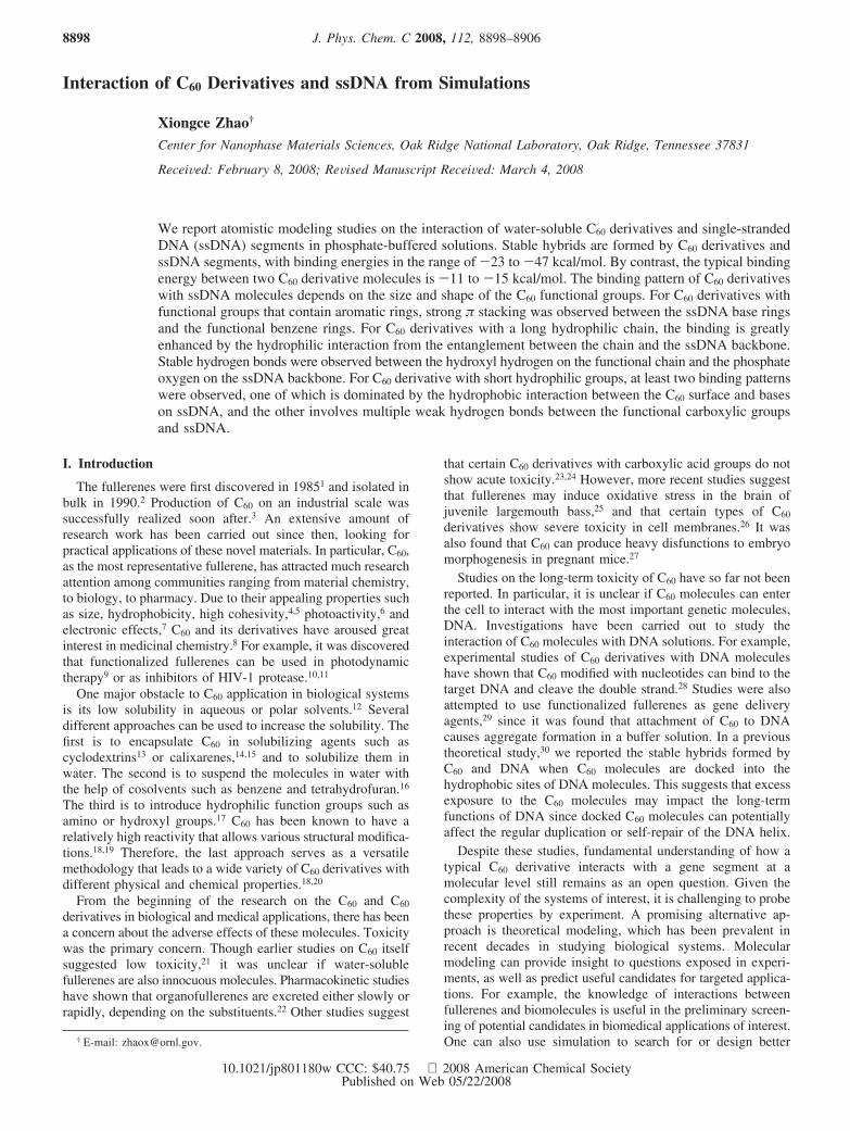
The water-soluble C60 derivatives selected in this study areshown in Figure 1. These three compounds represent some ofthe most commonly seen C60 derivatives synthesized and studiedin the past two decades.20 Each of these compounds containsone to several hydrophilic functional groups. Compound 1 hasone hydroxyl group and two benzene rings. Compound 2 is amonocarboxylic acid with a polyether chain that consists of twocarbonyl groups, two ether groups, and an acid group at theend. Compound 3 is a dicarboxylic acid with two -COOHgroups connected to the C60 surface. In this study, we focus onunderstanding the physical interaction between the C60 deriva-tives and DNA. This is warranted by the fact that a simplecarboxylic acid such as compound 3 is unlikely to reactchemically with DNA molecules.31
The single-strand DNA used in this study consists of 32 bases,which is composed of eight consecutive repeats of A-G-T-C(poly(AGTC)8). In a typical simulation, the ssDNA and fivebuckyballs (or their derivatives) were solvated in a preequili-brated TIP3P32 water box consisting of about 22 000 watermolecules, six KH2PO4 molecules (six K+ ions plus six H2PO4
-
ions), 10 Na2HPO4 molecules (20 Na+ plus 10 HPO42- ions),
and 580 NaCl molecules (580 Na+ ions plus 580 Cl- ions).The ratios between the number of water molecules and thenumber of various types of ions added in the system conformto the composition of the phosphate-buffered saline (PBS)solution at 300 K. Additionally, 31 K+ counterions were addedto electrically neutralize the negative charges from DNA.Periodic boundary conditions were applied in all three directions.Initial configurations were obtained by placing the C60 deriva-
tives along the DNA backbone. The nearest initial distancebetween any C60 molecule and DNA surfaces was about 9 Å,which is within the Lennard-Jones (LJ) cutoff distance used inthe simulation.
The DNA and Na+, Cl-, and K+ ions were modeled by theCHARMM22 force field.33 The phosphate ions were modeledby CHARMM22 plus the partial charges derived from a recentfirst-principles calculation.34 The force field parameters (bonds,angles, dihedrals, LJ parameters, partial charges) for the C60
and its derivatives were adopted from the existing CHARMM22atom types and interaction parameters, as has been donepreviously.35 The LJ interaction parameters between differentatoms were calculated by the standard Lorentz-Berthelotcombing rules, σij ) (σi + σj)/2 and εij ) (εiεj)1/2. The cutoffdistance for LJ interactions was 1.0 nm with smooth shift, andatom based pair lists with 1.2 nm were updated during thesimulation. The TIP3P water model was chosen based onprevious simulation works.36,37 The particle-mesh Ewald methodwith a fourth-order interpolation and direct space tolerance of10-6 was applied to evaluate electrostatic interactions. Ad-ditional potential parameters can be found in the SupportingInformation as well as in the original literature.33
Molecular dynamics (MD) simulations were performed withinthe constant pressure (1 bar) and constant temperature (300 K)ensemble.38 The NAMD39 software package was employed tointegrate the equations of motion. Each simulation included10 000 steps of energy minimization using a conjugate gradientalgorithm, followed by gradual heating from 0 to 300 K in 3ps, solvent equilibration for 5 ps with the DNA backbone atomsconstrained, and equilibration of 100 ps without any constraints.Typical production simulations lasted up to 15 ns. The integra-tion time step chosen was 2 fs. The SHAKE algorithm40 wasapplied to constrain the bonds involving hydrogen atoms. Thestructural configurations were saved every 1 ps for subsequentanalysis. Visualizations and analysis were performed using theVMD41 software package.
III. Results and Discussion
The association of C60 compounds and ssDNA segments ischaracterized by the binding energy for the formation ofC60-DNA hybrids. The binding energy between a pair ofmolecules is defined as30
∆E)EA+B -EA -EB -∆Edeform (1)
where EA+B, EA, and EB represent the potential energies of thebound A + B pair, molecule A in the bound pair, and moleculeB in the bound pair, respectively. ∆Edeform denotes the deforma-tion energy of the binding molecules. For ssDNA, the deforma-tion can be significant during the association. Therefore, thedeformation energy cannot be neglected. We define ∆Edeform
as
∆Edeform ) (EA -EA0 )+ (EB -EB
0 ) (2)
where EA0 and EB
0 represent the potential energies of moleculesA and B calculated from simulations without the presence oftheir binding pairs in the solution. The energetic informationwas calculated from the postsimulation analysis of the trajec-tories collected. The energies were obtained by averaging theenergetics computed from each frame.
A. C60 and DNA. We have simulated the binding of twoC60 molecules in PBS solution. We found that the bindingenergy of two C60 molecules in PBS solution is consistent withthat from our previous study,30 ∆E ) -7.2 kcal/mol. This
Figure 1. C60 derivatives investigated in this study, denoted ascompounds 1, 2, and 3 from top to bottom.
Interaction of C60 Derivatives and ssDNA J. Phys. Chem. C, Vol. 112, No. 24, 2008 8899
indicates that the presence of the excess salt ions has negligibleimpact on the association of two C60 molecules in the solution.
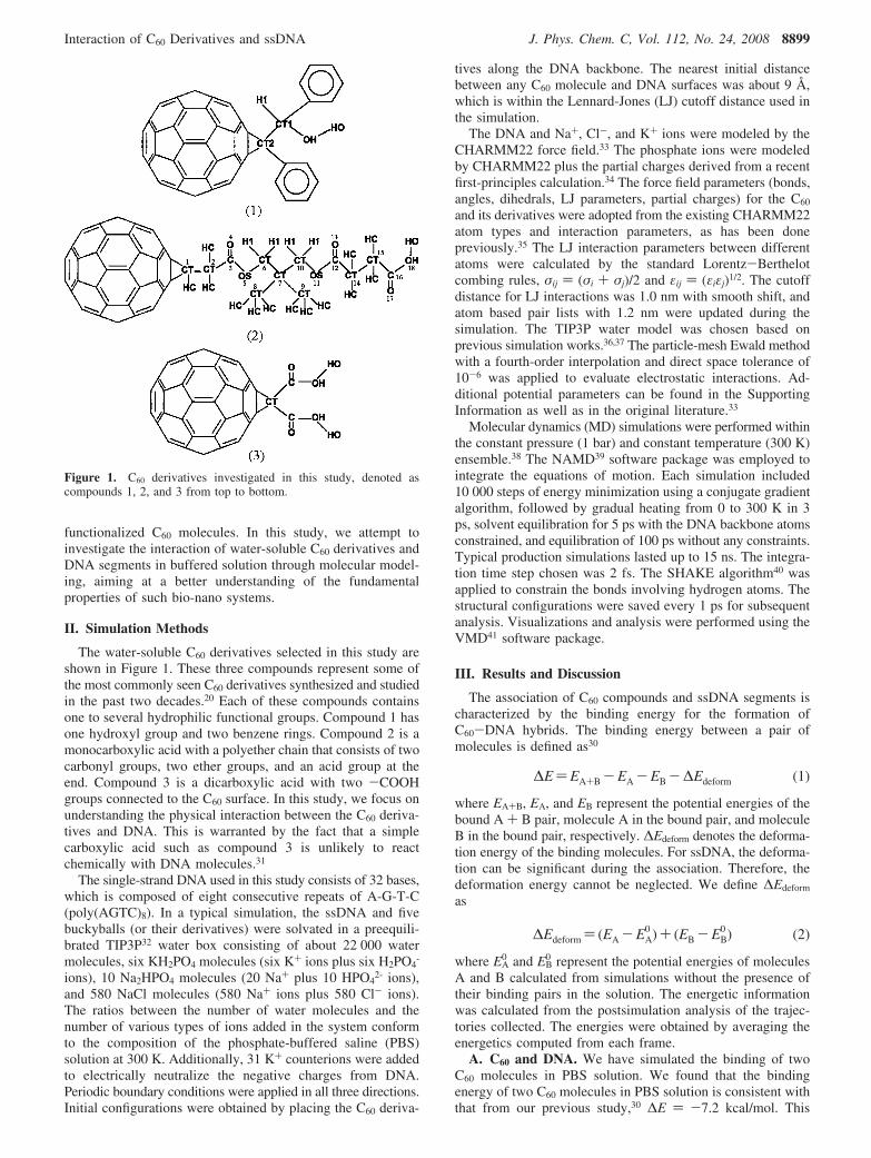
Simulations of C60 and DNA in PBS solution were performedto compare with the findings in our previous study.30 We foundthat buckyballs can bind to ssDNA to form stable hybrids inPBS solution. Typically, the binding of C60 with ssDNA occurswithin about 2 ns. Compared with the previous study,30 the newsimulation results indicate that the existence of phosphate ionsand a large amount of salt ions in the system does not affectthe hydrophobic nature of buckyball-DNA interaction. Onestabilized binding configuration is illustrated by the snapshotin Figure 2. The two bound buckyballs shown in the pictureare numbered “1” and “2” from the bottom of the picture. Theassociation of C60 1 with the ssDNA occurred at t ) 0.9-1.0ns, and C60 2 bound to the DNA at about t ) 2.2 ns.
The binding energies for C60 1 and C60 2 were found to be-22.8 and -24.4 kcal/mol, respectively. These values areslightly different from those obtained in our previous study ininfinitely diluted aqueous solution, but in the same range. Weobserved two different binding processes for buckyballs 1 and2. Buckyball 1 associated with the ssDNA by pushing aparttwo base planes on two neighboring nucleotides, T15 and C16,and then docked inside the two bases. Buckyball 2 was firstattracted into a palm-shaped hole formed by four consecutivenucleotides G10-T11-C12-A13, and was eventually wrapped bybases on C12 and A13 after a relaxation of a few nanoseconds.However, the final binding configurations for the two C60
molecules are similar (see Figure 2).The C60-DNA binding process through “wrapping” by base
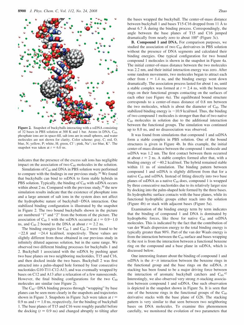
planes can be seen more clearly in the snapshots and trajectoriesshown in Figure 3. Snapshots in Figure 3a,b were taken at t )0.9 ns and t ) 1.0 ns, respectively, for the binding of buckyball1. The base planes of T15 and C16 were roughly parallel beforethe docking (t ) 0.9 ns) and changed abruptly to tilting after
the bases wrapped the buckyball. The center-of-mass distancebetween buckyball 1 and bases T15-C16 dropped from 11 Å toabout 6.7 Å during the binding process. Correspondingly, theangle between the base planes of T15 and C16 jumpeddramatically from nearly zero to about 100° (Figure 3c).
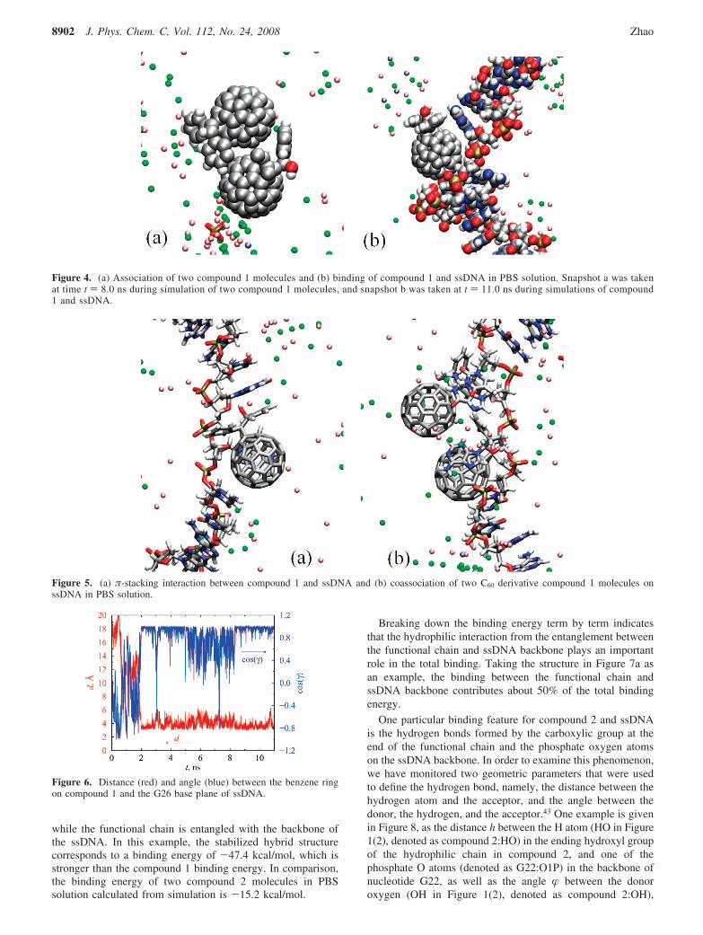
B. Compound 1 and DNA. For comparison purposes, westudied the association of two C60 derivatives in PBS solutionwithout the presence of DNA segments and calculated theirbinding energies. One typical configuration for two boundcompound 1 molecules is shown in the snapshot in Figure 4a.The initial center-of-mass distance between the two moleculeswas 2.2 nm, and their initial interaction energy was zero. Aftersome random movements, two molecules began to attract eachother from t ) 1.4 ns, and the binding energy went downdramatically. The association process lasted for about 1 ns, untila stable complex was formed at t ) 2.4 ns, with the benzenerings on their functional groups contacting on the surfaces ofeach other (see Figure 4a). The equilibrated bound structurecorresponds to a center-of-mass distance of 0.8 nm betweenthe two molecules, which is about the diameter of C60. Thestabilized binding energy is -10.9 kcal/mol. Thus, the bindingof two compound 1 molecules is stronger than that of two nativeC60 molecules in solution due to the additional interactionbetween the functional groups. The simulation was continuedup to 8.0 ns, and no disassociation was observed.
It was found from simulations that compound 1 and ssDNAform a stable complex in PBS solution. One of the boundstructures is given in Figure 4b. In this example, the initialcenter-of-mass distance between the compound 1 molecule andssDNA was 1.2 nm. The first contact between them occurredat about t ) 2 ns. A stable complex formed after that, with abinding energy of -40.2 kcal/mol. The hybrid remained stablewithin 11 ns of simulation. The binding pattern betweencompound 1 and ssDNA is slightly different from that for anative C60 and ssDNA. Instead of fitting directly into two baseplanes of ssDNA as a native C60 does, compound 1 is wrappedby three consecutive nucleotides due to its relatively larger sizeby docking into the palm-shaped hole formed by the three bases.Its hydrophobic surface contacts with the base planes, while itsfunctional hydrophilic groups either reach into the solution(Figure 4b) or stack with adjacent bases (Figure 5a).
Examination of the binding energy term by term indicatesthat the binding of compound 1 and DNA is dominated byhydrophobic forces, like those for native C60 and ssDNAmolecules. This is indicated by the fact that the contribution ofvan der Waals dispersion energy to the total binding energy istypically greater than 90%. Part of the van der Waals energy isfrom the interaction between the C60 surface and bases wrappingit; the rest is from the interaction between a functional benzenering on the compound and a base plane in ssDNA, which isdiscussed below.
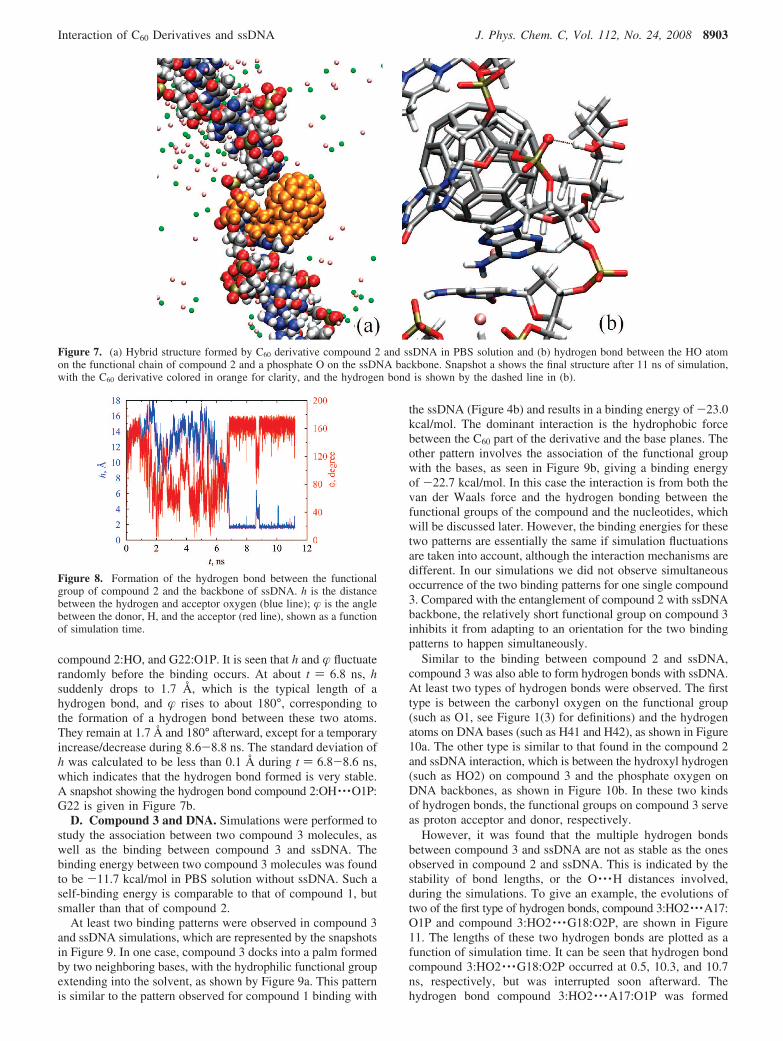
One interesting feature about the binding of compound 1 andssDNA is the π-π interaction between the benzene rings inthe functional group and the base rings on the ssDNA. πstacking has been found to be a major driving force betweenthe interaction of aromatic buckyball catchers and C60.42
Interestingly, we also observed very strong π-stacking associa-tion between compound 1 and ssDNA. One such observationis depicted in the snapshot shown in Figure 5a. It is seen thatone of the benzene rings in the functional groups of the C60
derivative stacks with the base plane of G26. The stackingpattern is very similar to that seen between two neighboringbases on DNA molecules. To examine this phenomenoncarefully, we monitored the evolution of two parameters that
Figure 2. Snapshot of buckyballs interacting with a ssDNA consistingof 32 bases in PBS solution at 300 K and 1 bar. Atoms in DNA, C60,phosphate ions are in space-fill, salt ions are in small spheres, and watermolecules are not shown for clarity. Color scheme: gray, C; red, O;blue, N; yellow, P; white, H; green, Cl-; pink, Na+; ice blue, K+. Thesnapshot was taken at t ) 6.0 ns.
8900 J. Phys. Chem. C, Vol. 112, No. 24, 2008 Zhao
were used to describe the stacking feature, i.e., the distancebetween the involved benzene plane and G26 base plane, d,and the angles formed by the two planes, γ. Plots of these twoparameters as a function of simulation time are given in Figure6. The value of d drops dramatically from random to 3.3 Åwhen the compound binds to the ssDNA at 2.0 ns. Simulta-neously, the angle γ between the benzene and base planes goesfrom random to near zero. The stabilized d of 3.3 Å is the sameas the distance between two neighboring base-pair planes in astandard B-DNA. Simulation was continued up to 11 ns. It canbe seen that d and γ remained around their stabilized valuesexcept for some fluctuations during 5-8.5 ns, suggesting thatthe π stacking observed is fairly stable. Further analysis indicatesthat the hydrophobic π-stacking interaction contributes morethan 25% of the total binding energy in this case.
Coadsorption of two compound 1 molecules on ssDNA wasalso observed. One example structure is presented in thesnapshot shown in Figure 5b. In this case, two compound 1molecules that adhered to each other were able to bind together
to ssDNA. The overall binding feature exhibited is that twoC60 derivatives are wrapped by a palm formed by fourconsecutive nucleotides in the ssDNA molecule, with C60
surfaces in contact with the base planes involved. However,when the coadsorption occurs, we have not observed clearevidence of π stacking like that seen in single compound1-ssDNA binding.
C. Compound 2 and DNA. Simulation of C60 derivativecompound 2 and ssDNA in PBS solution was performed forup to 12 ns. We found that compound 2 can also bind withssDNA to form stable structures in a nanosecond time scale.However, interaction of compound 2 and ssDNA is qualitativelydifferent from that of compound 1. Compound 2 has a long-chain functional group with a carboxylic tail. This enables it tointeract with the hydrophilic ssDNA backbones with moreflexibility and thus results in a structure with higher bindingenergy. One typical snapshot of the hybrid formed by compound2 and ssDNA is shown in Figure 7a. It can be seen that the C60
part of the compound is wrapped by bases on A21-G22-T23,
Figure 3. Docking of C60 between two base planes. Snapshots were taken at t ) 0.9 ns (a) and 1.0 ns (b), respectively. In (c), the blue line showsthe distance between the buckyball and bases, and the red line shows the change in the angle between base planes of T15-C16.
Interaction of C60 Derivatives and ssDNA J. Phys. Chem. C, Vol. 112, No. 24, 2008 8901
while the functional chain is entangled with the backbone ofthe ssDNA. In this example, the stabilized hybrid structurecorresponds to a binding energy of -47.4 kcal/mol, which isstronger than the compound 1 binding energy. In comparison,the binding energy of two compound 2 molecules in PBSsolution calculated from simulation is -15.2 kcal/mol.
Breaking down the binding energy term by term indicatesthat the hydrophilic interaction from the entanglement betweenthe functional chain and ssDNA backbone plays an importantrole in the total binding. Taking the structure in Figure 7a asan example, the binding between the functional chain andssDNA backbone contributes about 50% of the total bindingenergy.
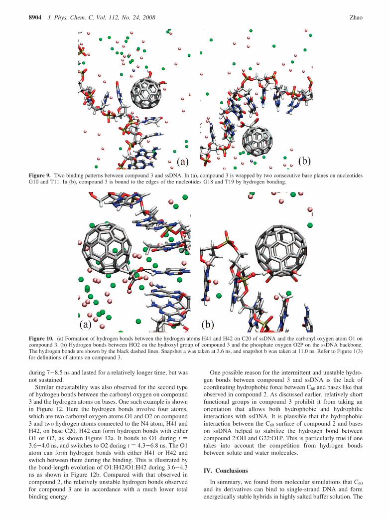
One particular binding feature for compound 2 and ssDNAis the hydrogen bonds formed by the carboxylic group at theend of the functional chain and the phosphate oxygen atomson the ssDNA backbone. In order to examine this phenomenon,we have monitored two geometric parameters that were usedto define the hydrogen bond, namely, the distance between thehydrogen atom and the acceptor, and the angle between thedonor, the hydrogen, and the acceptor.43 One example is givenin Figure 8, as the distance h between the H atom (HO in Figure1(2), denoted as compound 2:HO) in the ending hydroxyl groupof the hydrophilic chain in compound 2, and one of thephosphate O atoms (denoted as G22:O1P) in the backbone ofnucleotide G22, as well as the angle � between the donoroxygen (OH in Figure 1(2), denoted as compound 2:OH),
Figure 4. (a) Association of two compound 1 molecules and (b) binding of compound 1 and ssDNA in PBS solution. Snapshot a was takenat time t ) 8.0 ns during simulation of two compound 1 molecules, and snapshot b was taken at t ) 11.0 ns during simulations of compound1 and ssDNA.
Figure 5. (a) π-stacking interaction between compound 1 and ssDNA and (b) coassociation of two C60 derivative compound 1 molecules onssDNA in PBS solution.
Figure 6. Distance (red) and angle (blue) between the benzene ringon compound 1 and the G26 base plane of ssDNA.
8902 J. Phys. Chem. C, Vol. 112, No. 24, 2008 Zhao
compound 2:HO, and G22:O1P. It is seen that h and � fluctuaterandomly before the binding occurs. At about t ) 6.8 ns, hsuddenly drops to 1.7 Å, which is the typical length of ahydrogen bond, and � rises to about 180°, corresponding tothe formation of a hydrogen bond between these two atoms.They remain at 1.7 Å and 180° afterward, except for a temporaryincrease/decrease during 8.6-8.8 ns. The standard deviation ofh was calculated to be less than 0.1 Å during t ) 6.8-8.6 ns,which indicates that the hydrogen bond formed is very stable.A snapshot showing the hydrogen bond compound 2:OH · · ·O1P:G22 is given in Figure 7b.
D. Compound 3 and DNA. Simulations were performed tostudy the association between two compound 3 molecules, aswell as the binding between compound 3 and ssDNA. Thebinding energy between two compound 3 molecules was foundto be -11.7 kcal/mol in PBS solution without ssDNA. Such aself-binding energy is comparable to that of compound 1, butsmaller than that of compound 2.
At least two binding patterns were observed in compound 3and ssDNA simulations, which are represented by the snapshotsin Figure 9. In one case, compound 3 docks into a palm formedby two neighboring bases, with the hydrophilic functional groupextending into the solvent, as shown by Figure 9a. This patternis similar to the pattern observed for compound 1 binding with
the ssDNA (Figure 4b) and results in a binding energy of -23.0kcal/mol. The dominant interaction is the hydrophobic forcebetween the C60 part of the derivative and the base planes. Theother pattern involves the association of the functional groupwith the bases, as seen in Figure 9b, giving a binding energyof -22.7 kcal/mol. In this case the interaction is from both thevan der Waals force and the hydrogen bonding between thefunctional groups of the compound and the nucleotides, whichwill be discussed later. However, the binding energies for thesetwo patterns are essentially the same if simulation fluctuationsare taken into account, although the interaction mechanisms aredifferent. In our simulations we did not observe simultaneousoccurrence of the two binding patterns for one single compound3. Compared with the entanglement of compound 2 with ssDNAbackbone, the relatively short functional group on compound 3inhibits it from adapting to an orientation for the two bindingpatterns to happen simultaneously.
Similar to the binding between compound 2 and ssDNA,compound 3 was also able to form hydrogen bonds with ssDNA.At least two types of hydrogen bonds were observed. The firsttype is between the carbonyl oxygen on the functional group(such as O1, see Figure 1(3) for definitions) and the hydrogenatoms on DNA bases (such as H41 and H42), as shown in Figure10a. The other type is similar to that found in the compound 2and ssDNA interaction, which is between the hydroxyl hydrogen(such as HO2) on compound 3 and the phosphate oxygen onDNA backbones, as shown in Figure 10b. In these two kindsof hydrogen bonds, the functional groups on compound 3 serveas proton acceptor and donor, respectively.
However, it was found that the multiple hydrogen bondsbetween compound 3 and ssDNA are not as stable as the onesobserved in compound 2 and ssDNA. This is indicated by thestability of bond lengths, or the O · · ·H distances involved,during the simulations. To give an example, the evolutions oftwo of the first type of hydrogen bonds, compound 3:HO2 · · ·A17:O1P and compound 3:HO2 · · ·G18:O2P, are shown in Figure11. The lengths of these two hydrogen bonds are plotted as afunction of simulation time. It can be seen that hydrogen bondcompound 3:HO2 · · ·G18:O2P occurred at 0.5, 10.3, and 10.7ns, respectively, but was interrupted soon afterward. Thehydrogen bond compound 3:HO2 · · ·A17:O1P was formed
Figure 7. (a) Hybrid structure formed by C60 derivative compound 2 and ssDNA in PBS solution and (b) hydrogen bond between the HO atomon the functional chain of compound 2 and a phosphate O on the ssDNA backbone. Snapshot a shows the final structure after 11 ns of simulation,with the C60 derivative colored in orange for clarity, and the hydrogen bond is shown by the dashed line in (b).
Figure 8. Formation of the hydrogen bond between the functionalgroup of compound 2 and the backbone of ssDNA. h is the distancebetween the hydrogen and acceptor oxygen (blue line); � is the anglebetween the donor, H, and the acceptor (red line), shown as a functionof simulation time.
Interaction of C60 Derivatives and ssDNA J. Phys. Chem. C, Vol. 112, No. 24, 2008 8903
during 7-8.5 ns and lasted for a relatively longer time, but wasnot sustained.
Similar metastability was also observed for the second typeof hydrogen bonds between the carbonyl oxygen on compound3 and the hydrogen atoms on bases. One such example is shownin Figure 12. Here the hydrogen bonds involve four atoms,which are two carbonyl oxygen atoms O1 and O2 on compound3 and two hydrogen atoms connected to the N4 atom, H41 andH42, on base C20. H42 can form hydrogen bonds with eitherO1 or O2, as shown Figure 12a. It bonds to O1 during t )3.6-4.0 ns, and switches to O2 during t ) 4.3-6.8 ns. The O1atom can form hydrogen bonds with either H41 or H42 andswitch between them during the binding. This is illustrated bythe bond-length evolution of O1:H42/O1:H42 during 3.6-4.3ns as shown in Figure 12b. Compared with that observed incompound 2, the relatively unstable hydrogen bonds observedfor compound 3 are in accordance with a much lower totalbinding energy.
One possible reason for the intermittent and unstable hydro-gen bonds between compound 3 and ssDNA is the lack ofcoordinating hydrophobic force between C60 and bases like thatobserved in compound 2. As discussed earlier, relatively shortfunctional groups in compound 3 prohibit it from taking anorientation that allows both hydrophobic and hydrophilicinteractions with ssDNA. It is plausible that the hydrophobicinteraction between the C60 surface of compound 2 and baseson ssDNA helped to stabilize the hydrogen bond betweencompound 2:OH and G22:O1P. This is particularly true if onetakes into account the competition from hydrogen bondsbetween solute and water molecules.
IV. Conclusions
In summary, we found from molecular simulations that C60
and its derivatives can bind to single-strand DNA and formenergetically stable hybrids in highly salted buffer solution. The
Figure 9. Two binding patterns between compound 3 and ssDNA. In (a), compound 3 is wrapped by two consecutive base planes on nucleotidesG10 and T11. In (b), compound 3 is bound to the edges of the nucleotides G18 and T19 by hydrogen bonding.
Figure 10. (a) Formation of hydrogen bonds between the hydrogen atoms H41 and H42 on C20 of ssDNA and the carbonyl oxygen atom O1 oncompound 3. (b) Hydrogen bonds between HO2 on the hydroxyl group of compound 3 and the phosphate oxygen O2P on the ssDNA backbone.The hydrogen bonds are shown by the black dashed lines. Snapshot a was taken at 3.6 ns, and snapshot b was taken at 11.0 ns. Refer to Figure 1(3)for definitions of atoms on compound 3.
8904 J. Phys. Chem. C, Vol. 112, No. 24, 2008 Zhao
results on the binding of native C60 with ssDNA are consistentwith our previous simulations using a diluted aqueous solution.The binding of hydrophilically functionalized C60 derivativesto ssDNA segments has a stability similar to or higher thanthat of native C60 molecules.
From the standpoint of binding energy, the strongest bindingoccurred for a C60 with a long hydrophilic chain (compound2). The strong interaction came from two parts of contributions,the hydrophobic force between the C60 surface and base planes,and the hydrophilic interaction between the functional groupand the DNA backbones through entanglement. This gives abinding energy of about -47 kcal/mol. For C60 derivative withbenzene rings and hydroxyl group (compound 1), the bindingenergy is -40 kcal/mol, with part of the interaction from the πstacking between the benzene ring and the base planes of theDNA. The binding of compound 3, which has two shortcarboxylic acid groups on the surface, gives essentially the sameinteraction energy as that of native C60. However, at least twodifferent binding patterns were observed for compound 3. Inparticular, multiple intermittent hydrogen bonds were observedbetween the carboxylic groups on compound 3 and ssDNA.
The present findings suggest several effects in using C60
derivatives in biomedical applications. For instance, simulationsin this work indicate that different functional groups on a C60
result in different binding strength and binding features withDNA segments. Such information is useful in designing, forexample, target-specific gene delivery C60 agents by modifica-tions of the functional groups. On the other hand, very strongbinding between DNA and certain C60 derivatives implies
possible adverse effects on the gene. It is unknown if water-soluble C60 derivatives in the environment can readily enter thecell nucleus. If so, the association of C60 derivatives with DNAwould be strong enough to impair the regular functioning ofthe DNA.
In closing, we note that further studies can be performed inseveral aspects. First, the current results are based on relativelyshort time scales we are able to model with standard MD. Therecould be other interesting phenomena occurring well beyondnanoseconds which were not observed in this work. Simulationswith a much longer time scale are desired, but they will demandsignificant computing power. Second, the energetic informationgiven here is based on binding energy. Instead of binding energy,binding free energy is of more interest for practical applications.One could calculate the free energy of binding between C60
derivatives and DNA by computing the potential of mean forceassociated with binding of the molecule to DNA. However, suchan approach requires the identification of a set of conformationalcoordinates that span the binding process, which is not well-defined in C60/DNA interaction. Finally, the binding of C60
derivatives with double-strand DNA could be another point ofinterest for future studies.
Acknowledgment. The author thanks Peter T. Cummings formany helpful discussions. This research was conducted at theCenter for Nano phase Materials Sciences, which is sponsoredat Oak Ridge National Laboratory by the Division of ScientificUser Facilities, U.S. Department of Energy. This research usedresources of the National Energy Research Scientific ComputingCenter, which is supported by the Office of Science of the U.S.Department of Energy under Contract No. DE-AC02-05CH11231.
Supporting Information Available: Detailed description ofthe system setup, force field parameters, and simulation details.This information is available free of charge via the Internet athttp://pubs.acs.org.
References and Notes
(1) Kroto, H. W.; Heath, J. R.; O’Brien, S. C.; Curl, R. F.; Smalley,R. E. Nature 1985, 318, 162.
(2) Kratschmer, W.; Lamb, L. D.; Rostiropoulos, K.; Huffman, D. R.Nature 1990, 347, 354.
(3) Howard, J. B.; McKinnon, J. T.; Makarovsky, Y.; Lafleur, A. L.;Johnson, M. E. Nature 1991, 352, 139.
(4) Hamza, A. V.; Balooch, M. Chem. Phys. Lett. 1993, 201, 404.(5) Deguchi, S.; Alargova, R. G.; Tsuji, K. Langmuir 2001, 17, 6013.(6) Guldi, D. M.; Rato, M. Acc. Chem. Res. 2000, 33, 695.(7) Martin, N.; Sanchez, L.; Llescas, B.; Perez, I. Chem. ReV. 1998,
98, 2527.(8) Da Ros, T.; Prato, M. Chem. Commun. 1999, 663.(9) Tokuyama, H.; Yamago, S.; Nakamura, E.; Shiraki, T.; Sugiura,
Y. J. Am. Chem. Soc. 1993, 115, 7918.(10) Friedman, S. H.; DeCamp, D. L.; Sijbesma, R. P.; Srdanov, G.;
Wudl, R.; Kenyon, G. L. J. Am. Chem. Soc. 1993, 115, 6506.(11) Sijbesma, R.; Srdanov, G.; Wudl, F.; Castoro, J. A.; Wilkins, C.;
Friedman, S. H.; DeCamp, D. L.; Kenyon, G. L. J. Am. Chem. Soc. 1993,115, 6510.
(12) Korobov, M. V.; Smith, A. L. Fullerenes: Chemistry, Physics, andTechnology; Kadish, K. M., Ruoff, R. S., Eds.; Wiley-Interscience: NewYork, 2000; pp 53-89.
(13) Diederich, F.; Gomez-Lopez, M. Chem. Soc. ReV. 1999, 28, 263–277.
(14) Ikeda, A.; Hatano, T.; Kawaguchi, M.; Suenaga, H.; Shinkai, S.Chem. Comm. 1999, 15, 1403–1404.
(15) Atwood, J. L.; Koutsantonis, G. A.; Raston, C. L. Nature 1994,368, 229–231.
(16) Scrivens, W. A.; Tour, J. M.; Creek, K. E.; Pirisi, L. J. Am. Chem.Soc. 1994, 116, 4517.
(17) Chiang, L. Y.; Upasani, R. B.; Swirczewski, J. W. J. Am. Chem.Soc. 1992, 114, 10154–10157.
(18) Hirsch, A. Chemistry of Fullerenes; Thieme: Stuttgart, 1994.
Figure 11. Hydrogen bonds between HO2 on compound 3 andphosphate oxygen A17:O1P (blue) or G18:O2P (red). The anglesbetween the donor, H, and the acceptor are consistent with theevoluation of h, but not shown in the figure for clarity.
Figure 12. (a) Hydrogen bond between the C20:H42 atom and thecarbonyl oxygen atoms O1 (blue) and O2 (red) on compound 3. (b)Swapping hydrogen bonds between C20:H41 (blue)/C20:H42 (red) andthe carbonyl oxygen O1 on compound 3.
Interaction of C60 Derivatives and ssDNA J. Phys. Chem. C, Vol. 112, No. 24, 2008 8905
(19) Diederich, F.; Kessinger, R. Templated Organic Synthesis; Diede-rich, F., Stang, P., Eds.; Wiley-VCH: Weinheim, 2000; pp 189-218.
(20) Nakamura, E.; Isobe, H. Acc. Chem. Res. 2003, 36, 807–815.(21) Nelson, M. A.; Domann, F. E.; Bowden, G. T.; Hooser, S. B.;
Fernando, Q.; Carter, D. E. Toxicol. Ind. Health 1993, 9, 623–630.(22) Tabata, Y.; Murakami, Y.; Ikada, Y. Jpn. J. Cancer Res. 1997, 88,
11108–11116.(23) Yamago, S.; Tokuyama, H.; Nakamura, E.; Kikuchi, K.; Kananishi,
S.; Sueki, K.; Nakhara, H.; Enomoto, S.; Ambe, F. Chem. Biol. 1995, 2,385–389.
(24) Rancan, F.; Rosan, S.; Boehm, F.; Cantrell, A.; Brellreich, M.;Schoenberger, H.; Hirsch, A.; Moussa, F. J. Photochem. Photobiol. B: Biol.2002, 67, 157–162.
(25) Oberdorster, E. EnViron. Health Perspect. 2004, 112, 1058.(26) Bosi, S.; Feruglio, L.; Ros, T. D.; Spalluto, G.; Gregoretti, B.;
Terdoslavich, M.; Decorti, G.; Passamonti, S.; Moro, S.; Prato, M. J. Med.Chem. 2004, 47, 6711.
(27) Tsuchaya, T.; Oguri, I.; Yamakoshi, Y. N.; Miyata, N. FEBS Lett.1996, 393, 139.
(28) Boutorine, A. S.; Tokuyama, H.; Takasugi, M.; Isobe, H.; Naka-mura, E.; Helene, C. Angew. Chem., Int. Ed. Engl. 1994, 33, 2462–2465.
(29) Yamakoshi, Y. N.; Yagami, T.; Sueyoshi, S.; Miyata, N. J. Org.Chem. 1996, 61, 7236–7237.
(30) Zhao, X. C.; Striolo, A.; Cummings, P. T. Biophys. J. 2005, 89,3856.
(31) Nakamura, E.; Isobe, H.; Tomita, N.; Sawamura, M.; Jinno, S.;Okayama, H. Angew. Chem., Int. Ed. 2000, 39, 4254–4257.
(32) Jorgensen, W. L. J. Am. Chem. Soc. 1981, 103, 335.(33) MacKerell, A. D., Jr J. Phys. Chem. B 1998, 102, 3586.(34) Klahn, M.; Mathias, G.; Kotting, C.; Nonella, M.; Schlitter, J.;
Gerwert, K.; Tavan, P. J. Phys. Chem. A 2004, 108, 6186–6194.(35) Zhu, Z. W.; Schuster, D. I.; Tuckerman, M. E. Biochemistry 2003,
42, 1326–1333.(36) Cheatham, T. E., III; Kollman, P. A. Annu. ReV. Phys. Chem. 2000,
51, 435.(37) Cheatham, T. E., III; Young, M. A. Biopolymers 2001, 56, 232.(38) Berendsen, H. J. C.; Postma, J. P. M.; van Gunsteren, W. F.; Nola,
A. D.; Haak, J. R. J. Chem. Phys. 1984, 81, 3684.(39) Kale, L.; Skeel, R.; Bhandarkar, M.; Brunner, R.; Gursoy, A.;
Kraweta, N.; Phillips, J.; Shinozaki, A.; Varadarajan, K.; Schulten, K.J. Comput. Phys. 1999, 151, 283.
(40) Ryckaert, J. P.; Ciccotti, G.; Berendsen, H. J. C. J. Comput. Phys.1977, 23, 4613–4621.
(41) Humphrey, W.; Dalke, A.; Schulten, K. J. Mol. Graphics 1996,14, 1.
(42) Sygula, A.; Fronczek, F. R.; Sygula, R.; Rabideau, P. W.; Olmstead,M. M. J. Am. Chem. Soc. 2007, 129, 3842.
(43) Raschke, T. M.; Levitt, M. Proc. Natl. Acad. Sci. U.S.A. 2005, 102,6777.
JP801180W
8906 J. Phys. Chem. C, Vol. 112, No. 24, 2008 Zhao


![[Derivatives Consulting Group] Introduction to Equity Derivatives](https://img.pdfslide.us/doc/110x75/5525eed15503467c6f8b4b12/derivatives-consulting-group-introduction-to-equity-derivatives.jpg)
