Embed Size (px)
Citation preview
Introduction Activating mutations in fibroblast growth factor receptor-3(FGFR3) result in human dwarfisms ranging from the mildestcondition, hypochondroplasia, to lethal thanatophoricdysplasia (Passos-Bueno et al., 1999). Although themechanism of FGFR3 action remains obscure, bothchondrocyte proliferation and differentiation appear to bedisturbed leading to remarkably short growth plate cartilage(Wilcox et al., 1998). Recently, several reports have shed lighton FGFR3 action in cartilage by demonstrating that sustainedactivation of the Erk MAP kinase pathway accounts for theinhibitory effect of FGF signaling on chondrocyte proliferationand cartilage matrix production (Raucci et al., 2004; Krejci etal., 2004; Murakami et al., 2004; Yasoda et al., 2004).
Natriuretic peptides comprise a family of three structurallyrelated peptides: atrial natriuretic peptide, brain natriureticpeptide and C-natriuretic peptide (CNP) (Rosenzweig andSeidman, 1991). Unlike atrial and brain natriuretic peptidesthat act via the bloodstream (Mukoyama et al., 1991), CNPacts locally in a variety of tissues (Koller et al., 1992). CNPhas recently emerged as an important regulator oflongitudinal bone growth. Genetic ablation of CNP or itssignaling components results in severe skeletal dysplasias
caused by reduced chondrocyte proliferation anddifferentiation (Chusho et al., 2001; Miyazawa et al., 2002;Tamura et al., 2004). Loss-of-function mutations in the CNPcognate receptor, natriuretic peptide receptor (NPR)-B,causes an achondroplasia-like dwarfism in mice andacromesomelic dysplasia, type Maroteaux in humans (Tsujiand Kunieda, 2005; Bartels et al., 2004). In limb organculture, CNP upregulates chondrocyte proliferation andcartilage matrix production (Yasoda et al., 1998; Mericq etal., 2000). However, the molecular basis of CNP action incartilage remains unclear. Since the effects of CNP and FGFon endochondral bone growth appear to oppose each other, itis reasonable to propose a direct interaction of their signalingpathways. In fact, Yasoda et al. (Yasoda et al., 2004)demonstrated that overexpression of CNP in the cartilage ofa mouse model for achondroplasia partially rescues thedisease phenotype by correcting the decreased extracellularmatrix synthesis through inhibition of the Erk pathway.
Although the CNP-mediated reversal of FGF signaling incartilage was demonstrated (Yasoda et al., 2004), theunderlying mechanism remains unclear. In this study, wedetermined the molecular mechanism of FGF and CNPsignaling interactions in a chondrocyte environment as well as
5089
Overexpression of C-natriuretic peptide (CNP) in cartilagepartially rescues achondroplasia in the mouse. Here, westudied the interaction of fibroblast growth factor (FGF)and CNP signaling in chondrocytes. CNP antagonizedFGF2-induced growth arrest of rat chondrosarcoma (RCS)chondrocytes by inhibition of the Erk mitogen activatedprotein kinase pathway. This effect of CNP was proteinkinase G-dependent and was mimicked by the cGMPanalog pCPT-cGMP. FGF2-mediated activation of bothMEK and Raf-1 but not Ras or FRS2 was abolished byCNP demonstrating that CNP blocks the Erk pathway atthe level of Raf-1. CNP also counteracted the FGF2-mediated degradation of RCS extracellular matrix. CNP
partially antagonized FGF2-induced expression, releaseand activation of several matrix-remodeling moleculesincluding matrix metalloproteinase 2 (MMP2), MMP3,MMP9, MMP10 and MMP13. In addition, CNPcompensated for FGF2-mediated matrix loss byupregulation of matrix production independent of itsinterference with FGF signaling. We conclude that CNPutilizes both direct and indirect ways to counteract theeffects of FGF signaling in a chondrocyte environment.
Key words: Fibroblast growth factor, C-natriuretic peptide, Mitogen-activated protein kinase, Growth arrest, Extracellular Matrix,Chondrocyte
Summary
Interaction of fibroblast growth factor andC-natriuretic peptide signaling in regulation ofchondrocyte proliferation and extracellular matrixhomeostasisPavel Krejci1, Bernard Masri2, Vincent Fontaine2, Pertchoui B. Mekikian1, MaryAnn Weis3, Herve Prats2 andWilliam R. Wilcox1,4,*1Medical Genetics Institute, Cedars-Sinai Medical Center, Los Angeles, CA 90048, USA2INSERM U589, Institut Louis Bugnard, 31403 Toulouse, France3Orthopaedic Research Laboratories, University of Washington, Seattle, WA 98195, USA4Department of Pediatrics, UCLA School of Medicine, CA 90095, USA*Author for correspondence (e-mail: [email protected])
Accepted 8 August 2005Journal of Cell Science 118, 5089-5100 Published by The Company of Biologists 2005doi:10.1242/jcs.02618
Research Article
Jour
nal o
f Cel
l Sci
ence
5090
the effects of such interactions on chondrocyte proliferationand extracellular matrix homeostasis.
Materials and MethodsCell growth, cell cycle and cGMP analysesCells were propagated in DMEM media (Gibco, Gaithersburg, MD)containing 10% FBS (Atlanta Biological, Nordcross, GA) andantibiotics. For proliferation studies, cells were seeded in 24-wellplates and treated as desired for 30 minutes prior to FGF2 (R&DSystems, Minneapolis, MN) addition, cultivated for 72 hours andcounted. Chlorpromazine, EHNA, 4-{[3�,4�-(methylenedioxy)-benzyl]amino}-6-methoxyqinazoline, zaprinast, IBMX, CNP and Rp-8-pCPT-cGMPS were from Calbiochem (San Diego, CA); pCPT-cGMP and Rp-8-Br-PET-cGMPS were from Sigma (St Louis, MO);and heparin was from Gibco. For cell cycle analysis, cells treated withFGF2, CNP and pCPT-cGMP for 24 hours were analyzed for DNAcontent as described elsewhere (Krejci et al., 2004). IntracellularcGMP concentration was determined by ELISA (R&D Systems).
Western immunoblotting (WB) and immunoprecipitationCells were lysed in immunoprecipitation buffer (50 mM Tris-HCl pH7.4, 150 mM NaCl, 0.5% NP-40, 1 mM EDTA, 25 mM NaF, 0.1 mMDTT, 1 �g/ml leupeptin, 10 �g/ml soybean trypsin inhibitor, 1 mMPMSF, 8 mM �-glycerolphosphate, 10 mM Na3VO4, 1 �g/ml aprotinin).Lysates (20 �g) were resolved by SDS-PAGE and transferred onto aPVDF membrane. Blots were visualized by luminescence (Amersham,Piscataway, NJ). The following antibodies were used: actin, Erk2,MEK1, B-Raf, FRS2, NPR-B, NPR-C, MMP9, MMP13 (Santa CruzBiotechnology, Santa Cruz, CA); Raf-1 (Transduction Laboratories,Lexington, KY); P-Raf-1S338, Erk1/2, P-Erk1/2T183/Y185, P-MEKS217/221,P-FRS2Y196 (Cell Signaling, Beverly, MA); Ras, 4G10 (UpstateBiotechnology, Lake Placid, NY); MMP10 (Lab Vision, Fremont, CA);MMP2 and MMP3 (Chermicon, Temecula, CA). Forimmunoprecipitation, 500 �g of total protein was incubated with 2 �gof Raf-1 (Transduction Laboratories), B-Raf or FRS2 antibody (SantaCruz) for 2 hours at 4°C. Immunocomplexes were purified using A/G-agarose (Santa Cruz). Western blotting signal was quantitated bydetermining the integrated optical density (I.O.D.) of given band usingthe Scion Image software (Scion Corporation, Frederick, MA).
Signal transduction studiesCells were serum starved for 12 hours before treatment with FGF2 for30 minutes. When CNP, pCPT-cGMP, KT5823 (Calbiochem) or U0126(Cell Signaling) were used, cells were pretreated for 30 minutes priorto FGF2 treatment. The phosphorylation status of Raf-1, Erk and MEKwas detected by WB using the antibodies described above. Erk activitywas determined using an Erk kinase assay kit (Cell Signaling). Briefly,Erk was immunoprecipitated from 200 �g of total protein and incubatedwith recombinant Elk-1 in the presence of ATP. Phosphorylation of Elk-1 at Ser383 was determined by WB. For the Raf kinase assay, Raf-1 orB-Raf immunocomplexes were washed with kinase buffer (25 mM TrispH 7.5, 5 mM �-glycerolphosphate, 2 mM DTT, 0.1 mM Na3VO4, 10mM MgCl2). The kinase reaction was performed for 30 minutes at 30°Cin the presence of 20 �M ATP and 500 ng of recombinant MEK1 (SantaCruz) in 40 �l of kinase buffer. MEK1 phosphorylation at Ser217/221was determined by WB. Ras activation was determined using Rasactivation assay (Upstate Biotechnology). Briefly, active Ras waspurified from cell lysates using the GST fusion protein containing theRas-binding domain of Raf-1, and detected by WB.
RT-PCR and real-time RT-PCRTotal RNA was isolated using the RNeasy kit (Qiagen, Valencia, CA)including DNase I treatment. cDNA was synthesized from 2 �g of
total RNA. Real-time RT-PCR conditions are described in detailelsewhere (Krejci et al., 2004). The authenticity of real-time RT-PCRproducts was verified by agarose electrophoresis. Table 1 lists the PCRprimers used.
Extracellular matrix studiesFor Alcian blue staining, cells were treated with CNP and pCPT-cGMP for 30 minutes prior to FGF2 treatment for 72 hours, fixed with4% paraformaldehyde and stained with Alcian blue for 30 minutes.For MMP activity, cells were stimulated with FGF2 and CNP for 48hours in media containing 10% FBS followed by 24 hours ofcultivation in serum-free media. Conditioned culture media wasnormalized to cell number, concentrated ten times and subjected tonon-reducing SDS-PAGE using gels containing either 1 mg/ml gelatin(Sigma) or casein (Bio-Rad, Hercules, CA). Resolved proteins wererenatured by 2.5% Triton X-100 and incubated in activation buffer (50mM Tris-HCl pH 7.5, 10 mM CaCl2) for 19 hours (gelatin) or 96 hours(casein) at 37°C. Gels were stained with Coomassie Brilliant BlueR250 (Bio-Rad). MMP activity was confirmed by incubation of gelsin activation buffer containing either the MMP inhibitor EDTA (30mM) or the serine protease inhibitor PMSF (2 mM). For evaluationof matrix synthesis, cells were treated with FGF2 and CNP for 48 or72 hours in presence of 10 �Ci/ml of [35S]sulfate or 5 �Ci/ml of[3H]proline (Perkin Elmer, Boston, MA). For pulse-chaseexperiments, cells were labeled with [35S]sulfate or [3H]proline for 12hours, washed and treated with FGF2 and CNP for 48 or 72 hours.Following the cultivation period, cells were harvested andincorporated radioactivity was determined by liquid scintillation. Forevaluation of extracellular matrix mass, cells were treated with CNPfor 48 hours, scraped or detached by Trypsin-EDTA (Gibco; 10minutes, 37°C) and both the wet and dry mass of the cell pellets weredetermined.
Mass spectrometryThe protein band was excised from the acrylamide gel and subjectedto tandem mass spectrometric analysis after in-gel trypsin digestion.Recovered peptides were analyzed by nanobore LC (Vydac C8 MS0.3 mm � 15 cm; Vydac, Hesperia, CA) with electrospray sampleintroduction into an ion-trap mass spectrometer (Thermo FinniganLCQ Deca XP; Thermo, San Jose, CA). To identify peptides, thetandem spectra were analyzed by searching against a National Centerfor Biotechnology Information-derived database.
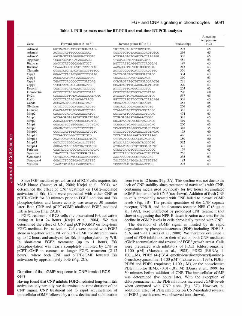
ResultsCNP counteracts FGF2-mediated growth arrest of RCScells via inhibition of the Erk MAP kinase pathwayRat chondrosarcoma (RCS) cells respond to FGF by arrestingtheir growth in the G1 phase of cell cycle (Aikawa et al., 2001).Treatment of cells with FGF2 together with CNP led to partialreversal of FGF2-mediated growth arrest as evidenced by bothcell counting and measurement of the cell cycle distribution(Fig. 1). C-natriuretic peptide binds and activates NPR-Bguanylyl cyclase resulting in rapid elevation of intracellularcGMP (Suga et al., 1992; Hagiwara et al., 1994). Consequently,the biological actions of CNP appear to be mediated by cGMP(Drewett et al., 1994). We therefore asked whether the CNPeffect on FGF2-mediated growth arrest can be reproduced by amembrane permeable cGMP analog pCPT-cGMP (Miller et al.,1973). Fig. 1 shows that pCPT-cGMP reversed, similarly toCNP, the FGF2-mediated growth arrest. For the subsequentexperiments, 10 ng/ml of FGF2, 0.2 �M CNP and 200 �MpCPT-cGMP were used unless otherwise noted.
Journal of Cell Science 118 (21)
Jour
nal o
f Cel
l Sci
ence
5091FGF and CNP signaling in chondrocytes
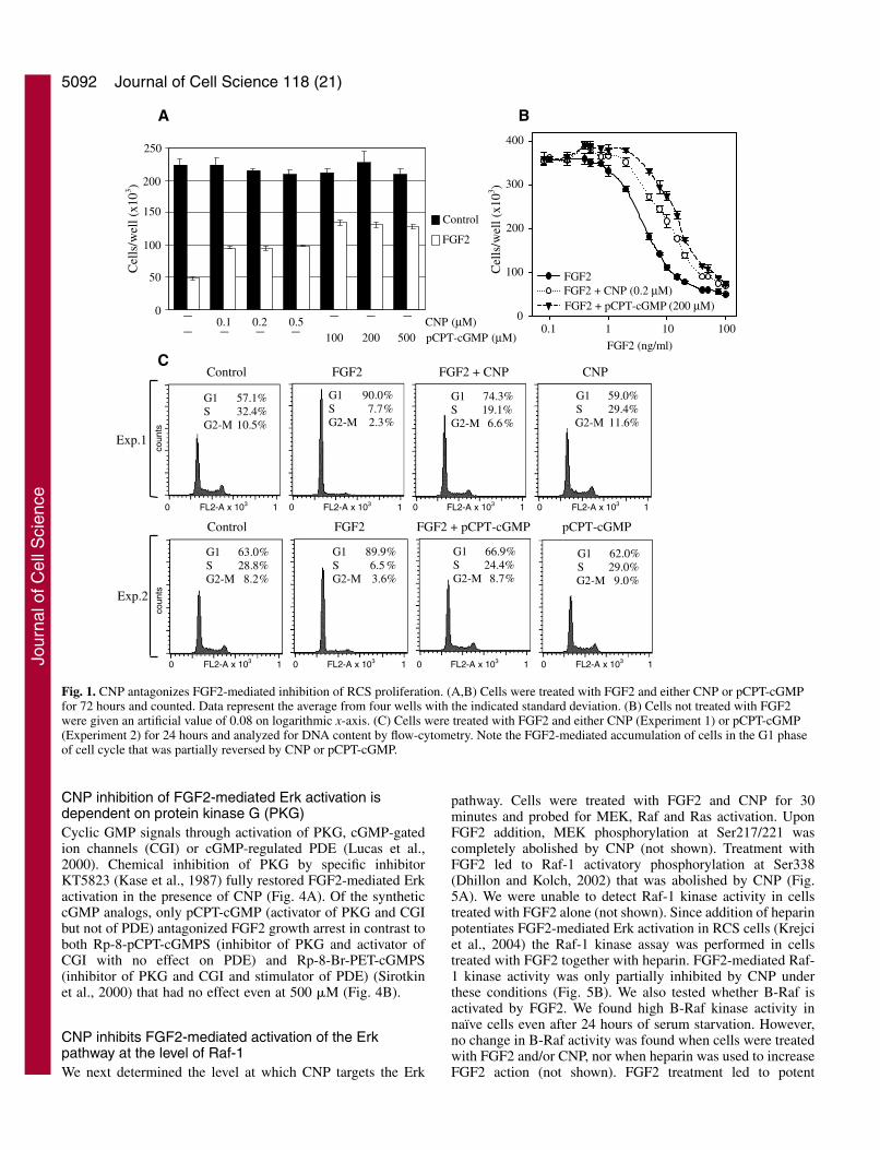
Since FGF-mediated growth arrest of RCS cells requires ErkMAP kinase (Raucci et al., 2004; Krejci et al., 2004), wedetermined the effect of CNP treatment on FGF2-mediatedactivation of Erk. Cells were pretreated with either CNP orpCPT-cGMP for 30 minutes prior to FGF2 addition and Erkphosphorylation and kinase activity was assayed 30 minuteslater. Both CNP and pCPT-cGMP abolished FGF2-mediatedErk activation (Fig. 2A,B).
FGF2 treatment of RCS cells elicits sustained Erk activationlasting at least 24 hours (Krejci et al., 2004). We thusdetermined the effect of CNP and pCPT-cGMP on long-termFGF2-mediated Erk activation. Cells were treated with FGF2alone or together with CNP or pCPT-cGMP for different timesup to 12 hours and analyzed for Erk phosphorylation by WB.In short-term FGF2 treatment (up to 1 hour), Erkphosphorylation was nearly completely inhibited by CNP orpCPT-cGMP in contrast to longer FGF2 treatment (2-12hours), where both CNP and pCPT-cGMP lowered Erkactivation by approximately 50% (Fig. 2C).
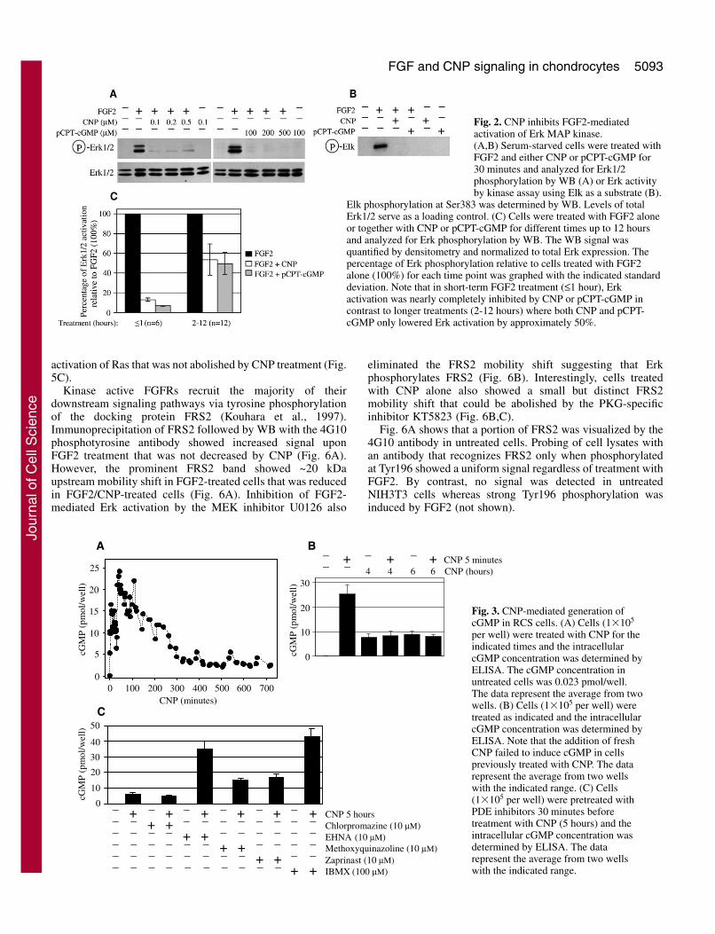
Duration of the cGMP response in CNP-treated RCScellsHaving found that CNP inhibits FGF2-mediated long-term Erkactivation only partially, we determined the time duration of theCNP signal. CNP treatment led to rapid accumulation ofintracellular cGMP followed by a slow decline and stabilization
from two to 12 hours (Fig. 3A). This decline was not due to thelack of CNP stability since treatment of naïve cells with CNP-containing media used previously for five hours accumulatedcGMP similar to fresh CNP (not shown). Addition of fresh CNPto cells chronically treated with CNP failed to elevate cGMPlevels (Fig. 3B). The protein quantities of the CNP cognatereceptor, NPR-B, and the clearance receptor, NPR-C (Suga etal., 1992), were unaffected by prolonged CNP treatment (notshown) suggesting that NPR-B desensitization accounts for thedecline in cGMP levels in cells chronically treated with CNP.
The duration of cGMP signal is limited by its rapiddegradation by phosphodiesterases (PDE) including PDE1-3,5, 6, and 9-11 (Lucas et al., 2000). We therefore evaluated apanel of PDE inhibitors for their effect on both CNP-mediatedcGMP accumulation and reversal of FGF2 growth arrest. Cellswere pretreated with inhibitors of PDE1 (chlorpromazine;1-100 �M) (Marshak et al., 1985), PDE2 (EHNA; 1-100 �M), PDE5 (4-{[3�,4’-(methylenedioxy)benzyl]amino}-6-methoxyqinazoline; 1-100 �M) (Takase et al., 1994), PDE5,PDE6 and PDE9 (zaprinast; 1-100 �M), or the nonselectivePDE inhibitor IBMX (0.01-1.0 mM) (Dousa et al., 1999) for30 minutes before addition of CNP. The intracellular cGMPwas determined five hours later. With the exception ofchlorpromazine, all the PDE inhibitors increased cGMP levelswhen compared with CNP alone (Fig. 3C). However, noadditional effect of PDE inhibitors on CNP-mediated reversalof FGF2 growth arrest was observed (not shown).
Table 1. PCR primers used for RT-PCR and real-time RT-PCR analysesAnnealing
temperatureGene Forward primer (5� to 3�) Reverse primer (5� to 3�) Product (bp) (°C)
Adamts1 GGTCACATCATTCCTGGACAACG TGTTCACACACTTGCCGCTG 293 65 Adamts4 AGTCCCATTTCCCGCAGAAC TGGTTTGTCTAAGAGGCAGTGTCG 216 65 Adamts5 AACGAGTTTACGGGGATGGGTC ATGGAAGAGTCAGCCACCAAGAGG 301 65 Aggrecan TGGGTGGATGCAGAGAGACG TTCGAGGCTCTTCCCAGTCC 481 63Biglycan AACCGTATCCGCAAAGTGCC AGTTCATTCAGGGTCTCAGGGAG 197 63Brevican TCAAAGGGGTCGTCTTCCTCTAC AACAGGCTTCTCGTGGGTTCTG 213 63Clusterin CTGGCATCATACATACGCTTTTCC ACTGTCGGGTCATCTTCACCTTC 301 63Cspg3 GGAACCTACAGTGGCTTTGGAAG TATCAGAGTGCTGGGGATGTCC 334 63Cspg4 ACCCTCATCAGGAAACCCTCAC TCACCGCCAATGTGGATAGG 310 63Cspg5 TGACTTCACCCCCTTTGATGAG CCAGAGTATGCTGTGGAGGAACTG 221 63Cspg6 TTCGTCCAGAGCAGCGATTG CCAGCACTTTCAAGGAGATTCATC 220 63Decorin TGATTGTCATAGAACTGGGCGG ATTCCTTTCAGGCTGGCTGC 205 63Fibronectin GCTCCTTCACAGATGTCCGAAC CCGTTTGAGTTGCCACCGTAAG 220 60Frs2� GAGCCCGTTGTAGAAAGGAATAGTC ATCGCTGTCATAGCCAGTGTCC 537 65Frs2� CCCTCCACAACAACAACAACG GGTTTCATCTTCCTCACCATCAGG 305 65Gapdh ACCACAGTCCATGCCATCAC TCCACCACCCTGTTGCTGTA 452 65 Glypican TCTGCTGCCCGATGACTATCTG TGACAGCCCGAGAACATTCTG 206 63Lumican CAACAATTTGACCGAGTCCGTG TTAAATGAGTTTCCAGGCACGC 381 63Mmp2 GACCTTGACCAGAACACCATCG GCTGTATTCCCGACCGTTGAAC 474 63 Mmp3 ACCAAGAGAGAGTGTGGATTCTGC TTGAGAGAGATGGAAACGGGC 385 65 Mmp7 AAGAGGGTTAGTTGGGGGACTGC GGAGTAAGTGTGGCTCAGGAGG 419 63 Mmp8 GGACATTCCTTGGGACTCTCTCAC TCAAATCTCAGGTGGGGGTCAC 200 63 Mmp9 AGAGATGTGCGTCTTCCCCTTC TCAGGTTTAGAGCCACGACCATAC 318 55 Mmp10 CCCTGGATTTTATGGAGATGCTC TTGGCTCGTGGAGAACCTGTAGAC 175 60 Mmp11 TTCAAGGCGGGCTTTGTGTG TCCACGAAGGAAGTAGGCATAGC 426 63 Mmp12 AGATTCATAAAGGCGAGGCTGAC TTTCACTGGGGCTCCATAGAGG 350 60Mmp13 GGTCTTCTGGCACACGCTTTTC ATGGCATCAAGGGATAGGGCTG 332 63 Mmp14 AGGAATAACCAAGTGATGGACGG ATGAATGAGCCTCTGGGAGACTC 371 60 Perlecan CAATACGGAGCCTACTTTCAGGAC CTAATGAAGTCTTTGCTGCGGC 179 63Syndecan1 TGACTTCACTGCTCGGGACAAG GTTTCATCCTCCACAACCTCTTTG 261 63Syndecan2 TCTGACAACATCCCAACTGATTCC AACTTCCGTCCGCTTGAACAG 235 63Syndecan4 GGACCTTCCCTGAGGTGATTTC TGCTGGACATGGACACTTTGTTG 183 63Versican TAACTGTGAAGCCAGCCGTAGC TCCCCCATCTGTGAAACTTGG 293 63
Jour
nal o
f Cel
l Sci
ence
5092
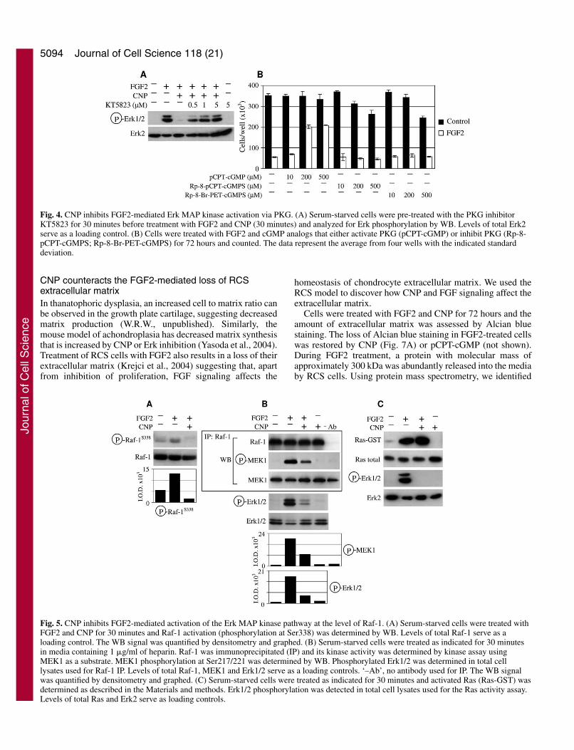
CNP inhibition of FGF2-mediated Erk activation isdependent on protein kinase G (PKG)Cyclic GMP signals through activation of PKG, cGMP-gatedion channels (CGI) or cGMP-regulated PDE (Lucas et al.,2000). Chemical inhibition of PKG by specific inhibitorKT5823 (Kase et al., 1987) fully restored FGF2-mediated Erkactivation in the presence of CNP (Fig. 4A). Of the syntheticcGMP analogs, only pCPT-cGMP (activator of PKG and CGIbut not of PDE) antagonized FGF2 growth arrest in contrast toboth Rp-8-pCPT-cGMPS (inhibitor of PKG and activator ofCGI with no effect on PDE) and Rp-8-Br-PET-cGMPS(inhibitor of PKG and CGI and stimulator of PDE) (Sirotkinet al., 2000) that had no effect even at 500 �M (Fig. 4B).
CNP inhibits FGF2-mediated activation of the Erkpathway at the level of Raf-1We next determined the level at which CNP targets the Erk
pathway. Cells were treated with FGF2 and CNP for 30minutes and probed for MEK, Raf and Ras activation. UponFGF2 addition, MEK phosphorylation at Ser217/221 wascompletely abolished by CNP (not shown). Treatment withFGF2 led to Raf-1 activatory phosphorylation at Ser338(Dhillon and Kolch, 2002) that was abolished by CNP (Fig.5A). We were unable to detect Raf-1 kinase activity in cellstreated with FGF2 alone (not shown). Since addition of heparinpotentiates FGF2-mediated Erk activation in RCS cells (Krejciet al., 2004) the Raf-1 kinase assay was performed in cellstreated with FGF2 together with heparin. FGF2-mediated Raf-1 kinase activity was only partially inhibited by CNP underthese conditions (Fig. 5B). We also tested whether B-Raf isactivated by FGF2. We found high B-Raf kinase activity innaïve cells even after 24 hours of serum starvation. However,no change in B-Raf activity was found when cells were treatedwith FGF2 and/or CNP, nor when heparin was used to increaseFGF2 action (not shown). FGF2 treatment led to potent
Journal of Cell Science 118 (21)
C
0 1 0 1 0 1 0 1FL2-A x 103 FL2-A x 103 FL2-A x 103 FL2-A x 103
coun
ts
G1 57.1%S 32.4%G2-M 10.5%
G1 90.0%S 7.7%G2-M 2.3%
G1 74.3%S 19.1%G2-M 6.6 %
G1 59.0%S 29.4%G2-M 11.6%
Exp.1
Control FGF2 FGF2 + CNP CNP
Cel
ls/w
ell(
x103 )
Series1Series2
0
50
100
150
200
250
Control
FGF2
0.1 1 10 1000
100
200
300
400
Cel
ls/w
ell(
x103 )
100
200
300
400
0.1 1 10 100
FGF2 (ng/ml)
0
FGF2FGF2 + CNP (0.2 μM)FGF2 + pCPT-cGMP (200 μM)_
0.1 0.2 0.5_ _ _
CNP (μM)_ _ _ _100 200 500 pCPT-cGMP (μM)
A B
0 1 0 1 0 1 0 1
Control FGF2 FGF2 + pCPT-cGMP pCPT-cGMP
G1 66.9%S 24.4%G2-M 8.7%
FL2-A x 103 FL2-A x 103 FL2-A x 103 FL2-A x 103
coun
tsExp.2
G1 63.0%S 28.8%G2-M 8.2%
G1 89.9%S 6.5 %G2-M 3.6%
G1 62.0%S 29.0%G2-M 9.0%
Fig. 1. CNP antagonizes FGF2-mediated inhibition of RCS proliferation. (A,B) Cells were treated with FGF2 and either CNP or pCPT-cGMPfor 72 hours and counted. Data represent the average from four wells with the indicated standard deviation. (B) Cells not treated with FGF2were given an artificial value of 0.08 on logarithmic x-axis. (C) Cells were treated with FGF2 and either CNP (Experiment 1) or pCPT-cGMP(Experiment 2) for 24 hours and analyzed for DNA content by flow-cytometry. Note the FGF2-mediated accumulation of cells in the G1 phaseof cell cycle that was partially reversed by CNP or pCPT-cGMP.
Jour
nal o
f Cel
l Sci
ence
5093FGF and CNP signaling in chondrocytes
activation of Ras that was not abolished by CNP treatment (Fig.5C).
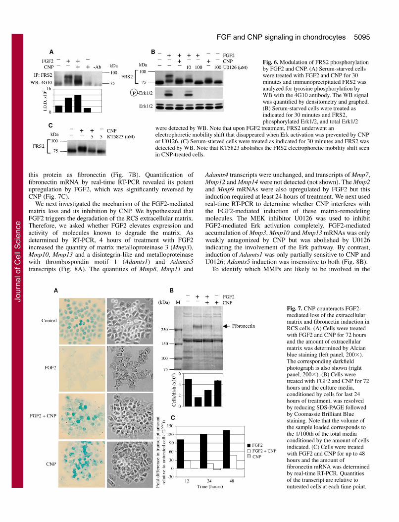
Kinase active FGFRs recruit the majority of theirdownstream signaling pathways via tyrosine phosphorylationof the docking protein FRS2 (Kouhara et al., 1997).Immunoprecipitation of FRS2 followed by WB with the 4G10phosphotyrosine antibody showed increased signal uponFGF2 treatment that was not decreased by CNP (Fig. 6A).However, the prominent FRS2 band showed ~20 kDaupstream mobility shift in FGF2-treated cells that was reducedin FGF2/CNP-treated cells (Fig. 6A). Inhibition of FGF2-mediated Erk activation by the MEK inhibitor U0126 also
eliminated the FRS2 mobility shift suggesting that Erkphosphorylates FRS2 (Fig. 6B). Interestingly, cells treatedwith CNP alone also showed a small but distinct FRS2mobility shift that could be abolished by the PKG-specificinhibitor KT5823 (Fig. 6B,C).
Fig. 6A shows that a portion of FRS2 was visualized by the4G10 antibody in untreated cells. Probing of cell lysates withan antibody that recognizes FRS2 only when phosphorylatedat Tyr196 showed a uniform signal regardless of treatment withFGF2. By contrast, no signal was detected in untreatedNIH3T3 cells whereas strong Tyr196 phosphorylation wasinduced by FGF2 (not shown).
Fig. 2. CNP inhibits FGF2-mediatedactivation of Erk MAP kinase.(A,B) Serum-starved cells were treated withFGF2 and either CNP or pCPT-cGMP for30 minutes and analyzed for Erk1/2phosphorylation by WB (A) or Erk activityby kinase assay using Elk as a substrate (B).
Elk phosphorylation at Ser383 was determined by WB. Levels of totalErk1/2 serve as a loading control. (C) Cells were treated with FGF2 aloneor together with CNP or pCPT-cGMP for different times up to 12 hoursand analyzed for Erk phosphorylation by WB. The WB signal wasquantified by densitometry and normalized to total Erk expression. Thepercentage of Erk phosphorylation relative to cells treated with FGF2alone (100%) for each time point was graphed with the indicated standarddeviation. Note that in short-term FGF2 treatment (≤1 hour), Erkactivation was nearly completely inhibited by CNP or pCPT-cGMP incontrast to longer treatments (2-12 hours) where both CNP and pCPT-cGMP only lowered Erk activation by approximately 50%.
0 100 200 300 400 500 600 700
0
5
10
15
20
25
cGM
P(p
mol
/wel
l)
0 100 200 300 400 500 600 700
10
5
0
15
20
25
CNP (minutes)
A B
10
0
20
30
cGM
P(p
mol
/wel
l)
_+
_+
_+ CNP 5 minutes_ _
4 4 6 6 CNP (hours)
C
cGM
P(p
mol
/wel
l)
_+
_+
_+
_+
_+
_+ CNP 5 hours_ _
+ +_ _ _ _ _ _ _ _
Chlorpromazine (10 μM)_ _ _ _+ +
_ _ _ _ _ _EHNA (10 μM)_ _ _ _ _ _
+ +_ _ _ _
_ _ _ _ _ _ _ _+ +
_ _Zaprinast (10 μM)_ _ _ _ _ _ _ _ _ _
+ + IBMX (100 μM)
10
0
20
30
50
40
Methoxyquinazoline (10 μM)
Fig. 3. CNP-mediated generation ofcGMP in RCS cells. (A) Cells (1�105
per well) were treated with CNP for theindicated times and the intracellularcGMP concentration was determined byELISA. The cGMP concentration inuntreated cells was 0.023 pmol/well.The data represent the average from twowells. (B) Cells (1�105 per well) weretreated as indicated and the intracellularcGMP concentration was determined byELISA. Note that the addition of freshCNP failed to induce cGMP in cellspreviously treated with CNP. The datarepresent the average from two wellswith the indicated range. (C) Cells(1�105 per well) were pretreated withPDE inhibitors 30 minutes beforetreatment with CNP (5 hours) and theintracellular cGMP concentration wasdetermined by ELISA. The datarepresent the average from two wellswith the indicated range.
Jour
nal o
f Cel
l Sci
ence
5094
CNP counteracts the FGF2-mediated loss of RCSextracellular matrixIn thanatophoric dysplasia, an increased cell to matrix ratio canbe observed in the growth plate cartilage, suggesting decreasedmatrix production (W.R.W., unpublished). Similarly, themouse model of achondroplasia has decreased matrix synthesisthat is increased by CNP or Erk inhibition (Yasoda et al., 2004).Treatment of RCS cells with FGF2 also results in a loss of theirextracellular matrix (Krejci et al., 2004) suggesting that, apartfrom inhibition of proliferation, FGF signaling affects the
homeostasis of chondrocyte extracellular matrix. We used theRCS model to discover how CNP and FGF signaling affect theextracellular matrix.
Cells were treated with FGF2 and CNP for 72 hours and theamount of extracellular matrix was assessed by Alcian bluestaining. The loss of Alcian blue staining in FGF2-treated cellswas restored by CNP (Fig. 7A) or pCPT-cGMP (not shown).During FGF2 treatment, a protein with molecular mass ofapproximately 300 kDa was abundantly released into the mediaby RCS cells. Using protein mass spectrometry, we identified
Journal of Cell Science 118 (21)
Fig. 4. CNP inhibits FGF2-mediated Erk MAP kinase activation via PKG. (A) Serum-starved cells were pre-treated with the PKG inhibitorKT5823 for 30 minutes before treatment with FGF2 and CNP (30 minutes) and analyzed for Erk phosphorylation by WB. Levels of total Erk2serve as a loading control. (B) Cells were treated with FGF2 and cGMP analogs that either activate PKG (pCPT-cGMP) or inhibit PKG (Rp-8-pCPT-cGMPS; Rp-8-Br-PET-cGMPS) for 72 hours and counted. The data represent the average from four wells with the indicated standarddeviation.
Fig. 5. CNP inhibits FGF2-mediated activation of the Erk MAP kinase pathway at the level of Raf-1. (A) Serum-starved cells were treated withFGF2 and CNP for 30 minutes and Raf-1 activation (phosphorylation at Ser338) was determined by WB. Levels of total Raf-1 serve as aloading control. The WB signal was quantified by densitometry and graphed. (B) Serum-starved cells were treated as indicated for 30 minutesin media containing 1 �g/ml of heparin. Raf-1 was immunoprecipitated (IP) and its kinase activity was determined by kinase assay usingMEK1 as a substrate. MEK1 phosphorylation at Ser217/221 was determined by WB. Phosphorylated Erk1/2 was determined in total celllysates used for Raf-1 IP. Levels of total Raf-1, MEK1 and Erk1/2 serve as a loading controls. ‘–Ab’, no antibody used for IP. The WB signalwas quantified by densitometry and graphed. (C) Serum-starved cells were treated as indicated for 30 minutes and activated Ras (Ras-GST) wasdetermined as described in the Materials and methods. Erk1/2 phosphorylation was detected in total cell lysates used for the Ras activity assay.Levels of total Ras and Erk2 serve as loading controls.
Jour
nal o
f Cel
l Sci
ence
5095FGF and CNP signaling in chondrocytes
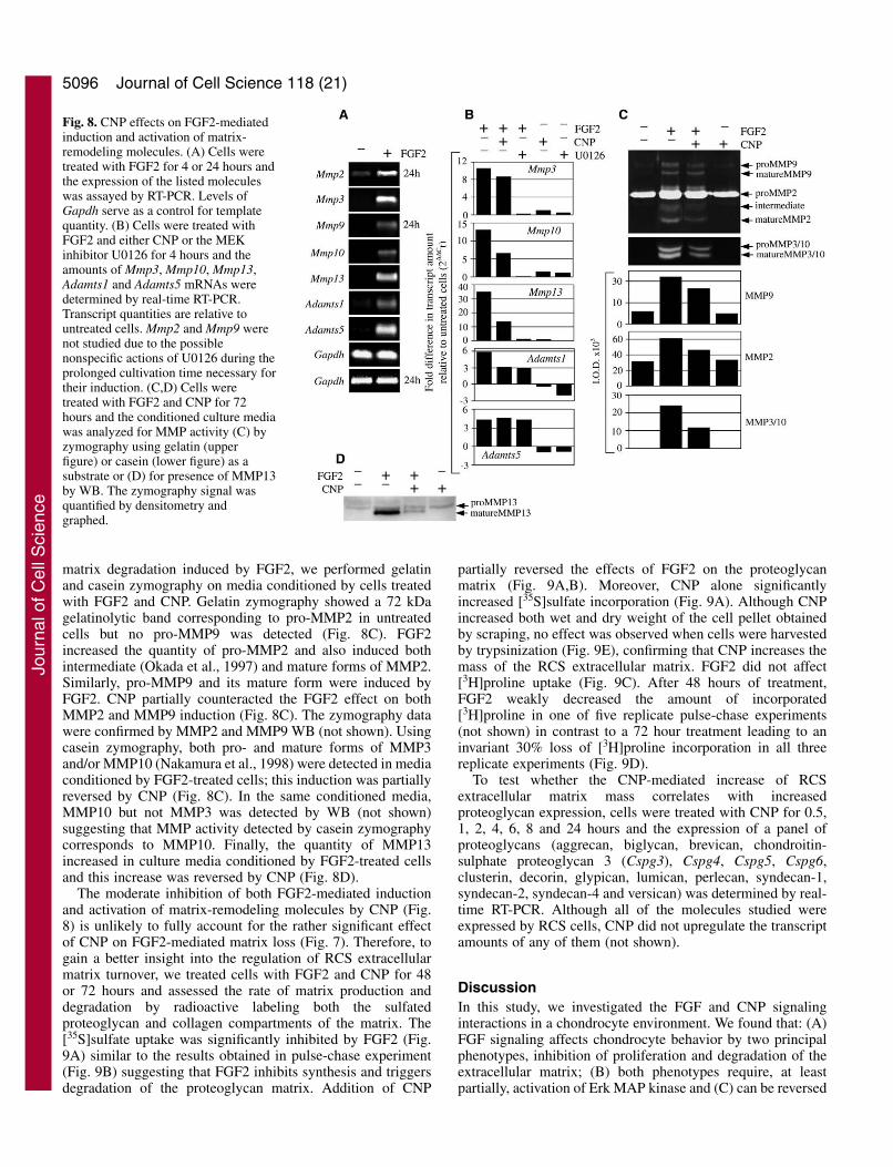
this protein as fibronectin (Fig. 7B). Quantification offibronectin mRNA by real-time RT-PCR revealed its potentupregulation by FGF2, which was significantly reversed byCNP (Fig. 7C).
We next investigated the mechanism of the FGF2-mediatedmatrix loss and its inhibition by CNP. We hypothesized thatFGF2 triggers the degradation of the RCS extracellular matrix.Therefore, we asked whether FGF2 elevates expression andactivity of molecules known to degrade the matrix. Asdetermined by RT-PCR, 4 hours of treatment with FGF2increased the quantity of matrix metalloproteinase 3 (Mmp3),Mmp10, Mmp13 and a disintegrin-like and metalloproteinasewith thrombospondin motif 1 (Adamts1) and Adamts5transcripts (Fig. 8A). The quantities of Mmp8, Mmp11 and
Adamts4 transcripts were unchanged, and transcripts of Mmp7,Mmp12 and Mmp14 were not detected (not shown). The Mmp2and Mmp9 mRNAs were also upregulated by FGF2 but thisinduction required at least 24 hours of treatment. We next usedreal-time RT-PCR to determine whether CNP interferes withthe FGF2-mediated induction of these matrix-remodelingmolecules. The MEK inhibitor U0126 was used to inhibitFGF2-mediated Erk activation completely. FGF2-mediatedaccumulation of Mmp3, Mmp10 and Mmp13 mRNAs was onlyweakly antagonized by CNP but was abolished by U0126indicating the involvement of the Erk pathway. By contrast,induction of Adamts1 was only partially sensitive to CNP andU0126; Adamts5 induction was insensitive to both (Fig. 8B).
To identify which MMPs are likely to be involved in the
Fig. 6. Modulation of FRS2 phosphorylationby FGF2 and CNP. (A) Serum-starved cellswere treated with FGF2 and CNP for 30minutes and immunoprecipitated FRS2 wasanalyzed for tyrosine phosphorylation byWB with the 4G10 antibody. The WB signalwas quantified by densitometry and graphed.(B) Serum-starved cells were treated asindicated for 30 minutes and FRS2,phosphorylated Erk1/2, and total Erk1/2
were detected by WB. Note that upon FGF2 treatment, FRS2 underwent anelectrophoretic mobility shift that disappeared when Erk activation was prevented by CNPor U0126. (C) Serum-starved cells were treated as indicated for 30 minutes and FRS2 wasdetected by WB. Note that KT5823 abolishes the FRS2 electrophoretic mobility shift seenin CNP-treated cells.
Fig. 7. CNP counteracts FGF2-mediated loss of the extracellularmatrix and fibronectin induction inRCS cells. (A) Cells were treatedwith FGF2 and CNP for 72 hoursand the amount of extracellularmatrix was determined by Alcianblue staining (left panel, 200�).The corresponding darkfieldphotograph is also shown (rightpanel, 200�). (B) Cells weretreated with FGF2 and CNP for 72hours and the culture media,conditioned by cells for last 24hours of treatment, was resolvedby reducing SDS-PAGE followedby Coomassie Brilliant Bluestaining. Note that the volume ofthe sample loaded corresponds tothe 1/100th of the total mediaconditioned by the amount of cellsindicated. (C) Cells were treatedwith FGF2 and CNP for up to 48hours and the amount offibronectin mRNA was determinedby real-time RT-PCR. Quantitiesof the transcript are relative tountreated cells at each time point.
Jour
nal o
f Cel
l Sci
ence
5096
matrix degradation induced by FGF2, we performed gelatinand casein zymography on media conditioned by cells treatedwith FGF2 and CNP. Gelatin zymography showed a 72 kDagelatinolytic band corresponding to pro-MMP2 in untreatedcells but no pro-MMP9 was detected (Fig. 8C). FGF2increased the quantity of pro-MMP2 and also induced bothintermediate (Okada et al., 1997) and mature forms of MMP2.Similarly, pro-MMP9 and its mature form were induced byFGF2. CNP partially counteracted the FGF2 effect on bothMMP2 and MMP9 induction (Fig. 8C). The zymography datawere confirmed by MMP2 and MMP9 WB (not shown). Usingcasein zymography, both pro- and mature forms of MMP3and/or MMP10 (Nakamura et al., 1998) were detected in mediaconditioned by FGF2-treated cells; this induction was partiallyreversed by CNP (Fig. 8C). In the same conditioned media,MMP10 but not MMP3 was detected by WB (not shown)suggesting that MMP activity detected by casein zymographycorresponds to MMP10. Finally, the quantity of MMP13increased in culture media conditioned by FGF2-treated cellsand this increase was reversed by CNP (Fig. 8D).
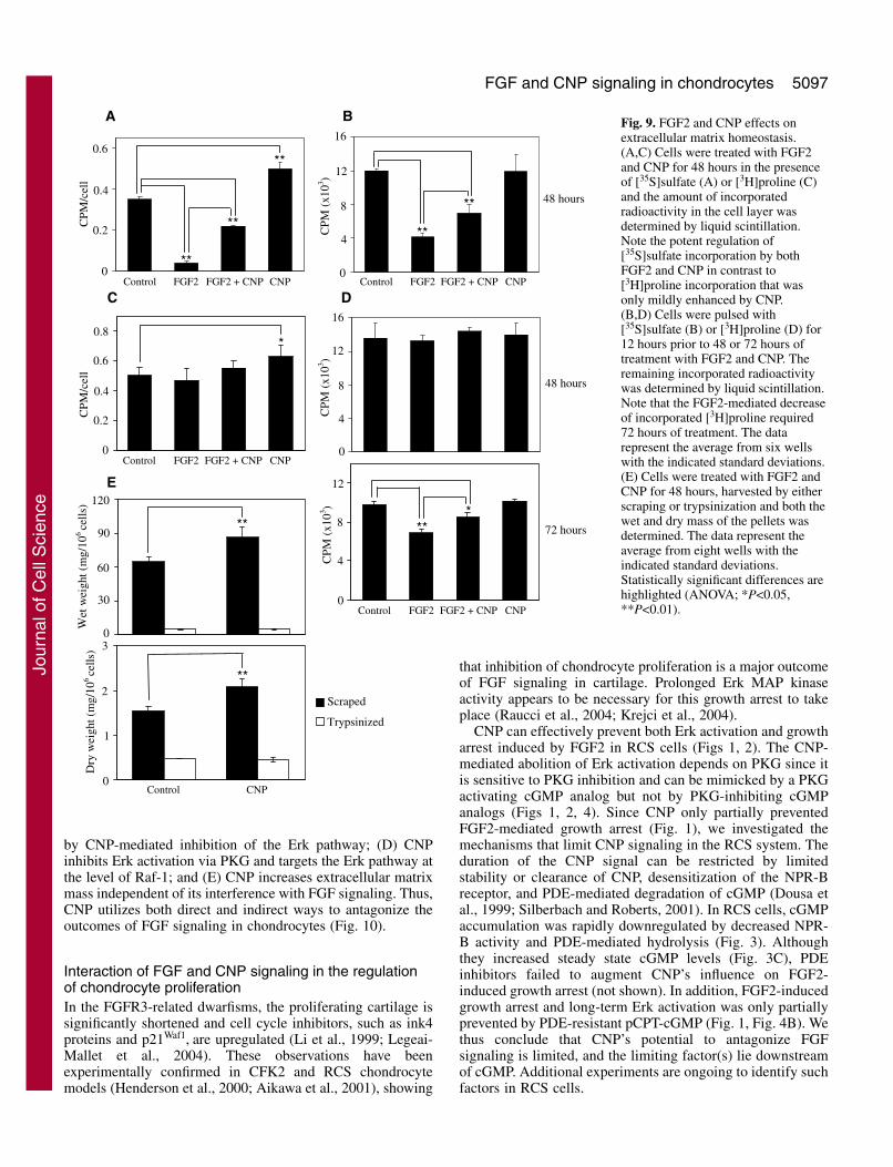
The moderate inhibition of both FGF2-mediated inductionand activation of matrix-remodeling molecules by CNP (Fig.8) is unlikely to fully account for the rather significant effectof CNP on FGF2-mediated matrix loss (Fig. 7). Therefore, togain a better insight into the regulation of RCS extracellularmatrix turnover, we treated cells with FGF2 and CNP for 48or 72 hours and assessed the rate of matrix production anddegradation by radioactive labeling both the sulfatedproteoglycan and collagen compartments of the matrix. The[35S]sulfate uptake was significantly inhibited by FGF2 (Fig.9A) similar to the results obtained in pulse-chase experiment(Fig. 9B) suggesting that FGF2 inhibits synthesis and triggersdegradation of the proteoglycan matrix. Addition of CNP
partially reversed the effects of FGF2 on the proteoglycanmatrix (Fig. 9A,B). Moreover, CNP alone significantlyincreased [35S]sulfate incorporation (Fig. 9A). Although CNPincreased both wet and dry weight of the cell pellet obtainedby scraping, no effect was observed when cells were harvestedby trypsinization (Fig. 9E), confirming that CNP increases themass of the RCS extracellular matrix. FGF2 did not affect[3H]proline uptake (Fig. 9C). After 48 hours of treatment,FGF2 weakly decreased the amount of incorporated[3H]proline in one of five replicate pulse-chase experiments(not shown) in contrast to a 72 hour treatment leading to aninvariant 30% loss of [3H]proline incorporation in all threereplicate experiments (Fig. 9D).
To test whether the CNP-mediated increase of RCSextracellular matrix mass correlates with increasedproteoglycan expression, cells were treated with CNP for 0.5,1, 2, 4, 6, 8 and 24 hours and the expression of a panel ofproteoglycans (aggrecan, biglycan, brevican, chondroitin-sulphate proteoglycan 3 (Cspg3), Cspg4, Cspg5, Cspg6,clusterin, decorin, glypican, lumican, perlecan, syndecan-1,syndecan-2, syndecan-4 and versican) was determined by real-time RT-PCR. Although all of the molecules studied wereexpressed by RCS cells, CNP did not upregulate the transcriptamounts of any of them (not shown).
DiscussionIn this study, we investigated the FGF and CNP signalinginteractions in a chondrocyte environment. We found that: (A)FGF signaling affects chondrocyte behavior by two principalphenotypes, inhibition of proliferation and degradation of theextracellular matrix; (B) both phenotypes require, at leastpartially, activation of Erk MAP kinase and (C) can be reversed
Journal of Cell Science 118 (21)
Fig. 8. CNP effects on FGF2-mediatedinduction and activation of matrix-remodeling molecules. (A) Cells weretreated with FGF2 for 4 or 24 hours andthe expression of the listed moleculeswas assayed by RT-PCR. Levels ofGapdh serve as a control for templatequantity. (B) Cells were treated withFGF2 and either CNP or the MEKinhibitor U0126 for 4 hours and theamounts of Mmp3, Mmp10, Mmp13,Adamts1 and Adamts5 mRNAs weredetermined by real-time RT-PCR.Transcript quantities are relative tountreated cells. Mmp2 and Mmp9 werenot studied due to the possiblenonspecific actions of U0126 during theprolonged cultivation time necessary fortheir induction. (C,D) Cells weretreated with FGF2 and CNP for 72hours and the conditioned culture mediawas analyzed for MMP activity (C) byzymography using gelatin (upperfigure) or casein (lower figure) as asubstrate or (D) for presence of MMP13by WB. The zymography signal wasquantified by densitometry andgraphed.
Jour
nal o
f Cel
l Sci
ence
5097FGF and CNP signaling in chondrocytes
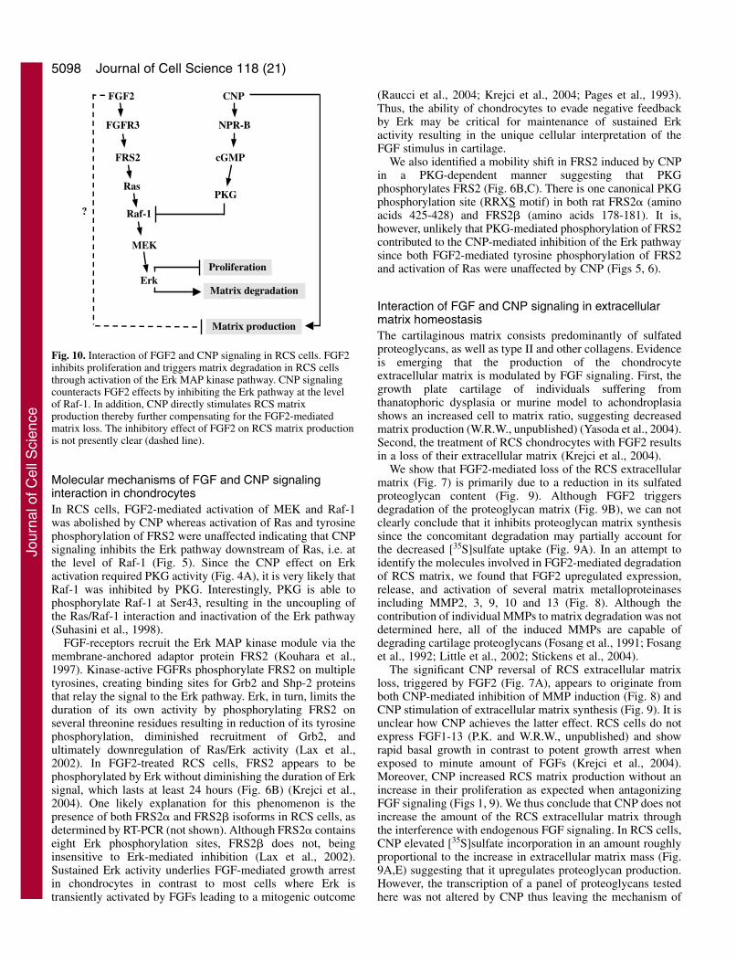
by CNP-mediated inhibition of the Erk pathway; (D) CNPinhibits Erk activation via PKG and targets the Erk pathway atthe level of Raf-1; and (E) CNP increases extracellular matrixmass independent of its interference with FGF signaling. Thus,CNP utilizes both direct and indirect ways to antagonize theoutcomes of FGF signaling in chondrocytes (Fig. 10).
Interaction of FGF and CNP signaling in the regulationof chondrocyte proliferationIn the FGFR3-related dwarfisms, the proliferating cartilage issignificantly shortened and cell cycle inhibitors, such as ink4proteins and p21Waf1, are upregulated (Li et al., 1999; Legeai-Mallet et al., 2004). These observations have beenexperimentally confirmed in CFK2 and RCS chondrocytemodels (Henderson et al., 2000; Aikawa et al., 2001), showing
that inhibition of chondrocyte proliferation is a major outcomeof FGF signaling in cartilage. Prolonged Erk MAP kinaseactivity appears to be necessary for this growth arrest to takeplace (Raucci et al., 2004; Krejci et al., 2004).
CNP can effectively prevent both Erk activation and growtharrest induced by FGF2 in RCS cells (Figs 1, 2). The CNP-mediated abolition of Erk activation depends on PKG since itis sensitive to PKG inhibition and can be mimicked by a PKGactivating cGMP analog but not by PKG-inhibiting cGMPanalogs (Figs 1, 2, 4). Since CNP only partially preventedFGF2-mediated growth arrest (Fig. 1), we investigated themechanisms that limit CNP signaling in the RCS system. Theduration of the CNP signal can be restricted by limitedstability or clearance of CNP, desensitization of the NPR-Breceptor, and PDE-mediated degradation of cGMP (Dousa etal., 1999; Silberbach and Roberts, 2001). In RCS cells, cGMPaccumulation was rapidly downregulated by decreased NPR-B activity and PDE-mediated hydrolysis (Fig. 3). Althoughthey increased steady state cGMP levels (Fig. 3C), PDEinhibitors failed to augment CNP’s influence on FGF2-induced growth arrest (not shown). In addition, FGF2-inducedgrowth arrest and long-term Erk activation was only partiallyprevented by PDE-resistant pCPT-cGMP (Fig. 1, Fig. 4B). Wethus conclude that CNP’s potential to antagonize FGFsignaling is limited, and the limiting factor(s) lie downstreamof cGMP. Additional experiments are ongoing to identify suchfactors in RCS cells.
CP
M(x
103 )
4
8
12
0
***
Control FGF2 FGF2 + CNP CNP
72 hours
8
12
16
4
00
0.2
0.4
0.6
0.8
48 hours
*
Control FGF2 FGF2 + CNP CNP
C D
A B
CP
M/c
ell
CP
M/c
ell
0
0.2
0.4
0.616
CP
M(x
103 )
CP
M(x
103 )
4
8
12**
**
**
**
**
0
48 hours
Control FGF2 FGF2 + CNP CNP Control FGF2 FGF2 + CNP CNP
E
0
Wet
wei
ght(
mg/
106ce
lls)
30
60
120
0
90
Series1Series2
Dry
wei
ght(
mg/
106ce
lls)
2
1
3
Control CNP
S e rie s1
S e rie s2
Trypsinized
Scraped
**
**
Fig. 9. FGF2 and CNP effects onextracellular matrix homeostasis.(A,C) Cells were treated with FGF2and CNP for 48 hours in the presenceof [35S]sulfate (A) or [3H]proline (C)and the amount of incorporatedradioactivity in the cell layer wasdetermined by liquid scintillation.Note the potent regulation of[35S]sulfate incorporation by bothFGF2 and CNP in contrast to[3H]proline incorporation that wasonly mildly enhanced by CNP.(B,D) Cells were pulsed with[35S]sulfate (B) or [3H]proline (D) for12 hours prior to 48 or 72 hours oftreatment with FGF2 and CNP. Theremaining incorporated radioactivitywas determined by liquid scintillation.Note that the FGF2-mediated decreaseof incorporated [3H]proline required72 hours of treatment. The datarepresent the average from six wellswith the indicated standard deviations.(E) Cells were treated with FGF2 andCNP for 48 hours, harvested by eitherscraping or trypsinization and both thewet and dry mass of the pellets wasdetermined. The data represent theaverage from eight wells with theindicated standard deviations.Statistically significant differences arehighlighted (ANOVA; *P<0.05,**P<0.01).
Jour
nal o
f Cel
l Sci
ence
5098
Molecular mechanisms of FGF and CNP signalinginteraction in chondrocytesIn RCS cells, FGF2-mediated activation of MEK and Raf-1was abolished by CNP whereas activation of Ras and tyrosinephosphorylation of FRS2 were unaffected indicating that CNPsignaling inhibits the Erk pathway downstream of Ras, i.e. atthe level of Raf-1 (Fig. 5). Since the CNP effect on Erkactivation required PKG activity (Fig. 4A), it is very likely thatRaf-1 was inhibited by PKG. Interestingly, PKG is able tophosphorylate Raf-1 at Ser43, resulting in the uncoupling ofthe Ras/Raf-1 interaction and inactivation of the Erk pathway(Suhasini et al., 1998).
FGF-receptors recruit the Erk MAP kinase module via themembrane-anchored adaptor protein FRS2 (Kouhara et al.,1997). Kinase-active FGFRs phosphorylate FRS2 on multipletyrosines, creating binding sites for Grb2 and Shp-2 proteinsthat relay the signal to the Erk pathway. Erk, in turn, limits theduration of its own activity by phosphorylating FRS2 onseveral threonine residues resulting in reduction of its tyrosinephosphorylation, diminished recruitment of Grb2, andultimately downregulation of Ras/Erk activity (Lax et al.,2002). In FGF2-treated RCS cells, FRS2 appears to bephosphorylated by Erk without diminishing the duration of Erksignal, which lasts at least 24 hours (Fig. 6B) (Krejci et al.,2004). One likely explanation for this phenomenon is thepresence of both FRS2� and FRS2� isoforms in RCS cells, asdetermined by RT-PCR (not shown). Although FRS2� containseight Erk phosphorylation sites, FRS2� does not, beinginsensitive to Erk-mediated inhibition (Lax et al., 2002).Sustained Erk activity underlies FGF-mediated growth arrestin chondrocytes in contrast to most cells where Erk istransiently activated by FGFs leading to a mitogenic outcome
(Raucci et al., 2004; Krejci et al., 2004; Pages et al., 1993).Thus, the ability of chondrocytes to evade negative feedbackby Erk may be critical for maintenance of sustained Erkactivity resulting in the unique cellular interpretation of theFGF stimulus in cartilage.
We also identified a mobility shift in FRS2 induced by CNPin a PKG-dependent manner suggesting that PKGphosphorylates FRS2 (Fig. 6B,C). There is one canonical PKGphosphorylation site (RRXS motif) in both rat FRS2� (aminoacids 425-428) and FRS2� (amino acids 178-181). It is,however, unlikely that PKG-mediated phosphorylation of FRS2contributed to the CNP-mediated inhibition of the Erk pathwaysince both FGF2-mediated tyrosine phosphorylation of FRS2and activation of Ras were unaffected by CNP (Figs 5, 6).
Interaction of FGF and CNP signaling in extracellularmatrix homeostasisThe cartilaginous matrix consists predominantly of sulfatedproteoglycans, as well as type II and other collagens. Evidenceis emerging that the production of the chondrocyteextracellular matrix is modulated by FGF signaling. First, thegrowth plate cartilage of individuals suffering fromthanatophoric dysplasia or murine model to achondroplasiashows an increased cell to matrix ratio, suggesting decreasedmatrix production (W.R.W., unpublished) (Yasoda et al., 2004).Second, the treatment of RCS chondrocytes with FGF2 resultsin a loss of their extracellular matrix (Krejci et al., 2004).
We show that FGF2-mediated loss of the RCS extracellularmatrix (Fig. 7) is primarily due to a reduction in its sulfatedproteoglycan content (Fig. 9). Although FGF2 triggersdegradation of the proteoglycan matrix (Fig. 9B), we can notclearly conclude that it inhibits proteoglycan matrix synthesissince the concomitant degradation may partially account forthe decreased [35S]sulfate uptake (Fig. 9A). In an attempt toidentify the molecules involved in FGF2-mediated degradationof RCS matrix, we found that FGF2 upregulated expression,release, and activation of several matrix metalloproteinasesincluding MMP2, 3, 9, 10 and 13 (Fig. 8). Although thecontribution of individual MMPs to matrix degradation was notdetermined here, all of the induced MMPs are capable ofdegrading cartilage proteoglycans (Fosang et al., 1991; Fosanget al., 1992; Little et al., 2002; Stickens et al., 2004).
The significant CNP reversal of RCS extracellular matrixloss, triggered by FGF2 (Fig. 7A), appears to originate fromboth CNP-mediated inhibition of MMP induction (Fig. 8) andCNP stimulation of extracellular matrix synthesis (Fig. 9). It isunclear how CNP achieves the latter effect. RCS cells do notexpress FGF1-13 (P.K. and W.R.W., unpublished) and showrapid basal growth in contrast to potent growth arrest whenexposed to minute amount of FGFs (Krejci et al., 2004).Moreover, CNP increased RCS matrix production without anincrease in their proliferation as expected when antagonizingFGF signaling (Figs 1, 9). We thus conclude that CNP does notincrease the amount of the RCS extracellular matrix throughthe interference with endogenous FGF signaling. In RCS cells,CNP elevated [35S]sulfate incorporation in an amount roughlyproportional to the increase in extracellular matrix mass (Fig.9A,E) suggesting that it upregulates proteoglycan production.However, the transcription of a panel of proteoglycans testedhere was not altered by CNP thus leaving the mechanism of
Journal of Cell Science 118 (21)
Raf-1
FGFR3 NPR-B
FRS2 cGMP
Ras
MEK
ErkProliferation
Matrix degradation
FGF2 CNP
Matrix production
PKG
?
Fig. 10. Interaction of FGF2 and CNP signaling in RCS cells. FGF2inhibits proliferation and triggers matrix degradation in RCS cellsthrough activation of the Erk MAP kinase pathway. CNP signalingcounteracts FGF2 effects by inhibiting the Erk pathway at the levelof Raf-1. In addition, CNP directly stimulates RCS matrixproduction thereby further compensating for the FGF2-mediatedmatrix loss. The inhibitory effect of FGF2 on RCS matrix productionis not presently clear (dashed line).
Jour
nal o
f Cel
l Sci
ence
5099FGF and CNP signaling in chondrocytes
CNP-mediated increase in the RCS extracellular matrix opento future investigation.
Upon FGF2 treatment, we found elevated expression andrelease of fibronectin into the culture media (Fig. 7B,C). Wepresently do not know the effect of increased fibronectin onRCS cells. Addition of fibronectin to hepatic stellate or lungcarcinoma cells results in Erk activation (Poulos et al., 1997;Han et al., 2005) and thus FGF2-induced fibronectin maycontribute to the maintenance of long-term Erk activity inRCS cells and thereby to FGF2-mediated growth arrest.Additional experiments are now ongoing to test thishypothesis.
We are grateful to P. Lin and R. Radha for assistance with the FACSdata analysis. This work was supported by Yang Seng Tang USACompany and NIH 5P01-HD22657.
ReferencesAikawa, T., Segre, G. V. and Lee, K. (2001). Fibroblast growth factor inhibits
chondrocytic growth through induction of p21 and subsequent inactivationof cyclin E-Cdk2. J. Biol. Chem. 276, 29347-29352.
Bartels, C. F., Bükülmez, H., Padayatti, P., Rhee, D. K., van Ravenswaaij-Arts, C., Pauli, R. M., Mundlos, S., Chitayat, D., Shih, L. Y., Al-Gazali,L. I. et al. (2004). Mutations in the transmembrane natriuretic peptidereceptor NPR-B impair skeletal growth and cause acromesomelic dysplasia,type Maroteaux. Am. J. Hum. Genet. 75, 27-34.
Chusho, H., Tamura, N., Ogawa, Y., Yasoda, A., Suda, M., Miyazawa, T.,Nakamura, K., Nakao, K., Kurihara, T., Komatsu, Y. et al. (2001).Dwarfism and early death in mice lacking C-type natriuretic peptide. Proc.Natl. Acad. Sci. USA 98, 4016-4021.
Dhillon, A. S. and Kolch, W. (2002). Untying the regulation of the Raf-1kinase. Arch. Biochem. Biophys. 404, 3-9.
Dousa, T. P. (1999). Cyclic-3�,5�-nucleotide phosphodiesterase isozymes incell biology and pathophysiology of the kidney. Kidney Int. 55, 29-62.
Drewett, J. G. and Garbers, D. L. (1994). The family of guanylyl cyclasereceptors and their ligands. Endocr. Rev. 15, 135-162.
Fosang, A. J., Neame, P. J., Hardingham, T. E., Murphy, G. and Hamilton,J. A. (1991). Cleavage of cartilage proteoglycan between G1 and G2domains by stromelysins. J. Biol. Chem. 266, 15579-15582.
Fosang, A. J., Neame, P. J., Last, K., Hardingham, T. E., Murphy, G. andHamilton, J. A. (1992). The interglobular domain of cartilage aggrecan iscleaved by PUMP, gelatinases, and cathepsin B. J. Biol. Chem. 267, 19470-19474.
Hagiwara, H., Sakaguchi, H., Itakura, M., Yoshimoto, T., Furuya, M.,Tanaka, S. and Hirose, S. (1994). Autocrine regulation of rat chondrocyteproliferation by natriuretic peptide C and its receptor, natriuretic peptidereceptor-B. J. Biol. Chem. 269, 10729-10733.
Han, S., Sidell, N. and Roman, J. (2005). Fibronectin stimulates human lungcarcinoma cell proliferation by suppressing p21 gene expression via signalsinvolving Erk and Rho kinase. Cancer Lett. 219, 71-81.
Henderson, J. E., Naski, M. C., Aarts, M. M., Wang, D., Cheng, L.,Goltzman, D. and Ornitz, D. M. (2000). Expression of FGFR3 with theG380R achondroplasia mutation inhibits proliferation and maturation ofCFK2 chondrocytic cells. J. Bone Miner. Res. 15, 155-165.
Kase, H., Iwahashi, K., Nakanishi, S., Matsuda, Y., Yamada, K.,Takahashi, M., Murakata, C., Sato, A. and Kaneko, M. (1987). K-252compounds novel and potent inhibitors of protein kinase C and cyclicnucleotide-dependent protein kinases. Biochem. Biophys. Res. Commun.142, 436-440.
Koller, K. J. and Goeddel, D. V. (1992). Molecular biology of the natriureticpeptides and their receptors. Circulation 86, 1081-1088.
Kouhara, H., Hadari, Y. R., Spivak-Kroizman, T., Schilling, J., Bar-Sagi,D., Lax, I. and Schlessinger, J. (1997). A lipid-anchored Grb2-bindingprotein that links FGF-receptor activation to the Ras/MAPK signalingpathway. Cell 89, 693-702.
Krejci, P., Bryja, V., Pachernik, J., Hampl, A., Pogue, R., Mekikian, P. andWilcox, W. R. (2004). FGF2 inhibits proliferation and alters the cartilage-like phenotype of RCS cells. Exp. Cell Res. 297, 152-164.
Lax, I., Wong, A., Lamothe, B., Lee, A., Frost, A., Hawes, J. and
Schlessinger, J. (2002). The docking protein FRS2alpha controls a MAPkinase-mediated negative feedback mechanism for signaling by FGFreceptors. Mol. Cell 10, 709-719.
Legeai-Mallet, L., Benoist-Lasselin, C., Munnich, A. and Bonaventure, J.(2004). Overexpression of FGFR3, Stat1, Stat5 and p21Cip1 correlates withphenotypic severity and defective chondrocyte differentiation in FGFR3-related chondrodysplasias. Bone 34, 26-36.
Li, C., Chen, L., Iwata, T., Kitagawa, M., Fu, X. Y. and Deng, C. X. (1999).A Lys644Glu substitution in fibroblast growth factor receptor 3 (FGFR3)causes dwarfism in mice by activation of STATs and ink4 cell cycleinhibitors. Hum. Mol. Genet. 8, 35-44.
Little, C. B., Hughes, C. E., Curtis, C. L., Janusz, M. J., Bohne, R., Wang-Weigand, S., Taiwo, Y. O., Mitchell, P. G., Otterness, I. G., Flannery, C.R. et al. (2002). Matrix metalloproteinases are involved in C-terminal andinterglobular domain processing of cartilage aggrecan in late stage cartilagedegradation. Matrix Biol. 21, 271-288.
Lucas, K. A., Pitari, G. M., Kazerounian, S., Ruiz-Stewart, I., Park, J.,Schulz, S., Chepenik, K. P. and Waldman, S. A. (2000). Guanylyl cyclasesand signaling by cyclic GMP. Pharmacol. Rev. 52, 375-414.
Marshak, D. R., Lukas, T. J. and Watterson, D. M. (1985). Drug-proteininteractions: binding of chlorpromazine to calmodulin, calmodulinfragments, and related calcium binding proteins. Biochemistry 24, 144-150.
Mericq, V., Uyeda, J. A., Barnes, K. M., De Luca, F. and Baron, J. (2000).Regulation of fetal rat bone growth by C-type natriuretic peptide and cGMP.Pediatr. Res. 47, 189-193.
Miller, J. P., Boswell, K. H., Muneyama, K., Simon, L. N., Robins, R. K.and Shuman, D. A. (1973). Synthesis and biochemical studies of various8-substituted derivatives of guanosine 3�,5�-cyclic phosphate, inosine 3�,5�-cyclic phosphate, and xanthosine 3�,5�-cyclic phosphate. Biochemistry 12,5310-5319.
Miyazawa, T., Ogawa, Y., Chusho, H., Yasoda, A., Tamura, N., Komatsu,Y., Pfeifer, A., Hofmann, F. and Nakao, K. (2002). Cyclic GMP-dependentprotein kinase II plays a critical role in C-type natriuretic peptide-mediatedendochondral ossification. Endocrinology 143, 3604-3610.
Mukoyama, M., Nakao, K., Hosoda, K., Suga, S., Saito, Y., Ogawa, Y.,Shirakami, G., Jougasaki, M., Obata, K., Yasue, H. et al. (1991). Brainnatriuretic peptide as a novel cardiac hormone in humans. Evidence for anexquisite dual natriuretic peptide system, atrial natriuretic peptide and brainnatriuretic peptide. J. Clin. Invest. 87, 1402-1412.
Murakami, S., Balmes, G., McKinney, S., Zhang, Z., Givol, D. and deCrombrugghe, B. (2004). Constitutive activation of MEK1 in chondrocytescauses Stat1-independent achondroplasia-like dwarfism and rescues theFgfr3-deficient mouse phenotype. Genes Dev. 18, 290-305.
Nakamura, H., Fujii, Y., Ohuchi, E., Yamamoto, E. and Okada, Y. (1998).Activation of the precursor of human stromelysin 2 and its interactions withother matrix metalloproteinases. Eur. J. Biochem. 253, 67-75.
Okada, A., Tomasetto, C., Lutz, Y., Bellocq, J. P., Rio, M. C. and Basset,P. (1997). Expression of matrix metalloproteinases during rat skin woundhealing: evidence that membrane type-1 matrix metalloproteinase is astromal activator of pro-gelatinase A. J. Cell Biol., 137, 67-77.
Pages, G., Lenormand, P., L’Allemain, G., Chambard, J. C., Meloche, S.,Pouyssegur, J. (1993). Mitogen-activated protein kinases p42mapk andp44mapk are required for fibroblast proliferation. Proc. Natl. Acad. Sci. USA90, 8319-8323.
Passos-Bueno, M. R., Wilcox, W. R., Jabs, E. W., Sertie, A. L., Alonso, L.G. and Kitoh, H. (1999). Clinical spectrum of fibroblast growth factorreceptor mutations. Hum. Mutat. 14, 115-125.
Poulos, J. E., Weber, J. D., Bellezzo, J. M., Di Bisceglie, A. M., Britton, R.S., Bacon, B. R. and Baldassare, J. J. (1997). Fibronectin and cytokinesincrease JNK, ERK, AP-1 activity, and transin gene expression in rat hepaticstellate cells. Am. J. Physiol. 273, G804-G811.
Raucci, A., Laplantine, E., Mansukhani, A. and Basilico, C. (2004).Activation of the ERK1/2 and p38 mitogen-activated protein kinasepathways mediates fibroblast growth factor-induced growth arrest ofchondrocytes. J. Biol. Chem. 279, 1747-1756.
Rosenzweig, A. and Seidman, C. (1991). Atrial natriuretic factor and relatedpeptide hormones. Annu. Rev. Biochem. 60, 229-255.
Silberbach, M. and Roberts, C. T., Jr (2001). Natriuretic peptide signalling:molecular and cellular pathways to growth regulation. Cell Signal. 13, 221-231.
Sirotkin, A. V., Makarevich, A. V., Genieser, H. G., Kotwica, J., Hetenyi,L. (2000). Effect of four cGMP analogues with different mechanisms ofaction on hormone release by porcine ovarian granulosa cells in vitro. Exp.Clin. Endocrinol. Diabetes 108, 214-219.
Jour
nal o
f Cel
l Sci
ence
5100
Stickens, D., Behonick, D. J., Ortega, N., Heyer, B., Hartenstein, B., Yu,Y., Fosang, A. J., Schorpp-Kistner, M., Angel, P. and Werb, Z. (2004).Altered endochondral bone development in matrix metalloproteinase 13-deficient mice. Development 131, 5883-5895.
Suga, S., Nakao, K., Hosoda, K., Mukoyama, M., Ogawa, Y., Shirakami,G., Arai, H., Kambayashi, Y., Inouye, K. et al. (1992). Receptorselectivity of natriuretic peptide family, atrial natriuretic peptide, brainnatriuretic peptide, and C-type natriuretic peptide. Endocrinology 130, 229-239.
Suhasini, M., Li, H., Lohmann, S. M., Boss, G. R. and Pilz, R. B. (1998).Cyclic-GMP-dependent protein kinase inhibits the Ras/Mitogen-activatedprotein kinase pathway. Mol. Cell. Biol. 18, 6983-6994.
Takase, Y., Saeki, T., Watanabe, N., Adachi, H., Souda, S. and Saito, I.(1994). Cyclic GMP phosphodiesterase inhibitors. 2. Requirement of 6-substitution of quinazoline derivatives for potent and selective inhibitoryactivity. J. Med. Chem. 37, 2106-2111.
Tamura, N., Doolittle, L. K., Hammer, R. E., Shelton, J. M., Richardson,J. A. and Garbers, D. L. (2004). Critical roles of the guanylyl cyclase B
receptor in endochondral ossification and development of femalereproductive organs. Proc. Natl. Acad. Sci. USA 101, 17300-17305.
Tsuji, T. and Kunieda, T. (2005). A loss-of-function mutation in natriureticpeptide receptor 2 (Npr2) gene is responsible for disproportionate dwarfismin cn/cn mouse. J. Biol. Chem. 280, 14288-14292.
Wilcox, W. R., Tavormina, P. L., Krakow, D., Kitoh, H., Lachman, R. S.,Wasmuth, J. J., Thompson, L. M. and Rimoin, D. L. (1998). Molecular,radiologic, and histopathologic correlations in thanatophoric dysplasia. Am.J. Med. Genet. 78, 274-281.
Yasoda, A., Ogawa, Y., Suda, M., Tamura, N., Mori, K., Sakuma, Y.,Chusho, H., Shiota, K., Tanaka, K. and Nakao, K. (1998). Natriureticpeptide regulation of endochondral ossification. Evidence for possible rolesof the C-type natriuretic peptide/guanylyl cyclase-B pathway. J. Biol. Chem.273, 11695-11700.
Yasoda, A., Komatsu, Y., Chusho, H., Miyazawa, T., Ozasa, A., Miura, M.,Kurihara, T., Rogi, T., Tanaka, S., Suda, M. et al. (2004). Overexpressionof CNP in chondrocytes rescues achondroplasia through a MAPK-dependent pathway. Nat. Med. 10, 80-86.
Journal of Cell Science 118 (21)
Jour
nal o
f Cel
l Sci
ence