Embed Size (px)
Citation preview
JOURNAL OF VIROLOGY, Apr. 2002, p. 3257–3266 Vol. 76, No. 70022-538X/02/$04.00�0 DOI: 10.1128/JVI.76.7.3257–3266.2002Copyright © 2002, American Society for Microbiology. All Rights Reserved.
Interaction between Parvovirus NS2 Protein and Nuclear ExportFactor Crm1 Is Important for Viral Egress from
the Nucleus of Murine CellsCathy L. Miller† and David J. Pintel*
School of Medicine, University of Missouri—Columbia, Columbia, Missouri 65212
Received 19 September 2001/Accepted 7 January 2002
A mutation that disrupts the interaction between the NS2 protein of minute virus of mice and the nuclearexport factor Crm1 results in a block to egress of mutant-generated full virions from the nucleus of infectedmurine cells. These mutants produce wild-type levels of monomer and dimer replicative DNA forms but areimpaired in their ability to generate progeny single-stranded DNA in restrictive murine cells in the first roundof infection. The NS2-Crm1 interaction mutant can be distinguished phenotypically from an NS2-null mutantand reveals a role for the Crm1-mediated export pathway at a late step in viral infection.
The autonomous parvovirus minute virus of mice (MVM)produces two nonstructural proteins that play critical roles inthe replication of the virus (6–8, 18). The large nonstructuralprotein, NS1, is a multifunctional protein with site-specificDNA binding, nickase, ATPase, and helicase activities thathave been mapped to specific regions of the NS1 protein andare critical for MVM replication (8, 17, 19, 20). The smallnonstructural protein, NS2, is also required for MVM replica-tion in a host-cell-specific manner; in murine cells NS2-nullmutants (18), as well as a number of other characterized NS2mutants (5), generate little double-stranded replicative inter-mediates and little to no progeny single-stranded DNA(ssDNA) is produced. Cells from a variety of other speciessupport MVM replication at near-wild-type levels in the ab-sence of NS2 (18).
The role(s) NS2 plays in MVM replication remains unclear.In the absence of NS2, wild-type levels of the VP1 and VP2structural proteins are synthesized at early times postinfection;however, they do not efficiently assemble into MVM capsids(5). Although the mechanisms are not well understood, theproduction of MVM progeny ssDNA is tightly connected tothe availability of assembled MVM capsids (8), and so it islikely that, at least to some extent, the lack of progeny ssDNAproduced during NS2 mutant infection is a consequence of thelack of proper capsid assembly. The lack of replication ofmonomer replicative DNA forms (mRF) and dimer replicativeDNA forms (dRF) in NS2-null mutant infection is more diffi-cult to explain, as this process is known to proceed efficiently inthe absence of capsid production (23).
NS2 was recently shown to interact with two members of the14–3–3 family of signaling proteins (3) and the nuclear exportfactor Crm1 (2, 21). Crm1 is a sequence-specific nuclear exportreceptor that binds leucine-rich sequences within proteins in
cooperation with RanGTP and actively transports these pro-teins from the nucleus to the cytoplasm (9, 24). The NS2interaction with Crm1 was previously shown to be important inproper localization of NS2 within the cell and was mapped toNS2 amino acids (aa) 81 to 103 by peptide analysis (N. Salome,personal communication) and glutathione S-transferase (GST)pull-down assays (21). The role of the NS2-Crm1 interactionduring MVM infection, however, has not been determined.
We have investigated the consequence of the NS2 interac-tion with Crm1 during viral replication by creating a mutationwithin NS2 that disrupts its interaction with Crm1. In contrastto MVM NS2-null mutant replication, NS2-Crm1� mutantsgenerate double-stranded replicative-form DNA at near-wild-type levels at early times postinfection; however, there is astriking decrease in the production of progeny ssDNA. Wefurther show that this defect in ssDNA production is associatedwith a severe defect in the export of full MVM virions from thenucleus to the cytoplasm of the cell. Export of MVM virionsfrom the nucleus is also blocked by the Crm1-specific inhibitorleptomycin B.
MATERIALS AND METHODS
Cells, viruses, and transfections. Murine A92L, murine ID5, and humanNB324K cells were maintained and passaged as described previously (22). Wild-type and mutant the MVM prototype (MVMp) virus was grown from transfec-tion and titers were determined on NB324K cell monolayers. All viral stockswere equilibrated by ssDNA content normalized to a wild-type multiplicity ofinfection (MOI) as indicated. NS2-null mutant (1989) virus, which contains asingle-nucleotide change at the MVM large splice acceptor site at nucleotide (nt)1989 and makes no NS2 protein, was previously described (18). A92L cells weredoubly blocked at the G1/S border where indicated by 44 h of growth in isole-ucine-deficient media followed by 12 h of growth in the presence of 12 �g ofaphidicolin/ml (Sigma Chemical, St. Louis, Mo.) as described elsewhere (18).Leptomycin B (Sigma Chemical) was added to cells at a final concentration of 10nM where indicated. Plaque assays were done as previously described (18), usingmethylene blue to stain fixed cells. Transient-transfection assays were performedby the standard CaPO4 method (5 �g of DNA/60-mm dish) as described previ-ously (23) or with Lipofectamine and Plus reagent (2 �g of DNA/60-mm dish) asrecommended by the supplier (Gibco BRL, Grand Island, N.Y.).
Plasmid constructs. The MVM wild-type and NS2-null (1989) infectiousclones were previously described (15, 18). The NS2-Crm1� MVM infectiousclone was made by overlap PCR mutagenesis (10), changing MVM nt 1990 to2008, such that the NS2 amino acids aa 84-FGTLJI-aa 89 were changed to aa84-VCPVAV-aa 89 without altering the NS1 amino acid sequence. The complete
* Corresponding author. Mailing address: Molecular Microbiologyand Immunology, M616 Medical Sciences Building, University of Mis-souri—Columbia School of Medicine, Columbia, MO 65212. Phone:(573) 882-3920. Fax: (573) 882-4287. E-mail: [email protected]
†Present address: Department of Microbiology and Molecular Ge-netics, Harvard Medical School, Boston, MA 02215.
3257
on June 5, 2018 by guesthttp://jvi.asm
.org/D
ownloaded from
coding region for the mutant protein was sequenced to ensure no additionalmutations were introduced. The pCMVNS2 plasmid was previously described(16). The pCMVNS2-Crm1� plasmid was constructed by overlap PCR mutagen-esis to incorporate the mutation of aa 84-FGTLJI-aa 89 to aa 84-VCPVAV-aa89. PGEX-5X-NS2 was previously described (17). pGEX-5X-NS2-Crm1� wasmade by inserting the EcoRV-XhoI fragment from pCMVNS2-Crm1� intopGEX-5X-NS2. pETCrm1 and pCIHACrm1 were previously described (11) andwere gifts of Mark Hannink, University of Missouri—Columbia.
Immunoprecipitation and Western analysis. A92L cells were transfected asindicated and collected 18 h posttransfection. Immunoprecipitation followed byWestern blotting was performed as described previously (16). Antibodies usedincluded rabbit polyclonal anti- (�)-NS2 C terminus, made against a peptidecontaining NS2 aa 156 to 182, and mouse monoclonal �-HA.11 (Covance, Rich-mond, Calif.).
Indirect IFAs. A92L, ID5, or NB324K cells were grown on either coverslips oreight-well slides (Labtech) and infected at the indicated wild-type MOI. Cover-slips or slides were processed for immunofluorescence assay (IFA) as describedelsewhere (18) at the indicated times postinfection. Images were generated usinga SPOT digital camera, and all images from each experiment were obtained atthe same exposure time. Primary antibodies were used at a 1:100 dilution andincluded rabbit polyclonal �-NS2 made against the internal sequences of NS2 (4)and mouse monoclonal �-assembled capsid (12). Secondary antibodies were
used at a 1:250 dilution and included fluorescein isothiocyanate (FITC)-conju-gated �-rabbit and FITC-conjugated �-mouse antibodies (ICN Biochemicals).
RanGAP protection assay. pGex-5X-NS2 and pGex-5X-NS2-Crm1� weretransformed into and grown in Escherichia coli strain BL21(DE3)pLysS. GST-NS2 and GST-NS2-Crm1� protein expression was induced with 1 mM isopropyl-�-D-thiogalactopyranoside (IPTG), and GST-conjugated proteins were purifiedwith glutathione-agarose as described previously (17). pETCrm1 was trans-formed into and grown in E. coli strain BL21(DE3)pLysS cells. HISCrm1 proteinexpression was induced with 1 mM IPTG and purified by metal chelate affinitychromatography (Invitrogen) as described elsewhere (11). RanGAP protein wasexpressed in BLR pREP4 cells and purified by metal chelate affinity chromatog-raphy (Invitrogen). RanGAP protection assays were then done as describedelsewhere (1, 11). Briefly, Ran protein was incubated with [�-32P]GTP (10mCi/ml) for 30 min on ice in buffer containing 20 mM HEPES (pH 7.3), 100 mMpotassium acetate, and 5 mM EDTA, followed by gel filtration on a Bio-Spin 6column (Bio-Rad). Reaction mixtures containing increasing amounts of purified,beaded GST-NS2 or GST-NS2-Crm1� were incubated at 15°C for 30 min with500 nM purified HisCrm1 and 50 pM [�-32P]RanGTP in 25 �l of hydrolysisbuffer to allow GST-NS2 or GST-NS2-Crm1�–HISCrm1–RanGTP ternary com-plexes to form. Ten nanomolar RanGAP was then added and the mixtures wereincubated at 30°C for 2 min to allow hydrolysis of Ran GTP that was notprotected in a ternary complex. The reaction mixtures were filtered through
FIG. 1. Mutation within the NS2 NES disrupts NS2 interaction with Crm1. (A) Graphical representation of the NS2 NES mutant sequence.(B) A92L cells were transfected with HA-Crm1 and either no plasmid, pCMVNS2, or pCMVNS2-Crm1�. At 18 h posttransfection, cells wereimmunoprecipitated with �-NS2 antibody followed by Western blotting with �-HA antibody (left panel). Parallel samples were subjected toWestern blot analysis with either �-NS2 (center panel) or �-HA (right panel) antibody to visualize NS2 and HA-Crm1 protein expression. Forreasons not yet determined, the mutant NS2 protein migrates slightly slower than the wild type. The complete coding region for the mutant proteinhas been sequenced and contains no additional changes. (C) Results of RanGAP protection assay using purified GST-NS2 (F) or GST-NS2-Crm1�
(�). Percent GTP hydrolysis is plotted against concentration of wild-type or mutant NS2 protein. (D) Localization of NS2 wild type andNS2-Crm1�. A92L cells were infected with either wild-type MVMp or NS2-Crm1� virus at an MOI of 5. At 24 h postinfection, cells were fixed andstained with �-NS2 rabbit primary antibody and FITC-conjugated �-rabbit secondary antibody. Images were obtained on a SPOT camera at thesame exposure time.
3258 MILLER AND PINTEL J. VIROL.
on June 5, 2018 by guesthttp://jvi.asm
.org/D
ownloaded from
nitrocellulose, the filters were washed, and the filtered lysates were counted inthe presence of 3 ml of scintillation fluid. Counts representing percent unbound(unprotected) hydrolyzed RanGTP were plotted against the concentration ofNS2 wild-type or mutant protein present.
Southern analysis. Wild-type and mutant MVM replication forms producedduring infection or transfection were visualized by Southern analysis of total cellextracts as previously described (18, 23). To equilibrate virus by ssDNA content,equivalent PFU of wild-type and mutant viral stocks extracted in 50 mM Tris–10mM EDTA (pH 8.7) were added to micrococcal nuclease buffer and treated withmicrococcal nuclease to remove associated MVM double-stranded DNA(dsDNA). Samples were then lysed in 2% sodium dodecyl sulfate (SDS) andtreated with proteinase K (0.5 mg/ml) to remove viral capsid proteins. Sampleswere run on a 1% agarose gel, and Southern blot analysis was performed asdescribed previously (18, 23). Blots were exposed to a Kodak phosphorimagingscreen, and the ssDNA was quantitated by using a Bio-Rad Imager FX andQuantity One software where indicated.
Western blot analysis. Cells collected 18 h posttransfection or viral stocksequilibrated by PFU or ssDNA were lysed in 2% SDS, and Western blot analysiswas performed as described elsewhere (16). Antibodies used included rabbitpolyclonal �-NS2 made against the NS2 C terminus (aa 156 to 182) and rabbitpolyclonal �-capsid antibody (�-allopeptide) that was previously shown to inter-act with individual MVM capsid proteins VP1 and VP2 and, potentially, assem-bly intermediates (5).
Hemagglutination assay. Hemagglutination assays of MVM wild-type andmutant viral stocks equilibrated by ssDNA were performed as described previ-ously (23).
NS1 infectious center assay. MVM wild-type and mutant viral stocks equili-brated by ssDNA and added at equivalent wild-type MOIs or cell lysates andmedia isolated at various times postinfection and added at equivalent volumeswere used to infect A92L, ID5, or NB324K cells. At 18 h postinfection, the cellswere treated for IFA and stained with rabbit polyclonal �-NS1 primary antibody(4) followed by FITC-conjugated goat �-rabbit secondary antibody (ICN Bio-chemicals). Cells from at least three microscope fields were counted, and thenumber of NS1-positive cells per 100 total cells was plotted.
Assembled capsid and ssDNA ratio assay. A92L cells were doubly blocked atthe G1/S border by incubation in isoleucine-deficient medium followed by treat-ment with 12 �g of aphidicolin/ml (18). During the aphidicolin incubation,wild-type and mutant viruses were added at equivalent wild-type MOIs. At 8 and
12 h postrelease from aphidicolin, 35S was added at 100 �Ci/ml. At 16 h post-release from aphidicolin, cells were collected and immunoprecipitated with �-as-sembled capsid antibody in nondenaturing buffer as previously described (16).After immunoprecipitation samples were split into two, with one half processedfor Southern blot analysis to visualize ssDNA while the other half was run onSDS-polyacrylamide gel electrophoresis (SDS-PAGE) to visualize labeled as-sembled capsid protein. DNA and protein gels were exposed to a phosphorim-aging screen and quantitated using a Bio-Rad Imager FX and Quantity Onesoftware. Ratios of ssDNA associated with assembled capsid were determinedand are shown below. Samples isolated at 8 h postrelease from aphidicolin wereimmunoprecipitated with the same antibody and processed for Southern blottingas a control for input viral ssDNA.
RESULTS
Mutation of NS2 aa 86 to 91 disrupts the NS2-Crm1 inter-action in vivo and functional interaction in vitro and results ina more nuclear-localized NS2. The region of NS2 involved ininteraction with Crm1 has previously been shown to span NS2aa 81 to 106 by both interfering peptide studies (N. Salome,personal communication) and in vitro GST-NS2 pull-downstudies (21). Mutation within the NS2-specific portion of thisregion, which changed NS2 aa 86 to 91 from FGTLTI toVCPVAV (Fig. 1A), was found to disrupt the NS2-Crm1 in-teraction. Following cotransfection of wild-type CMVNS2 anda hemagglutinin (HA)-tagged Crm1 expression construct intomurine A9 cells, HA-tagged Crm1 could be detected by West-ern blotting following immunoprecipitation with an NS2-spe-cific antibody (Fig. 1B). When similar levels of mutant NS2were expressed, however, little to no HA-tagged Crm1 wasimmunoprecipitated with �-NS2, suggesting that the NS2 in-teraction with Crm1 was significantly lost in vivo as a result ofthe 6-aa mutation.
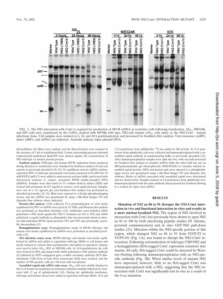
FIG. 2. The NS2 interaction with Crm1 is required for production of MVM ssDNA in restrictive cells following transfection. A92L, NB324K,and ID5 cells were transfected by the CaPO4 method with MVMp wild type, NS2-null mutant (A92L cells only), or the NS2-Crm1� mutantinfectious clone. Cell samples were isolated at 4, 24, and 48 h posttransfection and processed for Southern blot analysis. Viral monomer (mRF),dimer (dRF), and ssDNA are indicated. Asterisks indicate input plasmid DNA.
VOL. 76, 2002 MVM NS2-Crm1 INTERACTION MUTANT 3259
on June 5, 2018 by guesthttp://jvi.asm
.org/D
ownloaded from
Crm1 binds nuclear export signal (NES)-containing proteinscooperatively with RanGTP (9, 14). This interaction protectsRanGTP from hydrolysis by RanGAP in vitro, and the level ofprotection has been used as a quantitative method to measurethe functional interaction between Crm1 and NES-containingproteins (1, 11). While 0.063 �M purified wild-type GST-NS2was required to protect 50% of labeled RanGTP from hydro-lysis, it took 13-fold more GST-NS2-Crm1� (0.82 �M) to pro-tect equivalent levels of RanGTP from RanGAP hydrolysis(Fig. 1C). This difference in protection is similar to what hasbeen reported previously for a well-characterized Crm1-NESloss-of-interaction mutant of I�B� (11) and for studies of wild-type versus mutant NS2 NES peptide (1), suggesting that theNS2-Crm1� mutant loses functional interaction with Crm1.
The NS2-Crm1� interaction mutation was then introducedinto the infectious clone of MVMp. Similar to other NS2mutants, the NS2-Crm1� infectious clone was seen to replicate
very poorly following transfection of murine A9 cells yet rep-licated efficiently in NB324K cells, which allowed the genera-tion of mutant viral stocks on permissive cells. A more detailedcharacterization of the replication of the NS2-Crm1� mutantvirus is presented below.
While wild-type NS2 is found in both the nucleus and thecytoplasm of infected cells, it has been previously shown thatNS2 is relocalized to the nucleus in the presence of the Crm1inhibitor leptomycin B (2, 21). We therefore examined thecellular localization of the mutant NS2. Figure 1D shows thatthere was a significant redistribution of NS2-Crm1� mutant-generated NS2 to the nucleus, providing additional evidencethat the 6-aa mutation within the NS2 NES resulted in a loss ofin vivo interaction of NS2 with Crm1 during MVM infection.
Characterization of NS2-Crm1� mutant replication follow-ing transfection of permissive and nonpermissive cells. NS2has been shown to play a critical role in MVM replication in a
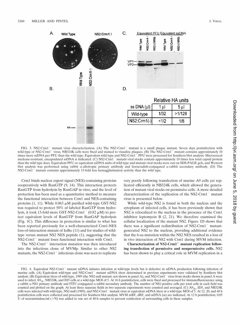
FIG. 3. NS2-Crm1� mutant virus characterization. (A) The NS2-Crm1� mutant is a small plaque mutant. Seven days postinfection withwild-type or NS2-Crm1� virus, NB324K cells were fixed and stained to visualize plaques. (B) The NS2-Crm1� mutant contains approximately 10times more ssDNA per PFU than the wild type. Equivalent wild-type and NS2-Crm1� PFU were processed for Southern blot analysis. Micrococcalnuclease-resistant, encapsidated ssDNA is indicated. (C) NS2-Crm1� mutant viral stocks contain approximately 10 times less total capsid proteinthan the wild type does. Equivalent PFU or equivalent ssDNA units of wild-type and mutant viral stocks were run on SDS-PAGE gels, and Westernblot analysis was performed using rabbit �-allotropic primary antibody and horseradish-conjugated �-rabbit secondary antibody. (D) TheNS2-Crm1� mutant contains approximately 15-fold less hemagglutination activity than the wild type.
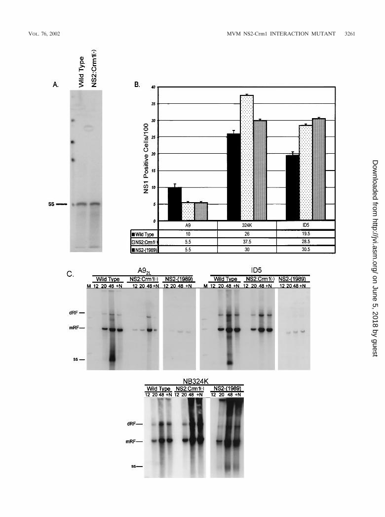
FIG. 4. Equivalent NS2-Crm1� mutant ssDNA initiates infection at wild-type levels but is defective in ssDNA production following infection ofmurine cells. (A) Equivalent wild-type and NS2-Crm1� mutant ssDNA titers determined in previous experiments were validated by Southern blotanalysis. (B) Equivalent titers of wild-type, 1989 (the NS2-null mutant; not shown in panel A), and NS2-Crm1� virus from stocks shown in panel A wereused to infect A92L, NB324K, and ID5 cells at a wild-type MOI of 5. At 16 h postinfection, cells were fixed and processed for immunofluorescence usinga rabbit �-NS1 primary antibody and FITC-conjugated �-rabbit secondary antibody. The number of NS1-positive cells per total cells in each field wascounted and plotted on the graph. At least three separate fields in two separate experiments were counted and averaged. (C) A92L, ID5, and NB324Kcells were infected with wild-type, NS2-null (1989), and NS2-Crm1� mutant virus at equivalent ssDNA titers at a wild-type MOI of 5. At 12, 20, and 48 hpostinfection cells were collected and processed for Southern blot analysis. MVM mRF, dRF, and ssDNA (ss) are indicated. At 12 h postinfection, 0.05U of neuraminidase/ml (�N) was added to one set of 48-h samples to prevent reinfection of surrounding cells in these samples.
3260 MILLER AND PINTEL J. VIROL.
on June 5, 2018 by guesthttp://jvi.asm
.org/D
ownloaded from
VOL. 76, 2002 MVM NS2-Crm1 INTERACTION MUTANT 3261
on June 5, 2018 by guesthttp://jvi.asm
.org/D
ownloaded from
host-cell-specific manner following either viral infection ortransfection of cloned NS2 mutants. There is little productionof NS2-mutant dsDNA replication intermediates, and no de-tectable accumulation of viral progeny ssDNA in murine cells;however, replication is more significant in a variety of permis-sive cell types of other species (18). In contrast to other de-scribed NS2 mutants, the NS2-Crm1� mutant generateddsDNA mRF and dRF at levels nearly equal to those of thewild type at early times following transfection of both permis-sive NB324K cells and restrictive A9 and ID5 murine cells (Fig.2). In restrictive murine cells, mutant mRF and dRF did notamplify at the same rate as with the wild type. In contrast, inpermissive NB324K cells, the NS2-Crm1� mutant generatedmRF and dRF at near-wild-type levels at both early and late
times posttransfection. Strikingly, even at early times post-transfection of restrictive murine cells, a dramatic decrease inthe accumulation of mutant viral progeny ssDNA was seen,and this decrease became relatively greater than wild-type lev-els as the transfection proceeded. We consistently detected atleast 50-fold less of both cell- or media-associated virus (asassayed by either DNA content or the ability to generate NS1-positive cells) following the transfection into restrictive murinecells of the mutant clone compared to the wild type at timeswhen levels of intracellularly replicating double-stranded rep-licative forms were similar (data not shown). The decrease inthe production of mutant ssDNA was not seen at early timesposttransfection of permissive NB324K cells; however, a slightdecrease in the production of ssDNA was apparent at later
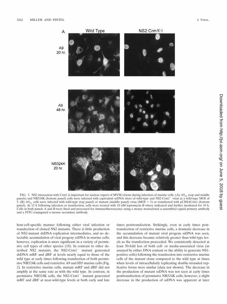
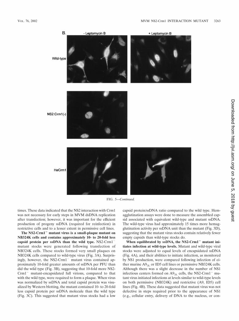
FIG. 5. NS2 interaction with Crm1 is important for nuclear export of MVM virions during infection of murine cells. (A) A92L (top and middlepanels) and NB324K (bottom panel) cells were infected with equivalent ssDNA titers of wild-type and NS2-Crm1� virus at a wild-type MOI of5. (B) A92L cells were infected with wild-type (top panel) or mutant (middle panel) virus (MOI 5) or transfected with pCIHACrm1 (bottompanel). At 12 h following infection or transfection, cells were treated with 10 nM leptomycin B where indicated and further incubated for 10 h.Cells in both panels A and B were fixed and processed for immunofluorescence using a mouse monoclonal �-assembled capsid primary antibodyand a FITC-conjugated �-mouse secondary antibody.
3262 MILLER AND PINTEL J. VIROL.
on June 5, 2018 by guesthttp://jvi.asm
.org/D
ownloaded from
times. These data indicated that the NS2 interaction with Crm1was not necessary for early steps in MVM dsDNA replicationafter transfection; however, it was important for the efficientproduction of progeny ssDNA (required for reinfection) inrestrictive cells and to a lesser extent in permissive cell lines.
The NS2-Crm1� mutant virus is a small-plaque mutant onNB324K cells and contains approximately 10- to 20-fold lesscapsid protein per ssDNA than the wild type. NS2-Crm1�
mutant stocks were generated following transfection ofNB324K cells. These stocks formed very small plaques onNB324K cells compared to wild-type virus (Fig. 3A). Surpris-ingly, however, the NS2-Crm1� mutant virus contained ap-proximately 10-fold greater amounts of ssDNA per PFU thandid the wild type (Fig. 3B), suggesting that 10-fold more NS2-Crm1� mutant-encapsidated full virions, compared to thatwith the wild type, were required to form a plaque. When viruswas normalized by ssDNA and total capsid protein was visu-alized by Western blotting, the mutant contained 10- to 20-foldless capsid protein per ssDNA molecule than the wild type(Fig. 3C). This suggested that mutant virus stocks had a low
capsid protein/ssDNA ratio compared to the wild type. Hem-agglutination assays were done to measure the assembled cap-sid associated with equivalent wild-type and mutant ssDNA.The wild-type virus had approximately 15 times more hemag-glutination activity per ssDNA unit than the mutant (Fig. 3D),suggesting that the mutant virus stocks contain relatively fewerempty capsids than wild-type stocks do.
When equilibrated by ssDNA, the NS2-Crm1� mutant ini-tiates infection at wild-type levels. Mutant and wild-type viralstocks were adjusted to equal levels of encapsidated ssDNA(Fig. 4A), and their abilities to initiate infection, as monitoredby NS1 production, were compared following infection of ei-ther murine A92L or ID5 cell lines or permissive NB324K cells.Although there was a slight decrease in the number of NS1infectious centers formed on A92L cells, the NS2-Crm1� mu-tant virus initiated infections at levels similar to wild-type levelson both permissive (NB324K) and restrictive (A9, ID5) celllines (Fig. 4B). These data suggested that mutant virus was notdefective in steps required prior to the appearance of NS1(e.g., cellular entry, delivery of DNA to the nucleus, or con-
FIG. 5—Continued.
VOL. 76, 2002 MVM NS2-Crm1 INTERACTION MUTANT 3263
on June 5, 2018 by guesthttp://jvi.asm
.org/D
ownloaded from
version of genomic ssDNA to mRF) but at a later step duringMVM infection.
Characterization of NS2-Crm1� mutant virus replicationon permissive and restrictive cells. When assayed for replica-tion following infection using viral stocks adjusted to containan equal ssDNA content, the NS2-Crm1� mutant generatedviral mRF to near-wild-type levels in the first round of infec-tion of both restrictive (A9, ID5) and permissive (NB324K)cell lines (Fig. 4C). This result is in contrast to results obtainedwith other NS2 mutants, which generate only very low levels ofdouble-stranded replicative forms in the first round of infec-tion in restrictive murine cells. This observation distinguishesthe NS2-Crm1� mutation from NS2-null mutants and suggeststhat this mutation abrogates only a subset of NS2 functions.Again, however, as seen following transfection of the mutantinfection clones, there was little to no detectable mutantssDNA produced late in infection in restrictive murine A9 orID5 cells. By 48 h postinfection it is clear that wild-type virushas exited and has been rebound to the surface of infected cells(compare 48-h lanes with and without neuraminidase). Thisfinding suggested that our assay had likely detected the major-ity of virus produced and further demonstrated that disruptingthe NS2 interaction with Crm1 resulted in a late defect causingdecreased accumulation of cell-associated progeny virus. Sim-ilar to what was seen following transfection, the mutant gen-erated wild-type levels of mRF and dRF in permissive NB324Kcells at both early and late time points following infection,suggesting that the mutant was generating near-wild-type lev-els of progeny ssDNA (Fig. 4C); however, for reasons not yetdetermined, we have observed that it is difficult to recoverprogeny ssDNA produced during infection of NB324K cells,and it is therefore difficult to measure ssDNA production inthese cells.
NS2-Crm1� mutant virus produces assembled MVM cap-sids that remain strikingly nuclear localized throughout infec-tion. Examination of MVM assembled capsids at 16 to 20 hpostinfection by IFA using an antibody shown to react exclu-sively with assembled capsids (13) showed that the mutantproduced assembled capsids in amounts that were qualitativelysimilar to wild-type amounts in restrictive A92L cells at earlytimes postinfection. However, whereas wild-type assembledvirions were localized in both the nucleus and the cytoplasm,NS2-Crm1� mutant assembled virions were found predomi-nantly in the nucleus of the infected cells (Fig. 5A, top panel).At later times during infection (48 h), the NS2-Crm1� mutantassembled capsids were still predominately nuclear, while wild-type assembled capsids were found primarily in the cytoplasm.Cytoplasm-localized wild-type virus seen at this time point arepresumed to be full assembled capsids that have moved into asecond round of infection of surrounding cells (Fig. 5A, middlepanel); they are not seen when neuraminidase is added toprevent reinfection (data not shown). In permissive cells atboth early and late times, there was also an increase in mutantnuclear-associated assembled capsids compared to that in thewild type. Some of the mutant virus, however, had clearlyescaped the nucleus, as the overall distribution of assembledcapsids in the permissive NB324K cells more closely resembledthat of wild-type virus infection (Fig. 5A, bottom panel). Ourresults suggested that the NS2-Crm1� mutant is defective inexport of assembled capsids out of the nucleus.
Treatment of wild-type infected cells with the Crm1-specificinhibitor leptomycin B 12 to 20 h postinfection also resulted inretention of assembled capsids in the nucleus of treated cells(Fig. 5B, top panel). This effect was specific, because similartreatment did not affect the typical whole-cell distribution ofexpressed Crm1 protein (Fig. 5B, bottom panel). These datacorroborate a role for the Crm1-mediated export pathway inthe nuclear export of MVM virions.
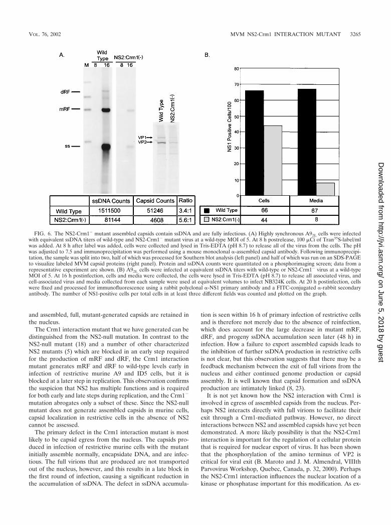
Nuclear-localized assembled capsids produced during NS2-Crm1� infection of restrictive cells contain ssDNA and remainfully infectious. Although at early times postinfection the NS2-Crm1� mutant capsids assembled at near-wild-type levels asmeasured by IFA, we could detect little progeny ssDNA onrestrictive cells as measured by whole-cell Southern blotting.This suggested that either the mutant assembled capsids didnot contain ssDNA or that the amount of ssDNA producedduring mutant infection was below the limit of detection of ourassay. Figure 6A shows that although there was an overalldecrease in ssDNA-containing full virions produced in infec-tion of A9 cells with the mutant compared to that with the wildtype, there was a significant amount of full virus present, sug-gesting that our inability to detect ssDNA following transfec-tion and infection of murine cells was likely to be because theamount of ssDNA produced in the mutant was below our limitof detection. However, there was also a concomitant decreasein the amount of assembled capsid protein produced on infec-tion with the mutant; the relative ratio of capsid protein tossDNA was approximately the same as that with the wild type.This suggested that at early times mutant virus encapsidationproceeded normally; however, when egress of capsids from thenucleus did not occur, continued production of assembled cap-sids and additional ssDNA was blocked.
The full virus made in infection of restrictive A9 cells withthe mutant was also shown to be fully infectious. Cell lysatesand media taken early (16 to 20 h postinfection) from mutantor wild-type infection of A9 cells were used at equivalentvolumes to infect NB324K cells, and resulting NS1 infectiouscenters were measured by IFA (Fig. 6B). The NS2-Crm1�
mutant generated levels of cell-associated infectious virus thatapproached 80% of wild-type levels, further demonstratingthat assembled capsids visible in nuclei by IFA in NS2-Crm1�
infection contain progeny ssDNA. Based on the experimentsshown in Fig. 4C, cell-associated virus included any virus thathad exited and rebound to cells. In addition, the culture mediafrom the mutant at early times postinfection was shown tocontain 10-fold less infectious virus than the culture mediacollected from the wild-type MVM-infected cells (Fig. 6B). Atlate times postinfection (48 h), as predicted from the largedifferences in ssDNA production, there were 50- to 100-foldless mutant cell- and media-associated infectious virions thanthat with the wild type (data not shown). This further sug-gested that viable mutant progeny are produced in A9 infec-tion; however, the full virions primarily remain cell associated.
DISCUSSION
In this study, we showed that the interaction between NS2and the nuclear export protein Crm1 is important for viralinfection. Mutants deficient in this interaction are impaired intheir ability to generate wild-type levels of progeny ssDNA,
3264 MILLER AND PINTEL J. VIROL.
on June 5, 2018 by guesthttp://jvi.asm
.org/D
ownloaded from
and assembled, full, mutant-generated capsids are retained inthe nucleus.
The Crm1 interaction mutant that we have generated can bedistinguished from the NS2-null mutation. In contrast to theNS2-null mutant (18) and a number of other characterizedNS2 mutants (5) which are blocked in an early step requiredfor the production of mRF and dRF, the Crm1 interactionmutant generates mRF and dRF to wild-type levels early ininfection of restrictive murine A9 and ID5 cells, but it isblocked at a later step in replication. This observation confirmsthe suspicion that NS2 has multiple functions and is requiredfor both early and late steps during replication, and the Crm1�
mutation abrogates only a subset of these. Since the NS2-nullmutant does not generate assembled capsids in murine cells,capsid localization in restrictive cells in the absence of NS2cannot be assessed.
The primary defect in the Crm1 interaction mutant is mostlikely to be capsid egress from the nucleus. The capsids pro-duced in infection of restrictive murine cells with the mutantinitially assemble normally, encapsidate DNA, and are infec-tious. The full virions that are produced are not transportedout of the nucleus, however, and this results in a late block inthe first round of infection, causing a significant reduction inthe accumulation of ssDNA. The defect in ssDNA accumula-
tion is seen within 16 h of primary infection of restrictive cellsand is therefore not merely due to the absence of reinfection,which does account for the large decrease in mutant mRF,dRF, and progeny ssDNA accumulation seen later (48 h) ininfection. How a failure to export assembled capsids leads tothe inhibition of further ssDNA production in restrictive cellsis not clear, but this observation suggests that there may be afeedback mechanism between the exit of full virions from thenucleus and either continued genome production or capsidassembly. It is well known that capsid formation and ssDNAproduction are intimately linked (8, 23).
It is not yet known how the NS2 interaction with Crm1 isinvolved in egress of assembled capsids from the nucleus. Per-haps NS2 interacts directly with full virions to facilitate theirexit through a Crm1-mediated pathway. However, no directinteractions between NS2 and assembled capsids have yet beendemonstrated. A more likely possibility is that the NS2-Crm1interaction is important for the regulation of a cellular proteinthat is required for nuclear export of virus. It has been shownthat the phosphorylation of the amino terminus of VP2 iscritical for viral exit (B. Maroto and J. M. Almendral, VIIIthParvovirus Workshop, Quebec, Canada, p. 32, 2000). Perhapsthe NS2-Crm1 interaction influences the nuclear location of akinase or phosphatase important for this modification. As ex-
FIG. 6. The NS2-Crm1� mutant assembled capsids contain ssDNA and are fully infectious. (A) Highly synchronous A92L cells were infectedwith equivalent ssDNA titers of wild-type and NS2-Crm1� mutant virus at a wild-type MOI of 5. At 8 h postrelease, 100 �Ci of Tran35S-label/mlwas added. At 8 h after label was added, cells were collected and lysed in Tris-EDTA (pH 8.7) to release all of the virus from the cells. The pHwas adjusted to 7.5 and immunoprecipitation was performed using a mouse monoclonal �-assembled capsid antibody. Following immunoprecipi-tation, the sample was split into two, half of which was processed for Southern blot analysis (left panel) and half of which was run on an SDS-PAGEto visualize labeled MVM capsid proteins (right panel). Protein and ssDNA counts were quantitated on a phosphorimaging screen; data from arepresentative experiment are shown. (B) A92L cells were infected at equivalent ssDNA titers with wild-type or NS2-Crm1� virus at a wild-typeMOI of 5. At 16 h postinfection, cells and media were collected, the cells were lysed in Tris-EDTA (pH 8.7) to release all associated virus, andcell-associated virus and media collected from each sample were used at equivalent volumes to infect NB324K cells. At 20 h postinfection, cellswere fixed and processed for immunofluorescence using a rabbit polyclonal �-NS1 primary antibody and a FITC-conjugated �-rabbit secondaryantibody. The number of NS1-positive cells per total cells in at least three different fields was counted and plotted on the graph.
VOL. 76, 2002 MVM NS2-Crm1 INTERACTION MUTANT 3265
on June 5, 2018 by guesthttp://jvi.asm
.org/D
ownloaded from
pected for a Crm1-dependent process, export of assembledMVM capsids from the nucleus was blocked in the presence ofleptomycin B.
It is important to note that the Crm1 interaction mutant-generated capsids also concentrate in the nucleus of permissiveNB324K cells. In this case, perhaps enough capsids exit thehuman cell nucleus to allow the continuation of single-strandproduction or new capsid assembly. Alternatively, the feedbackmechanism which halts single-strand production in murinecells may not be active in human NB324K cells. It is relevant tonote here that NS2-null mutant-generated capsids, which areproduced in NB324K cells, do not localize predominantly inthe nucleus. This may suggest that the NS2-Crm1� mutation isacting in a dominant-negative fashion in this cell type.
It seems that at least at early times in infection parvovirusescan exit from cells prior to cell lysis. Little is known about hownonenveloped viruses exit from the nucleus prior to cell lysis,and parvoviruses seem to provide a valuable system to inves-tigate this process.
ACKNOWLEDGMENTS
We thank Nathalie Salome (DKFZ-Heidelberg) for sharing infor-mation prior to publication. We also thank Mark Hannink and Sang-hyun Lee (University of Missouri—Columbia) for helpful discussion,reagents, and help with RanGAP functional assays, Greg Tullis forhelpful discussion, and Lisa Burger for excellent technical assistance.
This work was supported by Public Health Service grants AI21302and AI46458 from the National Institute of Allergy and InfectiousDiseases to D.J.P.
REFERENCES
1. Askjaer, P., A. Bachi, M. Wilm, F. R. Bischoff, D. L. Weeks, V. Ogniewski, M.Ohno, C. Niehrs, J. Kjems, I. W. Mattaj, and M. Fornerod. 1999. RanGTP-regulated interactions of CRM1 with nucleoporins and a shuttling DEAD-box helicase. Mol. Cell. Biol. 19:6276–6285.
2. Bodendorf, U., C. Cziepluch, J. C. Jauniaux, J. Rommelaere, and N. Salome.1999. Nuclear export factor CRM1 interacts with nonstructural proteins NS2from parvovirus minute virus of mice. J. Virol. 73:7769–7779.
3. Brockhaus, K., S. Plaza, D. J. Pintel, J. Rommelaere, and N. Salome. 1996.Nonstructural proteins NS2 of minute virus of mice associate in vivo with14–3–3 protein family members. J. Virol. 70:7527–7534.
4. Clemens, K. E., D. R. Cerutis, L. R. Burger, C. Q. Yang, and D. J. Pintel.1990. Cloning of MVM cDNAs and preliminary analysis of individual viralproteins expressed in murine cells. J. Virol. 64:3967–3973.
5. Cotmore, S. F., A. J. D’Abramo, Jr., L. F. Carbonell, J. Bratton, and P.Tattersall. 1997. The NS2 polypeptide of parvovirus MVM is required forcapsid assembly in murine cells. Virology 231:267–280.
6. Cotmore, S. F., L. Sturzenbecker, and P. Tattersall. 1983. The autonomousparvovirus encodes two nonstructural proteins in addition to its capsidpolypeptides. Virology 129:333–343.
7. Cotmore, S. F., and P. Tattersall. 1990. Alternate splicing in a parvoviralnonstructural gene links a common amino-terminal sequence to downstreamdomains which confer radically different localization and turnover charac-teristics. Virology 177:477–487.
8. Cotmore, S. F., and P. Tattersall. 1995. DNA replication in the autonomousparvoviruses. Semin. Virol. 6:271–281.
9. Fornerod, M., M. Ohno, M. Yoshida, and I. W. Mattaj. 1997. CRM1 is anexport receptor for leucine-rich nuclear export signals. Cell 90:1051–1060.
10. Haut, D. D., and D. J. Pintel. 1998. Intron definition is required for excisionof the minute virus of mice small intron and definition of the upstream exon.J. Virol. 72:1834–1843.
11. Lee, S. H., and M. Hannink. 2001. The N-terminal nuclear export sequenceof I�B� is required for RanGTP-dependent binding to CRM1. J. Biol.Chem. 276:23599–23606.
12. Lombardo, E., J. Ramirez, A. Agbandje-McKenna, and J. M. Almendral.2000. A beta-stranded motif drives capsid protein oligomers of the parvovi-rus minute virus of mice into the nucleus for viral assembly. J. Virology74:3804–3814.
13. Maroto, B., J. Ramirez, and J. M. Almendral. 2000. Phosphorylation statusof the parvovirus minute virus of mice particle: mapping and biologicalrelevance of the major phosphorylation sites. J. Virol. 74:10892–10902.
14. Melchior, F., and L. Gerace. 1998. Two-way trafficking with Ran. Trends CellBiol. 8:175–179.
15. Merchlinsky, M. J., P. J. Tattersall, J. J. Leary, S. F. Cotmore, E. M.Gardiner, and D. C. Ward. 1983. Construction of an infectious molecularclone of the autonomous parvovirus minute virus of mice. J. Virol. 47:227–232.
16. Miller, C. L., and D. J. Pintel. 2001. The NS2 protein generated by theparvovirus minute virus of mice is degraded by the proteasome in a mannerindependent of ubiquitin chain elongation or activation. Virology 285:346–355.
17. Mouw, M., and D. Pintel. 1998. Amino acids 16–275 of minute virus of miceNS1 include a domain that specifically binds (ACCA)2–3-containing DNA.Virology 251:123–131.
18. Naeger, L. K., J. Cater, and D. J. Pintel. 1990. The small nonstructuralprotein (NS2) of MVM is required for efficient DNA replication and infec-tious virus production in a cell-type-specific manner. J. Virol. 64:6166–6175.
19. Nuesch, J. P., S. F. Cotmore, and P. Tattersall. 1995. Sequence motifs in thereplicator protein of parvovirus MVM essential for nicking and covalentattachment to the viral origin: identification of the linking tyrosine. Virology209:122–135.
20. Nuesch, J. P., and P. Tattersall. 1993. Nuclear targeting of the parvoviralreplicator molecule NS1: evidence for self-association prior to nuclear trans-port. Virology 196:637–651.
21. Ohshima, T., T. Nakajima, T. Oishi, N. Imamoto, Y. Yoneda, A. Fukamizu,and K. Yagami. 1999. CRM1 mediates nuclear export of nonstructural pro-tein 2 from parvovirus minute virus of mice. Biochem. Biophys. Res. Com-mun. 264:144–150.
22. Tattersall, P., and J. Bratton. 1983. Reciprocal productive and restrictivevirus-cell interactions of immunosuppressive and prototype strains of minutevirus of mice. J. Virol. 46:944–955.
23. Tullis, G. E., L. R. Burger, and D. J. Pintel. 1993. The minor capsid proteinVP1 of the autonomous parvovirus MVM is not required for encapsidationof progeny single-strand DNA but is required for infectivity. J. Virol. 67:131–141.
24. Ullman, K., M. Powers, and D. Forbes. 1997. Nuclear export receptors: fromimportin to exportin. Cell 90:967–970.
3266 MILLER AND PINTEL J. VIROL.
on June 5, 2018 by guesthttp://jvi.asm
.org/D
ownloaded from

![RESEARCH Open Access Classic swine fever virus NS2 … · Classic swine fever virus NS2 protein leads to the ... (SUVEC) established in our lab previously [13] and increased the proportion](https://img.pdfslide.us/doc/110x75/5b2985ae7f8b9a47688b476e/research-open-access-classic-swine-fever-virus-ns2-classic-swine-fever-virus.jpg)