Embed Size (px)
Citation preview
Interaction between a cationic surfactant-like peptide and lipid vesicles and its relationship to antimicrobial activity Article
Published Version
Open Access (ACS Author Choice)
Dehsorkhi, A., Castelletto, V. and Hamley, I. (2013) Interaction between a cationic surfactant-like peptide and lipid vesicles and its relationship to antimicrobial activity. Langmuir, 29 (46). pp. 14246-14253. ISSN 0743-7463 doi: https://doi.org/10.1021/la403447u Available at https://centaur.reading.ac.uk/35336/
It is advisable to refer to the publisher’s version if you intend to cite from the work. See Guidance on citing .
To link to this article DOI: http://dx.doi.org/10.1021/la403447u
Publisher: American Chemical Society
All outputs in CentAUR are protected by Intellectual Property Rights law, including copyright law. Copyright and IPR is retained by the creators or other copyright holders. Terms and conditions for use of this material are defined in the End User Agreement .
www.reading.ac.uk/centaur
CentAUR

Central Archive at the University of Reading Reading’s research outputs online
Interaction between a Cationic Surfactant-like Peptide and LipidVesicles and Its Relationship to Antimicrobial ActivityAshkan Dehsorkhi, Valeria Castelletto, and Ian W. Hamley*
School of Chemistry, Pharmacy and Food Biosciences, University of Reading, Whiteknights, Reading, RG6 6AD, United Kingdom
Jani Seitsonen and Janne Ruokolainen
Department of Applied Physics, Aalto University School of Science, P.O. Box 15100, FI-00076 Aalto, Finland
*S Supporting Information
ABSTRACT: We investigate the properties of an antimicro-bial surfactant-like peptide (Ala)6(Arg), A6R, containing acationic headgroup. The interaction of this peptide withzwitterionic (DPPC) lipid vesicles is investigated using a rangeof microscopic, X-ray scattering, spectroscopic, and calori-metric methods. The β-sheet structure adopted by A6R isdisrupted in the presence of DPPC. A strong effect on thesmall-angle X-ray scattering profile is observed: the Braggpeaks from the DPPC bilayers in the vesicle walls areeliminated in the presence of A6R and only bilayer form factorpeaks are observed. All of these observations point to the interaction of A6R with DPPC bilayers. These studies provide insightinto interactions between a model cationic peptide and vesicles, relevant to understanding the action of antimicrobial peptides onlipid membranes. Notably, peptide A6R exhibits antimicrobial activity without membrane lysis.
■ INTRODUCTION
Surfactant-like peptides (SLPs) have a remarkable ability toself-assemble into different nanostructures, primarily due totheir amphiphilic nature. For example, they can aggregate intohigh aspect ratio structures while displaying bioactive peptides.SLPs are a class of amphiphilic peptide comprising a headgroupwhich is a short sequence of charged residues attached to atailgroup of neutral residues.1,2 Pioneering work on SLPs hasbeen conducted by the Zhang group including A6D, V6D, V6D2and L6D2.
3,4 We have recently investigated the self-assembly ofa cationic peptide which consists of six consecutive hydro-phobic alanine residues as a tailgroup with a cationic arginineheadgroup.5 We reported that this SLP can self-assemble intoultrathin sheets at low concentrations and at higherconcentrations the sheets wrap around to form nanotubesand helical ribbons.Peptides rich in arginine are known to have antimicrobial
activities.6−9 An example includes the transcription activatingpeptide, TAT [transactivator of transcription] from HIV-1,which has been reported to have antimicrobial properties.10,11
The TAT peptide is 11 amino acids long, and it is highly basicas it contains six arginine and two lysine residues. It was foundthat substitution of any of the basic residues with a neutralamino acid causes a reduction of antimicrobial activity, whicharises from its ability to bind to cell membranes.9 Argininecontains a guanidinium group which adopts a planar Y-shape,which can delocalize the cationic charge. As a result argininecan form bidentate hydrogen bonds with phosphates in lipid
headgroups as well as electrostatic interactions. As arginineinteracts with cell membranes, this can lead to negativecurvature and subsequently to cell leakage giving rise toantimicrobial properties.9,10,12
Our group previously investigated the self-assembly of apeptide amphiphile (PA) hexadecyl-β-alanine-histidine (C16-βAH) along with mixtures of multilamellar DPPC vesicles.13
We observed that the PA self-assembles into nanotapes basedon lamellae, that is, stacked bilayers. Mixing the PA with DPPCcaused a transition from multilamellar to unilamellar vesicles.Moshe et al. have studied the interactions of a designer cell-penetrating peptide (CPP) with phospholipids includingDOPC and DOPE.14 The peptide consisted of an arginineresidue with two short hydrophobic moieties either side tocreate hydrophobic and electrostatic interactions. At lowconcentrations, below the critical aggregation concentration,the peptide was reported to insert in the lipid bilayers and causea reduction in the membrane thickness. The CPP was found tochange the charge of the DOPC membrane and even cause aphase transition in DOPE from an inverted hexagonal to amultilamellar phase. These observations were ascribed to achange in the delicate balance of the hydrophobic, electrostaticinteractions and steric effects.
Received: September 9, 2013Revised: October 22, 2013Published: October 24, 2013
Article
pubs.acs.org/Langmuir
© 2013 American Chemical Society 14246 dx.doi.org/10.1021/la403447u | Langmuir 2013, 29, 14246−14253
Yaghmur et al. examined the effect of both anionic (A6D)and cationic (A6K) SLP’s on the bicontinuous cubic phase(Pn3 ̅m) of mono-olein.15 At low concentrations of A6D, abicontinuous cubic structure is retained, and only at highconcentrations of the peptide a phase transition to an invertedhexagonal phase occurs. The addition of A6K to mono-oleinhad no effect on the phase transition. These observations wereattributed to the ability of A6D to fully penetrate the membraneinterface in contrast to A6K which essentially does notpenetrate the membrane due to electrostatic repulsion. AsA6D is inserted, destabilization of the lipid bilayer occurs,leading to negative curvature in the membrane interface.Previously, our group studied the influence of anionic andnonionic surfactants (sodium dodecyl sulfate16 and PluronicP123,17 respectively) on the self-assembly of a collagenstimulating PA, C16-KTTKS. The PA is known to self-assembleinto extended nanotapes in solution.18 Both surfactantsinfluenced the self-assembly of C16-KTTKS since morpho-logical transitions from nanotapes to fibrils were observed.Here we report on the antimicrobial properties of A6R. Then
we attempt to understand by investigating the effect of thecationic peptide A6R on the structure of model zwitterioniclipid (DPPC) vesicles using a combination of microscopic,spectroscopic, and scattering techniques. DPPC vesicles wereselected as a model system, building on previous work in ourgroup on lipopeptide/vesicle interactions.19 Actually, DPPC is asuitable model for mammalian cell membranes, but notbacterial membranes.20 Other mixtures containing zwitterioniclipids have been used to model eukaryotic membranes.14,21
Bacterial membranes are typically rich in anionic lipids such asPOPG (oleoyl-1-palmitoyl-sn-glycero-3-phosphoglycerol) orDPPG (1,2-dipalmitoyl-sn-glycero-3-phosphoglycerol).20,22,23
The self-assembly of A6R has been thoroughly characterizedby us previously.5 It is therefore of practical as well asfundamental interest to characterize the peptide−lipid inter-actions to further enhance our understanding of the mechanismof binding and to improve the development of antimicrobialapplications for the future.
■ EXPERIMENTAL SECTIONMaterials. Peptide NH2-AAAAAAR-COOH, referred to as A6R,
was custom synthesized by CS Bio Company (Menlo Park, CA) andwas received as the TFA salt variant. The purity was 97.01% by HPLCin water/acetonitrile (0.1% TFA). Electrospray-ionization massspectroscopy (ESI-MS) indicated a molar mass 600.87 g mol−1
(600.69 g mol−1, expected).Control peptide A6D was purchased from CS Bio. Purity was
99.87% by HPLC in water/acetonitrile (0.1% TFA). ESI-MS indicateda molar mass 559.33 g mol−1 (559.59 g mol−1, expected).The phospholipid, 1,2-dipalmitoyl-sn-glycero-3-phosphocholine,
DPPC, was purchased from Sigma Aldrich, and has a molecularweight of 734.05 g mol−1.Bacterial Strains and Growth Media. Escherichia coli O157
strain BW25113, the parent strain of the Keio collection, was kindlyprovided by Professor H. Mori, Keio University, Japan. Staphylococcusaureus NCDO 949 was originally from the collection of the NationalInstitute for Research in Dairying, now listed as NCIMB 13062.Strains were maintained as frozen stocks at −70 °C on Cryobeads(Prolab Diagnostics, Neston, U.K.), which were plated onto nutrientagar (NA, Oxoid) and incubated at 37 °C overnight (16−18 h) toobtain single colonies before storage at 4 °C. Experimental cultureswere prepared by inoculating a single colony into 10 mL of tryptonesoy broth (TSB) supplemented with 0.3% (w/v) yeast extract (TSBY),and incubating statically for 6 h at 37 °C. This culture was thensubcultured into a fresh broth of TSB and incubated with shaking at
180 rev min−1 overnight at 37 °C before use. Viability was assessed bydiluting samples in Maximum Recovery Diluent (MRD, Oxoid), andplating 0.02 mL volumes onto nutrient agar. Plates were incubated at37 °C, and colonies were counted after 48 h. Colony counts werecalculated by colony forming units (CFU) equal to number of coloniestimes dilution factor times volume (CFU = N × dilution factor × V).
Preparation of Cell Suspensions. Cells were harvested bycentrifugation at 1300 rpm, 5 °C, for 5 min. The pellet wasresuspended in 1.5 mL of ice-cold phosphate-buffered saline (PBS, pH7.0, Sigma-Aldrich), and 20 μL of this solution was diluted into 200 μLof peptide solution (5, 2.5, 1, 0.5 mg/mL) to give approximately 107
cells mL−1. Control solutions were achieved by adding no peptidesolution but MRD instead. These solutions were vortexed for 3 s andleft for set time intervals (10, 20, 30, 40, 50, 60 min) before dilutingwith MRD and plating. Plates were then incubated at 37 °C overnightfor 18−24 h, followed by a cell count (CFU calculation).
Sample Preparation. Vesicle Preparation. DPPC vesicles wereprepared by the thin layer hydration method to ensure the formationof multilayered vesicles.24 A measured quantity of DPPC was dissolvedin ethanol and placed into a 100 mL round-bottom flask. The solventwas evaporated by using a rotary evaporator, which formed a thinDPPC film at the bottom of the flask. A measured quantity of waterwas added to the flask to make up the defined concentration. The flaskwas returned to the rotary evaporator, which was rotated while at 50°C by submerging it under a water bath. The solution was thenvortexed at 50 °C for approximately 5 min.
Solution Mixture Preparation. Solutions of 0.5, 1, and 2 wt %DPPC vesicles were first prepared followed by the addition of 1 wt %A6R. A measured quantity of A6R powder was added to a solution ofDPPC vesicles to make up the three different solutions with differentproportions of DPPC. The mixture was left to sonicate for 30 min at50 °C to dissolve A6R. Then the solution was left to equilibrate atroom temperature for a few days before any measurements.
Cryogenic-Transmission Electron Microscopy (Cryo-TEM).Imaging was carried out using a field emission cryo-electronmicroscope (JEOL JEM-3200FSC), operating at 200 kV. Imageswere taken in bright field mode and using zero loss energy filtering(omega type) with a slit width of 20 eV. Micrographs were recordedusing a Gatan Ultrascan 4000 CCD camera. The specimentemperature was maintained at −187 °C during the imaging. Vitrifiedspecimens were prepared using an automated FEI Vitrobot deviceusing Quantifoil 3.5/1 holey carbon copper grids with a hole size of3.5 μm. Just prior to use, grids were plasma cleaned using a GatanSolarus 9500 plasma cleaner and then transferred into an environ-mental chamber of a FEI Vitrobot at room temperature and 100%humidity. Thereafter, 3 mL of sample solution was applied on the gridand it was blotted twice for 5 s and then vitrified in a 1/1 mixture ofliquid ethane and propane at temperature of −180 °C. The grids withvitrified sample solution were maintained at liquid nitrogen temper-ature and then cryo-transferred to the microscope.
X-ray Diffraction (XRD). Measurements were performed on stalksprepared by drying filaments of solutions containing 1 wt % A6R mixedwith 0.5, 1, and 2 wt % DPPC. Solutions of the mixtures weresuspended between the ends of wax-coated capillaries and dried. Thestalks were mounted (vertically) onto the four axis goniometer of aRAXIS IV++ X-ray diffractometer (Rigaku) equipped with a rotatinganode generator. The sample−detector distance varied between 90and 100 mm depending on the sample. The X-ray wavelength was λ =1.54 Å. The wavenumber scale (q = 4π sin θ/λ, where 2θ is thescattering angle) was geometrically calculated using the size of eachpixel in the detector screen (0.0898 mm) and the sample−detectordistance. The XRD data was collected using a Saturn 992 CCDcamera.
Small-Angle and X-ray Scattering (SAXS). Solution SAXS datawas performed on the bioSAXS beamline BM29 at the ESRF,Grenoble, France. Solutions containing 1 wt % A6R and 1 wt % A6Rmixed with 0.5, 1, and 2 wt % DPPC were loaded in PCR tubes in anautomated sample changer. SAXS data was collected using a Pilatus 1M detector. The sample−detector distance was 2.84 m. The X-raywavelength was 0.99 Å.
Langmuir Article
dx.doi.org/10.1021/la403447u | Langmuir 2013, 29, 14246−1425314247
Circular Dichroism (CD). Spectra were obtained using a Chirascanspectropolarimeter (Applied Photophysics, UK). CD was performedon solution mixtures containing DPPC vesicles with A6R added later,with a 0.5 nm step, 1 nm bandwidth and 1 s collection time per step at20 °C. Data with absorbance A < 2 only are presented. Measurementswere repeated four times. Smoothing of the data was carried out usingthe supplied Chirascan software.Isothermal Titration Calorimetry (ITC). ITC experiments were
carried out using an iTC200 microcalorimeter from MicroCal Inc. Theworking cell was filled with 200 μL of 0.1 wt % DPPC solution(dissolved in water) and the reference cell was filled with deionizedwater. The titrant syringe was filled with a solution of 1 wt % A6R. TheITC experiment was programmed to run 20 injections of 2 μL volumeof the titrant solution (1 wt % A6R) into the working cell (0.1 wt %DPPC) with 300 s lag between each injection to ensure return to thebaseline. The syringe was stirred throughout the experiment at 500rpm and the working cell was set at 25 °C. The data was analyzedusing Origin 7 (MicroCal) by fitting the curve using the one set ofsites model.
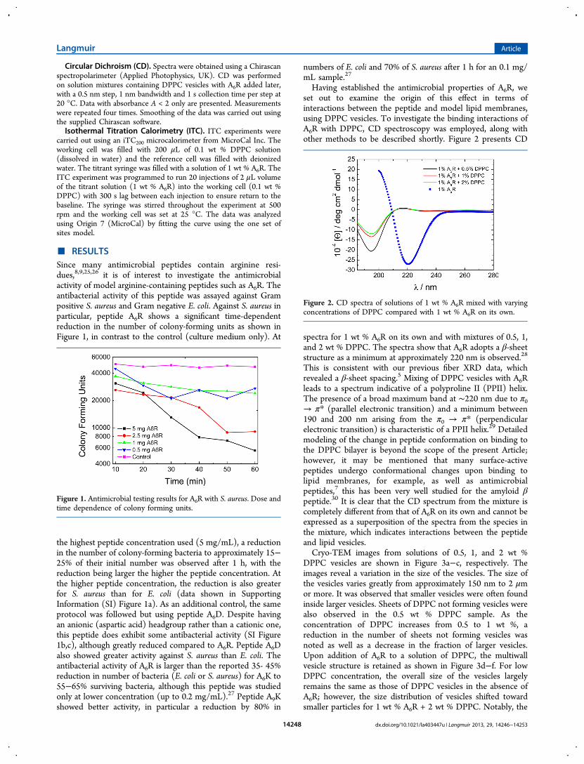
■ RESULTSSince many antimicrobial peptides contain arginine resi-dues,8,9,25,26 it is of interest to investigate the antimicrobialactivity of model arginine-containing peptides such as A6R. Theantibacterial activity of this peptide was assayed against Grampositive S. aureus and Gram negative E. coli. Against S. aureus inparticular, peptide A6R shows a significant time-dependentreduction in the number of colony-forming units as shown inFigure 1, in contrast to the control (culture medium only). At
the highest peptide concentration used (5 mg/mL), a reductionin the number of colony-forming bacteria to approximately 15−25% of their initial number was observed after 1 h, with thereduction being larger the higher the peptide concentration. Atthe higher peptide concentration, the reduction is also greaterfor S. aureus than for E. coli (data shown in SupportingInformation (SI) Figure 1a). As an additional control, the sameprotocol was followed but using peptide A6D. Despite havingan anionic (aspartic acid) headgroup rather than a cationic one,this peptide does exhibit some antibacterial activity (SI Figure1b,c), although greatly reduced compared to A6R. Peptide A6Dalso showed greater activity against S. aureus than E. coli. Theantibacterial activity of A6R is larger than the reported 35- 45%reduction in number of bacteria (E. coli or S. aureus) for A6K to55−65% surviving bacteria, although this peptide was studiedonly at lower concentration (up to 0.2 mg/mL).27 Peptide A9Kshowed better activity, in particular a reduction by 80% in
numbers of E. coli and 70% of S. aureus after 1 h for an 0.1 mg/mL sample.27
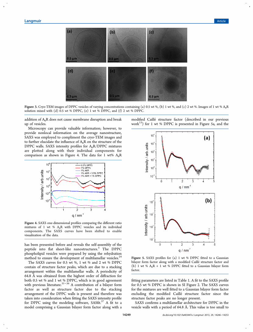
Having established the antimicrobial properties of A6R, weset out to examine the origin of this effect in terms ofinteractions between the peptide and model lipid membranes,using DPPC vesicles. To investigate the binding interactions ofA6R with DPPC, CD spectroscopy was employed, along withother methods to be described shortly. Figure 2 presents CD
spectra for 1 wt % A6R on its own and with mixtures of 0.5, 1,and 2 wt % DPPC. The spectra show that A6R adopts a β-sheetstructure as a minimum at approximately 220 nm is observed.28
This is consistent with our previous fiber XRD data, whichrevealed a β-sheet spacing.5 Mixing of DPPC vesicles with A6Rleads to a spectrum indicative of a polyproline II (PPII) helix.The presence of a broad maximum band at ∼220 nm due to π0→ π* (parallel electronic transition) and a minimum between190 and 200 nm arising from the π0 → π* (perpendicularelectronic transition) is characteristic of a PPII helix.29 Detailedmodeling of the change in peptide conformation on binding tothe DPPC bilayer is beyond the scope of the present Article;however, it may be mentioned that many surface-activepeptides undergo conformational changes upon binding tolipid membranes, for example, as well as antimicrobialpeptides,7 this has been very well studied for the amyloid βpeptide.30 It is clear that the CD spectrum from the mixture iscompletely different from that of A6R on its own and cannot beexpressed as a superposition of the spectra from the species inthe mixture, which indicates interactions between the peptideand lipid vesicles.Cryo-TEM images from solutions of 0.5, 1, and 2 wt %
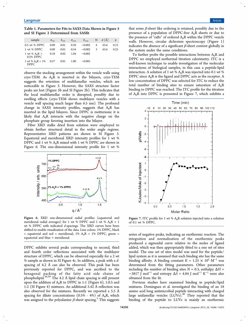
DPPC vesicles are shown in Figure 3a−c, respectively. Theimages reveal a variation in the size of the vesicles. The size ofthe vesicles varies greatly from approximately 150 nm to 2 μmor more. It was observed that smaller vesicles were often foundinside larger vesicles. Sheets of DPPC not forming vesicles werealso observed in the 0.5 wt % DPPC sample. As theconcentration of DPPC increases from 0.5 to 1 wt %, areduction in the number of sheets not forming vesicles wasnoted as well as a decrease in the fraction of larger vesicles.Upon addition of A6R to a solution of DPPC, the multiwallvesicle structure is retained as shown in Figure 3d−f. For lowDPPC concentration, the overall size of the vesicles largelyremains the same as those of DPPC vesicles in the absence ofA6R; however, the size distribution of vesicles shifted towardsmaller particles for 1 wt % A6R + 2 wt % DPPC. Notably, the
Figure 1. Antimicrobial testing results for A6R with S. aureus. Dose andtime dependence of colony forming units.
Figure 2. CD spectra of solutions of 1 wt % A6R mixed with varyingconcentrations of DPPC compared with 1 wt % A6R on its own.
Langmuir Article
dx.doi.org/10.1021/la403447u | Langmuir 2013, 29, 14246−1425314248
addition of A6R does not cause membrane disruption and breakup of vesicles.Microscopy can provide valuable information; however, to
provide nonlocal information on the average nanostructure,SAXS was employed to compliment the cryo-TEM images andto further elucidate the influence of A6R on the structure of theDPPC walls. SAXS intensity profiles for A6R/DPPC mixturesare plotted along with their individual components forcomparison as shown in Figure 4. The data for 1 wt% A6R
has been presented before and reveals the self-assembly of thepeptide into flat sheet-like nanostructures.5 The DPPCphospholipid vesicles were prepared by using the rehydrationmethod to ensure the development of multilamellar vesicles.24
The SAXS curves for 0.5 wt %, 1 wt % and 2 wt % DPPCcontain of structure factor peaks, which are due to a stackingarrangement within the multilamellar walls. A periodicity of64.8 Å was obtained from the highest order of diffraction forboth 0.5 wt % and 1 wt % DPPC, which is in good agreementwith previous literature.31−36 A contribution of a bilayer formfactor as well as structure factor due to the stackingarrangement of the DPPC walls is present and therefore wastaken into consideration when fitting the SAXS intensity profilefor DPPC using the modeling software, SASfit.37 A fit to amodel comprising a Gaussian bilayer form factor along with a
modified Caille ́ structure factor (described in our previouswork13) for 1 wt % DPPC is presented in Figure 5a, and the
fitting parameters are listed in Table 1. A fit to the SAXS profilefor 0.5 wt % DPPC is shown in SI Figure 2. The SAXS curvesfor the mixtures are well fitted to a Gaussian bilayer form factorexcluding the modified Caille ́ structure factor since thestructure factor peaks are no longer present.SAXS confirms a multilamellar architecture for DPPC in the
vesicle walls with a period of 64.8 Å. This value is too small to
Figure 3. Cryo-TEM images of DPPC vesicles of varying concentrations containing (a) 0.5 wt %, (b) 1 wt %, and (c) 2 wt %. Images of 1 wt % A6Rsolution mixed with (d) 0.5 wt % DPPC, (e) 1 wt % DPPC, and (f) 2 wt % DPPC.
Figure 4. SAXS one-dimensional profiles comparing the different ratiomixtures of 1 wt % A6R with DPPC vesicles and its individualcomponents. The SAXS curves have been shifted to enablevisualization of the data.
Figure 5. SAXS profiles for (a) 1 wt % DPPC fitted to a Gaussianbilayer form factor along with a modified Caille ́ structure factor and(b) 1 wt % A6R + 1 wt % DPPC fitted to a Gaussian bilayer formfactor.
Langmuir Article
dx.doi.org/10.1021/la403447u | Langmuir 2013, 29, 14246−1425314249
observe the stacking arrangement within the vesicle walls usingcryo-TEM. As A6R is inserted in the bilayers, cryo-TEMsuggests the retention of multilamellar vesicles, which arenoticeable in Figure 3. However, the SAXS structure factorpeaks are lost (Figure 5b and SI Figure 2b). This indicates thatthe local multilamellar order is disrupted, possibly due toswelling effects (cryo-TEM shows multilayer vesicles with avesicle wall spacing much larger than 6.5 nm). The profoundchange in SAXS intensity profiles, suggests that A6R hasinserted in the lipid bilayers. Since DPPC is zwitterionic it islikely that A6R interacts with the negative charge on thephosphate group favoring insertion into the bilayer.Fiber XRD stalks dried from solution were employed to
obtain further structural detail in the wider angle regions.Representative XRD patterns are shown in SI Figure 3.Equatorial and meridional XRD intensity profiles for 1 wt %DPPC and 1 wt % A6R mixed with 1 wt % DPPC are shown inFigure 6. The one-dimensional intensity profile for 1 wt %
DPPC exhibits several peaks corresponding to second, thirdand fourth order reflections associated with the multilayerstructure of DPPC, which can be observed especially for a 2 wt% sample as shown in SI Figure 4c. In addition, a peak with a d-spacing of 4.2 Å can also be observed. This peak has beenpreviously reported for DPPC, and was ascribed to thehexagonal packing of the fatty acid side chains ofphospholipid.38,39 The 4.2 Å lipid chain spacing is still presentupon the addition of A6R to DPPC in 1:1 (Figure 6), 1:0.5 and1:2 (SI Figure 4) mixtures. An additional 5.42 Å reflection wasalso observed for the mixtures. Recently we reported a 5.5 Åspacing for dilute concentrations (0.5% - 4%) of A6R, whichwas assigned to the polyalanine β-sheet spacing.5 This suggests
that some β-sheet like ordering is retained, possibly due to thepresence of a population of DPPC-free A6R sheets or due tothe presence of “rafts” of ordered A6R within the DPPC vesiclewalls. However, circular dichroism spectroscopy (Figure 1)indicates the absence of a significant β-sheet content globally inthe system under the same conditions.To further probe the possible interactions between A6R and
DPPC we employed isothermal titration calorimetry. ITC is awell-known technique to enable investigation of the molecularinteractions of biological samples, in this case a peptide-lipidinteraction. A solution of 1 wt % A6R was injected into 0.1 wt %DPPC since A6R is the ligand and DPPC acts as the receptor. Alow concentration of DPPC was selected for ITC to reduce thetotal number of binding sites to ensure saturation of A6Rbinding to DPPC was reached. The ITC profile for the titrationof A6R into DPPC is presented in Figure 7, which exhibits a
series of negative peaks, indicating an exothermic reaction. Theintegration and normalization of the exothermic peaksproduced a sigmoidal curve relative to the moles of ligandadded, which was then appropriately fitted to a one set of sitesmodel. The one set of sites model was used for the peptide/lipid system as it is assumed that each binding site has the samebinding affinity. A binding constant K = 1.25 × 10⁵ M−1 wasdetermined from the fitting parameters. Other parametersincluding the number of binding sites N = 0.3, enthalpy ΔH =−3817 J mol−1 and entropy ΔS = 4.84 J mol−1 K−1 were alsoobtained from the fit.Previous studies have examined binding in peptide/lipid
mixtures. Domingues et al. investigated the binding of an 18amino acid long antimicrobial peptide interacting with chargedlarge unilamellar vesicles (LUVs).40 They reported that thebinding of the peptide to LUVs is mainly an exothermic
Table 1. Parameters for Fits to SAXS Data Shown in Figure 5and SI Figure 2 Determined from SASfit
sample σout bout σcore bcore N d [Å] η
0.5 wt % DPPC 0.09 0.01 0.59 −0.002 4 63.6 0.151 wt % DPPC 0.09 0.01 0.54 −0.002 3 63.6 0.251 wt % A6R +0.5% DPPC
0.10 0.02 1.04 −0.002
1 wt % A6R + 1%DPPC
0.57 0.01 1.80 −0.005
Figure 6. XRD one-dimensional radial profiles (equatorial andmeridional radial averages) for 1 wt % DPPC and 1 wt % A6R + 1wt % DPPC with indicated d-spacings. The XRD curves have beenshifted to enable visualization of the data. Line colors: 1% DPPC, black= equatorial and red = meridional; 1% A6R + 1% DPPC, green =equatorial and blue = meridional.
Figure 7. ITC profile for 1 wt % A6R solution injected into a solutionof 0.1 wt % DPPC.
Langmuir Article
dx.doi.org/10.1021/la403447u | Langmuir 2013, 29, 14246−1425314250
process. The binding of a cationic pentapeptide composed ofanalogs of lysine resides to negatively charged phospholipid,DPPG was investigated.41 It was reported that an exothermicreaction occurs upon binding. A binding constant, K = 5.4 ×10⁵ M−1, was determined, which is a similar value to the one weobtained. The Vogel group also investigated the binding of anantimicrobial peptide, Ac-FRWWHR-NH2 to POPG vesicles,which reveal an exothermic reaction and a binding constant, K= 3.13 × 10⁵ M−1.42 The mentioned examples are in goodagreement with our observations that an exothermic processoccurs during binding and the binding constant values aresimilar to those we obtained.
■ SUMMARY AND DISCUSSIONIn summary, A6R interacts with DPPC vesicles leading tochanges in the vesicle wall layer spacing such that the SAXSstructure factor peaks present for DPPC vesicles are eliminatedand only the form factor of isolated bilayers is observed. This issimilar to what is observed for the interaction of the peptideamphiphile C16-βAH (βAH: β-alanine-histidine dipeptide,known as L-carnosine) with DPPC.13 X-ray diffraction indicatesthe presence of a fraction of tightly packed alanine-rich β-sheetstructures in the A6R/DPPC mixtures, although circulardichroism spectroscopy shows the suppression of global β-sheet ordering of A6R in the presence of DPPC. Some A6R mayform separate β-sheet assemblies or “rafts” of ordered A6R maybe present in the vesicle walls. Discriminating between thesepossibilies is a challenge for future work.Interestingly, A6R does not seem to permeabilize DPPC
vesicles despite its insertion into the vesicle walls (as inferredfrom dramatic changes in the SAXS intensity profiles). Additionof the peptide leads to the loss of structure factor peaks. This isthe opposite of the behavior observed by Moshe et al. for theirGFfWG (f: D-phenylalanine) peptide interacting with modelcell membranes (DOPS, DOPC, DOPE mixture) since a seriesof Bragg reflections were observed in the presence of thepeptide, but only form factor (similar to that shown in ourFigure 4b) features were observed for the membrane/lipidmixture.14
Hoernke et al. reported that short basic pentapeptides suchas K5 insert into lipid (DPPG) membranes,41 in contrast to thefindings of Ben-Tal et al.43 However, in neither of these studieswas imaging of vesicles or permeabilization measurementsperformed. The hexapeptide FRWWHR, identified bycombinatorial screening methods to have strong antimicrobialactivity, does not cause substantial leakage from vesicles, andRezansoff et al. suggested that the bactericidal action of thepeptide may involve translocation across the membrane.42
However, Blondelle et al. did observe lysis of model DPPC-containing membranes in the presence of lysine-rich 18-merpeptides.44 Natural antimicrobial peptides such as magainin45,46
and gomesin40 permeabilize membranes and lead to lysis. Somecationic peptides are known to cause fusion of cell membranesand have been studied in particular in the context of viralinfection where they mediate fusion of the host cell membraneand the enveloped virus. The fusogenic TAT proteintransduction domain has been used to deliver a wide range ofbiologically active cargo (DNA, proteins, liposomes, andothers).47 The initial model for cellular uptake involves directpenetration across the lipid membrane, however it has beenshown that TAT-fusion proteins are rapidly internalized bylipid-raft dependent macropinocytosis47 (pinocytosis is non-specific endocytosis within vesicles). As mentioned above,
substitution of any of the basic residues in the TAT peptidewith a neutral amino acid causes a reduction of antimicrobialactivity, reflecting the influence of charge and hydrophobicity.9
Thus, prior work indicates that membrane permeabilizationcan occur for peptides with more than one cationic residue, butthis alone is not sufficient. The sequence and length of thepeptide is also important, as is the nature and composition ofthe lipid membrane. This was highlighted by Chen et al. in theircomparison of the antibacterial properties of A3K, A6K, andA9K.
27 They found that the latter, which has the longesthydrophobic alanine block and the strongest aggregationtendency, has the highest antimicrobial activity. This peptidealso did not significantly disrupt DPPC vesicles, althoughDPPG membranes were broken up. Even relatively shortpeptides righ in arginine and/or tryptophan have potentantimicrobial activity.26 Other factors influencing the activity ofantimicrobial peptides are discussed elsewhere.48 As discussedin the Introduction, models for bacterial cell membranes shouldconsist of anionic lipids although as discussed above manystudies have used DPPC as model membranes.We have shown that A6R is a model antimicrobial cationic
peptide containing a single arginine residue attached to ahydrophobic hexa-alanine sequence to drive self-assembly. Thisstudy provides insight into its interaction with model lipidmembranes. It also introduces the concept of addition of SLPsto modulate the structure of lipid vesicles. Remarkably, A6Rexhibits antimicrobial activity without zwitterionic lipidmembrane lysis.
■ ASSOCIATED CONTENT*S Supporting InformationAntimicrobial activity data for control peptide A6D, additionalSAXS and XRD data. This material is available free of charge viathe Internet at http://pubs.acs.org.
■ AUTHOR INFORMATIONNotesThe authors declare no competing financial interest.
■ ACKNOWLEDGMENTSWe thank Claire Moulton, Cristina Arroyo and Dr BernardMackey for the antimicrobial activity measurements. This workwas supported by EPSRC Grant EP/G067538/1 to I.W.H.
■ REFERENCES(1) Hamley, I. W. Self-Assembly of Amphiphilic Peptides. Soft Matter2011, 7, 4122.(2) Zhao, X.; Pan, F.; Xu, H.; Yaseen, M.; Shan, H.; Hauser, C. A.;Zhang, S.; Lu, J. R. Molecular Self-Assembly and Applications ofDesigner Peptide Amphiphiles. Chem. Soc. Rev. 2010, 39, 3480.(3) Vauthey, S.; Santoso, S.; Gong, H.; Watson, N.; Zhang, S.Molecular Self-Assembly of Surfactant-Like Peptides to FormNanotubes and Nanovesicles. Proc. Natl. Acad. Sci. U.S.A. 2002, 99,5355.(4) Maltzahn, G. V.; Vauthey, S.; Santoso, S.; Zhang, S. PositivelyCharged Surfactant-Like Peptides Self-Assemble into Nanostructures.Langmuir 2003, 19, 4332.(5) Hamley, I. W.; Dehsorkhi, A.; Castelletto, V. Self-AssembledArginine-Coated Peptide Nanosheets in Water. Chem. Commun. 2013,49, 1850.(6) Nicolas, P.; Mor, A. Peptides as Weapons against Microorganismsin the Chemical Defense System of Vertebrates. Annu. Rev. Microbiol.1995, 49, 277.
Langmuir Article
dx.doi.org/10.1021/la403447u | Langmuir 2013, 29, 14246−1425314251
(7) Chan, D. I.; Prenner, E. J.; Vogel, H. J. Tryptophan- andArginine-Rich Antimicrobial Peptides: Structures and Mechanisms ofAction. Biochim. Biophys. Acta, Biomembr. 2006, 1758, 1184.(8) Reddy, K. V.; Yedery, R. D.; Aranha, C. Antimicrobial Peptides:Premises and Promises. Int. J. Antimicrob. Agents 2004, 24, 536.(9) Schmidt, N.; Mishra, A.; Lai, G. H.; Wong, G. C. Arginine-RichCell-Penetrating Peptides. FEBS Lett. 2010, 584, 1806.(10) Mishra, A.; Lai, G. H.; Schmidt, N. W.; Sun, V. Z.; Rodriguez, A.R.; Tong, R.; Tang, L.; Cheng, J. J.; Deming, T. J.; Kamei, D. T.;Wong, G. C. L. Translocation of HIV TAT Peptide and AnaloguesInduced by Multiplexed Membrane and Cytoskeletal Interactions.Proc. Natl. Acad. Sci. U.S.A. 2011, 108, 16883.(11) Piantavigna, S.; McCubbin, G. A.; Boehnke, S.; Graham, B.;Spiccia, L.; Martin, L. L. A Mechanistic Investigation of Cell-Penetrating TAT Peptides with Supported Lipid Membranes. Biochim.Biophys. Acta 2011, 1808, 1811.(12) Zhao, K.; Choe, U.-J.; Kamei, D. T.; Wong, G. C. L. EnhancedActivity of Cyclic Transporter Sequences Driven by Phase Behavior ofPeptide−Lipid Complexes. Soft Matter 2012, 8, 6430.(13) Castelletto, V.; Cheng, G.; Stain, C.; Connon, C. J.; Hamley, I.W. Self-Assembly of a Peptide Amphiphile Containing L-Carnosineand Its Mixtures with a Multilamellar Vesicle Forming Lipid. Langmuir2012, 28, 11599.(14) Moshe, L.; Saper, G.; Szekely, O.; Linde, Y.; Gilon, C.; Harries,D.; Raviv, U. Modulating the Structure and Interactions of Lipid−Peptide Complexes by Varying Membrane Composition and SolutionConditions. Soft Matter 2013, 9, 7117.(15) Yaghmur, A.; Laggner, P.; Zhang, S.; Rappolt, M. TuningCurvature and Stability of Monoolein Bilayers by Designer Lipid-LikePeptide Surfactants. PLoS One 2007, e479.(16) Castelletto, V.; Hamley, I. W.; Adamcik, J.; Mezzenga, R.;Gummel, J. Modulating Self-Assembly of a Nanotape-Forming PeptideAmphiphile with an Oppositely Charged Surfactant. Soft Matter 2012,8, 217.(17) Dehsorkhi, A.; Castelletto, V.; Hamley, I. W.; Lindner, P.Influence of a Non-Ionic Amphiphilic Copolymer on the Self-Assembly of a Peptide Amphiphile That Forms Nanotapes. Soft Matter2012, 8, 8608.(18) Castelletto, V.; Hamley, I. W.; Perez, J.; Abezgauz, L.; Danino,D. Fibrillar Superstructure from Extended Nanotapes Formed by aCollagen-Stimulating Peptide. Chem. Commun. 2010, 46, 9185.(19) Castelletto, V.; Cheng, G.; Stain, C.; Connon, C. J.; Hamley, I.W. Self-Assembly of a Peptide Amphiphile Containing L-Carnosineand Its Mixtures with a Multilamellar Vesicle Forming Lipid. Langmuir2012, 28, 11599.(20) Hu, J.; Chen, C. X.; Zhang, S. Z.; Zhao, X. C.; Xu, H.; Zhao, X.B.; Lu, J. R. Designed Antimicrobial and Antitumor Peptides withHigh Selectivity. Biomacromolecules 2011, 12, 3839.(21) Zhao, K.; Choe, U. J.; Kamei, D. T.; Wong, G. C. L. EnhancedActivity of Cyclic Transporter Sequences Driven by Phase Behavior ofPeptide-Lipid Complexes. Soft Matter 2012, 8, 6430.(22) Domingues, F. S.; Riske, K. A.; Miranda, A. Revealing the LyticMechanism of the Antimicrobial Peptide Gomesin by Observing GiantUnilamellar Vesicles. Langmuir 2010, 26, 11077.(23) Taheri-Araghi, S.; Ha, B. Y. Cationic Antimicrobial Peptides: APhysical Basis for Their Selective Membrane-Disrupting Activity. SoftMatter 2010, 6, 1933.(24) Szoka, F.; Papahadjopoulos, D. Procedure for Preparation ofLiposomes with Large Internal Aqueous Space and High Capture byReverse-Phase Evaporation. Proc. Natl. Acad. Sci. U.S.A. 1978, 75,4184.(25) Nicolas, P.; Mor, A. Peptides as Weapons. Annu. Rev. Microbiol.1995, 49, 277.(26) Chan, D. I.; Prenner, E. J.; Vogel, H. J. Tryptophan- andArginine-Rich Antimicrobial Peptides: Structures and Mechanisms ofAction. Biochim. Biophys. Acta 2006, 1758−1184.(27) Chen, C. X.; Pan, F.; Zhang, S. Z.; Hu, J.; Cao, M. W.; Wang, J.;Xu, H.; Zhao, X. B.; Lu, J. R. Antibacterial Activities of Short DesignerPeptides: A Link between Propensity for Nanostructuring and
Capacity for Membrane Destabilization. Biomacromolecules 2010, 11,402.(28) Woody, R. W. Circular Dichroism of Peptides and Proteins; NewYork, 1994.(29) Wallace, B. A.; Janes, R. W. Synchrotron Radiation CircularDichroism Spectroscopy of Proteins: Secondary Structure, FoldRecognition and Structural Genomics. Curr. Opin. Chem. Biol. 2001,5, 567.(30) Hamley, I. W. The Amyloid Beta Peptide: A Chemist’sPerspective. Role in Alzheimer’s and Fibrillization. Chem. Rev. 2012,112, 5147.(31) Tristram-Nagle, S.; Zhang, R.; Suter, R. M.; Worthington, C. R.;Sun, W. J.; Nagle, J. F. Measurement of Chain Tilt Angle in FullyHydrated Bilayers of Gel Phase Lecithins. Biophys. J. 1993, 64, 1097.(32) McManus, J. J.; Radler, J. O.; Dawson, K. A. Phase Behavior ofDppc in a DNA-Calcium-Zwitterionic Lipid Complex Studied bySmall-Angle X-Ray Scattering. Langmuir 2003, 19, 9630.(33) Sun, W. J.; Tristam-Nagle, S.; Suter, R. M.; Nagle, J. F. Structureof Gel Phase Saturated Lecithin Bilayers: Temperature and ChainLength Dependence. Biophys. J. 1996, 71, 885.(34) Tenchov, B. G.; Yao, H.; Hatta, I. Time-Resolved X-RayDiffraction and Calorimetric Studies at Low Scan Rates. Biophys. J.1989, 56, 757.(35) Hauet, N.; Artzner, F.; Boucher, F.; Grabielle-Madelmont, C.;Cloutier, I.; Keller, G.; Lesieur, P.; Durand, D.; Paternostre, M.Interaction between Artificial Membranes and Enflurane, a GeneralVolatile Anesthetic: DPPC-Enflurane Interaction. Biophys. J. 2003, 84,3123.(36) Quinn, P. J.; Takahashi, H.; Hatta, I. Characterization ofComplexes Formed in Fully Hydrated Dispersions of DipalmitoylDerivatives of Phosphatidylcholine and Diacylglycerol. Biophys. J.1995, 68, 1374.(37) http://kur.web.psi.ch/sans1/SANSSoft/sasfit.html, in 2013.(38) Mckersie, B. D.; Thompson, J. E. Influence of Plant Sterols onthe Phase Properties of Phospholipid Bilayers. Plant Physiol. 1979, 63,802.(39) Katsaras, J.; Yang, D. S.-C.; Epand, R. M. Fatty-Acid Chain TiltAngles and Directions in Dipalmitoyl Phosphatidylcholine Bilayers.Biophys. J. 1992, 63, 1170.(40) Domingues, T. M.; Mattei, B.; Seelig, J.; Perez, K. R.; Miranda,A.; Riske, K. A. Interaction of the Antimicrobial Peptide Gomesin withModel Membranes: A Calorimetric Study. Langmuir 2013, 29, 8609.(41) Hoernke, M.; Schwieger, C.; Kerth, A.; Blume, A. Binding ofCationic Pentapeptides with Modified Side Chain Lengths toNegatively Charged Lipid Membranes: Complex Interplay of Electro-static and Hydrophobic Interactions. Biochim. Biophys. Acta 2012,1818, 1663.(42) Rezansoff, A. J.; Hunter, H. N.; Jing, W.; Park, I. Y.; Kim, S. C.;Vogel, H. J. Interactions of the Antimicrobial Peptide Ac-FRWWHR-NH2 with Model Membrane Systems and Bacterial Cells. J. Pept. Sci.2005, 65, 491.(43) Ben-Tal, N.; Honig, B.; Peitzsch, R. M.; Denisov, G.;McLaughlin, S. Binding of Small Basic Peptides to MembranesContaining Acidic Lipids: Theoretical Models and ExperimentalResults. Biophys. J. 1996, 71, 561.(44) Blondelle, S. E.; Takahashi, E.; Houghten, R. A.; Perez-Paya,́ E.Rapid Identification of Compounds Having Enhanced AntimicrobialActivity Using Conformationally Defined Combinatorial Libraries.Biochem. J. 1996, 313, 141.(45) Matsuzaki, K.; Sugishita, K.; Ishibe, N.; Ueha, M.; Nakata, S.;Miyajima, K.; Epand, R. M. Relationship of Membrane Curvature tothe Formation of Pores by Magainin 2. Biochemistry 1998, 37, 11856.(46) Papo, N.; Shai, Y. Exploring Peptide Membrane InteractionUsing Surface Plasmon Resonance: Differentiation between PoreFormation Versus Membrane Disruption by Lytic Peptides.Biochemistry 2003, 42, 458.(47) Wadia, J. S.; Stan, R. V.; Dowdy, S. F. Transducible Tat-HaFusogenic Peptide Enhances Escape of TAT-Fusion Proteins afterLipid Raft Macropinocytosis. Nature 2004, 10, 310.
Langmuir Article
dx.doi.org/10.1021/la403447u | Langmuir 2013, 29, 14246−1425314252
(48) Brogden, K. A. Antimicrobial Peptides: Pore Formers orMetabolic Inhibitors in Bacteria? Nat. Rev. Microbiol. 2005, 3, 238.
Langmuir Article
dx.doi.org/10.1021/la403447u | Langmuir 2013, 29, 14246−1425314253


![Influence of copper nanoparticles capped by cationic surfactant … · 2017. 1. 17. · oxidation [20–24]. Organic coatings are widely used to prevent corrosion of metal structures](https://img.pdfslide.us/doc/110x75/60d9f73f1f82944250661db5/influence-of-copper-nanoparticles-capped-by-cationic-surfactant-2017-1-17-oxidation.jpg)


![Predictive model of cationic surfactant binding to humic … · 2019. 10. 9. · Koopal et al. [29] have shown the influence of pH and cationic surfactant aliphatic chain length on](https://img.pdfslide.us/doc/110x75/6125e8328ab15b5cd9535070/predictive-model-of-cationic-surfactant-binding-to-humic-2019-10-9-koopal-et.jpg)