Embed Size (px)
Citation preview

Integumentary System and Body Membranes
Structure and Function

2
Love the Skin You’re In!
Skin..Cutaneous Membrane– Largest, most visible, primary
organ– Composes 16% of body weight– Superficial layer is epithelial,
underlying layer is connective

3
Structure of the Skin: Layers
EPIDERMISDERMISSUBCUTANEOUS TISSUE
4
Structure of the Skin:
Epidermis..outermost layer of the skin constantly replaced from underneath layer.
Dermis..deeper,thicker layer, composed of connective tissue
– Basis of fingerprints– Contain nerve endings, muscle fibers, hair follicles, sweat &
sebaceous glands, & blood vessels

5
Structure of the Skin:
Subcutaneous tissue..thicker layer of loose connective tissue & fat – Insulates from heat & cold– Shock absorbing pad to protect– Stored source of food & energy

6
Structure of the Skin:
Keratin..hard protein substance in nails, hair, skin,
Pigment ..produced by deepest layer of epidermis, gives color to the skin,
Melanin..Brown pigment produced by melanocytes in the epidermis
FYI: Millions of epithelial cells reproduced & shed daily

7
SKIN STRUCTURE:
8
FUNCTION OF SKIN:
Protection..first line of defense against– infection by microbes– ultraviolet rays from sun– harmful chemicals– cuts & tears
Temperature regulation– Regulation of sweat secretion– Regulation of blood flow close to the body surface
Sensory organ detects all changes in environment

9
Appendages of the skin:
Appendage..something that is attached Hair Receptors Nails Skin glands
– Sweat– Sebaceous

10
Appendages of the Skin:
Body Hair– Lanugo, extremely fine soft hair on newborn
infants– Follicles.. small tubes where hair growth
occurs– Hair root hidden in the follicle,visible hair
called shaft– Only hairless areas are lips, palms, and
soles

11
HAIR:

12
Appendages of the Skin:
Receptors– Make it possible for body surface to sense
touch,pain,temperature, & pressure– Meissner’s corpuscle ..close to the skin surface
make light touch sensation possible– Pacinian corpuscle.. deep in the dermis, detect
pressure on the skin surface– Free nerve endings ..respond to pain &
temperature changes – Krause’s end bulbs.. detect touch sensation & low
freq. vibrations

13
Appendages of the Skin:
Nails– Nail body..visible part of the nail– Root..lies in a groove , hidden by a fold of skin– Cuticle..fold of skin which holds the nail body in
place– Lunula..crescent-shaped white area nearest the
root– Nail bed.. Under the nail, a layer of epithelium
containing abundant blood vessels

14
NAILS:

15
Appendages of the Skin:
Sudoriferous or Sweat Glands– Eccrine.. produce perspiration, (most numerous &
wide spread)– Apocrine sweat glands .. secretes thick, milky
secretions which breakdown & cause odor (found in axilla & around genitalia)
Sebaceous or Oil glands– Secrete oil or sebum for hair & skin– Secretion increases during adolescence– Sebum may darken in gland ducts to form
blackheads

16
SWEAT GLAND:

What is a Membrane?
Simple thin, sheetlike cell structures – Cover & protect body surface– Lines body cavities– Cover inner surfaces of hollow organs– Secrete lubricating fluids
17

18
Two Types of Membranes:
Epithelial membranes– Cutaneous– Serous– Mucous
Connective tissue membranes– Synovial

19
Serous Membrane:
Found on the surface of closed cavities Composed of two distinct layers
– Parietal portion,lines the walls of body cavities
– Visceral portion,covers organs found in body cavities
Squamous epithelium & connective tissue called basement membrane

20
REVIEW BODY CAVITIES:

21
Examples of Serous Membranes:
Pleura: membrane of the thoracic cavity– Pleurisy : very painful inflammation of the pleura
that line the chest cavity & cover the lungs
Peritoneum: membrane in the abdominal cavity – Peritonitis : inflammation of the abdominal cavity
membranes, can be secondary to infected or ruptured appendix

22
Mucous Membranes are:
Epithelial membranes that line body surfaces opening directly to the exterior of the body.
The mucocutaneous junction is where the skin & the mucous membranes meet.– Examples: eyelids, nasal openings, vulva ,and anus
have mucocutaneous junctions

23
Connective Tissue Membranes:
Synovial membranes line the spaces between bones and joints
Secrete a thick, colorless lubricating fluid, synovial fluid, to reduce friction
Also line the bursae, small cushionlike sacs found between moving body parts.
FYI: Do not contain epithelial tissue

24
The Skin in Perspective:
Architectural Marvel In one (1) inch of skin there are:
– 500 sweat glands– Over 1000 nerve endings– Nearly 100 sebaceous glands– 150 sensors for pressure– 75 sensors for heat– 10 sensors for cold– Millions of cells
FUN FACTS:
Humans shed and regrow outer skin cells about every 27 days.
Every square inch of skin has about 9.5 million skins cells
By age 70, the average person will have lost 105 pounds of skin.
25

Disorders of the Skin
Skin Lesions
Burns
Skin Infections
Skin Cancer

27
Disorders of the Skin
Dermatosis..any disorder of the skin, skin condition
Dermatitis..
involves inflammation
of the skin

28
Skin Lesions
Lesion..any measurable variation from the normal structure of a tissue– Not necessarily signs of disease,Freckles are
lesions, but benign– May be elevated, flat,or depressed– Abnormal coloration, density,
calcification
Herpes Zoster lesion

29
Burns
Skin Damage or destruction caused by:
Fire or contact with hot surfacesOverexposure to ultraviolet light
(sunburn)Contact with electric currentHarmful chemicals (acids)

Surface Area of Burns:
The Rule of “9’s”
30

Burns:
31 Third degree sunburn

32
Classification of Burns:
First-degree– Minimal tissue destruction– No blistering– Minor discomfort
Second-degree– Involves the deep epidermal layers– Damages sweat glands, hair follicles, sebaceous
glands– Blisters, severe pain, generalized swelling, fluid loss– Scarring is common, injures/destroys epidermal
layers

33
Classification of Burns:
Third Degree – Full thickness burn– Complete destruction of epidermis & dermis– May extend into subcutaneous, muscle, and
bone– Serious fluid loss problem– Insensitive to pain immediately due to
destruction of nerve endings
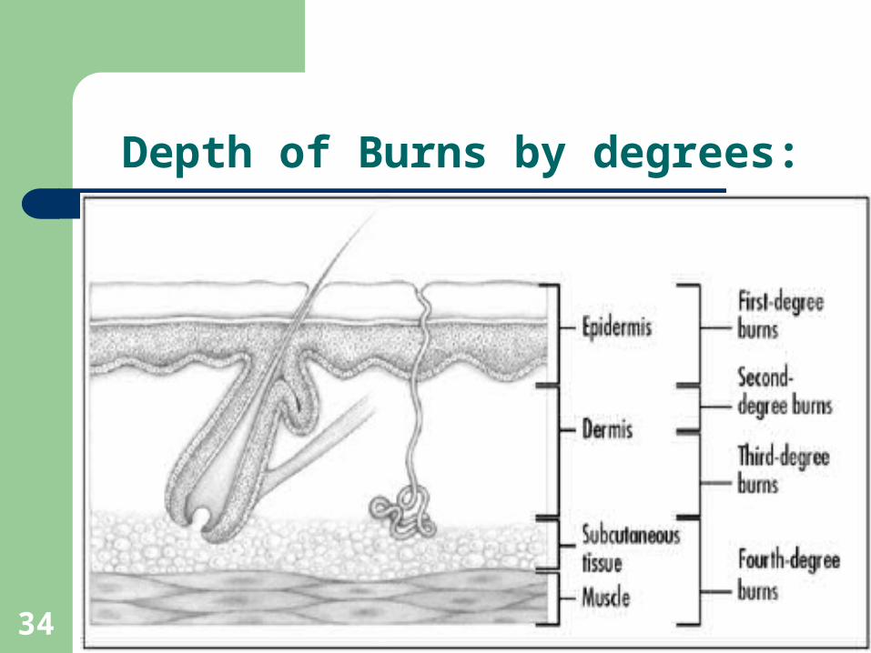
Depth of Burns by degrees:
34

35
Skin Infections Facts:
The skin is the first defense against Infection
Skin is commonly a site of infection
Conditions may be caused by viruses, bacteria, fungi or larger parasites.
36
Skin Infections:
Impetigo..staph or strep infection in young children,starts with erythema, vesicles, then yellowish crusts
Tinea..fungal skin infections includes ringworm,
jock itch, and athlete’s foot. Signs include erythema,scaling and crusting, with itching.
Warts.. papilloma virus,benign neoplasm, contagious by direct contact. Removed by freezing, drying, laser therapy, chemical applications

Skin Infections:
37Ringworm warts

38
Skin Infections:
Boils/Furuncles..staph infections of hair follicles. Large inflamed pustules.
Carbuncles.. group of untreated boils that fuse into larger pus-filled lesion.
Scabies.. Contagious condition
caused by itch mites. Young
mites hatch causing rash,
erythema

39
Vascular/Inflammatory Skin Disorders:
Decubitus..pressure sores caused by lying in one position for long periods. Common sites include heels, and over bony prominences.
Urticaria..hives/itching,red raised lesions called wheals, caused by hypersensitivity or allergic reaction.
Scleroderma..autoimmune disease,mild localized inflammed patches which later turns yellowish, hardened skin.

Decubitus Ulcer:
40

41
Vascular/Inflammatory Skin Disorders:
Psoriasis..common,
chronic inflammatory disorder of cutaneous tissue characterized by scaly plaques of epithelial tissue.
Eczema..most common inflammatory disorder, characterized by papules, vesicles, and crusts. It is a symptom of underlying condition. Example: Poison ivy causes contact dermatitis and eczema

42
Skin Cancer
Squamous cell carcinomaBasal cell carcinomaMelanomaKaposi’s sarcoma
43
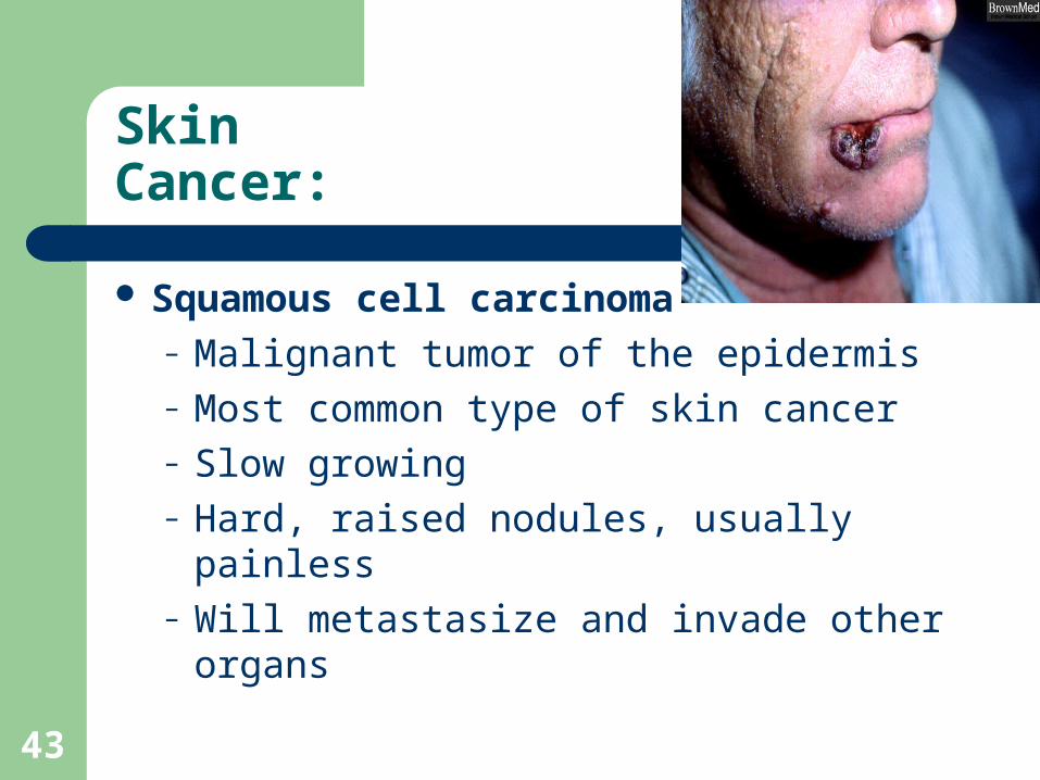
Skin Cancer:
Squamous cell carcinoma– Malignant tumor of the epidermis– Most common type of skin cancer– Slow growing– Hard, raised nodules, usually painless– Will metastasize and invade other organs

44
Skin Cancer:
Basal cell carcinoma– Occurs on upper face– Malignant, begins in base of
the epidermis– Begin as papule– Form bleeding, crusted
crater– Less likely to metastasize
than other types
45
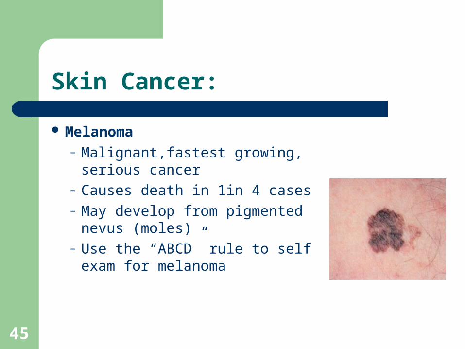
Skin Cancer:
Melanoma– Malignant,fastest growing,
serious cancer– Causes death in 1in 4 cases– May develop from pigmented
nevus (moles)– Use the “ABCD” rule to self
exam for melanoma

46
Skin Cancer:
Kaposi’s Sarcoma– Rarer skin cancer– Appears in AIDS and
other immune deficiencies– Appears as purple
papules– Spreads quickly to lymph
nodes and internal organs
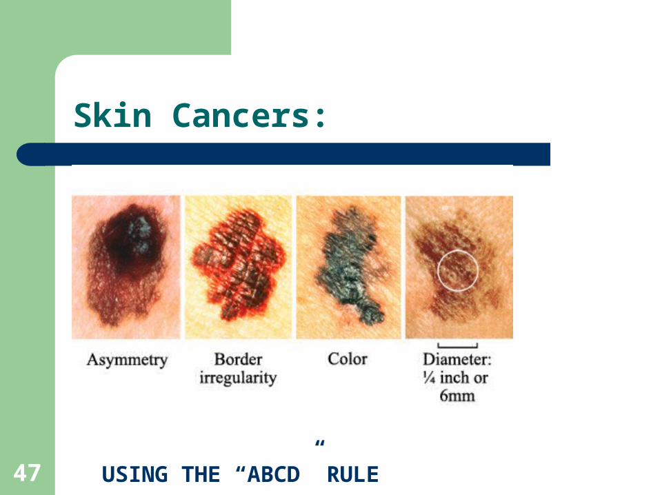
Skin Cancers:
47 USING THE “ABCD” RULE